1 2 3 4 5 6 7 8 9 10 11 12 13 14 15 16 17 18 19 20 21 22 23 24 25 26 27 28 29 30 31 32 33 34 35 36 37 38 39 40 41 42 43 44 45 46 47 48 49 50 51 52 53 54 55 56 57
DPP-4 Cleaves α/β-Peptide Bonds: Substrate Specificity and Half-Lives
Amila Turalić,
[a]Jasmina Đeđibegović,
[a]Zsófia Hegedüs,*
[b]and Tamás A. Martinek*
[b, c]The incorporation ofβ-amino acids into a peptide sequence has gained particular attention asβ- andα/β-peptides have shown remarkable proteolytic stability, even after a single homologa- tion at the scissile bond. Several peptidases have been shown to cleave such bonds with high specificity but at a much slower rate compared to α-peptide bonds. In this study, a series of analogs of dipeptidyl peptidase-4 (DPP-4) substrate inhibitors were synthesized in order to investigate whetherβ-amino acid homologation at the scissile bond could be a valid approach to improving peptide stability towards DPP-4 degradation. DPP-4
cleaved theα/β-peptide bond after the N-terminal penultimate Pro with a broad specificity and retained full activity regardless of theβ3-amino acid side chain and peptide length. Significantly improved half-lives were observed forβ3Ile-containing peptides.
Replacing the penultimate Pro with a conformationally con- strained Pro mimetic led to proteolytic resistance. DPP-4 cleavage of α/β-peptide bonds with a broad promiscuity represents a new insight into the stability of peptide analogs containingβ-amino acids as such analogs were thought to be stable towards enzymatic degradation.
Introduction
Rapid proteolytic degradation of peptides by endogenous enzymes is one of the major obstacles to their application in humans.[1] Peptide backbone modification by introduction of unnatural amino acids is an effective method for preparation of stable and peptidase resistant peptide analogs, without altering their activity. Of many unnatural amino acids known,β-amino acids gained much attention. Oligomers built from β-amino acids show tendency to adopt well-defined structures[2,3]
capable of exerting biological function and having increased proteolytic stability at the same time.[3,4]β-Peptides have been shown to have remarkable stability against a wide range of proteases[5–8] which can be explained by changes of the electronic environment around the amide bond or lack of substrate recognition resulting from the altered side-chain spacing or conformation of the peptide backbone.[9,10]
Besides pure β-peptides, it has been shown that α/β- peptides in which the β-amino acid replacement does not change the relative orientation of side chains around the scissile bond (Scheme 1), thus mimicking α-peptide environment, are still stable against a wide range of proteases.[9] This was
confirmed by many examples of peptide analogs where a strategic replacement of a single P1’ amino acid by a β- homolog (Scheme 1b) abolished the activity of pepsin, chymo- trypsin, plasmepsin II,[11] metalloproteinase EP24.15 and EP24.16,[12]neprylisin,[13]pronase E[14]or ACE-2,[15]and improved the stability of the whole peptide. With this kind of substitution, minimal modifications are possible in order to provide peptide stability, while maintaining α-peptide structural and spatial characteristics required for interaction with receptor.
Reports on α/β-bond cleavage are not unprecedented.
Pronase, a mixture of fungal endo- and exopeptidases could cleave the N-terminal Ala-β3Ala peptide bond although with a much slower rate in comparison with the natural sequence and it was attributed to its exopeptidase activity.[9] In the same study, pronase did not cleave Arg-β3Ala or after N-terminal βGly, pointing towards this is not a general feature of the enzyme. Angiotensin-converting enzyme cleaved α/β-peptide [a] A. Turalić, Prof. J. Đeđibegović
Department of Pharmaceutical Analysis University of Sarajevo, Faculty of Pharmacy
Zmaja od Bosne 8, 71 000 Sarajevo (Bosnia and Herzegovina) [b] Dr. Z. Hegedüs, Prof. T. A. Martinek
Department of Medical Chemistry University of Szeged, Faculty of Medicine 8 Dóm tér, 6720, Szeged (Hungary) E-mail: hegedus.zsofia@med.u-szeged.hu
martinek.tamas@med.u-szeged.hu [c] Prof. T. A. Martinek
MTA-SZTE Biomimetic Systems Research Group University of Szeged
Dóm tér 8, Szeged, 6720 (Hungary)
Supporting information for this article is available on the WWW under https://doi.org/10.1002/cbic.202000050
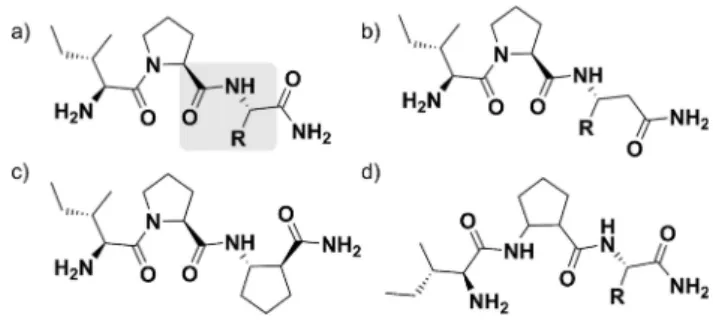
Scheme 1.(a) Naturalα-peptide bond and (b-d) three types of homologation usingβ2(side chain on the second carbon atom) orβ3(side chain on the third carbon atom) amino acids that do not change environment around the peptide bond, thus mimicking anα-peptide environment: (b)αXaa-β3Xaa, (c) β2Xaa-β3Xaa and (d)β2Xaa-αXaa.
1 2 3 4 5 6 7 8 9 10 11 12 13 14 15 16 17 18 19 20 21 22 23 24 25 26 27 28 29 30 31 32 33 34 35 36 37 38 39 40 41 42 43 44 45 46 47 48 49 50 51 52 53 54 55 56 57
bond in substance P analog withβ3- andβ2Ala modification at P1’, resulting in prolonged half-life of the analog,[16] but β- peptidase activity was not later confirmed as well. Another enzyme, proteinase K showed aminopeptidase activity on N- terminal Val-Glu peptide bond when Val was replaced by β3- homolog. Although cleaved, peptide half-life was increased from 1.6 to 300 min.[17]
Since β-amino acids gained huge attention to produce metabolically stable bioactive peptide analogs or enzyme inhibitors,[18] it is important to understand which factors can potentially influence their degradation in humans. This poses the question whether enzymes that have broad substrate specificity cleave certainα/β-peptide bonds or a singleβ-amino acid subsitution at the cleavage site is sufficient to create stable analogs. In the present study, the ability of the human dipeptidyl peptidase-4 (DPP-4) to cleave α/β-peptides was investigated with the primary aim of determining their stability against such an enzyme.
DPP-4 is a human exopeptidase that can be found in soluble circulating or membrane bound form and it is one of the few enzymes that cleaves peptide bonds primarily after Pro.[19] By cleaving dipeptides from N-terminus of proteins and oligopeptides, DPP-4 regulates a variety of physiological functions including immune regulation, endocrine activity and degradation of peptide hormones.[20]Besides well-known Xaa-Ala DPP-4 substrates GLP-1 and GIP, there are over 40 physiological and pharmacological substrates,[21] such as neuropeptides (substance P, bradykinin, endomorphin-1, neuropeptide Y, gastrin releasing peptide), gastric hormones (PYY, enterostatin) various chemokines, brain natriuretic peptide, erythropoietin, granulocyte colony-stimulating fac- tor, and other various endogenous peptides with penultimate (i. e., the next to last amino acid from the N-terminus) Pro in their structure.[22–24] Thus, the investigation of β-amino acid substitution after Pro could provide an insight for rational design of peptidic analogs of these ligands.
Results
In order to investigate how differentβ-amino acid substitutions at positions P1 and P1’of DPP-4 substrates affect their stability, following the substitution strategy shown in Scheme 1b, a series of α/β-peptide analogs of known DPP-4 ligands were synthesized (Scheme 2) and tested using an enzymatic cleavage assay. Proline containing tripeptides IPI and VPL are among the strongest known DPP-4 peptide inhibitors[25] whereas IPA and IPM have been shown to have inhibitory activity as well.[26,27]
These peptides are also substrates for the enzyme hence acting as competitive inhibitors, and their inhibitory activity is a result of kinetic artefact.[28,29] Based on these sequences, tripeptides having a general IPXaa structure were chosen as a model, where Xaa can represent any amino acid side chain. All selected tripeptides have Ile at position P2, in order to exclude possible influence of different amino acids at this position on DPP-4 cleavage and preserve effective docking to the enzyme.
First, IPA, IPI and IPM were prepared by solid-phase peptide synthesis and subjected to a DPP-4 cleavage test. The peptides were incubated with the enzyme, and the amount of the intact peptide and the cleavage products were determined by HPLC- MS monitored for 24 h. The resulting degradation curves were fitted to determine peptide half-lives (Figure 1a, Table 1, Table S1 in the Supporting information).
In all three cases, the cleavage product IP could be detected, showing that the cleavage occurs at the anticipated site. The obtained half-lives of IPA, IPI and IPM were quite similar (~ 5, 6 and 4 min respectively, Table 1), showing no significant effect of the P1’ amino acid on the peptide degradation.
In order to test whether β-amino acid replacement at position P1’ of these peptides influence stability, the α/β- analogs (IPβ3A, IPβ3I and IPβ3M) were synthesized and tested following the same protocol. IPβ3A and IPβ3M showed only a slightly increased resistance against DPP-4 cleavage, on the other hand, for IPβ3I a significantly prolonged half-life (t1/2
~ 274 min) was observed compared to the natural sequence IPI (t1/2~ 6 min) (Figure 1b, Tables 1 and S2). The sequence IP was detected as a degradation product for all of the peptides, pointing towards that DPP-4 is able to cleave the peptide bond between the α-amino acid at P1 and the β3-amino acid at position P1’, but the rate of cleavage is depending on the nature of the amino acid side chain at P1’.
Scheme 2.DPP-4 ligands and the investigatedα/β-peptide analogs. Sche- matic figure of IPXaa series of peptides. (a) DPP-4 cleaves any peptide bond after proline (highlighted grey), except for Pro at position P1’. Schematic figure ofα/β-peptides with (b)β3-amino acids, (c) cyclicβ-amino acid substitution at position P1’and (d) Pro to cyclicβ-amino acid substitution at position P1.
Table 1. Approximate half-lives of α-peptides and corresponding α/β- peptide analogs (obtained from best-fit nonlinear curve).
No. Peptide t1/2 [min]
No. Peptide t1/2
[min]
1 IPA 5 11 IPβ3S 6
2 IPI 6 12 IPβ3W 7
3 IPM 4 13 IP-(1S,2S)ACPC 12
4 IPβ3A 12 14 IPIQY 7
5 IPβ3I 274 15 IPβ3IQY 216
6 IPβ3M 8 16 LPQNIPPL 9
7 IPβ3E 15 17 LPβ3QNIPPL 27
8 IPβG 3 18 I-(1R,2R)ACPC-I >1440
9 IPβ3K 17 19 I-(1S,2S)ACPC-A >1440
10 IPβ3Q 7 20 I-(1R,2R)ACPC-A >1440
1 2 3 4 5 6 7 8 9 10 11 12 13 14 15 16 17 18 19 20 21 22 23 24 25 26 27 28 29 30 31 32 33 34 35 36 37 38 39 40 41 42 43 44 45 46 47 48 49 50 51 52 53 54 55 56 57
To further investigate the substrate specificity of DPP-4 for α/β-peptide bond cleavage, a series of IPβ3Xaa tripeptides were synthesized having all classes of amino acid side chains at P1’ namely β3E, βG,β3K, β3Q, β3S, β3W and a cyclic β-amino acid (1S,2S)-aminocyclopentane-carboxylic acid (ACPC). Cleavage product IP was observed for all of the tested peptides, with almost no intact peptide after 1 h (Figure 2, Table S3), resulting in similar half-lives (Table 1). Analogs having charged side chains or ACPC showed a slightly increased stability in comparison with the other sequences, with half-lives of approximately 11–17 min. These results suggested that DPP-4 retains its broad side chain specificity for β3-amino acid substitutions except for the bulky, hydrophobic β3Ile substitu- tion, which slows down the cleavage significantly.
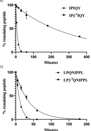
To investigate further this new insight into DPP-4 cleavage of α/β-peptides, previously reported longer DPP-4 peptide inhibitors IPIQY and LPQNIPPL and their analogs IPβ3IQY and LPβ3QNIPPL were synthesized. Results of DPP-4 cleavage test for these longer peptides were in correlation with the tripeptide series. Cleavage products IP and LP, as well as IQY/β3IQY and QNIPPL/β3QNIPPL were observed for testedα andα/β-peptide
analogs (Figure 3, Table S4). Theβ3Ile substitution had the same profound effect on stability, while substitution with β3Gln resulted only in 3-fold increase in half-life.
Further, we wanted to examine DPP-4 cleavage when Pro is substituted with a Pro mimicking, conformationally constrained Figure 1.DPP-4 degradation curves of (a) IPA, IPI and IPM and (b)β3-amino
acid-modified peptides. Remaining peptide quantities were determined based on LCMS peak integration. Peptides (50μM final concentration) were incubated with DPP-4 in TBS buffer (pH 8.0) at 37°C for 24 h. Curves were fitted by using a one-phase exponential decay fit with a variable plateau valuey=(y0–yP) ekx+yP. Note that thex-axes cover a different range. Data points show the average of two individual experiments, error bars represent the standard deviation. In several cases the error bars are smaller than the symbol size.
Figure 2.DPP-4 degradation curves of tripeptides of IPβ3Xaa series. Curves were fitted by using a one-phase exponential decay fit with a variable plateau valuey=(y0–yP)ekx+yP. Data points show the average of two individual experiments, error bars represent the standard deviation. In several cases the error bars are smaller than the symbol size.
Figure 3.(a) DPP-4 degradation curves of IPIQY and IPβ3IQY and (b) LPQNIPPL and LPβ3QNIPPL. Curves were fitted by using a one-phase exponential decay fit with a variable plateau valuey=(y0–yP)ekx+yP. Data points show the average of two individual experiments, error bars represent the standard deviation. In several cases the error bars are smaller than the symbol size.
1 2 3 4 5 6 7 8 9 10 11 12 13 14 15 16 17 18 19 20 21 22 23 24 25 26 27 28 29 30 31 32 33 34 35 36 37 38 39 40 41 42 43 44 45 46 47 48 49 50 51 52 53 54 55 56 57
β-amino acid ACPC. In contrast to other tested peptides with α!β3 replacement after Pro, I-(1R,2R)ACPC-I, I-(1S,2S)ACPC-A and I-(1R,2R)ACPC-A were stable to degradation by DPP-4, since DPP-4 cleavage products were not observed within 24 h.
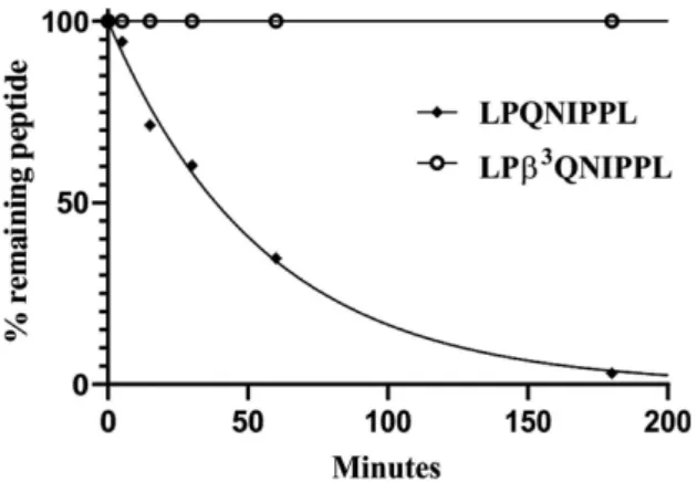
To demonstrate the unique ability of DPP-4 cleaving α/β- peptide bonds we compared half-lives with a nonspecific protease. The peptide LPQNIPPL and itsα/β-analog LPβ3QNIPPL were tested against a broad-spectrum serine protease protei- nase K. Small peptides were excluded due to peptide size limits for proteinase K cleavage.[30]LPQNIPPL was completely cleaved within a few hours, with half-life of ~ 39 min (cleavage products LPQ and NIPPL were observed), while LPβ3QNIPPL remained stable, without any cleavage products observed within 24 h (Figure 4, Table S5). This is consistent with previous studies[11]
demonstrating that the amino acid replacement at the cleavage site abolishes proteinase K action which contrasts with the activity of DPP-4 shown here.
Discussion
β-Amino acid replacement is a recognized strategy to obtain metabolically stable analogs of bioactive peptides, thus it is important to investigate every aspect of their metabolic route in humans. Here we investigated the effect of a singleβ-amino acid replacements in substrates of DPP-4, a human exopepti- dase playing the key role in a wide variety of physiological functions.[20]
The main cleavage product of the IPβXaa ligand series was the IP sequence, indicating that DPP-4 is one of the few enzymes that can cleave a bond between anαand aβ-amino acid. Knownβ- aminopeptidases in humans cleave only a specific bond, such as the βAla/His bond in case of carnosinase.[31,32] In contrast, our results indicate that the cleavage specificity of DPP-4 for anα/β- bond is not limited to certain residues, thus DPP-4 is the only
human peptidase that have a broad substrate specificity forα/β- bond cleavage and is able to retain its full activity, when a β- amino acid is present at P1’.
It has been shown that DPP-4 does not have a strong preference for amino acids at P1’, and is capable of cleaving free amino group containing structures,[33] chromophoric amines[23] after Pro or D-amino acids after Ser or Ala.[34,35]
Homologation at position P1’ has a main effect on the C- terminal part of the peptide, but since P1’ carbonyl or interactions of P2’-Pn’with the enzyme are not prerequisite for recognition,[36]this retained promiscuity can be explained. While peptides with Pro or hydroxyproline at position P1’are resistant to DPP-4 cleavage,[20,37] interestingly, the cyclic amino acid containing analog (IP-(1S,2S)ACPC) is not stable. The difference between the two substitution strategies is that the nitrogen of ACPC at P1’can be protonated, which is a requirement for the serine protease mechanism of DPP-4,[38] thus explaining the instability of this analog.
The only exception to the broad substrate specificity in our series of IPβXaa ligands was the substitution withβ3Ile, which resulted in a significantly increased stability, which was not depending on peptide length, pointing towards that this is a unique and general effect of the β3Ile substitution. Since the electronic environment around the α/β-bond is similar in all tested analogs, one explanation could be theβ-branched side chain. Side-chain branching can have a profound effect on the local conformation of the peptide,[39] which might result in a spatial arrangement incompatible with enzyme recognition. To support this, β3Ile, β3Ala and ACPC containing analogs were tested in a DPP-4 inhibitory assay. IPA and IPβ3A showed similar DPP-4 inhibitory activity (Figure S1), whereas analogs having substitution withβ3Ile and ACPC did not inhibit the enzyme as effectively as their natural counterparts. Thus, slower cleavage is associated with less inhibitory activity, pointing towards that these substitutions are detrimental to binding to the enzyme and including these amino acids in a sequence would provide stability against DPP-4. Since the side chain of the P1’ amino acid of the natural sequence IPI is not involved in binding to the active site of DPP-4[29,36]another explanation could be that upon substitution a more favorable stabilizing interaction is formed between the C-terminal amide of the peptide and the enzyme. This kind of interaction is proposed to stabilize degradation byproducts by preventing P1’ amino acid leaving from the active site.[29]
Due to the lack of specificity for β-amino acids at position P1’ shown here, one could speculate that similar substrates with penultimate Ser or Ala could also be cleaved. Bai et al.[40]
reported that substitution of glutamic acid after Ala (Glu3) in GLP-1 molecule with corresponding β-amino acid lead to increased stability towards DPP-4 degradation and half-life
~ 96 h. In contrast, the peptide IPβ3E investigated here was cleaved almost as fast as the natural sequences, pointing towards this property of DPP-4 might be specific to penultimate Pro and not Ala. This feature should be further investigated because mechanism of DPP-4 cleavage is slightly different for Pro and Ala containing DPP-4 substrates.[23]
Figure 4.Proteinase K degradation curves of LPQNIPPL and LPβ3QNIPPL.
Remaining peptide quantities were determined based on LCMS peak integration. Peptides (50μM final concentration) were incubated with proteinase K (24μg/mL final concentration) in TBS buffer (pH 8.0) at 37°C for 24 h. Curves were fitted by using a one-phase exponential decay fit with a variable plateau valuey=(y0–yP)ekx+yP. Data points show the average of two individual experiments, error bars represent the standard deviation. In several cases error bars are smaller than the symbol size
1 2 3 4 5 6 7 8 9 10 11 12 13 14 15 16 17 18 19 20 21 22 23 24 25 26 27 28 29 30 31 32 33 34 35 36 37 38 39 40 41 42 43 44 45 46 47 48 49 50 51 52 53 54 55 56 57
Our findings suggest that care should be taken when designingα/β-peptide analogs including Pro residues, since all peptides with penultimate Pro are considered as good sub- strates for DPP-4.[37]Possible obstacles regarding the proteolytic stability of such analogs could occur, moreover because DPP-4 is present at the absorption site in gastrointestinal tract and other tissues, as well as in circulating form, which raises issues about bioavailability of such peptides. As an example, DPP-4 is one of the enzymes involved in endomorphin metabolism,[41]
which, together with our findings could explain the degradation of a previously reported endomorphin analog withα-hydroxy- β-phenylalanine (AHPBA) substitution at position P1’.[42]
DPP-4 resistant peptide analogs can be created by substitut- ing amino acid at position P1 with amino acids such as 2- aminoisobutyric acid (Aib).[43] Here we show that substitution with the Pro mimicking β-amino acid, ACPC can also result in highly stable analogs. Thus, using this strategy DPP-4 resistant analogs can be prepared, assuming that the binding prefer- ences to the target receptor allow such kind of substitution.
Furthermore, exploiting our findings, a selective proteolytic profile can be achieved by rational design of the peptide sequence resulting in cleavage only by DPP-4 and not by other enzymes, as we have shown for LPQNIPPL and its analog (Figure 4), or in an opposite way controlled slow cleavage by DPP-4 can be encoded in the sequence by incorporatingβ3Ile residues after Pro.
Conclusion
In summary, we have shown that DPP-4 cleaves α/β-peptide bond when anα-amino acid after penultimate N-terminal Pro is replaced with aβ3- or constrained (cyclic)β-amino acid, and it retains its broad substrate specificity and full enzymatic activity regardless of the chemical properties of the side chain. This activity is unique compared to other human enzymes involved in peptide cleavage known so far. The only exception to this is when β3Ile is present at position P1’ which increases the stability significantly, thus this replacement can be used to create analogs stable against DPP-4 cleavage. Cleavage does not occur either when Pro at position P1 is replaced with ACPC, which provides an option to generate stable ligands in which this substitution is tolerated. While Pro or Ala are preferred at position P1, α/β-peptide analogs containing Ser, Gly, Val and Leu at this position could also be potentially cleaved.[20,22]
Exploiting these findings, a selective and programmable proteolytic profile can be designed, leading to a sequence- controlled degradation pattern, thus extending the scope of the utilization ofα/β-peptides.
Experimental Section
Peptides synthesis, purification and identification. Peptides were synthesized manually by standard solid-phase peptide synthesis using Fmoc chemistry. Tentagel S RAM (for peptide IP-(1S,2S)ACPC), Fmoc-Rink-Amid AM (for peptides IPβG (IPβA), IPβ3K, IPβ3Q and IPβ3W) and Tentagel R RAM (for all other peptides) resins were used
as a solid supports. Couplings were carried out using 3 equivalent amino acid excess, COMU (3 equivalent) as a coupling reagent and DIPEA (3 equivalent) as base for 3 h at room temperature.
Deprotection was carried out using 2 % piperidine and 2 % DBU (1,8-diazabicycloundec-7-ene) in DMF (N,N-dimethylformamide).
Peptides were cleaved from the resin using TFA/H2O/DTT/TIS (90 : 5 : 2.5 : 2.5) or TFA/H2O (95 : 5) for 3 h. After the cleavage, TFA was evaporated, peptides LPQNIPPL and LPβ3QNIPPL were precipi- tated in ice-cold diethlyether and re-dissolved in acetic acid, diluted with water and freeze dried. All other peptides were dissolved after TFA evaporation without the precipitation step and freeze dried.
Peptides were purified by RP-HPLC on a C18 column (Phenomenex Luna 5μm C18(2) 100 Å, 250 × 10 mm) using 0.1 % TFA in water and 0.1 % TFA, 80 % ACN in water as HPLC eluents. Pure fractions were pooled together and freeze dried. Purity of the peptides was confirmed by ESI-MS measurements and analytical RP-HPLC on Aeris Peptide XB-C18 column (250 × 4.6 mm). Eluents were A: 0.1 % TFA in water and B: 0.1 % TFA, 80 % ACN in water using a gradient of 5–80 % B over 15 min at 50°C at 1.2 mL/min flow rate (for IPβ3K), 0–40 % B over 25 min at 50°C at 1.2 mL/min flow rate (for IPβ3Q and IPβ3S) and 0–60 % B over 25 min at 50°C at 1.2 mL/min flow rate (for all other peptides).
Peptides were identified by HPLC-MS on analytical HPLC column Aeris Peptide XB-C18 (250 × 4.6 mm). Eluents were A: 0.1 % HCOOH in water and B: 0.1 % HCOOH in ACN using a gradient of 5–80 % B over 15 min at 50°C at 0.7 mL/min flow rate (for IPβ3K), 0–40 % B over 20 min at 50°C at 0.7 mL/min flow rate (for IPβ3Q and IPβ3S) and 0–60 % B over 20 min at 50°C at 0.7 mL/min flow rate (for all other peptides).
DPP-4 cleavage protocol. Peptide stock solutions (1 mM) were prepared by dissolving peptides in 20 % DMSO/TBS buffer (45 mM Tris · HCl, pH 8.0) and diluted to 500μM using TBS buffer. DPP-4 was purchased from Sigma-Aldrich (D3446), supplied as a solution in 45 mM Tris · HCl, pH 8.0, 124 mM NaCl, 2.4 mM KCl, 225 mM imidazole, 10 % glycerol and 3 mM DTT. From the DPP-4 stock solution (� 0.01 mg/mL,�4500 units/μg), a diluted enzyme sol- ution was prepared using 1.7μL of the stock solution and 110.3μL of TBS buffer. Cleavage reactions were performed using 35μL of diluted peptide solution (50μM final concentration), 52.5μL of diluted enzyme solution and 262.5μL of TBS buffer and incubated at 37°C for 24 h. Controls containing diluted peptide solution (50μM final concentration) without the enzyme in TBS buffer were also prepared and measured att=0 and after 24 h. At eight time points (0, 5, 15, 30, 60, 180, 360 and 1440 min), an aliquot of 20μL was removed from the incubation mixtures and pipetted into 80μL of 5 % TFA solution to quench the reaction, from which 20μL was injected to the HPLC. All samples were prepared in duplicates. The amounts of intact peptides and DPP-4 cleavage products at selected time points were analyzed by HPLC-MS.
Proteinase K cleavage protocol. Peptide stock solutions (1 mM) were prepared by dissolving peptides in DMSO and diluted to 100μM using TBS buffer (50 mM Tris · HCl, 5 mM CaCl2, pH 8.0).
Proteinase K stock solution (5 mg/mL) was prepared in 20 % glycerol/TBS, which was further diluted to 120μg/mL in TBS buffer.
Cleavage reactions were performed using 550μL of diluted peptide solution (50μM final concentration), 220μL of diluted enzyme solution (24μg/mL final concentration) and 330μL of TBS buffer and incubated at 37°C for 24 h. Controls containing diluted peptide solution (50μM final concentration) without the enzyme in TBS buffer were also prepared and measured att=0 and after 24 h. At eight time points (0, 5, 15, 30, 60, 180, 360 and 1440 min), an aliquot of 50μL was removed from the incubation mixtures and pipetted into 100μL of 5 % TFA solution to quench the reaction, from which 20μL was injected to the HPLC. All samples were
1 2 3 4 5 6 7 8 9 10 11 12 13 14 15 16 17 18 19 20 21 22 23 24 25 26 27 28 29 30 31 32 33 34 35 36 37 38 39 40 41 42 43 44 45 46 47 48 49 50 51 52 53 54 55 56 57
prepared in duplicates. The amounts of intact peptides and proteinase K cleavage products at selected time points were analyzed by HPLC-MS.
HPLC-MS methods and parameters for identification of intact peptides and cleavage products. HPLC-MS analysis was performed on a Thermo Scientific Dionex UltiMate 3000 HPLC system interfaced to LTQ ion trap mass spectrometer (Thermo Electron Corp., San Jose, CA, USA). Measurements were performed using analytical HPLC column Aeris Widepore XB-C18 (250 × 4.6 mm) with 0.1 % HCOOH in water and 0.1 % HCOOH in ACN as HPLC eluents.
Operating conditions were 0–60 % ACN over 20 min at 50°C and 0–
40 % ACN over 20 min at 50°C (for peptides IPβ3K, IPβ3Q and IPβ3S) at 0.7 mL/min flow rate. Mass spectra were acquired in a positive ion full scan mode from 110 to 2000 m/z range.
HPLC-MS quantification of intact peptides and cleavage products.
Thermo Xcalibur 2.2 software was used for peak identification and integration. Using ICIS peak detection algorithm, the general detection and integration criteria were as follows – smoothing points: 5–15, baseline window: 50, area noise factor: 5, peak noise factor: 10.
Intact peptides and DPP-4/Proteinase K cleavage products were identified based on their molecular weights and retention times, thereafter areas under the curves (AUC) were determined. The percentage of the intact peptides at each of the time points was determined using the following equation [Eq. (1)]:
intact peptideð%Þ ¼AUCt=AUCt0�100 (1) where AUCtis the area under the curve at a specific time point and AUCt0area under the curve att=0. The % of intact peptide IPβ3K was calculated based on the amount of cleavage product IP using the following equation: 100 – (AUCIP,t/AUCIP,6h× 100), with the assumption that the peptide is fully cleaved after 6 h. The percentages of the remaining intact peptides were plotted against time in order to assess the half-life values based on the fitted curves. The data was fitted to one-phase exponential decay fit with a variable plateau valuey=(y0–yP)e kx+yPusing GraphPad Prism to generate a calculated half-life.
Acknowledgements
A.T. has been awarded an EU ERASMUS+Credit Mobility student grant (15-1/KA1ICM/IN/142/SMS-028) at the University of Szeged.
Prof. Fabio Altieri is gratefully acknowledged for discussions on DPP-4 cleavage protocol and Dr. Éva Bartus for helping in peptide synthesis. Ministry of Human Capacities, Hungary grant 20391-3/
2018/FEKUSTRAT and grant GINOP-2.3.2-15-2016-00014 (EVOMER) are acknowledged.
Conflict of Interest
The authors declare no conflict of interest.
Keywords: DPP-4 · enzyme inhibitors · proteolysis · peptide bond cleavage·α/β-peptides
[1] L. Diao, B. Meibohm,Clin. Pharmacokinet.2013,52, 855–868.
[2] R. De Marco, A. Tolomelli, E. Juaristi, L. Gentilucci,Med. Res. Rev.2016, 36, 389–424.
[3] Z. Hegedüs, T. A. Martinek In:Comprehensive Supramolecular Chemistry II (Ed.: J. L. Atwood), Elsevier, Amsterdam,2017, pp. 511–537.
[4] J. W. Checco, S. H. Gellman,Curr. Opin. Struct. Biol.2016,39, 96–105.
[5] J. Frackenpohl, P. I. Arvidsson, J. V. Schreiber, D. Seebach,ChemBioChem 2001,2, 445–455.
[6] D. Seebach, M. Overhand, F. N. Kühnle, B. Martinoni, L. Oberer, U.
Hommel, H. Widmer,Helv. Chim. Acta1996,79, 913–941.
[7] T. Hintermann, D. Seebach,Chimia1997,51, 244–247.
[8] E. A. Porter, B. Weisblum, S. H. Gellman,J. Am. Chem. Soc.2002,124, 7324–7330.
[9] D. F. Hook, P. Bindschädler, Y. R. Mahajan, R. Šebesta, P. Kast, D.
Seebach,Chem. Biodiversity2005,2, 591–632.
[10] H. M. Werner, C. C. Cabalteja, W. S. Horne,ChemBioChem2016,17, 712–
718.
[11] H. N. Gopi, G. Ravindra, P. P. Pal, P. Pattanaik, H. Balaram, P. Balaram, FEBS Lett.2003,535, 175–178.
[12] R. A. Lew, E. Boulos, K. M. Stewart, P. Perlmutter, M. F. Harte, S. Bond, M. I. Aguilar, A. I. Smith,J. Pept. Sci.2000,6, 440–445.
[13] D. Steer, R. Lew, P. Perlmutter, A. I. Smith, M. I. Aguilar, Biochemistry 2002,41, 10819–10826.
[14] C. A. McDonald, N. L. Payne, G. Sun, D. J. Clayton, M. P. Del Borgo, M. I.
Aguilar, P. Perlmutter, C. C. Bernard,J. Neuroimmunol.2014,277, 67–76.
[15] D. Clayton, I. Hanchapola, N. Hausler, S. Unabia, R. A. Lew, R. E. Widdop, A. I. Smith, P. Perlmutter, M. I. Aguilar,J. Mol. Recognit.2011,24, 235–
244.
[16] S. Sagan, T. Milcent, R. Ponsinet, O. Convert, O. Tasseau, G. Chassaing, S.
Lavielle, O. Lequin,Eur. J. Biochem.2003,270, 939–949.
[17] H. S. Haase, K. J. Peterson-Kaufman, S. K. Lan Levengood, J. W. Checco, W. L. Murphy, S. H. Gellman,J. Am. Chem. Soc.2012,134, 7652–7655.
[18] D. L. Steer, R. A. Lew, P. Perlmutter, A. Smith, M. I. Aguilar,Curr. Med.
Chem.2002,9, 811–822.
[19] Y. Sanz In:Industrial enzymes(Eds.: J. Polaina, A. P. MacCabe), Springer, Dordrecht2007, pp. 243–260.
[20] A. M. Lambeir, C. Durinx, S. Scharpé, I. De Meester,Crit. Rev. Clin. Lab.
Sci.2003,40, 209–294.
[21] E. E. Mulvihill, E. M. Varin, B. Gladanac, J. E. Campbell, J. R. Ussher, L. L.
Baggio, B. Yusta, J. Ayala, M. A. Burmeister, D. Matthews, K. A. Bang, J. E.
Ayala, D. J. Drucker,Cell Metab.2017,25, 152–165.
[22] E. E. Mulvihill, D. J. Drucker,Endocr. Rev.2014,35, 992–1019.
[23] A. Yaron, F. Naider, S. Scharpe,Crit. Rev. Biochem. Mol. Biol.1993,28, 31–81.
[24] M. S. Glover, E. P. Bellinger, P. Radivojac, D. E. Clemmer, Anal. Chem.
2015,87, 8466–8472.
[25] A. B. Nongonierma, R. J. FitzGerald,Food Chem.2014,145, 845–852.
[26] A. B. Nongonierma, R. J. FitzGerald,Peptides2016,79, 1–7.
[27] G. Tulipano, V. Sibilia, A. M. Caroli, D. Cocchi,Peptides2011,32, 835–
838.
[28] J. Rahfeld, M. Schierborn, B. Hartrodt, K. Neubert, J. Heins, Biochim.
Biophys. Acta Protein Struct. Mol. Enzymol.1991,1076, 314–316.
[29] R. Thoma, B. Löffler, M. Stihle, W. Huber, A. Ruf, N. Hennig,Structure 2003,11, 947–959.
[30] E. V. Petrotchenko, J. J. Serpa, D. B. Hardie, M. Berjanskii, B. P. Suriya- mongkol, D. S. Wishart, C. H. Borchers,Mol. Cell. Proteomics2012,11, M111–013524.
[31] M. C. Jackson, C. M. Kucera, J. F. Lenney,Clin. Chim. Acta1991,196, 193–
205.
[32] M. Teufel, V. Saudek, J. P. Ledig, A. Bernhardt, S. Boularand, A. Carreau, N. J. Cairns, C. Carter, D. J. Cowley, D. Duverger, A. J. Ganzhorn, C.
Guenet, B. Heintzelmann, V. Laucher, C. Sauvage, T. Smirnova,J. Biol.
Chem.2003,278, 6521–6531.
[33] A. Diez-Torrubia, C. García-Aparicio, S. Cabrera, I. De Meester, J. Balzarini, M. J. Camarasa, S. Velázquez,J. Med. Chem.2009,53, 559–572.
[34] S. A. Hinke, J. A. Pospisilik, H. U. Demuth, S. Mannhart, K. Kühn-Wache, T.
Hoffmann, E. Nishimura, R. A. Pederson, C. H. McIntosh,J. Biol. Chem.
2000,275, 3827–3834.
[35] K. Kühn-Wache, S. Manhart, T. Hoffmann, S. A. Hinke, R. Gelling, R. A.
Pederson, C. H. McIntosh, H. U. Demuth InCellular Peptidases in Immune Functions and Diseases 2. Adv Exp Med Biol., 477(Eds.: J. Langner, S.
Ansorge), Springer, Boston, MA,2002, pp. 187–195.
[36] M. J. Ojeda-Montes, A. Gimeno, S. Tomas-Hernández, A. Cereto-Massag- ué, R. Beltrán-Debón, C. Valls, M. Mulero, S. Garcia-Vallvé,Med. Res. Rev.
2018,38, 1874–1915.
1 2 3 4 5 6 7 8 9 10 11 12 13 14 15 16 17 18 19 20 21 22 23 24 25 26 27 28 29 30 31 32 33 34 35 36 37 38 39 40 41 42 43 44 45 46 47 48 49 50 51 52 53 54 55 56 57
[37] R. Mentlein,Regul. Pept.1999,85, 9–24.
[38] W. Brandt, O. Ludwig, I. Thondorf, A. Barth,Eur. J. Biochem.1996,236, 109–114.
[39] T. L. Raguse, J. R. Lai, S. H. Gellman,Helv. Chim. Acta,2002,85, 4154–
4164.
[40] X. Bai, Y. Niu, J. Zhu, A. Q. Yang, Y. F. Wu, X. S. Ye,Bioorg. Med. Chem.
2016,24, 1163–1170.
[41] C. Sakurada, S. Sakurada, T. Hayashi, S. Katsuyama, K. Tan-No, T.
Sakurada,Biochem. Pharmacol.2003,66, 653–661.
[42] M. Hu, M. A. Giulianotti, J. P. McLaughlin, J. Shao, G. Debevec, L. E.
Maida, P. Geer, M. Cazares, J. Misler, C. Dooley, M. L. Ganno, S. O. Eans,
E. Mizrachi, R. G. Santos, A. B. Yongye, R. A. Houghten, Y. Yu,Eur. J. Med.
Chem.2015,92, 270–281.
[43] M. V. Hager, L. M. Johnson, D. Wootten, P. M. Sexton, S. H. Gellman,J.
Am. Chem. Soc.2016,138, 14970–14979.
Manuscript received: January 29, 2020 Revised manuscript received: February 19, 2020 Version of record online: March 31, 2020