THE KEY ELEMENTS OF CONDUCTING LOAD-TO-FRACTURE MECHANICAL TESTING ON RESTORATION-TOOTH UNITS IN
RESTORATIVE DENTISTRY
1Balázs Szabó P., 2Tekla Sáry, 3Balázs Szabó
1University of Szeged, Faculty of Engineering, Department of Food Engineering, Moszkvai krt. 9., 6725, Szeged, Hungary
2University of Szeged, Faculty of Dentistry, Department of Operative and Esthetic Dentistry, Tisza Lajos krt. 64., 6726, Szeged, Hungary
3University of Szeged, Faculty of Dentistry, Department of Periodontology, Tisza Lajos krt. 64., 6726, Szeged, Hungary e-mail: szabobalazs77@gmail.hu, teklasary@gmail.com, szpb@mk.u-szeged.hu
ABSTRACT
Biomimetic restorative dentistry strives to replace lost tooth tissue by biomaterials with similar physical properties. In order to do so, mechanical testing of dental restorative materials on their own and later in dental cavities is highly important. During this process dentists and engineers are collaborating aiming to set the indications of certain restorative materials and application techniques. In vitro fracture resistance testing of a restored tooth is one of the most important tests to be carried out during the indication setting process. However, for this specific test and received results to be valid for clinicians, the group conducting the tests must pay attention to mimic intraoral conditions as much as possible. The article aims at identifying the key elements of fracture resistance tests in dentistry. Adequately conducting this test is a prerequisite for later testing in in vivo conditions the restorative techniques that produced the best results among the in vitro tests.
Keywords: mechanical testing, fracture resistance, fracture pattern, restored tooth, load-to-fracture 1. INTRODUCTION
The aim of restorative dentistry is to restore the function and esthetic apperance of teeth after previous dental hard tissue loss due to caries, trauma or parafunctional jaw movements. Any dental restorative or prosthetic material used for this purpose must have sufficient mechanical integrity to function in the oral cavity for an extended period of time. Thus, testing the mechanical properties of these materials on their own and also luted to dental structures is highly clinically relevant. This is supported by the fact that the two main causes of failure identified in case of dental restorations are fracture (restoration and/or tooth) and secondary caries. Complete cusp fracture of posterior restored teeth is a common phenomenon in dental practices with incidence rates varying from approx. 20 to 71 per 1000 person-years at risk [1].
Fracture shows a higher tendency in vital teeth with large direct restorations, in root canal treated teeth restored with direct restoration, and in neglected cariously infected teeth.
Thus, the restoration should provide adequate fracture resistance to the dammaged tooth and also, if fracture occurs, should direct the fracture away from the center of the tooth for it be restorable later on.
This means that some restorative materials (placed on the outside) must be hard and wear resistant, while some materials (placed in deeper layers) must be more elastic and have adequate fracture toughness to be able to stop possible crack propagation [2].
Once a restorative material has been analyzed and tested regarding its mechanical properties, testing it in certain in vitro dental indications should be performed. For this purpose intact human teeth extracted for other reasons (periodontal infection or orthodontic treatment planning) are used widely. One disadvantage when using human teeth for testing is the large variation among individual teeth, e.g. in the mechanical and physical properties, and existing microcracks in the dentin may not always be seen before testing. On general this may lead to large standard deviations.
Despite the mentioned shortcommings, the use of natural human teeth is a reliable methodology in fracture testing and still represent the first choice for in vitro tests [3]. When utilizing human teeth, setting strict exclusion and inclusion criteria are mandatory. For mechanical testing the most often used inclusion criteria are the following: visual absence of caries or root cracks, absence of previous endodontic treatment, posts or crown or resorptions. Teeth with severe polymorphism of the coronal structures should be excluded from the study and careful attention must be paid during measuring the coronal dimensions of the tooth and/or the prepared cavity to insure standardisation [4]. Also, teeth should be used within 2-6 months after extraction and they must be kept in specific solutions before usage.
Usually the first and most important step when testing tooth-restoration units is measuring frature resistance. This can be performed in a static load-to-fracture setup with a universal testing machine. As stated by Ref. [5] static loading is usually the first step in the evaluation process of a novel dental materials and related techniques and is commonly used in order to obtain basic knowledge regarding the fracture behaviour and load capacity of a restored tooth. There are a number of factors that may interfere with resistance to fracture, such as the differences between specimens, tooth embedment method, type and direction of load application, crosshead speed, and simulation of thermal or mechanical fatiguing [6].
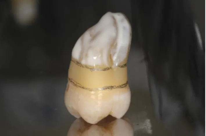
During this test intraoral conditions should be mimicked as much as possible. During the embedding of the samples two important elements should be emphasized: simulating the periodontal ligaments and simulating the bone level around the roots. In order the best mimic the elastic property of the periodontal ligaments, which normally connects the roots to the bone in a healthy clinical situation, the root part of the samples is covered with a layer of liquid latex separating material prior to embedding [7] (Figure 1.).
Figure 1.A layer of later separator applied on the root surface before embedding.
This is highly important as the ligaments provide a 0.05 mm movement for the teeth in the bony socket, thus it absorbs stress to a certain amount before transferring it to the bone. This is also visible in mechanical testing if the latex is applied on the roots of the specimen. Although some studies do not use any separating agent on the root surfaces before embedding them, most studies highlight the lack of periodontal ligament simulation as a limitation of the design of these studies with regard to their clinical relevance [8]. During the embedding procedure teeth are embedded in metacrylate resin, usually 2 mm apical from the cemento-enamel junction (CEJ) to simulate the bone level. This represents a healthy situation regarding the amount of bone supporting the tooth. To our knowledge only Szabó et al. examined the effect of altered bone level on the fracture resistance of root amputated molar teeth [9, 10]. In their studies the embedding was positioned at a more apical level to simulate a root furcation involved situation, which develops in periodontally compromised patients.
Once the samples are embedded, they need to be positioned under the rounded cylyndrical bar delivering the loading, according to the type of tooth to be tested. In molar teeth the bar is positioned at the centre of the occlusal surface of the tooth crown between the buccal and oral cusps. This way a tripod contact is created by simultaneous and equal contact of the bar to the mesiobuccal, distobuccal and lingual cusps while the tooth is loaded from a vertical direction paralel to the long axis of the tooth. Contrary to molar teeth, an oblique load (45 degrees to the long axis of the tooth) is applied to the occlusal incline of the buccal cusp in premolar teeth [11]. This pattern of loading is intended to simulate normal working side occlusal contacts. This should represent a worst-case occlusal loading scenario for these teeth and test the integrity of the tested restorations and tooth structure. In case of anterior teeth the loading tip is positioned at a 30-45 degrees to the long axis of the tooth in order to simulate the normal relationship of the front teeth. The bar delivering the loading is usually a rounded, 6 mm diameter stainless-steel ball-shaped stylus [12]. Some research groups were trying to incorporate a more realistic relationship and contact with the opposing dentition into their mechanical studies. With this intent Soares et al. are using a 6-7 mm diameter composite resin sphere as a loading tip [13]. As composite shows similar wear to human enamel, the utilization of such sphere for loading could be benefitial, but more when dynamic loading is applied rather than static load-to-fracture.
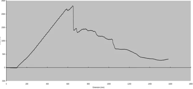
The crosshead speed (speed of loading) also needs to be set prior to loading. Molar teeth are usually loaded at 2 mm/min. [14], whereas premolar and front teeth are usually loaded at 0.5 mm/min. speed [15]. One can diverge from these numbers, as all specimen will be loaded at the same pace the difference between the groups are important, not the actual speed itself [16]. During the test a force vs. distance curve is dynamically plotted for each tooth. Failure load, which can be defined as the load at which the tooth- restoration complex exhibited the first fracture and results in a peak formation on the force versus distance curve, is recorded in
Newtons (N). In each test the specific failure load is determined when the force versus distance curve showed a sudden change in load, which indicates an abrupt decrease in the specimen's resistance to compressive loading (Figure 2.).
Figure 2. Force (load) versus distance (extension) curve
Lloyd Instruments Data Analysis Package RControl
-500 0 500 1000 1500 2000 2500
0 200 400 600 800 1000 1200 1400 1600 1800
Extension (mm)
Load (N)
This sudden drop in load does not necessarily refer to the fracture load in all cases. If the two values are not the same, then the initial failure is a much more significant and useful parameter than the fracture resistance from a clinical standpoint. This is due to the fact that once a detectable crack (indicated by the drop of load) occurs, penetration of oral fluids would lead to decay formation and a reduction in the longevity of the restoration in the oral environment. Therefore, initial failure must be detected when present.
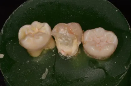
Furthermore, apart from determination of the load required for fracture, evaluation of the fracture type is important from a clinical point of view. Thus, whenever conducting a load-to-fracture test, not only the fracture resistance, but also the failure mode should be evaluated. After the mechanical testing, the fractured specimens are examined regarding their fracture patterns. According to Scotti and co-workers, distinction is made between restorable or nonrestorable fractures under optical microscope with a two- examiner agreement. Based upon their evaluation system, a restorable fracture is above the CEJ, meaning that in case of fracture the tooth can be restored, while a nonrestorable fracture extends below the CEJ and the tooth is likely to be extracted [14] (Figure 3).
Figure 3. Fracture pattern deemed unrestorable as it extends under the CEJ.
Fracture resistance and fracture pattern must be interpretted together as a restoration that shows only high fracture resistance but unfavorable fracture pattern in case of fracture cannot be deemed the best solution among other restorative materials and techniques. The ideal solution will be the one with fracture resistance not different from an intact tooth (the control group in the studies) and with dominantly favorable fracture pattern [17].
The drawback of load-to-failure tests is that the continually increasing load applied to the teeth is not typical of the type of loading that occurs in clinical conditions, in which failures occur primarily due to fatigue. Also the load at which the specimen fracture are sometimes way higher than the values registered at a normal biting event. In the front region the average biting force is between 100-300 N, this increases to 400-500 N in the premolar teeh, and further increases to 600-800 N in the molar region. Although forces range from 8 to 880 N during normal mastication, but it must be emphasized that greater loads have been described in bruxism, and teeth in this region may be exposed to extremely high forces when accidentally biting on a hard object or in trauma.
Such extreme forces cannot be completely absorbed, therefore the restorations pland and made in teeth, especially in the posterior ones, should be able to withstand much higher loading than the ones manifesting during normal biting forces and movement. Surh high forces will continue to rise with the high prevalence of temporomandibular disorders leading to bruxism in modern Western societies [18]. As pointed out by Deliperi et al., during a tooth’s lifetime, a wide range of overload events may happen, including those from bruxism, trauma (high extrinsic loads), or during dynamic loading (intrinsic chewing strokes in a small area due to a hard foreign body such as a stone or seed) [19]. Therefore load-to-fracture tests are resembling a possible traumatic event, or biting on a foreign object, or just the condition potentially present in a buxing patients mouth.
4. CONCLUSIONS
Despite its limitations, fracture testing remains a common experimental method of evaluating restorative procedures for restored teeth. In the future, load-to-fracture tests should be accompanied by dynamic loading and fatigue testing, which better mimics intraoral loading conditions.
REFERENCES
[1] Bader JD, Martin JA, Shugars DA. Incidence rates for complete cusp fracture. Community Dent Oral Epidemiol. 2001 Oct;29(5):346-53.
[2] Lassila L, Keulemans F, Säilynoja E, Vallittu PK, Garoushi S. Mechanical properties and fracture behavior of flowable fiber reinforced composite restorations. Dent Mater. 2018 Apr;34(4):598-606.
[3] Alshahrani FA, Yilmaz B, Seidt JD, McGlumphy EA, Brantley WA. A load-to-fracture and strain analysis of monolithic zirconia cantilevered frameworks. J Prosthet Dent. 2017 Dec;118(6):752-758.
[4] Sáry T, Garoushi S, Braunitzer G, Alleman D, Volom A, Fráter M. Fracture behaviour of MOD restorations reinforced by various fibre-reinforced techniques - An in vitro study. J Mech Behav Biomed Mater. 2019 Oct;98:348-356.
[5] Le Bell-Rönnlöf AM, Lassila LV, Kangasniemi I, Vallittu PK. Load-bearing capacity of human incisor restored with various fiber-reinforced composite posts. Dent Mater. 2011 Jun;27(6):e107-15.
[6] Plotino G, Buono L, Grande NM, Lamorgese V, Somma F. Fracture resistance of endodontically treated molars restored with extensive composite resin restorations. J Prosthet Dent. 2008 Mar;99(3):225-32.
[7] Fráter M, Forster A, Keresztúri M, Braunitzer G, Nagy K. In vitro fracture resistance of molar teeth restored with a short fibre-reinforced composite material. J Dent. 2014 Sep;42(9):1143-50.
[8] Hannig C, Westphal C, Becker K, Attin T. Fracture resistance of endodontically treated maxillary premolars restored with CAD/CAM ceramic inlays. J Prosthet Dent. 2005 Oct;94(4):342-9.
[9] Szabó B, Eördegh G, Szabó PB, Fráter M. In vitro fracture resistance of root amputated molar teeth restored with overlay: a pilot study. Fogorv Szle. 2017:111-116.
[10] Szabó B, Garoushi S, Braunitzer G, Szabó PB, Baráth Z, Fráter M. Fracture behavior of root- amputated teeth at different amount of periodontal support – a preliminary in vitro study. BMC Oral Health accepted manuscript.
[11] Forster A, Sáry T, Braunitzer G, Fráter M (2016) In vitro fracture resistance of endodontically treated premolar teeth restored with a direct layered fiber-reinforced composite post and core. J Adhes Sci Technol 31:1454–1466. https://doi.org/10.1080/01694243.2016.1259758
[12] Forster A, Braunitzer G, Tóth M, Szabó BP, Fráter M. In Vitro Fracture Resistance of Adhesively Restored Molar Teeth with Different MOD Cavity Dimensions. J Prosthodont. 2019 Jan;28(1):e325- e331.
[13] Soares LM, Razaghy M, Magne P. Optimization of large MOD restorations: Composite resin inlays vs. short fiber-reinforced direct restorations. Dent Mater. 2018 Apr;34(4):587-597.
[14] Scotti N, Coero Borga FA, Alovisi M, et al. Is fracture resistance of endodontically treated mandibular olars restored with indirect onlay composite restorations influenced by fiber post insertion? J Dent.
2012;40:814–820.
[15] Fráter M, Forster A, Jantyik Á, Braunitzer G, Nagy K, Grandini S. In vitro fracture resistance of premolar teeth restored with fibre-reinforced composite posts using a single or a multi-post technique.
Aust Endod J. 2017 Apr;43(1):16-22.
[16] Fráter M, Forster A, Jantyik Á, Braunitzer G, Nagy K. Fracture strength of elastic and conventional fibre-reinforced composite intraradicular posts - an in vitro pilot study. Fogorv Szle. 2015 Dec;108(4):115-9.
[17] Fráter M, Lassila L, Braunitzer G, Vallittu PK, Garoushi S. Fracture resistance and marginal gap formation of post-core restorations: influence of different fiber-reinforced composites. Clin Oral Investig. 2019 May 16.
[17] Volom A, Fráter M. Transmural fiber reinforcement in order to restore the fracture resistance of large MOD cavities – a technical report. Fogorv Szle. 2019 Sept;112(3):82-86.
[19] Deliperi S, Alleman D, Rudo D. Stress-reduced Direct Composites for the Restoration of Structurally Compromised Teeth: Fiber Design According to the "Wallpapering" Technique. Oper Dent. 2017 May/Jun;42(3):233-243.