Utilizing digital pathology applications in teaching, routine surgical pathology and in research
PhD thesis
László Fónyad, MD.
Doctoral School of Clinical Medicine Semmelweis University
Supervisor: Dr. Béla Molnár, D.Sc.
Official reviewers: Dr. Attila Doros, Ph.D., associate professor Dr. Richárd Szmola, Ph.D., assistant professor Head of the Final
Examination Committee: Dr. József Tímár, D.S.c., professor Members of the Final
Examination Committee: Dr. Pál Ákos Deák, Ph.D., assistant lecturer Dr. Simon Károly, Ph.D., departmend head
Budapest
2015
I. Introduction
Using digital still images in pathology for various purposes is increasingly popular as an easy way to archive and share medical information. The first telepathology networks to provide pathological diagnosis and consultation to remote sites used still images as well. The limitations of these telepathology networks (i.e. the lack of sufficient number and quality of images) were obvious and resulted in diagnostic errors. Later hybrid dynamic/store-and- forward telepathology systems were tested, where pathologists remotely controlled robotised microscopes. Results were impressive, though disadvantages of this solution, such as the dependence on the assistance in situ to handle the glass slides, interfered with the real success of this solution. Digital whole slides (DS) that offer dynamic access for the entire tissue section overcome the limitations imposed by static preselected microscopical frames.
As digital pathology solutions became more and more sophisticated their spread in histology teaching, routine surgical pathology and in the scientific field were unstoppable.
Utilizing DS in histopathology courses started in the United States. Pioneers in this field were pathologists at the University of Iowa. A not-for-profit Virtual Slidebox of histology and pathology was developed and all digital material were uploaded to servers that can be used by other educators and students. Course evaluations at the University of Iowa indicated that both students and faculty favor the use of virtual over glass slides. They also claim that in the long run using DS in education is less expensive than traditional microscopy.
Using digital images in routine pathology practice is related to the evolution of telemedicine. Telepathology itself is the practice of pathology at a distance, viewing images on a video monitor rather than directly through a light microscope. The first telediagnosis system connected the Massachusetts General Hospital and Logan International Airport Medical Station. In a 17-month period 1000 patient transaction were performed. Using a telemicroscope still images of peripheral blood smear and urine sediment were shared and diagnosed at the central lab of the hospital. Relevant publications specifically dealing with telepathology were published from the mid-eighties. One of the first publication gaining media attention as well described the use of telemicroscopy for the remote diagnosis of fresh frozen sections between the Armed Forces Institute of Pathology and the El Paso Hospital in Texas. Among others, pioneer in the field is Ronald Weinstein from Chicago. In 1986. his group developed a dynamic robotic microscopy system. From the early nighties validation studies were carried out to evaluate the diagnostic accuracy of the different digital pathology
applications. Dunn et al. at the Veterans Affairs Medical Center, Milwaukee implemented a regional telepathology network, in Europe Eusebi et al. started using static images for consultation. Later as DS technique became accessible validation studies reported an improved, 94-98 % diagnostic accuracy compared to the gold standard optical microscopy diagnosis. However both hardware and software tools of digital pathology developed significantly during the past decade the 2-6 % of incoherency rate does not seem to be disappear. It is a debate whether this error rate is the effect of the non standardized preanalitical processes and not the issue of digital pathology itself.
Adventages of digital microscopy in research is unquestionable. One field where using DS could give us a better understanding of diseases is the three dimensional (3D) tissue reconstruction. 3D imaging in pathology especially using DS is relatively new and fields of useful adaptations are being examined. Chronic allograft vasculopathy (CAV) is a major mechanism of graft failure of transplanted organs in humans. Morphometric analysis of coronary arteries enables the quantitation of CAV in mouse models of heart transplantation.
However, conventional histological procedures using single 2-dimensional sections limit the accuracy of CAV quantification.
II. Aims and scopes
● Introducing digital microscopy in graduate histopathology teaching in Hungary and establish an open access digital slide set for medical students.
● Validation of diagnostic accuracy using digital slides in routine histopathology, estimate the type of errors that cause misdiagnosis and to reveal any preventable errors that endanger patient safety.
● Reconstruction of the coronary system of murines in 3 dimensions using digital slides, to accurately measure the Neointimal Index (described later) to evaluate the severity of the rejection associated chronic allograft vasculopathy in transplanted mice.
III. Methods
All Mirax and Pannoramic hardware and software tools are products of 3DHISTECH Ltd., Budapest, Hungary.
III.1. Introducing digital microscopy in graduate histopathology teaching in Hungary
„Pilot study” and infrastructural developments
After a testing period lasted for almost a year, including software tests and pilot histology practices, we decided to replace all optical microscopes by computers for the 2007 academic year. We set up a digital histology lab with 40 commercially available PCs (Intel 3.06 GHz processor, 1 GB DDRII RAM, TFT 17" LCD monitor), an internal slide server (AMD Phenom 9550 Quad Core 2.2 GHz processor, 2 GB FBD DDR2 RAM) and built up the intranet (Cisco 2970G 24TS-E switch, 1000 Mbps) that connects the PCs with the teacher's laptop. Continuously upgraded versions of Mirax Viewer on the client PCs and Mirax Slide Server were installed on. Slides were scanned using Mirax Scan equipped with a Hitachi 3-chip camera and a Plan-Apochromat objective with 20× magnification, 0.465 µm/pixel resolution, 0.63 numeric aperture, 0.5× camera adapter magnification and 1×
optovar magnification.
The feedback process
We have composed a Student satisfaction questionnaire and a Tutor satisfaction questionnaire, both to be completed voluntarily. The main interest was to disclose the students'/teachers' opinion about the user friendliness of the software, the image quality of DS and how they make use of the features DS offer. We counted an overall satisfaction rate, by adding the results of every questions from the questionnaire and correlated it to a maximum possible score, naming this rate "attitude". The page load statistics of the external slide server were evaluated.
III.2. Validation of diagnostic accuracy using digital slides in routine histopathology
Participants
Seven experienced pathologists and a junior pathologist participated in the study. The seniors have been working as consultants for 13-28 years and they are all specialized in various fields of pathology. Two technicians were responsible for slide scanning, database and network management.
Case selection
306 cases (1858 slides) from 1998 to 2007 were selected from our archive. Case selection was randomised by the SNOMED-L/M codes. A notable washout period of 24 months was applied.
Hardware and software tools
Slides were scanned using MiraxScan 1.11 equipped with a Hitachi 3-chip camera, a Plan-Apochromat objective (20× magnification) and 0.5× camera adapter magnification, resulting in 0.465 µm/pixel resolution. For data management we used 3DH DataBase (3DH) software. No direct connection were built between the database application and the hospital information system. Participants used their office PCs for evaluation.
The evaluation process
Pathologists rendered microscopic descriptions and diagnosis to each case and filled a research form. After all data were available a consensus session was held, consensus diagnosis were given for the cases and the missed cases were graded according to the clinical significance of the error. A diagnostic error was defined relevant when it had therapeutic or prognostic consequence. Diagnostic uncertainty related to case complexity or poor image quality was also recorded.
III.3. Reconstruction of the coronary system of murines in 3 dimensions using digital slides
Tissue samples
Mouse tissue samples, native heart and transplanted hearts with chronic allograft vasculopathy were collected. Explanation of the procedure is detailed in the dissertation.
Sample preparation
The murine hearts were fixed in 10% formalin. Two transplanted hearts and one control heart were used, named as Sample A, B and C, respectively. Sample A, B was embedded in high melting point paraffin (58°C, Tissue-Prep, Fisher Scientific, Idaho, USA) required for automated sectioning. For sectioning the Kurabo AS-200S Automated Tissue Sectioning System (Kurabo Industries, Osaka, Japan) was used. Sample C was embedded in regular paraffin and sectioned manually. The thickness of the sections was 4µm each. The sections were hematoxylin-eosin (H&E)-stained manually in batches.
Slide digitization and 3D reconstruction
Bright field scanning was performed with Pannoramic Scan using Plan-Apochromat 20x magnification objective, a 0.63x camera adapter magnification and 1x Optovar magnification with a Hitachi HV F22CL camera, resulting in 0.369 µm/pixel resolution. For fluorescence scanning a Pannoramic Scan device with HXP 120 illuminator was used. The µCore software (Microdimensions Ltd., Münich, Germany) was used to generate 3D reconstructions. The software allows performance of virtual rotation and re-sectioning of a reconstructed sample, thus generating virtual, true cross sections of the coronary arteries.
Neointimal Index (NI - neointimal area divided by neointimal area plus luminal area multiplied by 100) was calculated on these virtual sections.
Statistical analysis
Statistical analysis was performed using IBM SPSS Statistics 20 Software Package (IBM, Inc., Armonk, NY, USA). For the correlation of the continuous variables paired- samples t-test was utilized /t(df)/. Independent samples t-test and general linear model multivariate test was used to assess difference in distribution between the groups of the continuous value groups. P-value of less than 0.05 was considered significant.
IV. Results
IV.1. Introducing digital microscopy in graduate histopathology teaching in Hungary
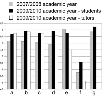
After the first year 116/268 students, after the third year 112/334 students and 8/12 tutors filled the questionnaires. The results are detailed in Figure 1. and show a great satisfaction with digital slides. There was a correlation between the teachers' and the students' attitude towards digital applications. The more the teachers were confident the better student attitude was recorded. (Data not shown.)
Figure 1. Results from the Student/Tutor satisfaction questionnaire.
Answers for all questions were scaled between 1-10, where the higher score preferred digital over conventional microscopy and 5 denoted no difference between the methods. a- How much have you liked the practices with digital slides? b- Comparing to a conventional microscope, how much have you liked working with digital slides? c- Have you found the application user-friendly? d- Comparing to a conventional microscope, how user- friendly the application was? e- How do you rate the quality of the digital slides? f- How do you rate the speed of opening a slide during practices? g- How useful the Pathonet was for preparing the examination? (1-10: not useful - very useful).(In 2007/2008 only students filled in the questionnaire.)
The increasing number of page loads from the slide server supports the results of the somehow subjective results of the queries. In average 97-98% of the students used the digital slides from home during the exam periods, In 2010. we had a record number of 10.000 page downloads in a day.
IV.2. Validation of diagnostic accuracy using digital slides in routine histopathology
Digital or optical diagnosis and the consensus diagnosis were different in 63 (20.6%) cases (discordant case). In 36 (11.7%) cases (incoherent case) the optical and in 27 (8.82%) cases (reassessed case) the digital method yielded the correct diagnoses (Table 1.).
Uncertainty due to case complexity was recorded in 48 (15.7%) cases, due to poor image quality in 15 (4.9%) cases.
Table 1. Four types of incoherency
Type of error Description n=36
Type I. non relevant incoherence - uncertainty recorded 5
Type II. non relevant incoherence - uncertainty not recorded 7
Type III. relevant incoherence - uncertainty recorded 17
Type IV. relevant incoherence - uncertainty not recorded 7
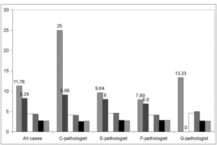
We investigated the relation between the diagnostic results and the experience of pathologist and the impact of the pathologist's competence on various fields of histopathology (Figure 2.). Excluding the non-field specific cases from each pathologists' record resulted in
~30% decrease in incoherency and in ~23% of Type-4 errors. Result of the most experienced pathologist excluding the non-field specific cases was faultless. Second in this rank with 96%
coherency was the second most experienced pathologist.
Figure 2. Importance of pathologists' competence.
Excluding the non-field specific cases from each pathologists' record resulted in better coherency. No significant differences were found in the diagnostic confidence and how the pathologists rated the quality of the slides.
IV.3. Reconstruction of the coronary system of murines in 3 dimensions using digital slides
After detecting areas of interest and creating correct virtual cross sections, the NI of the coronary arteries, spanning 0.4 mm (Sample A) and 0.8 mm (Sample B) length of artery were calculated on altogether four times twenty-eight non-corrected and corrected images (two samples, two different method, 28 different levels). The results obtained from the corrected images were statistically different from those obtained from the non-corrected images [t(55)=-5.343, p=2.0e-06]. Both conventional measurement method and reconstruction and virtual sectioning demonstrated greater NI in proximal segments of the coronary arteries relative to distal segments. Conventional measurements of both samples had neointimal indices of 86.31 ± 12.06 and 72.26 ± 10.00 for the proximal and distal parts, respectively (p=1.7e-05). On the contrary, measurements on the reoriented sections demonstrate the NI decreasing from 88.71 ± 10.28 proximally to 88.51 ± 10.03 distally (p=0.940). When considering both methods we have seen a significant difference between the measurements of the distal segment (conventional 70.62 ± 10.35 vs. reconstructed 97.80 ± 1.39) of the second sample (p<0.001). In Sample-B the examined coronary portion was cut obliquely and as we hypothesized applying the conventional method resulted in greater variability of the NI, while the reconstruction gave accurate results.
Volumes of the neointima and lumen can be calculated once 3D images of the samples are reconstructed to obtain a novel assessment of CAV (Figure 3.). Based on the original NI, we describe a new parameter: the neointimal volume index (NVI). This value is defined as neointimal volume divided by neointimal volume plus luminal volume multiplied by 100.
Figure 3.
Extracted lumen (right) and neointima (left) of a distal coronary portion from sample A.
V. Discussion
The success of digital microscope applications have brought a new era to medicine, especially to anatomic pathology, both in the field of research, education and routine practice.
The spreading of whole slide imaging or digital slide systems in pathology as an innovative technique seems to be unstoppable now.
V.1. Introducing digital microscopy in graduate histopathology teaching in Hungary
Considering the fact that the most obvious advantages of digital pathology appear in education it is not surprising that spreading of digital microscopes have started in the histopathology classrooms. Our results additionally show that digital slides could easily be integrated to histopathology education. The results emphasize the importance of the "human factor" in the efficiency of the installation of any new techniques confirmed by how the tutors' attitude towards digital slides affected the students' attitude.
In order to understand this success in pathology education we have to inspect the advantages of digital microscopes over optical microscopes.
● Simplified slide handling – instead of dealing with thousands of glass slides, applying easy-to-use digital materials.
● Standardisation of educational materials – as students have access to the exact same quality material.
● Easier orientation on slides – is crucial in the learning phase.
● Parallel visualization of slides – enables comparing different slides or different areas on the same slide.
● Placing annotations on digital slides – a feature that gives the possibility to further explain the alteration on the slide to enhance self-learning.
● Continous slide access via the internet – and the students are able to review the slides before or after practices or preparing for examinations
The growing importance of informatics in health care and the expected capability of medical doctors to handle many sources of digital materials raise further questions. Medical training programs must add informatics to their curriculum and training materials need to be
developed. In special fields of postgraduate training, such as pathology and radiology it is worth considering to organize short courses on digital imaging and photography
V.2. Validation of diagnostic accuracy using digital slides in routine histopathology
In this study the diagnostic reliability of a fully digital slide based system, comparing it with the routine conventional optical microscope procedure was evaluated. Our results are in line with previously published studies in the field, where authors reported 94-98% accuracy of digital diagnoses. Besides analyzing the results according to the origin of the samples and the types of errors we measured the effects of the pathologists' interpretative skills and experience on the diagnostic results.
According to our results one of the most important factor of the diagnostic accuracy using DS, is the pathologist's experience in a specific field. There is an increase of diagnostic accuracy signing out only field-specific cases by the pathologists. There was significant negative correlation between diagnostic confidence and individual pathologist's experience.
These results indirectly suggests that the impact of the pathologist's age is a major factor for dislike and mistrust DS, as usually more experienced the pathologists the older they are.
We found that quality of the digital slide is important to achieve the best possible diagnostic accuracy but the failure of proper scanning and expected image quality will not endanger patient safety as such errors are detectable by the examiner.
In our series in 27/306 - 8.82% - the consensus diagnoses were coherent with the digital diagnoses and overwrote the original OM-based diagnoses (reassessed case). As the difference between the ratio of these reassessed and incoherent cases is insignificant we believe this supports our assumption that diagnostic errors using digital slides are related to intrinsic (human) factors rather than technical problems.
V.3. Reconstruction of the coronary system of murines in 3 dimensions using digital slides
The techniques of 3D image reconstruction are newly introduced modalities in the fields of medical imaging and provide increased diagnostic accuracy compared with conventional 2D imaging methods, especially in the positional relationship between the lesions and surrounding structures. The field of radiology has pioneered the use of 3D reconstruction from computed tomography and magnetic resonance images, offering intuitive and informative visualizations for diagnosing disease, surgical planning, radiation oncology treatment and other clinical applications. Pathology is another field in which 3D reconstruction offers the potential to serve as a useful scientific tool, revolutionize diagnosis, and improve clinical management. Technical advances in whole slide imaging (WSI) in pathology offer high throughput, high magnification and high resolution. These technical breakthroughs permit the generation of high quality 3D reconstructions able to display the physical relationships between microscopic structures. In the recent years, with the improvement of WSI and with the design of automated serial sectioning devices, more papers are being published in this field. 3D reconstruction has been employed in the classification of lung adenocarcinomas, diagnosis of colorectal biopsies and metastasis of breast cancer to lymph nodes.
Currently, the challenges limiting the further adoption of 3D imaging of serial sections are physical artifacts from sectioning, digital artifacts resulting from the assembly and manipulation of 2D structures and their conversion to 3D image, and the time consuming and laborious process of cutting sections and digital reconstruction. These limitations require the researchers’ careful consideration on estimating costs and benefits when choosing 3D imaging of serial sections as a method to investigate scientific questions.
In the current study, we demonstrate the feasibility of applying the WSI-3D technique to the reconstruction of the coronary vasculature in mouse hearts and demonstrate the applicability of this method to the research of chronic allograft rejection and the development of CAV. We show that virtual image rotation can obtain a true vascular cross-section, reducing artifacts encountered during conventional 2-dimensional image assessments to measure NI, and therefore increasing scientific accuracy. By integrating the neointimal index along a length of vessel to obtain a “neointimal volume index” (NVI), 3D reconstruction further improves accuracy over conventional NI by eliminating sampling bias.
VI. Conclusion
● We have successfully introduced digital microscopy in graduate histopathology teaching in Hungary.
● We have established and continuously maintain an open access digital slide set for medical students.
● We have proven that failure of proper scanning and expected image quality will not endanger patient safety as such errors are detectable by the examiner.
● We found the most important factor of diagnostic accuracy is the pathologist's experiance in a specific field.
● Using digital whole slides we reconstructed the coronary vasculature of mouse hearts in 3D and demonstrated the applicability of this method to the research of allograft vasculopathy during chronic rejection.
● We described the “neointimal volume index” to measure the severity of chronic allograft vasculopathy and defined it as neointimal volume divided by neointimal volume plus luminal volume multiplied by 100.
VII. Publication list
VII. I. Publications related to the dissertation
Fónyad L, Krenács T, Nagy P, Zalatnai A, Csomor J, Sápi Z, Pápay J, Schönléber J, Diczházi C, Molnár B. (2012) Validation of diagnostic accuracy using digital slides in routine histopathology Diagn Pathol, 7:35
IF: 1,85
Fónyad L, Gerely L, Cserneky M, Molnár B, Matolcsy A. (2010) Shifting gears higher-- digital slides in graduate education--4 years experience at Semmelweis University. Diagn Pathol, 5:73
IF: 1,388
Andocs G, Renner H, Balogh L, Fonyad L, Jakab C, Szasz A. (2009) Strong synergy of heat and modulated electromagnetic field in tumor cell killing. Strahlenther Onkol, 185(2):120-6.
IF: 3,767
Cserneky M, Szende B, Fonyad L, Krenacs T. Telepathology in Hungary. Könyvfejezet:
In: Telepathology. (Ed: Kumar S). Springer-Verlag Berlin, (ISBN 978-3-540-85785-3), 2009:127-147.
IF: - (Book chapter)
Krenacs T, Zsakovics I, Micsik T, Fonyad L, Varga VS, Ficsor L, Kiszler G, Molnar B.
Digital microscopy – the upcoming revolution in histopathology teaching, diagnostics, research and quality assurance. Könyvfejezet: In Microscopy: Science, Technology, Applications and Education, (Ed. A. Méndez-Vilas and J. Díaz), Formatex Research Center, Badajoz, Spain, Volume 2 (ISBN 13: 978-84-614-6190-5), 2010:965-977.
IF: - (Book chapter)
VII. II. Other publications
Changchien YC, Tátrai P, Papp G, Sápi J, Fónyad L, Szendrői M, Pápai Z, Sápi Z. (2012) Poorly differentiated synovial sarcoma is associated with high expression of enhancer of zeste homologue 2 (EZH2). J Transl Med, 10:216.
IF: 3,474
Changchien YC, Haltrich I, Micsik T, Kiss E, Fónyad L, Papp G, Sápi Z. (2012) Gonadoblastoma: Case report of two young patients with isochromosome 12p found in the dysgerminoma overgrowth component in one case. Pathol Res Pract, 15;208(10):628-32.
IF: 1,213
Changchien YC, Katalin U, Fillinger J, Fónyad L, Papp G, Salamon F, Sápi Z. (2012) A challenging case of metastatic intra-abdominal synovial sarcoma with unusual immunophenotype and its differential diagnosis. Case Rep Pathol, 2012:786083.
IF: -
Balogh Z, Szemlaky Z, Szendroi M, Antal I, Pápai Z, Fónyad L, Papp G, Changchien YC, Sápi Z. (2011) Correlation between DNA ploidy, metaphase high-resolution comparative genomic hybridization results and clinical outcome of synovial sarcoma. Diagn Pathol, 6:107.
IF: 1,638