Repercussion of Megakaryocyte-Specific Gata1 Loss on Megakaryopoiesis and the Hematopoietic Precursor Compartment
Marjolein Meinders1, Mark Hoogenboezem2, Maaike R. Scheenstra1, Iris M. De Cuyper1, Petros Papadopoulos3, Tamás Németh4,5, Attila Mócsai4,5, Timo K. van den Berg1, Taco W. Kuijpers1,6, Laura Gutiérrez1,3*
1Dept. of Blood Cell Research, Sanquin Research and Landsteiner Laboratory, Academic Medical Centre (AMC), University of Amsterdam (UvA), Amsterdam, the Netherlands,2Dept. of Molecular Cell Biology, Sanquin Research and Landsteiner Laboratory, AMC, UvA, Amsterdam, the Netherlands,3Dept. of Hematology, Hospital Clínico San Carlos, Instituto de Investigación Sanitaria San Carlos (IdISSC), Madrid, Spain,4Dept. of Physiology, Semmelweis University School of Medicine, Budapest, Hungary,5MTA-SE
“Lendület”Inflammation Physiology Research Group of the Hungarian Academy of Sciences and
Semmelweis University, Budapest, Hungary,6Emma Children’s Hospital, Academic Medical Centre (AMC), UvA, Amsterdam, the Netherlands
*lgutierrezg@salud.madrid.org
Abstract
During hematopoiesis, transcriptional programs are essential for the commitment and differ- entiation of progenitors into the different blood lineages. GATA1 is a transcription factor expressed in several hematopoietic lineages and essential for proper erythropoiesis and megakaryopoiesis. Megakaryocyte-specific genes, such asGP1BA, are known to be directly regulated by GATA1. Mutations in GATA1 can lead to dyserythropoietic anemia and pseudogray-platelet syndrome. Selective loss of Gata1 expression in adult mice results in macrothrombocytopenia with platelet dysfunction, characterized by an excess of immature megakaryocytes. To specifically analyze the impact of Gata1 loss in mature committed megakaryocytes, we generated Gata1-Lox|Pf4-Cre mice (Gata1cKOMK). Consistent with previous findings, Gata1cKOMKmice are macrothrombocytopenic with platelet dysfunction.
Supporting this notion we demonstrate that Gata1 regulates directly the transcription of Syk, a tyrosine kinase that functions downstream of Clec2 and GPVI receptors in megakaryo- cytes and platelets. Furthermore, we show that Gata1cKOMKmice display an additional aberrant megakaryocyte differentiation stage. Interestingly, these mice present a misbal- ance of the multipotent progenitor compartment and the erythroid lineage, which translates into compensatory stress erythropoiesis and splenomegaly. Despite the severe thrombocy- topenia, Gata1cKOMKmice display a mild reduction of TPO plasma levels, and Gata1cK- OMKmegakaryocytes show a mild increase in Pf4 mRNA levels; such a misbalance might be behind the general hematopoietic defects observed, affecting locally normal TPO and Pf4 levels at hematopoietic stem cell niches.
a11111
OPEN ACCESS
Citation:Meinders M, Hoogenboezem M, Scheenstra MR, De Cuyper IM, Papadopoulos P, Németh T, et al. (2016) Repercussion of Megakaryocyte-Specific Gata1 Loss on
Megakaryopoiesis and the Hematopoietic Precursor Compartment. PLoS ONE 11(5): e0154342.
doi:10.1371/journal.pone.0154342
Editor:Kevin D Bunting, Emory University, UNITED STATES
Received:December 12, 2015 Accepted:April 11, 2016 Published:May 6, 2016
Copyright:© 2016 Meinders et al. This is an open access article distributed under the terms of the Creative Commons Attribution License, which permits unrestricted use, distribution, and reproduction in any medium, provided the original author and source are credited.
Data Availability Statement:All relevant data are within the paper.
Funding:This study was funded by the Center for Translational Molecular Medicine (CTMM,www.
ctmm.nl), project Innovative Coagulation Diagnostics (INCOAG, grant 01C-201), the Dutch Heart Foundation, a Ramón y Cajal Fellowship (RYC-2013- 12587; LG; Ministerio de Economía y Competitividad -Spain-), and an I+D Excelencia 2014 project grant (SAF2014-55231-P; LG and PP; Ministerio de Economía y Competitividad -Spain- and Fondos
Introduction
GATA1 is a critical transcription factor for the differentiation of several hematopoietic lineages [1]. It is expressed in megakaryocytes [2], primitive and definitive erythroid cells [3], eosino- phils [4], mast cells [5], dendritic cells [6] and Sertoli cells of the testis [7]. It belongs to the GATA family of zinc-finger transcription factors, which recognize the W(A/T)GATAR(A/G) DNA motif, and is located on the X-chromosome [8].
GATA1 regulates the transcription of lineage-specific genes contributing to differentiation, cell cycle regulation and survival of the hematopoietic lineages that express it [9–12]. In addi- tion, it regulates the expression of target genes either by repressing or activating their transcrip- tion, depending on the transcription factor complex where it acts [13]. As an additional dimension of regulation, timely GATA1 levels during lineage commitment and differentiation are crucial for its proper action as master transcription regulator, and reciprocal regulation with the other hematopoietic GATA factors, such as GATA2 and GATA3, during lineage com- mitment and specification needs to be properly orchestrated [14–18].
In humans, several mutations affecting the GATA1 gene have been described, which lead to a broad spectrum of defects, including X-linked thrombocytopenia (XLT), X-linked thrombo- cytopenia with thalassemia (XLTT) and transient myeloproliferative disorder (TMD) [19,20].
All disorders have in common mutations mostly located in the amino (N)-zinc finger region of GATA1, which might influence either DNA binding directly and/or the interaction with Friend of Gata1 (FOG1), leading to macrothrombocytopenia. Besides these deficiencies, patients with Down syndrome are more likely to develop an acquired,i.e. somatic,GATA1mutation leading to a truncated form of GATA1 (GATA1s). This short GATA1 isoform increases the possibility to develop acute megakaryoblastic leukemia (AMKL) [21].
Gata1 knockout mice die around day E10.5 of gestation due to severe anemia [22] and con- ditional ablation of Gata1 has shown its requirement in the erythro-megakaryocytic lineages in adult mice [23]. Mice with targeted mutations on theGata1promoter affecting its expression, such as Gata-1.05/X female mice andΔneoΔHS mice, display severe macrothrombocytopenia, an increased proliferation of megakaryocytes, and platelets with a defective response to colla- gen and vWF [2,24]. From these studies, megakaryocytic Gata1 target genes were unveiled, such asGP1BA,GP1BBandPF4[25]. However, these mouse models are not tissue/lineage spe- cific or display downregulation of Gata1 instead of complete loss.
The generation of tissue specific conditional mouse model tools,i.e. Pf4-Cre mice, makes possible the study of Gata1 loss specifically in the megakaryocytic lineage [26]. We have gener- ated megakaryocyte-specific Gata1-Lox|Pf4-Cre mice in order to study not only the direct con- sequences of Gata1 depletion in committed megakaryocytes but its repercussion on the general hematopoietic compartment. Consistent with previous findings, late ablation of Gata1 in the megakaryocytic lineage resulted in macrothrombocytopenia and platelets that are functionally impaired. Some of the defective responses in platelets were due to a defect on receptor expres- sion,i.e. integrinβ1, vWF-R. However, other defects appeared to happen downstream other receptors,i.e. Clec2 and GPVI. We have identified Syk, a pivotal tyrosine kinase that functions downstream of Clec2 and GPVI receptors in megakaryocytes and platelets, as abona fide Gata1 target. Interestingly, we describe an additional aberrant megakaryocyte differentiation stage in bone marrow and spleen. Furthermore, we show that these mice, and most likely due to the platelet production defect and concomitant stress megakaryopoiesis caused by Gata1 loss in the megakaryocytic lineage, display misbalance of their multipotent progenitor and ery- throid compartments and present with compensatory extramedullary hematopoiesis with splenomegaly.
Feder). This project was supported by the János Bolyai Research Scholarship of the Hungarian Academy of Sciences (to TN). The funders had no role in study design, data collection and analysis, decision to publish, or preparation of the manuscript.
Competing Interests:The authors have declared that no competing interests exist.
Materials and Methods Mice
Gata1-lox mice [27] were crossed with Pf4-Cre mice [26] and maintained in the Dutch Cancer Center (NKI) animal facility under specific pathogen-free conditions and following the institu- tional ethical committee guidelines.
Syk-lox animals (Syktm1.2Tara) [28], obtained from Professor Alexander Tarakhovsky (The Rockefeller University, New York, USA), were crossed to Pf4-Cre mice [26]. The genotyping was carried out by using allele-specific PCR. The efficacy and specificity of cell-specific Syk deletion was tested on cell lysates by Western blotting (data not shown). The experimental ani- mals were kept in individually sterile ventilated cages (Tecniplast) under specific pathogen-free conditions until use, when they were transferred to the conventional facility. Animal experi- ments made on these cell-specific Syk-deleted mice were approved by the Animal Experimen- tation Review Board of the Semmelweis University.
Mice were anesthetized with isofluoran, bled by heart puncture, euthanized by cervical dis- location and dissected for bone marrow and spleen collection.
Blood analysis and platelet functional assays
Blood was drawn by heart puncture and collected in heparin-coated vials (Sarstedt, Nϋm- brecht, Germany). Blood parameters were determined on a scil Vet abc Plus+. FCA was per- formed as described [29]. Platelet adhesion to collagen at physiological shear rate was performed as described [30]. In brief, whole blood was reconstituted to the same platelet con- centration amongst genotypes, and was perfused over collagen-coated slides (Horm-Collagen type I, 100μg/ml) at a shear rate of 1300s-1. After labeling platelets with CD61-FITC conju- gated antibody, photos were taken at 200x magnification with an EVOS microscope. Coverage of fluorescence was quantified with ImageJ software.
Flow cytometry
Platelets or bone marrow single cell suspensions were stained for flow cytometry analysis as previously described [30,31]. Antibodies used were: CD61-FITC, CD41-PE, Sca1-PECy7, CD34-FITC, Lin-cocktail-APC, CD16/CD32-PE, cKit-PerCP, CD71-FITC, Ter119-V405 (BD Pharmingen, Oxford, United Kingdom), Clec2–FITC (AbD Serotec, Kidlington, United King- dom), CD42a-FITC, CD42b-DL649, CD42c-FITC (Emfret, Wurzburg, Germany), GPVI-PE (R&D, Abingdon, United Kingdom), CD9-PE, CD31-PECy7 (Abcam, Cambridge United Kingdom) and CD49b-PB (BioLegend, San Diego, CA).
Mouse plasma TPO ELISA
TPO levels were measured on the plasma of Gata1cKOMKand WTloxmice with a Quantikine Mouse Thrombopoietin ELISA Kit (R&D Systems), following the manufacturer´s guidelines.
Bone marrow-derived megakaryocyte cultures
Megakaryocyte cultures were performed as previously described [30,31].
RNA
CD61+CD41+CD49b+cells were sorted from megakaryocyte cultures on a FACS ARIA (BD Bioscience) and used for RNA extraction using Trizol (Ambion, Life Technologies). cDNA was prepared from 1μg RNA with Superscript III first strand (Invitrogen). Expression levels of
mRNAs were analyzed by quantitative real-time PCR (qPCR) using SYBR green on an Applied Biosystems StepOne RT-qPCR system (Life Technologies). All reactions were performed in triplo.
Gene expression levels were calculated with the 2-ΔCTmethod [32]. Target gene expression was normalized to Gapdh expression. Primers used were:
Gata15’-CAGTCCTTTCTTCTCTCCCAC-3’and5’-GCTCCACAGTTCACACACT-3’;
PU.15’-TCTTCACCTCGCCTGTCTT-3’and5’-TCCAGTTCTCGTCCAAGCA-3’; Syk5’-TCACAACAGGAAGGCACAC-3’and5’-GAGTGGTAATGGCAGAGGTC-3’;
Pf45’-CTGCGGTGTTTCGAGGCCTCC-3’and5’-accagcgctggtgacagcaa-3’; Gapdh5’-CCTGCCAAGTATGATGACAT-3’and5’-GTCCTCAGTGTAGCCCAAG-3’.
Protein
Approximately 109platelets were lysed in 500μl RIPA buffer (150 mM NaCl, 1% NP-40, 0.5%
Deoxycolate, 0.1% SDS, 50 mM Tris HCL, pH 7.5). Proteins were separated by SDS-PAGE gel electrophoresis and transferred to PVDF membranes and incubated following standard proce- dures. Antibodies used were Anti-Gata1 (sc-265, Santa Cruz), Anti-Syk (sc-573, Santa Cruz), Anti-Gapdh (MAB374, Merck Millipore), and secondary antibodies IRDye 680 goat anti- mouse IgG and IRDye 800CW donkey anti-mouse IgG (926–32220 and 926–32212, respec- tively, LI-COR Biosciences). Western blot membranes were quantified using Odyssey LI-COR Imaging system.
Chromatin Immunoprecipitation (ChIP)
Chromatin Immunoprecipitations were performed as previously described [33] using anti- Gata1 antibody (ab11963, Abcam) and protein A magnetic beads (10002D, Life Technologies).
Enrichment of a DNA region of interest was measured by qPCR. Primers used were:
Syk GATA positive (+) amplicon:
5’-TCACAACAGGAAGGCACAC-3’and5’-GAGTGGTAATGGCAGAGGTC-3’;
Gp1ba GATA positive (+) amplicon:
5’-GCTGATAAGAGCCTTTGCC-3’and5’-GGGAGGAAATGACAACCTG-3; Cd9 GATA negative (-) amplicon:
5’-ACCGTGCTCAACTAAGTGCG-3’and5’-AGATTCCCGAGGCAAGTCTG-3’.
Statistical Analysis
We represent average and standard error of the mean (SEM) of at least three mice per genotype or experiments unless otherwise indicated. We applied two-tailed Student’st-tests to calculate statistical significance.
Results
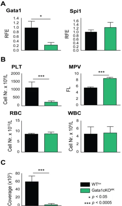
To investigate the role of Gata1 in late megakaryopoiesis, Gata1-lox|Pf4-Cre mice were gener- ated [26,27]. From now on we refer to these mice as Gata1cKOMK, and to control Gata1-lox littermates as WTlox. To quantify Gata1 reduction in Gata1cKOMKmice,Gata1mRNA levels were quantified in cultured bone marrow megakaryocytes. On average, an 80% reduction of Gata1in megakaryocytes was observed, while the levels of transcription factor PU.1 (Spi1) were not affected (Fig 1a). Consistent with previous reports [2,24,25], Gata1cKOMKmice suf- fer from severe macrothrombocytopenia, while the red and white blood cell numbers are unaf- fected (Fig 1b). Furthermore, platelets were impaired to adhere and to form thrombi over collagen-coated slides at physiological shear rate (Fig 1c).
Malfunction of Gata1cKOMKplatelets
It has been previously reported that platelets derived fromΔneoΔHS mice are dysfunctional [34], and consistently, we observed a dramatic defect of Gata1cKOMKplatelets using perfusion assay (Fig 1c). To increase knowledge on the platelet dysfunction caused by Gata1 loss in
Fig 1. Gata1cKOMKmice display severe macrothrombocytopenia and platelet dysfuncion while other blood cell counts are unaffected. (a)Gata1 and PU.1 (Spi1) mRNA expression levels in Gata1cKOMKand WTloxcultured megakaryocytes. PU.1 (Spi1) levels are normal in Gata1cKOMKmice. RFE, relative fold enrichment. (b) Blood parameters of Gata1cKOMKand WTloxmice at 8–12 weeks of age. PLT, platelets;
MPV, mean platelet volume; RBC, red blood cells; WBC, white blood cells. (c) Platelet adhesion to collagen under physiological shear rate. Whole blood from WTloxand Gata1cKOMKmice was perfused over collagen- coated slides. Adhered platelets were visualized with CD61-FITC conjugated antibody, and
immunofluorescence coverage of slides was quantified, and is represented.
doi:10.1371/journal.pone.0154342.g001
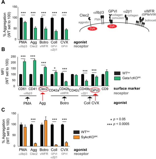
megakaryocytes in Gata1cKOMKmice, we measured platelet aggregation using FCA, which allows the study of the contribution of single receptors to the aggregation process [29]. We observed that upon stimulation with aggretin (Clec2 agonist), collagen (GPVI andα2ß1 ago- nist), convulxin (GPVI agonist), and botrocetin (GPIb/V/IX agonist), platelet aggregation responses were severely reduced, whereas responses were only mildly affected when stimulated with PMA (PKC agonist that triggersαIIbß3 integrin-dependent aggregation;Fig 2a). The reduced aggregation capacity upon stimulation with botrocetin and collagen could be
Fig 2. Functional analysis of platelets from Gata1cKOMKand SykcKOMKmice show overlapping defects.(a) Flow cytometry-based platelet aggregation assay (FCA) shows the aggregation capacity of platelets when stimulated with different agonists. Gata1cKOMKand WTloxplatelets were studied. PMA, phorbol myristate acid; Agg, aggretin; Botro.
Botrocetin; Coll, collagen; CVX, convulxin. (b) MFI of receptors expressed on Gata1cKOMKplatelets, relative expression of a given receptor in WTloxplatelets was set to 100. For clarification: CD61 (Itgb3), CD41 (Itga2b), CD42a (GPIX), CD42b (Gp1ba), CD42c (Gp1bb), CD49b (Itga2). (c) Flow cytometry-based platelet aggregation assay (FCA) shows the aggregation capacity of SykcKOMKand WTloxplatelets when stimulated with various agonists as described above.
doi:10.1371/journal.pone.0154342.g002
explained by the decreased expression of receptors/subunits, as previously reported (CD42b and CD49b;Fig 2b) [25,35,36]. In addition, the lower collagen response might also be due to a defective GPVI signaling response, as GPVI is described as the major collagen receptor [37].
Interestingly, the expression of Clec2 was not decreased, and GPVI expression was only mini- mally affected, which could therefore not explain the prominent reduction in aggregation when stimulated with aggretin, convulxin or collagen, respectively.
Since GPVI and Clec2 receptors share Syk-dependent signaling pathways [38], we next aimed at evaluating whether those responses are indeed Syk-dependent. In order to do that, we generated Syk-lox|Pf4-Cre mice. From now on we refer to these mice as SykcKOMK, and to control Syk-lox littermates as WTlox. We measured platelet aggregation using FCA as per- formed above, and observed that upon stimulation with aggretin (Clec2), collagen (GPVI and α2ß1, being GPVI the major collagen receptor) and convulxin (GPVI), platelet aggregation responses were almost absent, while the aggregation response was normal when stimulated with PMA (αIIbß3) and botrocetin (GPIb/V/IX;Fig 2c). This result corroborates the notion that Clec2 and GPVI share Syk-dependent signaling pathways [38], and confirms the overlap of Clec2- and GPVI-dependent responses with Gata1cKOMKplatelets, which prompted us to investigate whether Syk expression was affected in Gata1cKOMKmice.
Syk transcription is regulated by Gata1
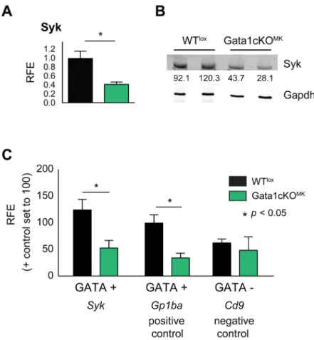
To investigate if Syk transcription is affected in the megakaryocytic lineage of Gata1cKOMK mice, mRNA and protein expression levels were measured in cultured sorted Gata1cKOMK megakaryocytes and platelets, respectively (Fig 3a and 3b). A significant reduction in both Syk mRNA and protein levels was observed, suggesting that Gata1 could regulate its expression.
We identified a WGATAR binding site600bp upstream of theSykgene start codon and per- formed Gata1 ChIP experiments on WTloxand Gata1cKOMKmegakaryocytes (as background control) to elucidate whether Gata1 interacts directly with theSykpromoter.
We took as positive control a GATA positive (+) amplicon (-300bp) at theGp1bapro- moter (known Gata1 target) and as a negative control a GATA negative (−) amplicon (-600bp) at theCd9promoter. Gata1 ChIP results show enrichment at the GATA site in the Sykpromoter in WTloxmegakaryocytes, similarly to the positive control, while it does not enrich above background at the GATA negative (−) site in theCd9promoter (Fig 3c). There- fore, this data corroborates the notion that Syk is abona fideGata1 target.
Aberrant megakaryocyte maturation in Gata1cKOMKmice
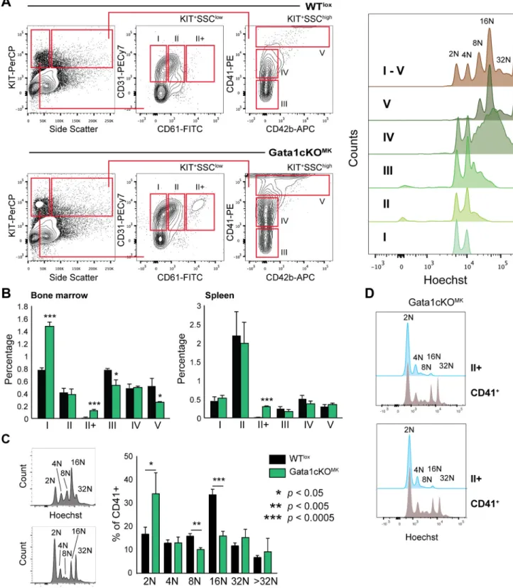
Megakaryocyte maturation was determined based on receptor expression profile in the bone marrow and the spleen. Five consecutive stages (I-V) of megakaryocyte maturation could be identified, as previously described, whereby subtype I contains the most immature and V the most mature megakaryocytes (for gating strategy seeFig 4a) [31].
To validate the differentiation status of our megakaryocyte subpopulations based on flow cytometry, the ploidy status of the individual subpopulations was analyzed. We observed a pos- itive correlation of ploidy level with maturation progression following stages I to V, which in our view validates this megakaryocyte differentiation gating strategy [31].
Compared to WTloxlittermates, and as previously reported, we identified a shift towards more immature megakaryocytes in the bone marrow,i.e. a relative increase in population I and a decrease in populations IV-V (Fig 4b). Interestingly, we identified an additional subpopula- tion exclusively in the Gata1cKOMKsamples, indicated as II+, in which megakaryocytes with low SSC characteristics express higher levels of CD61. In concordance with this, and similarly
to what has been described in other Gata1 deficient mouse models, when analyzing the ploidy status of CD41+ megakaryocytes in the bone marrow, we observed that there was an increase of 2N megakaryocytes, and the normal 16N profile was lost (Fig 4c). Furthermore, ploidy anal- ysis of the II+ population revealed that this population was composed of mainly of megakaryo- cytes at 2N and 4N ploidy level, with a low percentage of 8N-16N, similarly to what we observe in population II in WTloxsamples, supporting the notion that the II+ population identified in the Gata1cKOMKsamples are megakaryocytes at stage II that start expressing higher (timely inappropriate) levels of CD61 (Fig 4d).
In contrast to human, the mouse spleen is known to be a secondary hematopoietic organ for megakaryopoiesis and erythropoiesis, whereby the output of erythroid cells and platelets increases during stress hematopoiesis [39]. Although we did not notice a change in any of the megakaryocyte progenitor subpopulations in the spleen, we did identify the II+ subpopulation, comparable to the bone marrow (Fig 4b). However, we cannot conclude whether this aberrant megakaryocytic stage is dependent on Gata1 loss, or a consequence of the thrombocytopenia or stress megakaryopoiesis itself.
Fig 3. Gata1 regulates Syk expression. (a)Syk mRNA expression levels in Gata1cKOMKand WTlox cultured megakaryocytes measured by qRT-PCR. (b) Syk protein levels in Gata1cKOMKand WTloxplatelets, analyzed by Western blotting. Syk expression level normalized to loading control Gapdh is indicated, setting the average expression levels of Syk in WTloxplatelets to 100. (c) Chromatin immunoprecipitation (ChIP) assay showsGata1 binding to theSykpromoter in WTloxcompared to Gata1cKOMK(background control) cultured megakaryocytes. A GATA positive (+) site on the promoter of the known targetGp1ba(CD42b) was used as positive control and a GATA negative (−) site on the promoter ofCd9was used as negative control.
doi:10.1371/journal.pone.0154342.g003
Fig 4. Gata1cKOMKmice have a defect in megakaryopoiesis.(a) Gating strategy to identify megakaryocytes at consecutive stages of differentiation in the bone marrow and the spleen based on surface marker expression CD31, CD61, CD41 and CD42b. The dot plot depicts the extra population, named II+ found exclusively in Gata1cKOMKbone marrow. On the right we show the ploidy status of the individual
subpopulations, thereby justifying our gating strategy. (b) Percentage of megakaryocytes at consecutive stages of differentiation (I-V) of nucleated bone marrow and spleen cells. (c) Ploidy staining of CD41-positive bone marrow and spleen megakaryocytes. The right bar graph depicts ploidy staining of CD41+ in bone marrow. (d) Ploidy status of gated II+ megakaryocyte differentiation stage vs total CD41+ cells in two representative Gata1cKOMKbone marrow samples.
doi:10.1371/journal.pone.0154342.g004
Misbalance of multipotent progenitors in the bone marrow of Gata1cKOMKmice
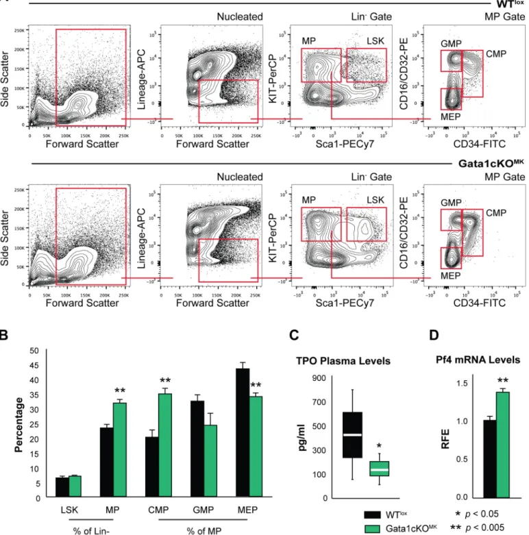
To investigate the repercussion of Gata1 deletion in megakaryocytes on the hematopoietic compartment, we analyzed the distribution of hematopoietic progenitors in the bone marrow based on receptor surface expression whereby nucleated, Lin-|Sca-1+|Kit+cells were identified as LSK and Lin-|Kit+|Sca-1-as multipotent progenitors (MP). A further subdivision of MPs was made based on CD16/CD32 and CD34 expression, whereby we could differentiate between the common myeloid progenitor (CMPs), granulocyte-macrophage progenitor (GMP) and megakaryocyte-erythroid progenitor (MEP) (seeFig 5afor gating strategy).
This revealed that Gata1cKOMKmice display a misbalance in hematopoietic progenitors,i.
e. an increase in the total number of myeloid progenitors (MP). This increase is mainly due to an increase in CMP cells at the expense of GMP and MEP cells (Fig 5b). This could be an indi- rect effect of the chronic thrombocytopenia displayed by Gata1cKOMKmice, however, we could not exclude other possibilities implicit to the genetic strategy as to affect the balance of hematopoietic progenitors in our system. Severe thrombocytopenia is accompanied by a raise in plasma thrombopoietin (TPO) levels, when the megakaryocyte-platelet mass is reduced, and it is known that excess TPO has a direct effect on hematopoietic stem cells [40]. Contrary to what could be anticipated, TPO plasma levels were mildly reduced in Gata1cKOMKas com- pared to WTloxmice (Fig 5c). This suggests that despite the severe thrombocytopenia, the megakaryocyte-platelet mass is increased, due to the overgrowth of immature megakaryocyte populations identified in the bone marrow in Gata1cKOMKas compared to WTloxmice (Fig 4b). On the other hand, it has been reported that Pf4 produced by megakaryocytes in the bone marrow niche affects hematopoietic stem cell quiescence and therefore performance [41], and since Pf4 is a Gata1 target gene [25,42], we aimed at analyzing Pf4 mRNA levels in bone mar- row derived Gata1cKOMKmegakaryocytes. As shown inFig 5d, Pf4 was significantly, although mildly, upregulated in Gata1cKOMKcultured megakaryocytes. Therefore, we can conclude that Pf4 production is not deficient in these megakaryocytes.
Twinned compensatory stress erythropoiesis that accompanies the stress megakaryopoiesis of Gata1cKOMKmice
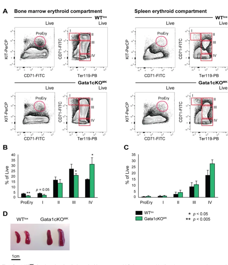
As show inFig 5, we identified alterations in the hematopoietic compartment, where we noticed and increase in CMP and a decrease in GMPs and MEPs. Although the red blood cell count in Gata1cKOMKmice was normal (Fig 1b), we were interested on the repercussion of the defective platelet production on erythroid differentiation.
Based on surface expression of CD71 and Ter119, as described by Socolovskyet al[43], four consecutive erythroid differentiation stages can be identified, whereby I is the most immature erythroid progenitor and IV the most mature erythroid progenitor; addition of KIT to the panel allows the detection of proerythroblasts, which are KIT+CD71+(for gating strategy see Fig 6a). The bone marrow erythroid compartment showed a clear shift towards more mature erythroid cells,i.e. in population IV and a decrease in the more immature erythroid popula- tions,i.e. I-III (Fig 6b). The splenic erythroid compartment did not show overt changes in the distribution of I-IV stages, although there was a tendency to an increase in mature erythroid cells (stage IV,Fig 6c). However, and although the RBC counts were normal, these mice pres- ent with splenomegaly. This suggests that there is compensatory extramedullary hematopoie- sis/erythropoiesis, probably due to the indirect exhaustion in MEPs caused by the described thrombocytopenia that suffer these mice (Fig 6d). Therefore, incapacitated megakaryopoiesis on a chronic manner (stress megakaryopoiesis) due to Gata1 loss in mature megakaryocytes,
Fig 5. Gata1cKOMKmice have a defect in the hematopoietic early precursor compartment(a) Flow cytometry analysis of the stem cell and committed progenitor compartment. LSK, Lin-|Sca-1+|Kit+cells; MP (Lin-|Sca-1-|Kit+), multipotent progenitors; CMP (MP gate—CD34+|CD16/
CD32mid), common myeloid progenitor; GMP (MP gate—CD34-|CD16/CD32+), granulocyte-monocyte progenitor; MEP (MP gate—CD34-|CD16/
CD32-), megakaryocyte-erythroid progenitor. (b) Percentage of the different hematopoietic progenitors. Absolute cell number of bone marrow megakaryocytes at consecutive stages of differentiation [31]. The dot plot depicts the extra population, named II+ found exclusively in Gata1cKOMK bone marrow. (c) Whisker/Box plot depicts plasma TPO levels from Gata1cKOMKand WTloxblood samples, as measured by ELISA. At least 5 mice were analyzed per genotype. (d) qPCR analysis of Pf4 mRNA expression levels in cultured bone marrow derived Gata1cKOMKand WTlox
megakaryocytes.
doi:10.1371/journal.pone.0154342.g005
Fig 6. Gata1cKOMKmice show alterations in the erythroid compartment.(a) Gating strategy to identify erythrocytes at consecutive stages of differentiation in the bone marrow and the spleen based on surface marker expression KIT, CD71 and Ter119. (b) Percentage of reticulocytes at consecutive stages of differentiation of live cells. Left graph depicts the bone marrow compartment, right the splenic compartment. (c) Photograph of representative spleens from Gata1cKOMKand control mice shows the splenomegaly that Gata1cKOMKdevelop.
doi:10.1371/journal.pone.0154342.g006
influences the early hematopoietic and erythroid compartments inducing compensatory extra- medullary hematopoiesis/erythropoiesis.
Discussion
Gata1 is a key transcription factor involved in both erythroid and megakaryocyte development and essential for terminal differentiation of these lineages. Since Gata1 knockout mice are lethal due to severe anemia [22], differentGata1targeted mouse models were generated to investigate the role of Gata1 in adult mice, includingΔneoΔHS [2] and Gata1.05 mice [24]. Although these models were fundamental in identifying the critical role of Gata1 in megakaryopoiesis, they are not restricted to the megakaryocytic lineage or they pose a model for the downregula- tion of the expression levels of Gata1 without gene ablation. Therefore, we generated Gata1- lox|Pf4-Cre mice, since Pf4-Cre are a generally accepted Cre mouse model to induce megakar- yocyte/platelet specific gene recombination [26], to investigate the function of Gata1 in mature megakaryocytes. Previous reports have shown that this system could have some leakage in a minor population of circulating leukocytes, a subset of monocytes and macrophages and in epi- thelial cells of the colon; however, Gata1 is scarcely expressed in these cells, thus we exclude major effects from recombination of Gata1 in these lineages [44,45].
Comparable to theΔneoΔHS and Gata1.05 mouse models, Gata1cKOMKmice show severe macrothrombocytopenia accompanied by platelet dysfunction, while in Gata1cKOMKmice the red blood cells and white blood cell counts are unaffected. Our platelet functionality tests showed that our mouse model has similar platelet defects shown in previous Gata1 deficient mouse models, including a defect in aggregation when platelets are stimulated with botrocetin and collagen. This aggregation defect could be explained by the defective expression of respec- tive receptors/subunits (i.e. vWF-R and CD49b). Besides these known platelet defects we show that platelets from Gata1cKOMKmice have an aggregation defect when stimulated with aggre- tin and convulxin. Since the defect could not be explained by downregulation of Clec2 or GPVI receptor expression (or at least considering the extent of downregulation) on these platelets, we next aimed at studying whether a common signaling protein, Syk, could be involved in the lower response. In fact, FCA performed on SykcKOMKplatelets confirmed an essential role of Syk kinase on the platelet aggregation responses induced by aggretin, convulxin and collagen, but not on those induced by PMA or botrocetin. In addition, pre-treatment of platelets from WT animals with a Syk kinase inhibitor prior platelet aggregation induction results in the same FCA profile when using the same battery of agonists,i.e. defective aggretin, convulxin and col- lagen responses, normal PMA and botrocetin responses [29]. Syk, is essential during megakar- yocyte migration and spreading and involved in platelet aggregation, and thus essential for normal megakaryocyte function [46]. We therefore found it of relevance to study whether Gata1 could be directly involved in the transcriptional regulation of Syk.
A reduced expression of Syk was noticed in mRNA of megakaryocytes and protein extracts of platelets from Gata1cKOMKmice and we found that Gata1 directly regulates the transcrip- tion of Syk by employing ChIP assays. These data support the notion that Syk is a direct Gata1 target in megakaryocytes and explain the GPVI/Clec2 dependent aggregation defects shown in Gata1cKOMKplatelets. The lowered, but not complete absence of this protein could be explained by the fact that Gata1 recombination occurs in mature megakaryocytes, and we can- not exclude the possibility that combinatorial interactions between several transcription factors during megakaryopoiesis allows for basal transcription [12,15,18].
The severe thrombocytopenia caused a mild shift in megakaryocyte maturation in the bone marrow compartment, which could be explained by the increased demand in output of plate- lets. Interesting, however, is the new II+ population we identified in both the bone marrow and
the splenic compartment. This SSClowCD61highpopulation could be a consequence of the increased pressure on megakaryocyte maturation. Of note, although the ploidy profile of this II+ megakaryocyte population in Gata1cKOMKmice resembles mostly the WTloxII megakar- yocyte ploidy profile, we have to consider that the 8N in the Gata1cKOMKmegakaryocytes dis- plays a characteristic double peak, which does not appear in WTloxsamples.
Analysis of the hematopoietic precursor compartment revealed a shift in total number of MPs, CMPs, GMPs and MEPs. The shift could potentially be explained by the severe and chronic thrombocytopenia suffered by Gata1cKOMKmice; still we could not discard Gata1-de- pendent or -independent alterations in relevant factors such as Pf4 or TPO to be causative of the misbalance observed at the progenitor level. Thrombocytopenia is normally accompanied by raised TPO levels (when the megakaryocyte-platelet mass is reduced), and it has been previ- ously reported that besides a direct effect of TPO on megakaryocyte proliferation and matura- tion, TPO also influences directly hematopoietic stem cell (HSC) quiescence and cycling [40, 47]. Contrary to what could be anticipated, TPO levels were mildly lower in Gata1cKOMK mice. This supports the notion that despite the severe thrombocytopenia, the megakaryocyte- platelet mass is increased due to the overgrowth of immature megakaryocytes. In concordance with these results, it has been previously reported that Gata1-low mice display normal TPO levels [48]. Reduction of MEPs could be a consequence of a higher demand of platelets, which leads to exhaustion of the most committed progenitors towards the demanded lineage, through TPO stimulation. In addition, we examined mRNA levels of Gata1-target Pf4 in bone marrow derived cultured megakaryocytes, as it has been recently reported the important and local role of this megakaryocyte-secreted factor on HSC senescence/performance [41]. Pf4 levels were increased in Gata1cKOMKmegakaryocytes. Interestingly, megakaryocytes from Gata1-low mice have increased Pf4 levels [49], profile that is reversed (Gata1 increases and Pf4 decreases) in Gata1-low mice treated with a TGFβinhibitor. Furthermore, Gata1 timely represses Pf4 in immature megakaryocytes in cooperation with Eto2 [42]. It is important to acknowledge the complexity of Gata1 transcription regulation, which includes its participation in activating as well as repressing transcription factor complexes, as it has been well characterized in the ery- throid lineage [13,50].
A closer look at the erythroid compartment, revealed a change in erythroid maturation in the bone marrow and the spleen, and splenomegaly. Both organs showed an increased percent- age of mature erythroid cells, which together with the splenomegaly, suggest compensatory stress erythropoiesis or extramedullary hematopoiesis/erythropoiesis that develops as a direct consequence of the (chronic) thrombocytopenia.
In summary, Gata1 loss in the megakaryocytic lineage leads to -chronic- thrombocytopenia and dysfunctional platelets. The platelet dysfunction is a direct consequence of Gata1 loss, since several receptors and signaling molecules arebona fideGata1 targets, amongst which, Syk is demonstrated in the present manuscript. Megakaryopoiesis in these mice do not follow the normal paths, and Gata1 null megakaryocytes seem to proliferate rather than differentiate, as has been previously suggested. In the present manuscript we identify an aberrant megakar- yocyte differentiation stage, misbalance of hematopoietic precursor progenitors and extrame- dullary hematopoiesis. We think that these latter events are a consequence of the -chronic- thrombocytopenia, rather thanGata1gene recombination in the megakaryocytic lineage itself.
However, whether these events occur only in the context of Gata1 loss or are merely due to the severe prolonged thrombocytopenia (or both), needs to be further investigated. In particular, whether a local recruitment of TPO or overproduction of Pf4 by immature megakaryocytes would exert locally an effect on hematopoietic stem cells at the microenvironment level, remains to be elucidated.
Acknowledgments
This study was funded by the Center for Translational Molecular Medicine (CTMM,www.
ctmm.nl), project Innovative Coagulation Diagnostics (INCOAG, grant 01C-201), and the Dutch Heart Foundation, a Ramón y Cajal Fellowship (RYC-2013-12587; LG; Ministerio de Economía y Competitividad -Spain-) and an I+D Excelencia 2014 project grant (SAF2014- 55231-P; LG and PP; Ministerio de Economía y Competitividad -Spain- and Fondos Feder).
This project was supported by the János Bolyai Research Scholarship of the Hungarian Acad- emy of Sciences (to TN). The funders had no role in study design, data collection and analysis, decision to publish, or preparation of the manuscript. We thank Edina Simon for expert tech- nical assistance and Gábor Bánhegyi for access to the BD FACSCalibur flow cytometer. We thank Dr. Dulcenombre Gómez Garré for access to the ELISA microplate reader. We thank Prof. J. Eble for kindly providing aggretin.
Author Contributions
Conceived and designed the experiments: MM LG TN AM. Performed the experiments: MM MH MRS LG IMDC PP TN AM. Analyzed the data: MM LG. Contributed reagents/materials/
analysis tools: TN AM. Wrote the paper: MM LG. Participated in discussions and reviewed the manuscript: MM LG TKvdB TWK IMDC PP TN AM.
References
1. Crispino JD. GATA1 in normal and malignant hematopoiesis. Semin Cell Dev Biol. 2005; 16(1):137–47.
doi:10.1016/j.semcdb.2004.11.002PMID:15659348.
2. Shivdasani RA, Fujiwara Y, McDevitt MA, Orkin SH. A lineage-selective knockout establishes the criti- cal role of transcription factor GATA-1 in megakaryocyte growth and platelet development. EMBO J.
1997; 16(13):3965–73. doi:10.1093/emboj/16.13.3965PMID:9233806; PubMed Central PMCID:
PMC1170020.
3. Simon MC. Transcription factor GATA-1 and erythroid development. Proc Soc Exp Biol Med. 1993; 202 (2):115–21. PMID:8424101.
4. Yu C, Cantor AB, Yang H, Browne C, Wells RA, Fujiwara Y, et al. Targeted deletion of a high-affinity GATA-binding site in the GATA-1 promoter leads to selective loss of the eosinophil lineage in vivo. J Exp Med. 2002; 195(11):1387–95. PMID:12045237; PubMed Central PMCID: PMC2193547.
5. Harigae H, Takahashi S, Suwabe N, Ohtsu H, Gu L, Yang Z, et al. Differential roles of GATA-1 and GATA-2 in growth and differentiation of mast cells. Genes Cells. 1998; 3(1):39–50. PMID:9581981.
6. Gutierrez L, Nikolic T, van Dijk TB, Hammad H, Vos N, Willart M, et al. Gata1 regulates dendritic-cell development and survival. Blood. 2007; 110(6):1933–41. doi:10.1182/blood-2006-09-048322PMID:
17505015; PubMed Central PMCID: PMC1975828.
7. Yomogida K, Ohtani H, Harigae H, Ito E, Nishimune Y, Engel JD, et al. Developmental stage- and sper- matogenic cycle-specific expression of transcription factor GATA-1 in mouse Sertoli cells. Develop- ment. 1994; 120(7):1759–66. PMID:7924983.
8. Ko LJ, Engel JD. DNA-binding specificities of the GATA transcription factor family. Mol Cell Biol. 1993;
13(7):4011–22. PMID:8321208; PubMed Central PMCID: PMC359950.
9. Weiss MJ, Orkin SH. Transcription factor GATA-1 permits survival and maturation of erythroid precur- sors by preventing apoptosis. Proc Natl Acad Sci U S A. 1995; 92(21):9623–7. PMID:7568185;
PubMed Central PMCID: PMC40854.
10. Dubart A, Romeo PH, Vainchenker W, Dumenil D. Constitutive expression of GATA-1 interferes with the cell-cycle regulation. Blood. 1996; 87(9):3711–21. PMID:8611696.
11. Whyatt DJ, Karis A, Harkes IC, Verkerk A, Gillemans N, Elefanty AG, et al. The level of the tissue-spe- cific factor GATA-1 affects the cell-cycle machinery. Genes Funct. 1997; 1(1):11–24. PMID:9680325.
12. Aird WC, Parvin JD, Sharp PA, Rosenberg RD. The interaction of GATA-binding proteins and basal transcription factors with GATA box-containing core promoters. A model of tissue-specific gene expres- sion. J Biol Chem. 1994; 269(2):883–9. PMID:8288642.
13. Rodriguez P, Bonte E, Krijgsveld J, Kolodziej KE, Guyot B, Heck AJ, et al. GATA-1 forms distinct acti- vating and repressive complexes in erythroid cells. EMBO J. 2005; 24(13):2354–66. doi:10.1038/sj.
emboj.7600702PMID:15920471; PubMed Central PMCID: PMC1173143.
14. Ferreira R, Ohneda K, Yamamoto M, Philipsen S. GATA1 function, a paradigm for transcription factors in hematopoiesis. Mol Cell Biol. 2005; 25(4):1215–27. doi:10.1128/MCB.25.4.1215-1227.2005PMID:
15684376; PubMed Central PMCID: PMC548021.
15. Ferreira R, Wai A, Shimizu R, Gillemans N, Rottier R, von Lindern M, et al. Dynamic regulation of Gata factor levels is more important than their identity. Blood. 2007; 109(12):5481–90. doi:10.1182/blood- 2006-11-060491PMID:17327407.
16. Weiss MJ, Orkin SH. GATA transcription factors: key regulators of hematopoiesis. Exp Hematol. 1995;
23(2):99–107. PMID:7828675.
17. McDevitt MA, Shivdasani RA, Fujiwara Y, Yang H, Orkin SH. A "knockdown" mutation created by cis- element gene targeting reveals the dependence of erythroid cell maturation on the level of transcription factor GATA-1. Proc Natl Acad Sci U S A. 1997; 94(13):6781–5. PMID:9192642; PubMed Central PMCID: PMC21235.
18. Pimkin M, Kossenkov AV, Mishra T, Morrissey CS, Wu W, Keller CA, et al. Divergent functions of hematopoietic transcription factors in lineage priming and differentiation during erythro-megakaryopoi- esis. Genome Res. 2014; 24(12):1932–44. doi:10.1101/gr.164178.113PMID:25319996; PubMed Central PMCID: PMC4248311.
19. Freson K, Devriendt K, Matthijs G, Van Hoof A, De Vos R, Thys C, et al. Platelet characteristics in patients with X-linked macrothrombocytopenia because of a novel GATA1 mutation. Blood. 2001; 98(1):85–92.
PMID:11418466.
20. Greene ME, Mundschau G, Wechsler J, McDevitt M, Gamis A, Karp J, et al. Mutations in GATA1 in both transient myeloproliferative disorder and acute megakaryoblastic leukemia of Down syndrome.
Blood Cells Mol Dis. 2003; 31(3):351–6. PMID:14636651.
21. Wechsler J, Greene M, McDevitt MA, Anastasi J, Karp JE, Le Beau MM, et al. Acquired mutations in GATA1 in the megakaryoblastic leukemia of Down syndrome. Nat Genet. 2002; 32(1):148–52. doi:10.
1038/ng955PMID:12172547.
22. Fujiwara Y, Browne CP, Cunniff K, Goff SC, Orkin SH. Arrested development of embryonic red cell pre- cursors in mouse embryos lacking transcription factor GATA-1. Proc Natl Acad Sci U S A. 1996; 93 (22):12355–8. PMID:8901585; PubMed Central PMCID: PMC37995.
23. Gutierrez L, Tsukamoto S, Suzuki M, Yamamoto-Mukai H, Yamamoto M, Philipsen S, et al. Ablation of Gata1 in adult mice results in aplastic crisis, revealing its essential role in steady-state and stress eryth- ropoiesis. Blood. 2008; 111(8):4375–85. doi:10.1182/blood-2007-09-115121PMID:18258797.
24. Takahashi S, Komeno T, Suwabe N, Yoh K, Nakajima O, Nishimura S, et al. Role of GATA-1 in prolifer- ation and differentiation of definitive erythroid and megakaryocytic cells in vivo. Blood. 1998; 92 (2):434–42. PMID:9657742.
25. Vyas P, Ault K, Jackson CW, Orkin SH, Shivdasani RA. Consequences of GATA-1 deficiency in mega- karyocytes and platelets. Blood. 1999; 93(9):2867–75. PMID:10216081.
26. Tiedt R, Schomber T, Hao-Shen H, Skoda RC. Pf4-Cre transgenic mice allow the generation of line- age-restricted gene knockouts for studying megakaryocyte and platelet function in vivo. Blood. 2007;
109(4):1503–6. doi:10.1182/blood-2006-04-020362PMID:17032923.
27. Lindeboom F, Gillemans N, Karis A, Jaegle M, Meijer D, Grosveld F, et al. A tissue-specific knockout reveals that Gata1 is not essential for Sertoli cell function in the mouse. Nucleic Acids Res. 2003; 31 (18):5405–12. PMID:12954777; PubMed Central PMCID: PMC203309.
28. Saijo K, Schmedt C, Su IH, Karasuyama H, Lowell CA, Reth M, et al. Essential role of Src-family protein tyrosine kinases in NF-kappaB activation during B cell development. Nat Immunol. 2003; 4(3):274–9.
doi:10.1038/ni893PMID:12563261.
29. De Cuyper IM, Meinders M, van de Vijver E, de Korte D, Porcelijn L, de Haas M, et al. A novel flow cytometry-based platelet aggregation assay. Blood. 2013; 121(10):e70–80. doi:10.1182/blood-2012- 06-437723PMID:23303822.
30. Drabek K, Gutierrez L, Vermeij M, Clapes T, Patel SR, Boisset JC, et al. The microtubule plus-end tracking protein CLASP2 is required for hematopoiesis and hematopoietic stem cell maintenance. Cell Rep. 2012; 2(4):781–8. doi:10.1016/j.celrep.2012.08.040PMID:23084744.
31. Meinders M, Kulu DI, van de Werken HJ, Hoogenboezem M, Janssen H, Brouwer RW, et al. Sp1/Sp3 transcription factors regulate hallmarks of megakaryocyte maturation and platelet formation and func- tion. Blood. 2015; 125(12):1957–67. doi:10.1182/blood-2014-08-593343PMID:25538045.
32. Schmittgen TD, Livak KJ. Analyzing real-time PCR data by the comparative C(T) method. Nat Protoc.
2008; 3(6):1101–8. PMID:18546601.
33. Borg J, Papadopoulos P, Georgitsi M, Gutierrez L, Grech G, Fanis P, et al. Haploinsufficiency for the erythroid transcription factor KLF1 causes hereditary persistence of fetal hemoglobin. Nat Genet. 2010;
42(9):801–5. doi:10.1038/ng.630PMID:20676099; PubMed Central PMCID: PMC2930131.
34. Hughan SC, Senis Y, Best D, Thomas A, Frampton J, Vyas P, et al. Selective impairment of platelet activation to collagen in the absence of GATA1. Blood. 2005; 105(11):4369–76. doi:10.1182/blood- 2004-10-4098PMID:15701726.
35. Hashimoto Y, Ware J. Identification of essential GATA and Ets binding motifs within the promoter of the platelet glycoprotein Ib alpha gene. J Biol Chem. 1995; 270(41):24532–9. PMID:7592671.
36. Zutter MM, Santoro SA, Painter AS, Tsung YL, Gafford A. The human alpha 2 integrin gene promoter.
Identification of positive and negative regulatory elements important for cell-type and developmentally restricted gene expression. J Biol Chem. 1994; 269(1):463–9. PMID:8276836.
37. Kehrel B, Wierwille S, Clemetson KJ, Anders O, Steiner M, Knight CG, et al. Glycoprotein VI is a major collagen receptor for platelet activation: it recognizes the platelet-activating quaternary structure of col- lagen, whereas CD36, glycoprotein IIb/IIIa, and von Willebrand factor do not. Blood. 1998; 91(2):491– 9. PMID:9427702.
38. Watson SP, Herbert JM, Pollitt AY. GPVI and CLEC-2 in hemostasis and vascular integrity. J Thromb Haemost. 2010; 8(7):1456–67. doi:10.1111/j.1538-7836.2010.03875.xPMID:20345705.
39. Brodsky I, Dennis LH, Kahn SB, Brady LW. Normal mouse erythropoiesis. I. The role of the spleen in mouse erythropoiesis. Cancer Res. 1966; 26(2):198–201. PMID:5903171.
40. de Graaf CA, Metcalf D. Thrombopoietin and hematopoietic stem cells. Cell Cycle. 2011; 10(10):1582– 9. PMID:21478671; PubMed Central PMCID: PMC3127159.
41. Bruns I, Lucas D, Pinho S, Ahmed J, Lambert MP, Kunisaki Y, et al. Megakaryocytes regulate hemato- poietic stem cell quiescence through CXCL4 secretion. Nat Med. 2014; 20(11):1315–20. doi:10.1038/
nm.3707PMID:25326802; PubMed Central PMCID: PMC4258871.
42. Hamlett I, Draper J, Strouboulis J, Iborra F, Porcher C, Vyas P. Characterization of megakaryocyte GATA1-interacting proteins: the corepressor ETO2 and GATA1 interact to regulate terminal megakar- yocyte maturation. Blood. 2008; 112(7):2738–49. doi:10.1182/blood-2008-03-146605PMID:
18625887; PubMed Central PMCID: PMC2556610.
43. Socolovsky M, Nam H, Fleming MD, Haase VH, Brugnara C, Lodish HF. Ineffective erythropoiesis in Stat5a(-/-)5b(-/-) mice due to decreased survival of early erythroblasts. Blood. 2001; 98(12):3261–73.
PMID:11719363.
44. Calaminus SD, Guitart AV, Sinclair A, Schachtner H, Watson SP, Holyoake TL, et al. Lineage tracing of Pf4-Cre marks hematopoietic stem cells and their progeny. PLoS One. 2012; 7(12):e51361. doi:10.
1371/journal.pone.0051361PMID:23300543; PubMed Central PMCID: PMC3531453.
45. Pertuy F, Aguilar A, Strassel C, Eckly A, Freund JN, Duluc I, et al. Broader expression of the mouse platelet factor 4-cre transgene beyond the megakaryocyte lineage. J Thromb Haemost. 2015; 13 (1):115–25. doi:10.1111/jth.12784PMID:25393502.
46. Mazharian A, Thomas SG, Dhanjal TS, Buckley CD, Watson SP. Critical role of Src-Syk-PLC{gamma}
2 signaling in megakaryocyte migration and thrombopoiesis. Blood. 2010; 116(5):793–800. doi:10.
1182/blood-2010-03-275990PMID:20457868.
47. de Graaf CA, Kauppi M, Baldwin T, Hyland CD, Metcalf D, Willson TA, et al. Regulation of hematopoi- etic stem cells by their mature progeny. Proc Natl Acad Sci U S A. 2010; 107(50):21689–94. doi:10.
1073/pnas.1016166108PMID:21115812; PubMed Central PMCID: PMC3003054.
48. Vannucchi AM, Bianchi L, Paoletti F, Pancrazzi A, Torre E, Nishikawa M, et al. A pathobiologic pathway linking thrombopoietin, GATA-1, and TGF-beta1 in the development of myelofibrosis. Blood. 2005; 105 (9):3493–501. doi:10.1182/blood-2004-04-1320PMID:15665119.
49. Zingariello M, Martelli F, Ciaffoni F, Masiello F, Ghinassi B, D'Amore E, et al. Characterization of the TGF-beta1 signaling abnormalities in the Gata1low mouse model of myelofibrosis. Blood. 2013; 121 (17):3345–63. doi:10.1182/blood-2012-06-439661PMID:23462118; PubMed Central PMCID:
PMC3637011.
50. Schuh AH, Tipping AJ, Clark AJ, Hamlett I, Guyot B, Iborra FJ, et al. ETO-2 associates with SCL in ery- throid cells and megakaryocytes and provides repressor functions in erythropoiesis. Mol Cell Biol.
2005; 25(23):10235–50. doi:10.1128/MCB.25.23.10235-10250.2005PMID:16287841; PubMed Cen- tral PMCID: PMC1291220.
