Article
A Candida parapsilosis Overexpression Collection Reveals Genes Required for Pathogenesis
Sára E. Pál1, Renáta Tóth1 , Joshua D. Nosanchuk2 , Csaba Vágvölgyi1 , Tibor Németh1,†
and Attila Gácser1,3,*,†
Citation: Pál, S.E.; Tóth, R.;
Nosanchuk, J.D.; Vágvölgyi, C.;
Németh, T.; Gácser, A. ACandida parapsilosisOverexpression Collection Reveals Genes Required for Pathogenesis.J. Fungi2021,7, 97.
https://doi.org/10.3390/jof7020097
Academic Editors: Thomas Lehrnbecher and Hector M. Mora-Montes Received: 4 December 2020 Accepted: 25 January 2021 Published: 29 January 2021
Publisher’s Note:MDPI stays neutral with regard to jurisdictional claims in published maps and institutional affil- iations.
Copyright: © 2021 by the authors.
Licensee MDPI, Basel, Switzerland.
This article is an open access article distributed under the terms and conditions of the Creative Commons Attribution (CC BY) license (https://
creativecommons.org/licenses/by/
4.0/).
1 Department of Microbiology, University of Szeged, Közép Fasor, 6726 Szeged, Hungary;
palsara0713@gmail.com (S.E.P.); renatatth@gmail.com (R.T.); csaba@bio.u-szeged.hu (C.V.);
narvaltm@gmail.com (T.N.)
2 Departments of Medicine and Microbiology and Immunology, Albert Einstein College of Medicine, New York, NY 10461, USA; josh.nosanchuk@einsteinmed.org
3 MTA-SZTE Lendület Mycobiome Research Group, University of Szeged, 6726 Szeged, Hungary
* Correspondence: gacsera@bio.u-szeged.hu
† T.N. and A.G. contributed equally to this work.
Abstract: Relative to the vast data regarding the virulence mechanisms ofCandida albicans, there is limited knowledge on the emerging opportunistic human pathogenCandida parapsilosis. The aim of this study was to generate and characterize an overexpression mutant collection to identify and explore virulence factors inC. parapsilosis. With the obtained mutants, we investigated stress tolerance, morphology switch, biofilm formation, phagocytosis, and in vivo virulence inGalleria mel- lonellalarvae and mouse models. In order to evaluate the results, we compared the data from the C. parapsilosisoverexpression collection analysis to the results derived from previous deletion mutant library characterizations. Of the 37 overexpressionC. parapsilosismutants, we identified eight with altered phenotypes compared to the controls. This work is the first report to identifyCPAR2_107240, CPAR2_108840,CPAR2_302400,CPAR2_406400, andCPAR2_602820as contributors toC. parapsilosis virulence by regulating functions associated with host-pathogen interactions and biofilm forma- tion. Our findings also confirmed the role ofCPAR2_109520,CPAR2_200040,andCPAR2_500180in pathogenesis. This study was the first attempt to use an overexpression strategy to systematically assess gene function inC. parapsilosis, and our results demonstrate that this approach is effective for such investigations.
Keywords:Candida parapsilosis; gene overexpression; mutant collection; virulence factors
1. Introduction
Candidaspecies are responsible for the vast majority of systemic fungal nosocomial infection cases. Numerous studies from different parts of the world report increasing rates of systemic candidiasis [1–4]. Non-albicans Candida(NAC) species such asC. glabrata, C. parapsilosis,C. krusei,C. auris, andC. tropicalishave significantly contributed to this continuous rise in the number of candidiasis cases, which highlights the importance of these emerging species [5–7]. Remarkable changes in medical practice that subvert host effector responses, such as the robust use of immunosuppressive therapies, can also affect the epidemiology of a pathogenic species and increase the incidence of fungal infections [8–10].
There is an enormous amount of data available in the literature on the virulence mech- anisms ofC. albicans, while our knowledge lags behind for emerging NAC opportunistic human pathogens, such asC. parapsilosis[11]. AlthoughC. parapsilosisis a commensal yeast of the normal human skin, it can, in contrast to the strict association ofC. albicanswith humans, also survive in diverse environments.C. parapsilosis’increasing role in low birth weight neonatal infections highlights the importance of performing extensive surveys of its physiology and virulence factors [12–15].C. parapsilosisis able to form pseudohypha,
J. Fungi2021,7, 97. https://doi.org/10.3390/jof7020097 https://www.mdpi.com/journal/jof
but not true hypha, and its elevated tolerance to echinocandins are additional specific attributions with clinical relevance [16,17]. So far, several groups have explored specific gene functions inC. parapsilosis[18–20]. The first genome sequence of aC. parapsilosisstrain (CDC317) was published in 2009 [21]. Later, three more strains derived from human and environmental sources were also sequenced and comprehensively analyzed, enabling fur- ther investigations of large scale targeted genomic alterations in this species [21,22]. These and other studies also revealed genomic and phenotypic alterations betweenC. albicans andC. parapsilosis, suggesting that these two pathogenic species have evolved different strategies to invade the host [23,24]. According to the Candida Genome Database (CGD) only 1.35% of the protein coding genes ofC. parapsilosishave been verified to date, while this number is 27.9% in the case ofC. albicans[25,26]. Taking into account the unique and distinctive interspecies characteristics ofC. parapsilosisin theCandidaclade, further investi- gation is urgently needed to reveal factors associated with the pathobiological mechanisms of the species.
The conventional method to reveal gene function is the deletion of a given gene and observation of the phenotype of the knock out (KO) mutant. Comprehensive KO mutant libraries and their characterizations are accessible for many organisms, including C. parapsilosis[27–30]. Pathogenesis associated gene functions such as biofilm formation, phenotypic profile, and transition or genes responsible for drug resistance or transcriptional regulator networks have also been identified in bothC. albicansandC. parapsilosisusing KO methods. Even in silico gene deletion for drug development has been carried out with C. albicans[31]. This approach was aided by the rapid evolution of the molecular tech- niques replacing the laboriousCaSAT1flipper, or the double auxotrophy complementation approaches with CRISPR/Cas9 system that simplified the generation of loss of function mutations [18,28,32–35]. However, in some cases, the resulting phenotype achieved by gene deletion has been uninformative, or the KO approach has failed, especially when essential genes are in question. Furthermore, in diploid organisms deletion of both alleles for the targeted gene is required. The artificial overexpression (OE) of a gene can circumvent the aforementioned drawbacks of the KO method. In addition to this, an OE strategy can be utilized to map regulatory rate-limiting steps in gene interaction studies [36]. Systematic library analysis as well as specific gene examination studies have been published using OE mutant strains in many organisms [37–41]. Genes involved in biological processes associated with fungal pathogenesis, such as biofilm formation, invasive hyphal growth or drug resistance, have also been successfully identified using this approach inC. albicans and otherCandidaspecies [42–44]. Moreover, the OE approach can also complement the KO library screens to explore and validate the function of a gene [36,45–48].
Although extensive studies in gene OE inSaccharomyces cerevisiaeandC. albicanshave enriched our knowledge on their fitness and virulence [49–52], no such an effort has yet been achieved in the case of the neonatal pathogenC. parapsilosis. Therefore, we have generated and characterized an OE collection inC. parapsilosisusing a recently adapted system by Németh and co-workers [53] and explored the collection in order to reveal novel virulence traits.
2. Materials and Methods 2.1. Strains and Growth Conditions
C. parapsilosisandEscherichia colistrains used in this study are listed in Table S1. Yeast strains were stored at−80◦C in YPD (0.5% (m/v) yeast extract, 1% (m/v) peptone, and 1% (m/v) D-glucose) media supplemented with 20% (v/v) glycerol. Yeast strains were routinely maintained on YPD plates supplemented with 2% (m/v) agarose and 100 unit/mL Penicillin-Streptomycin (PS) solution and maintained at 4◦C. For the experiments, fungal strains were grown in YPD media supplemented with 100 unit/mL PS in an orbital shaker at 30◦C overnight, then 1µL of the overnight culture was transferred into 3 mL of fresh YPD media and cultivated under the same conditions overnight. Two milliliters of the
suspension was collected and washed twice with sterile 1×PBS, diluted then counted with a Burker-chamber. Specific cell concentrations were set using 1×PBS.
Prototroph transformants were selected and maintained on solid minimal media, containing YNB (0.19%m/vYeast Nitrogen Base without amino acids) media supplemented with 2% (m/v) glucose, 2% (m/v) agar, 100 unit/mL PS, and 10% (v/v) 10×Dropout (DO) amino acid solution (without leucine) [28].
E. colistrains were grown on LB plates (1% (m/v) NaCl, 1% (m/v) tryptone, 0.5% (m/v) yeast extract, and 2% (m/v) agar) supplemented with 10µg/mL tetracycline for strain 2T1.
Selection of colonies bearing the plasmids was carried out on LB plates supplemented with 50µg/mL kanamycin A or 100µg/mL ampicillin. Plasmid extraction from 2T1 or DB3.1 E. colistrains was performed by cultivating transformants in 2 mL LB liquid media in an orbital shaker at 37◦C with the appropriate antibiotics overnight.
Dulbecco’s modified eagle’s medium (DMEM) + 10% (v/v) heat-inactivated fetal bovine serum (HI FBS) + 100 unit/mL PS media was used for pseudohypha detection experiments and for the maintenance of the J774.2 murine cell line.
2.2. Construction of the Overexpression Cassettes
Overexpression constructs were generated using GatewayTMcloning method with pDONRTM221 for BP and pDCpOE-L-N5L for LR cloning [53]. Target ORFs were amplified with an attB1 site at the 50end, and an attB2 site at the 30end. Additionally, the 30end also contained a 20 nt long unique bar code sequence to enable rapid identification by molecular methods (Table S1). BP and LR cloning were performed according to the manufacturer’s instructions.E. colistrain 2T1 was transformed with BP and LR cloning products and used to propagate pENTRY and pECpOE, respectively, while pDONRTM221 and pDCpOE-L- N5L plasmids were propagated in strain DB3.1.
2.3. Generation of the Overexpression Mutants
The pECpOE-ORF-L-N5L plasmids were linearized byStuI digestion overnight, pre- cipitated, washed with 70% (v/v) ethanol, air-dried, and suspended in 10µL distilled water.
Three micrograms of the linearized plasmid was used to transform the leucine auxotroph derivative of C. parapsilosis(CPL2) using the ssDNA/LiAc/PEG mediated heat-shock method as described by Holland and co-workers [28]. Cells were plated onto minimal media and incubated for three days at 30◦C.
2.4. Validation of the Mutant Strains
To verify the proper integration of the cassettes, we first performed rapid DNA isola- tion followed by colony PCR involving primers specific to the given ORF and downstream from the site of the integration [28]. See primer sequences in Table S1. Further verification was managed using Southern blot. Briefly, 10µg of total genomic DNA was digested with EcoRI overnight. A digoxigenin labeled CpN5L downstream probe was generated by ampli- fying the given region fromC. parapsilosisCLIB214 genomic DNA with CpN5LDo1SouFor and CpN5LDo2SouRev primers using the DIG DNA Labeling Kit (Roche) according to the manufacturer’s instructions (Table S1). For real-time PCR analysis, extraction and reverse transcription of the RNA to cDNA was performed using Ambion, RibopureTM-Yeast RNA Isolation Kit, and the RevertAid First Strand cDNA Synthesis Kit (ThermoFisher), respec- tively, according to the instructions of the manufacturers. Real-time qPCR was carried out as previously described [29] in a Bio-Rad Real-Time PCR detection system. Data were normalized to CLIB214 wild type strain, and relative transcription levels were determined using the housekeeping geneCpTUB4(CPAR2_500510) as an internal control. The RNA extraction was performed from the pool of three independent cultures mixed in an even ratio. To evaluate the results, the 2−∆∆Cqcomparison method was used with Bio-Rad CFX Manager software. Primers for these experiments are listed in Table S1.
2.5. Characterization of the OE Mutant Strains
2.5.1. Growth Kinetics Measurements in Liquid Cultures
The experiments were performed three times except for those mutants that showed no difference compared to the control in the first two experiments. Three different control C. parapsilosisstrains-the wild type strain (CLIB214), the reintegrated strain of the double auxotroph strain (CPRI), and the mCherryOEmutant-were used in growth kinetics, stress tolerance, and antifungal susceptibility experiments.
To monitor the growth capabilities of the generated mutants, kinetic studies were performed in YPD + 100 unit/mL PS and YNB (0.67% Yeast Nitrogen Base without amino acids) + 0.5% glucose + 100 unit/mL PS liquid medium under static conditions at 30◦C for 24 h, and the OD600was determined every hour after one-minute of shaking (200 rpm) (SpectroStar Nano). Tests were performed three times in triplicates in 48-well plates. Initial concentrations were determined at 5×10⁵cells/mL, where 100µL of the suspension was added to each well loaded with 900µL liquid medium.
2.5.2. Viability Tests under Different Stress Conditions on Solid Media
Viability of the strains was tested under the stress conditions listed in Table S2.
Four step ten-fold dilutions were prepared with 105to 102cells per 5µL. Growth was examined at 30 and 37◦C after 48 h of incubation. Heat dependency was examined by incubating the cells at 20, 25, 30, 37, and 40◦C on YPD solid medium. In addition, growth was investigated on solid minimal media (0.19% YNB + 0.5% glucose + 100 unit/mL PS) with or without the addition of 10% (v/v) HI FBS at 30 and 37◦C.
2.5.3. Morphology Switch and Pseudohypha Forming Capacity
The morphology of OE strains was examined under a light microscope. Prior to examination, each strain was grown in liquid YPD + 100 unit/mL PS media, collected, washed three times, suspended in 1×PBS solution, and pipetted onto glass slides.
The pseudohypha forming capacity was investigated after growing the cells in liquid YPD + 100 unit/mL PS and DMEM + 10% (v/v) HI FBS + 100 unit/mL PS media at 37◦C, 5% (v/v) CO2for 24–48 h. After one day of incubation, the samples were examined by light microscopy. After two days, each sample was measured with Amnis®FlowSight®flow cytometer. Data were analyzed by the IDEAS Software (Amnis).
2.5.4. Biofilm Assay
Biofilm inducing conditions were 0.67% (m/v) YNB + 0.5% (m/v) glucose + 100 unit/mL PS liquid media incubated at 37◦C in the presence of 5% (v/v) CO2for 48 h without shaking.
Experiments were carried out in 10% FBS precoated 96-well plates (polystyrene). After two washing steps, 20–20µL 5 mg/mL 3-(4,5-dimethylthiazol-2-yl)-2,5-diphenyltetrazolium bromide (MTT) solution was added to the suspensions and incubated for 5 h under the same conditions followed by two washing steps with 1×PBS. Insoluble (formazan) residue was formed, which was dissolved in DMSO for 10–15 min, and OD540were detected by spectrophotometry (SpectroStar Nano). Cell-free culturing media with MTT solution and MTT-free cell suspension samples were applied as controls. At least eight parallels were used in at least three experiments.
2.5.5. Antifungal Susceptibility
Strains were prepared as described above then the concentration was adjusted to 3×10⁴cell/mL by suspending the cells in Roswell Park Memorial Institute 1640 medium supplemented with 34.53 g/L 4-Morpholinepropanesulfonic acid (RPMI-MOPS). Two-fold dilutions of anidulafungin, caspofungin, and micafungin were prepared. Final drug con- centrations were from 8µg/mL to 0.0156µg/mL with 3×10³fungal cells in 200µL final volume of RPMI-MOPS. Cells were incubated at 30◦C without shaking and monitored after 24 and 48 h. Minimum inhibitory concentration (MIC) values were determined according to the M27-A3 protocol, and MIC values were defined by the M27-S4 supplementary docu-
ment [54,55]. MIC values for the applied drugs were defined as the lowest concentrations that resulted in at least 50% growth reduction. Experiments were performed in triplicates.
In addition to mCherryOE, CPRI, and CLIB214C. parapsilosisstrains, aC. kruseistrain with known antifungal susceptibility was also included as a control.
2.5.6. Phagocytosis Assay
The murine macrophage-like cell line J774.2 and each mutant strain were grown as previously described. The fungi were stained with Alexa FluorTM488 succinimidyl ester dye (Invitrogen) prior to the experiments, according to Papp and colleagues [17]. We then infected J774.2 macrophages with stained yeast cells using the multiplicity of infection (MOI) of 5:1 (pathogen:host) and co-incubated the cells at 37◦C, 5% (v/v) CO2for 1 h. The wild type strain and non-infected J774.2 macrophages were used as controls to exclude autofluorescence. Samples were measured with Amnis®FlowSight®flow cytometer.
2.5.7. In VivoGalleria mellonellaInfection Model
The larvae (BioSystems Technology-TruLarvTM,Exeter, Devon, England) were kept at 4◦C and transferred to 30◦C one day before the experiment. Mutant yeast strains were tested at least two times with 20 larvae per sample. Concentrations of OE strains were adjusted to 5×10⁷cell/mL. Each larva was injected with 10µL suspension containing 5×10⁵cells and incubated at 30◦C in dark. Survival was monitored daily for 10 days.
Larvae injected with wild type strain, non-injected (witness)G. mellonella larvae, and 1×PBS injected larvae were used as controls.
2.5.8. Cell Wall Composition Assay
To investigate putative alterations in certain cell wall components of the selected mu- tants (CPAR2_107240OE, CPAR2_108840OE, CPAR2_109520OE, CPAR2_200040OE, CPAR2_
302400OE, CPAR2_406400OE, CPAR2_500180OE, and CPAR2_602820OE), ConA-FITC (Sigma- Aldrich, St. Louis, MO, USA), Calcofluor white (Sigma-Aldrich, St. Louis, MO, USA), and WGA-FITC (Sigma-Aldrich, St. Louis, MO, USA) were applied. Samples were prepared as previously described by Tóth and colleagues [29]. The first fluorescent dye mix contained 4µL 2.5 mg/mL ConA-FITC and 0.5µL 1 mg/mL Calcofluor white while the second contained 0.25µL 2 mg/mL WGA-FITC in the final 100µL volume of 1% BSA + 1×PBS so- lution. For detection, we used a Zeiss Axio Observer fluorescent microscope, and the means of the different fluorescent dyes were recorded with an Amnis®FlowSight®flow cytometer.
2.5.9. In Vivo Mouse Infection Model
Selected mutant and wild type strains were prepared as described above, and cell concentrations were set to 2×10⁸/mL.Mus musculusBALB/c 7–8-week-old females (BRC, Szeged, Hungary, XVI./2015) were infected with 100µL suspensions through their lateral tail veins. Animals were monitored daily and sacrificed after three days. Brain, liver, spleen, and kidneys were isolated, weighed, and homogenized under sterile conditions in 1x sterile PBS. Following subsequent dilutions, 50–50µL of samples were plated on YPD plates and incubated for two days at 30◦C. Colony-forming units (CFUs) were determined, and the results were calculated and expressed in CFU/g tissue units. Four or five parallel animals were tested in two independent experiments.
2.6. Ethics Statement
Animal experiments were performed according to Hungarian national animal ethics guidelines (guideline 1998, XXVIII; 40/2013) and European animal ethics guidelines (guide- line 2010/63/EU). The procedures were licensed by the Animal Experimentation and Ethics Committee of the Biological Research Centre of the Hungarian Academy of Sciences and the Hungarian National Animal Experimentation and Ethics Board (clearance number XVI./03521/2011), with the University of Szeged granting permission XII./00455/2011 and XVI./3646/2016 to work with mice.
2.7. Statistical Analysis
Statistical analysis was performed using GraphPad Prism 7 software. For the biofilm assay and the phagocytosis analyses, we used one-way ANOVA, Dunnett’s multiple comparisons test. To evaluate the results of theG. mellonellainfection experiments Log-rank (Mantel–Cox) tests and, for the assessment of fungal infection in mice, Mann–Whitney tests were used. Statistically significant differences were considered atp-values of≤0.05 (*p≤0.05; **p≤0.01; ***p≤0.001; ****p≤0.0001).
3. Results
3.1. Generation of an Overexpression Mutant Collection in C. parapsilosis
The examined ORFs were preselected according to our preliminary experiments and related data from the literature (Table S3). Transcriptional analysis of fungi co-incubated with THP-1 human monocytes was performed as previously described by Tóth and co- workers in order to identify genes related to pathogenesis based on their altered expression patterns during infection [29]. We selected 18 differentially expressed genes and 19 ad- ditional genes that also have known virulence-related ortholog functions inC. albicans or inS. cerevisiaebased on literature prior to generating the OE mutant collection. The 18 differentially expressed genes were also tested in previous KO mutant library analyses inC. parapsilsosis[19,28,29].
To generate overexpression mutants, the GatewayTMsystem was used. Our work is based on the previous work of Chauvel and colleagues withC. albicans[50], which we modified and optimized forC. parapsilosis[53]. Selected ORFs were amplified along with a 20 nt long bar code (See Table S1 TAG sequences) in the non-transcribed region to enable subsequent identification by molecular methods. Within the expression vector, the cloned ORFs were under the regulation of the TDH3 promoter ofC. albicans(Figure S1). The plasmid was then linearized withStuI digestion. The linearized plasmid (or integrative cassette) was then integrated into the CpNEUT5L (CpN5L) intergenic region ofC. parapsilo- sis(Figure S2). We targeted the CpN5L region because, in contrast to the ortholog ofRPS1, this locus not only provides a strong homogenous expression for exogenous constructs, but it does not affect the fitness of the transformed/modified strain [53,56]. The expression vector utilizes theC. maltosa LEU2marker for selection that is compatible with the leucine auxotroph laboratory strain ofC. parapsilosisCPL2 [28]. A total of 37 ORFs were ampli- fied, BP cloned, and then sequenced to ensure that there were no SNPs (single-nucleotide polymorphism) present causing any amino acid alterations. The correct integration of the linearized plasmids was verified by colony PCR and Southern-blot analysis (Figure S2).
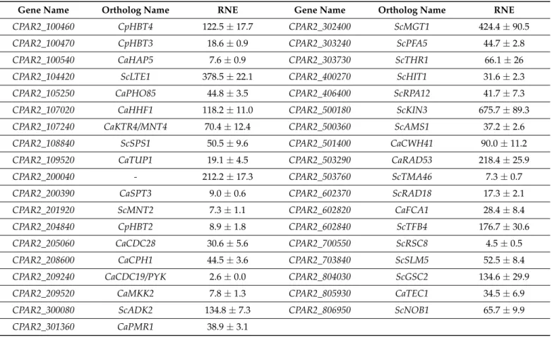
The relative expression of each gene was measured using qPCR and compared to CLIB214.
We found that the expression levels varied across a wide range with increases from 2.6-fold to 675.7-fold (Table1).
3.2. Characterization of the Generated OE Mutant Strains
3.2.1. Overexpression Does Not Affect Growth in either Complete or in Minimal Liquid Media
We applied three control strains in these experiments. CPRI is theC. maltosa LEU2 complemented derivative of the leucine auxotroph CPL2 strain, which was used to generate the mutant collection [28]. CLIB214 is a prototroph isolate and the parental strain of CPL2.
We also included an mCherry fluorescent protein-expressing strain as a control OE strain (mCherryOE), which was generated using the same procedure. We found this pertinent because expressing an irrelevant gene (coding a “neutral” protein) in terms of virulence would imitate the same load on the expression machinery, which itself might have an effect on the physiological and virulence properties of the fungus [53].
Table 1. Real-time qPCR-based overexpression data (fold-change, relative normalized expression—RNE) of the created mutant strains. Data were normalized to the CLIB214 wild type strain. Relative transcription levels were determined using the housekeeping geneCpTUB4(CPAR2_500510) as an internal control. Measurements were performed in three statistical parallels. The 2−∆∆Cq comparison method was used with Bio-Rad CFX Manager software. (Cp—Candida parapsilosis;
Ca—C. albicans;Sc—Saccharomyces cerevisiae).
Gene Name Ortholog Name RNE Gene Name Ortholog Name RNE
CPAR2_100460 CpHBT4 122.5±17.7 CPAR2_302400 ScMGT1 424.4±90.5
CPAR2_100470 CpHBT3 18.6±0.9 CPAR2_303240 ScPFA5 44.7±2.8
CPAR2_100540 CaHAP5 7.6±0.9 CPAR2_303730 ScTHR1 66.1±26
CPAR2_104420 ScLTE1 378.5±22.1 CPAR2_400270 ScHIT1 31.6±2.3
CPAR2_105250 CaPHO85 44.8±3.5 CPAR2_406400 ScRPA12 41.7±7.3
CPAR2_107020 CaHHF1 118.2±11.0 CPAR2_500180 ScKIN3 675.7±89.3
CPAR2_107240 CaKTR4/MNT4 70.4±12.4 CPAR2_500360 ScAMS1 37.2±2.6
CPAR2_108840 ScSPS1 50.5±9.6 CPAR2_501400 CaCWH41 90.0±11.2
CPAR2_109520 CaTUP1 19.1±4.5 CPAR2_503290 CaRAD53 218.4±25.9
CPAR2_200040 - 212.2±17.3 CPAR2_503760 ScTMA46 7.3±0.7
CPAR2_200390 CaSPT3 9.0±0.6 CPAR2_602370 ScRAD18 17.3±2.1
CPAR2_201920 ScMNT2 7.3±1.1 CPAR2_602820 CaFCA1 28.4±8.4
CPAR2_204840 CpHBT2 8.9±1.8 CPAR2_602840 ScTFB4 176.7±30.6
CPAR2_205060 CaCDC28 30.6±5.6 CPAR2_700550 ScRSC8 4.5±0.5
CPAR2_208600 CaCPH1 44.5±3.6 CPAR2_703840 ScSLM5 52.5±8.4
CPAR2_209240 CaCDC19/PYK 2.6±0.0 CPAR2_804030 ScGSC2 134.6±29.9
CPAR2_209520 CaMKK2 7.8±1.3 CPAR2_805930 CaTEC1 34.5±6.9
CPAR2_300080 ScADK2 134.8±7.3 CPAR2_806950 ScNOB1 65.7±9.9
CPAR2_301360 CaPMR1 38.9±3.1
First, we tested if the overexpression of the selected genes had any effect on the viability of the mutants. To achieve this, we cultivated the strains in YPD and YNB minimal liquid media for 24 h and compared the growth curves to that of the control strains. As a result, we found no significant differences between the growth capacity of the generated mutants compared to the three control strains (Figures S3 and S4), suggesting that the overexpression of these genes did not alter the viability of the OE mutants. Therefore, we included all the strains in the subsequent experiments to examine potential alterations in their virulence and fitness under different stress-inducing conditions, and as a control, we included the wild type CLIB214 strain unless otherwise stated.
3.2.2. Overexpression of Certain Genes Affects Viability under Specific Stress Conditions As a surrogate for the yeast’s ability to resist severe growth restrictive conditions such as elevated temperatures, different pH levels, or oxidative stressors in the host, a wide variety of the aforementioned stress conditions were applied in vitro to the OE collection on YPD solid media (Table S2). Growth was monitored both at 30 and 37◦C, which represent the regular cultivation temperature of the fungus and the physiological body temperature of humans, respectively. The viability of the mutants was also tested at different temperatures, including 20, 25, 30, 37, and 40◦C on YPD agar plates and at 30, 37◦C on YNB and YNB + FBS solid agar. In this range of temperature, we could not detect any difference in the growth of the examined mutants in comparison to the applied controls, either on complete or minimal media (Figure S5).
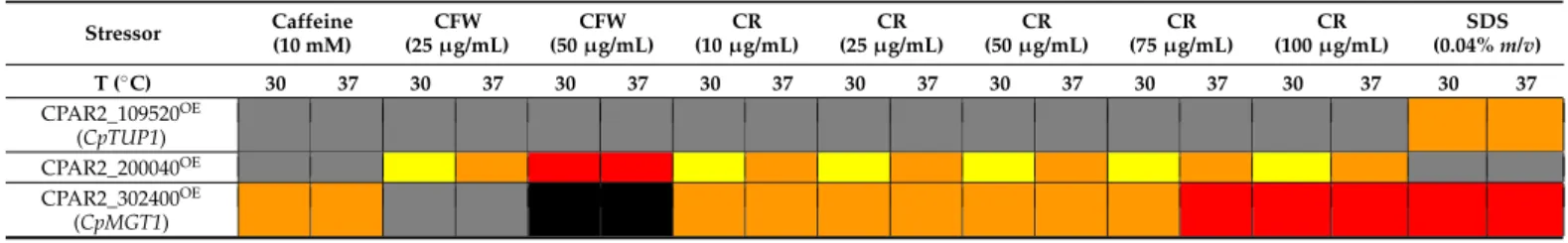
Three out of the 37 overexpression mutants showed alteration in their fitness under at least one condition (Table2). The three control strains had similar growth patterns under different stress conditions (Figure S6). Reduced growth was detected in the presence of Calcofluor white (CFW) or Congo red (CR) with CPAR2_200040OEand CPAR2_302400OE. Susceptibility to sodium dodecyl sulfate (SDS) occurred in the case of CPAR2_109520OE and CPAR2_302400OE, while caffeine intolerance was detected with the CPAR2_302400OE mutant (Table2). The applied stressors like CFW, CR, and caffeine have negative effects on the wall biogenesis and activate cell wall integrity (CWI) signaling via different components of the pathway. SDS is a membrane perturbing agent, and it has an effect on the integrity of the cell wall as well. Also, SDS can induce protein denaturation and cell lysis [57].
Table 2.The heat map shows the results of the spot assay analyses (CFW—Calcofluor white; CR—Congo red; black—no growth; red—strong defect; orange—medium defect; yellow—slight defect; grey—no difference).
Stressor Caffeine (10 mM)
CFW (25µg/mL)
CFW (50µg/mL)
CR (10µg/mL)
CR (25µg/mL)
CR (50µg/mL)
CR (75µg/mL)
CR (100µg/mL)
SDS (0.04%m/v)
T (◦C) 30 37 30 37 30 37 30 37 30 37 30 37 30 37 30 37 30 37
CPAR2_109520OE (CpTUP1) CPAR2_200040OE CPAR2_302400OE
(CpMGT1)
To evaluate the spot assay experiment, the following categories were assessed to compare the growth of the mutants to that of the wild type: No growth (in contrast to the wild type, the mutant did not form any colonies); strong defect (reduced growth was seen in the 104–105spots, and there was usually no growth at lower cell concentrations);
medium defect (reduced growth capacity was observed with the 103–104spots, the 105spot showed normal growth, and, usually, the 102had no growth); slight defect (reduced growth capacity was observable at 102–103spots, and, usually, the 104–105spots showed normal growth); no difference (the viability of the mutant was indistinguishable from that of the control).
The fitness of the CPAR2_302400OEstrain decreased upon exposure to each of the cell wall–and membrane perturbing agents tested (Figure1). The viability of this strain in the presence of CR or CFW was reduced in a concentration-dependent manner. Increasing concentration of CR resulted in medium (10, 25, 50, 75µg/mL) to strong (75 and 100µg/mL) growth defect. No growth or strong growth defect was observed when CFW was applied in a concentration of 50µg/mL, while no defect was found at lower stressor concentrations.
CPAR2_302400OEwas the only mutant that showed a growth defect in the presence of caffeine. A strong growth defect was observed when the cells were incubated in the presence of the membrane perturbing agent SDS.
Slight, medium, or strong reductions in fitness were observed when cells were exposed to CR or CFW agents during the investigation of the CPAR2_200040OEmutant (Figure1).
The CPAR2_109520OEshowed a medium growth defect only in the presence of SDS (Figure1).
3.2.3. The Examined OE Mutants Did Not Display Any Alterations in Morphology or Pseudohypha Formation
In contrast withC. albicans, C. parapsilosisis unable to form true hypha, although it is able to produce pseudohypha. The capability of reversible switching between yeast and filamentous form is an important virulence factor as it can enhance yeast surface/size and facilitate adhesion. Adhesion can lead to biofilm formation and colonization, which contribute to the intrusion of the fungus into the host cells and subsequent dissemination inside the host. It can also play a protective role for the fungus against the host immune cells and can be a tool to avoid phagocytosis [58]. Genetic elements responsible for dimorphic transition have been explored inC. albicansby gene deletion or expression analysis, and there have been further attempts to reveal morphological and filamentation regulatory
functions in otherCandidaspecies [59–63]. Studies have concluded that the filamentary regulation pathways revealed inC. albicansare partially (evolutionary) conserved compared to otherCandidaspecies (C. parapsilosis,C. tropicalis,C. guilliermondii,C. dubliniensis) [61,64].
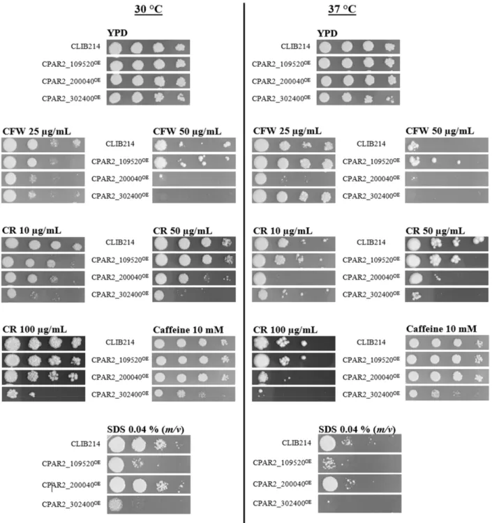
Figure 1.Representative pictures present CPAR2_109520OE, CPAR2_200040OE, and CPAR2_302400OEstrains with altered fitness compared to CLIB214 control strain in spot assay analysis. Tenfold dilutions of the cell suspensions ranging from 105cell/5µL to 102cell/5µL were pinned onto the surface of the solid media and kept for 48 h at 30 or 37◦C. Except for CFW (50µg/mL) and CR (50, 100µg/mL) at 37◦C, that was incubated for 3–4 days.
The screen of the OE strain collection for potential alterations in pseudophypha formation in two different liquid media at 37◦C and 5% (v/v) CO2using a light microscope (qualitative analysis) as well as the Amnis® FlowSight® flow cytometer (quantitative analysis) revealed no detectable alterations in the mutants’ capacity to form pseudohypha (Table S4 and Figure S7).
3.2.4. Overexpression of theCPAR2_302400Gene Resulted in Attenuated Biofilm Formation Adherence and morphology switching can facilitate or strengthen biofilm formation and the virulence of a pathogenic fungal species, andC. parapsilosisforms biofilms on the surfaces of various medical devices, enabling nosocomial systemic infections [65]. Holland et al. examinedC. albicansandC. parapsilosisbiofilm-associated genes and concluded that different factors are responsible for biofilm development in these two species. WhileCZF1, UME6,CPH2, andGZF3are the major regulators forC. parapsilosisbiofilm formation, in C. albicans BRG1andTEC1were found to be the central regulators of this process [28,66].
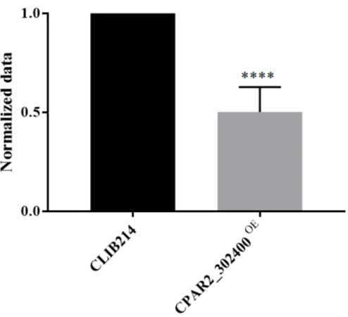
Building on the data gained fromC. parapsilosistranscriptome and KO collection analyses, we aimed to utilize the complementary OE strategy to investigate this attribute of the fungus under biofilm-inducing conditions by monitoring their metabolic activity. We found that only CPAR2_302400OEshowed attenuated biofilm formation compared to the control strain (Figure2and Figure S8).
Figure 2. Biofilm formation analysis. OD540values of the overexpression (OE) strains were nor- malized to the CLIB214 control strain,n= 3 with eight parallel samples per experiment. Statistical analysis: one-way ANOVA, Dunnett’s multiple comparisons test (****p< 0.0001).
3.2.5. Overexpression of Selected Genes Did Not Affect Tolerance to Echinocandins Echinocandins represent the primary therapy to treat candidaemia in clinical practice;
however,C. parapsilosisisolates show higher tolerance to these drugs compared to other Candidaspecies [2,17,67]. A possible explanation for this is thatC. parapsilosisisolates tend to accumulate point mutations in the hot spot region of antifungal resistance-related genes, causing a gain-of-function phenotype leading to increases in the tolerance to echinocan- dins [16]. To test how the overexpression of the selected genes affects the susceptibility of the mutants to echinocandins, the MICs of each of the strains to the three major members of this class of drugs were determined. The OE mutants displayed similar MICs to those of the control strains independently of whether anidulafungin (ANF), caspofungin (CAF), or micafungin (MIF) was applied. After 48 h of incubation, the MICANFvalues of the mutants and theC. parapsilosiscontrol strains were 2–4µg/mL, both MICCAFand MICMIFvalues
were 1–2µg/mL, while higher sensitivity was recorded in the case ofC. krusei(MICANF: 0.125–0.25µg/mL; MICCAF: 0.5µg/mL; MICMIF: 0.25µg/mL). These results are in line with the literature [68,69] (Table S5).
3.2.6. Overexpression of Certain Genes Interferes with Resistance to Phagocytosis by J774.2 Cells
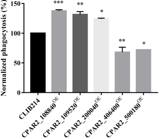
Phagocytes represent the primary cellular components of the innate immune system to fight againstCandidacells reaching the tissues. Macrophages are among the first line of cellular responders that combat fungal invasion by rapidly infiltrating to the site of the infection, where they attempt to ingest and eliminate yeast cells. However, successful fungal pathogens have evolved specific mechanisms through which they are capable of interfering with macrophage responses, and some can even survive within macrophages, indicating that the capability of an intruding pathogen to avoid endocytosis and lysis by the host immune system is an important virulence trait [70–73]. To investigate if the OE strains had altered interactions with host phagocytic cells, we fluorescently labeled the OE strains with Alexa FluorTM488 succinimidyl ester dye (Invitrogen) and co-cultured them with J774.2 murine macrophage-like cells in vitro. We monitored the phagocytic efficiency after 1 h using Amnis® FlowSight® flow cytometer by determining the percentages of the phagocytic activity of each sample and normalized to CLIB214 control values. We identified three strains (CPAR2_108840OE, CPAR2_109520OE, CPAR2_200040OE) that were more efficiently phagocytosed and two strains (CPAR2_406400OE, CPAR2_500180OE) that were less effectively phagocytosed by murine macrophages (Figure3).
Figure 3.J774 phagocytosis analysis. Phagocytic activity (%) of the J774 macrophages during the co-incubation with OE strains were normalized to the values of the phagocytosed CLIB214 control strain,n= 3. Statistical analysis: one-way Anova, Dunnett’s multiple comparisons test (*p≤0.05;
**p≤0.01; ***p≤0.001).
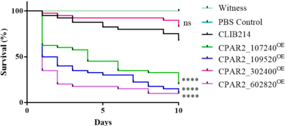
3.2.7. Overexpression of Certain Genes Regulate Virulence Related Processes inGalleria mellonellaLarvae Model
Galleria mellonellalarvae possess similar humoral and cellular innate immune mecha- nisms as mammalian hosts; therefore, this insect is widely used as a substitute model for vertebrate infection experiments in order to reduce mammalian consumption. To reveal if the overexpression of the selected genes had an effect on the virulence ofC. parapsilosis in this model,G. mellonellalarvae were infected with the OE strains, and their survival was monitored. Shortly after the injection, the larvae became brownish due to a process called melanization. This is a common and rapid reaction of insects against any kind of foreign particles (including pathogens), whereby the hemocytes surround the foreign particle and start producing melanin via a multistep biochemical process [74]. In the case ofG. mellonella,the melanization occurs within minutes after injection withC. parapsilosis.
Melanization failed to occur following infection with CPAR2_302400OE, but there were similar survival rates to that of the control strain. However, we identified three melanin- inducing strains (CPAR2_107240OE, CPAR2_109520OE, and CPAR2_602820OE) that were more virulent compared to the control strain (Figure4)
Figure 4.Summarized results of the in vivoGalleria. mellonellainfection experiments,n= 3, separate experiments with 20 larvae per strain and experiment. Statistical analysis: Log-rank (Mantel–Cox) test (****p< 0.0001; ns is not significant).
3.2.8. Overexpression of the Selected Genes Does Not Affect Certain Components of the Cell Wall
Based on the results of the aforementioned experiments, certain components of the cell wall (chitin and its oligomer compounds and alpha mannan content) were further examined using specific cell wall stains. We found that none of the examined mutant strains showed an alteration compared to the control strains, neither in the distribution nor in the amount of the examined elements of the cell wall (Figure S9).
3.2.9.CPAR2_109520andCPAR2_302400Differentially Alter Fungal Burden in Organs withinMus Musculus
We next examined the virulence of two OE mutant strains in a mouse infection model, which represents the “gold” standard of in vivo infection models. We selected CPAR2_109520OEbecause it was more virulent than the control strain in theG. melonella model. We also chose CPAR2_302400OE, which was not significantly different from the wild type in terms of the survival ofG. mellonella, but it did not induce melanization in the larvae.
In the case of CPAR2_302400OE, we detected elevated fungal burdens in the brain and kidney tissues, while there were lower CFUs in the spleens and livers. CPAR2_109520OE also had higher CFU levels in brain samples, while there was a reduced splenic fungal burden (Figure5).
Figure 5.Murine infection experiments. The graphs show the fungal burden (CFU values) of brain, spleen, liver, and kidney in CFU/g organ values. Animals were infected with 2×10⁷fungal cells through their lateral tail veins and sacrificed after 3 days,n= 2 separate experiments, with four to five animals per strain and experiment were used. For the statistical analysis Mann-Whitney tests were used (*p< 0.05; **p< 0.01; ns: not significant).
4. Discussion
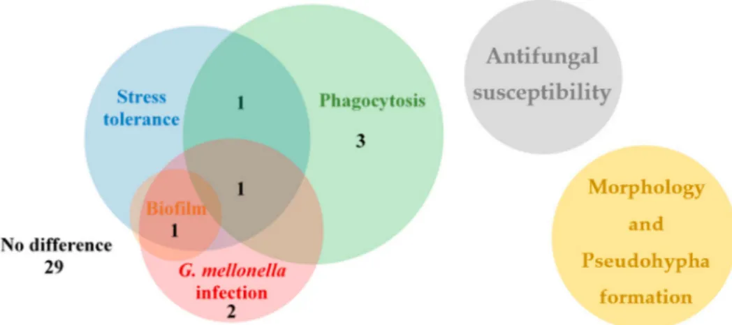
The aim of this study was to generate and rigorously examine an OE mutant strain col- lection with the purpose of identifying virulence factors inC. parapsilosis. We investigated stress tolerance, morphology switch, biofilm-forming capacity, phagocytosis, and in vivo virulence inG. mellonellalarvae and BALB/c mice. During our examinations, we found eight OE mutants with altered phenotype compared to the applied control strains. Both the CPAR2_109520OEand CPAR2_302400OEmutants showed an altered phenotype under three conditions, the CPAR2_200040OEstrain under two conditions, while the other five mutant strains under a single condition compared to the control strains (Figure6, Table S6).
In addition, the CPAR2_109520OEand CPAR2_302400OEmutants were selected for murine experiments, and alterations in CFU values in different tissues were found relative to the wild type control strain. During the investigation of morphology, pseudohypha formation, and antifungal susceptibility, we detected no alterations in the OE strains compared to controls (Figure6).
Figure 6.The Venn diagram shows summarized results of the OE mutant collection analysis separated into distinct experiments. The numbers display the number of mutant strains with alteration(s) in the exact experiment(s).
Recently, the function of three of the examined 37 genes was determined by Cillingova and co-workers [19]. TheCPAR2_100460(CpHBT4), theCPAR2_100470(CpHBT3), and the CPAR2_204840(CpHBT2) genes were confirmed as hydroxybenzoate transporters. The investigators reported that theCpHBT4deletion mutant showed resistance to caffeine and altered sensitivity to caspofungin and fluconazole; however, we found no alteration under any tested conditions in the case of the OE parallel mutant strains. The function of 18 of the 37 genes’ orthologs has already been confirmed inS. cerevisiae, while 15 orthologs functions have been examined inC. albicans[25]. Notably,CPAR2_200040has no confirmed functional ortholog in either of these other yeast species.
In order to further evaluate the results, we compared the data from the OE collection analysis to the results derived from previous KO mutant library characterizations. Accord- ing to Prelich, during comparison of the KO and the OE phenotype of the same gene, three phenomena can be observed: opposite (hypermorph or hypomorph) phenotypes (−/+;
+/−); similar phenotypes (−/−; +/+; 0/0); no phenotype versus altered (neomorph or antimorph) properties (−/0 or 0/−; +/0 or 0/+) [36]. Amongst the studied OE mutant strains here, there are 12 [29], four [28], and three [19] (18 different genes in total) KOs available inC. parapsilosisthat were characterized with similar methods, and importantly, they have common genetic background enabling proper comparison of the mutant pairs (Table S3).
The ortholog gene toCPAR2_109520encodes a transcriptional corepressor protein, CaTUP1, which represses filamentous growth [75]. The CPAR2_109520OEmutant displayed alterations in stress tolerance, phagocytosis, and both in vivo experiments. This OE mutant showed a medium growth defect only in the presence of SDS at 30 and 37◦C and was more avidly endocytosed by macrophages. A similar reduction in growth was also observed in aC. parapsilosisdeletion mutant of the gene in question (with additional sensitivity to other conditions as well) [28]. TheC. albicans TUP1null mutant showed abnormal colony morphology and increased filamentous growth compared to its originating strain [75–78].
A previous study also examining virulence in aC. albicans TUP1KO found reductions in adhesion, invasion, and damage with oral epithelial cells [79]. Although we also recorded reduced virulence properties of the CPAR2_109520OEmutant in in vitro experiments, we found this mutant to be more virulent than the wild typeC. parapsilosiscontrol strain in G. mellonellamodel.
The putative ortholog of theCPAR2_302400gene functions as a DNA repair methyl- transferase (ScMGT1) inS. cerevisiae[80,81]. The null mutant of this gene inS. cerevisiae showed increased growth in competitive fitness analysis [30], but there was no significant alteration in a biofilm assay [82]. AC. albicansnull mutant also displayed normal phe- notype in fitness analysis [27]; however, aC. parapsilosisKO showed decreased adhesive
properties, but there was not any difference in its biofilm-forming capacity compared to the control strain [83]. In the current study, theG. mellonellamodel confirmed the importance of melanization against invading pathogens. The only OE strain that was coupled with the absence of melanization was CPAR2_302400OE, which produced a survival curve similar to that of the control strain. Although we could not detect any alteration in phagocytosis efficiency, the mutant showed severe growth defects in other important virulence prop- erties such as stress tolerance or biofilm formation. The fitness of the CPAR2_302400OE strain decreased upon exposure to each of the cell wall and membrane perturbing agents (CFW, CR, SDS, and caffeine). This mutant was the only one out of the 37 tested whose growth was reduced in the presence of caffeine and the only one that showed decreased biofilm production. Biofilm formation enables invasion while concomitantly providing protection of the pathogen from different antifungal agents and helps avoid recognition by host immune cells.C. parapsilosisis able to form biofilm on the surfaces of various medically used devices, thus it is a notable virulence factor [65]. This is the first report to connect the CPAR2_302400gene function to the biofilm regulatory network ofC. parapsilosis, and thus to the pathogenicity of this species.
To further investigate the virulence properties of the strains, we selected two OE strains for study in a mammalian model. We chose a strain with increased virulence in the larval model, CPAR2_109520OE, and one that did not induce larval melanization but had virulence similar to the control strain, CPAR2_302400OE. TheG. mellonellalarvae and murine data suggest different invasion efficiencies of the mutants in diverse tissues or vari- ous mechanism or capacity of fungi depletion in the examined organs. Significantly higher fungal burdens were enumerated in the brains of mice infected with the CPAR2_109520OE mutant, while lower CFU numbers were observed in the spleen. For CPAR2_302400OE, which was similar to wild type control yeast inG. mellonella,significantly higher CFUs were found in both the brains and kidneys of challenged mice, while lower counts were enumerated in the spleens and livers. Other comparative studies examining the correlation between the results derived from the wax moth and murine infections have also shown discordant results [84–86]. Nevertheless, monitoring survival in mice is not optimal in the case of many NAC species likeC. parapsilosis, since they do not cause lethal infections in this species, while lethality can be measured in insect models [87]. Interestingly, in a mouse systemic infection model, theC. albicans TUP1(CPAR2_109520gene’s ortholog) null mutant, showed decreased virulence [78], whereas another study reported no mortality 30 days after i.v. infection with the same mutant [88]. Further,C. albicans MGT1 (the ortholog ofCPAR2_302400) null mutant displayed no altered phenotype in competitive experiments during i.v. infection in mice [27]. These findings underscore the imperative to explore pathogenesis studies in individualCandidaspecies as determinations made with C. albicanscannot routinely be extrapolated to have similar behaviors in these biologically different pathogens.
The CPAR2_107240OE and CPAR2_602820OE mutants were also more virulent in G. mellonellaexperiments. TheCaKTR4/MNT4(CPAR2_107240gene’s ortholog) gene was verified as a mannosyltranferase inC. albicanswhere it has a role in N-linked glycosylation and cell wall regeneration [89]. The null mutant of the gene showed normal growth capacity, morphology, and competitive fitness in pooled mice infection model [27]. Only the double, triple, quadruple, or quintuple deletion of theCaMNT1-2-3-4-5genes in different combinations (but not the single ones) resulted in mutants with altered cell wall content and elevated sensitivity to cell wall perturbing agents (CFW or SDS), indicating thatMNTgenes ofC. albicanshave redundant functions [89]. Changes in the composition of the cell wall imply alterations in host-pathogen interactions as well [89]. TheCPAR2_602820ortholog gene (CaFCA1) encodes a cytosine deaminase, an enzyme of pyrimidine salvage [90,91].
TheC. albicansnull mutant was resistant to flucytosine [92]. However, there has been no information about the ortholog genes in connection with host-pathogen interactions in C. parapsilosis.
The CPAR2_200040OEmutant displayed defects in stress tolerance and phagocytosis compared to control strains. The mutant showed slight, medium or strong reduction in fitness in the presence of CR and CFW and displayed elevated uptake by murine macrophages. The function of this gene ortholog’s function is unknown. Interestingly, the corresponding KO mutant of this strain was resistant to CFW compared to the control strain, further confirming the role ofCPAR2_200040in cell wall assembly regulation of C. parapsilosis[29].
The CPAR2_108840OE, CPAR2_406400OE, and the CPAR2_500180OEmutants showed altered phenotypes only in the context of phagocytosis experiments. The CPAR2_108840OE mutant was more efficiently phagocytosed by macrophages. The function ofScSPS1 (CPAR2_108840gene’s ortholog) is putatively a serine/threonine kinase, which is involved in spore packaging inS. cerevisiae[93]. Its KO parallel was sensitive to SDS inC. parap- silosis[29], while it displayed a normal phenotype inC. albicans[27]. However, the gene has not previously been linked to virulence. We also recorded lower phagocytic events with the CPAR2_406400OEmutant strain. The ortholog gene,ScRPA12,was identified in S. cerevisiaeas an RNA polymerase I subunit [94]. Chaillot and colleagues found that this gene has a role in cell size homeostasis inC. albicans[95], but it has not been associated with virulence regulation. The CPAR2_500180OEmutant displayed a more virulent phenotype in phagocytosis experiments compared to the control strain. Its gene ortholog isScKIN3, which encodes a nonessential serine/threonine protein kinase that has a possible role in DNA damage responses [96–98]. Its KO parallel was susceptible to Congo red and caffeine inC. parapsilosis[29] and showed increased cell size and decreased resistance to caspofungin inC. albicans[99,100]. InS. cerevisiae,its overexpression caused a reduction in vegetative growth, abnormal cellular morphology, and cell cycle [45]; however, we did not observe any of the aforementioned effects. According to the phagocytosis experiments in this study, we monitored alterations occurring at the phases of recognition and uptake.
Both recognition and endocytosis primarily depend on the recognition of specific cell wall components of the pathogen. Fungal evasion of recognition occurs via masking these structures or changing morphology type by the fungi [70]. Our results suggest that dif- ferences in cell wall composition of these OE mutant strains cause the observed altered phagocytic efficiencies.
During the OE and KO phenotype comparisons, we found variations in phenotypes in the case of CPAR2_200040OEwhen we examined CFW tolerance and in vitro host-pathogen interactions [29]. Similar phenotypes were observed with two OE mutants and their KO counterparts. TheC. parapsilosisKO pair of the examined CPAR2_109520OEmutant was sensitive to SDS. In addition to this, the CPAR2_302400OEmutant strain showed decreased adhesive properties, although there was no difference in its biofilm-forming capacity compared to the control strain, while the OE mutant displayed less biofilm formation [29].
We also found absences of phenotypic alterations in OE, KO, or both parallel mutants based on the literature. TheCPAR2_108840deletion mutant was resistant to SDS, while we did not notice such alterations in the OE strain [29]. Neither the CPAR2_500180OEmutant nor its KO pair displayed any alterations under similar experimental conditions [29]. For the CPAR2_602820OEmutant, we found no changes in its fitness during the spot assay analysis, while its deletion strain showed mild and strong defects in growth on minimal media at 30 and 37◦C (Table S6). When onlyS. cerevisiaeorC. albicansmutant collection analysis data were available, comparisons were more challenging. Additionally, it was necessary to consider whether the overexpressed genes were not unique or correctly manifest when a lack of gain-of-function or no reversed phenotype was observed [36]. In several cases, the OE and the KO phenotypes of a given gene did not complement each other. In addition to this, the applied control strains could influence the interpretation of the results. Therefore, in approaching this task, we also assessed the use of the reintegrated CPRI and an overexpressing strain mCherryOE as controls in addition to the wild type CLIB214 in fitness-related experiments. We found that it was more appropriate to apply the neutral gene carrier OE strain mCherryOE, rather than a mutant bearing an empty
cassette as a control in order to exclude any alteration in fitness derived from the integration and/or the continuous operation of the expression cassette itself (due to the presence of a constitutive promoter) [42,46,53]. As we found no alteration between the three control strains that we examined in preliminary investigations, we selected CLIB214 wild type as the control for subsequent experiments.
Although we examined diverse effects in our OE collection, several important addi- tional areas remain to be explored if genes are examined in a targeted manner using gene deletion or overexpression strategies. These include the (1) gene dosage effect, (2) infor- mation about their interactions with other genes and (3) the subsequent transcriptomic effects, (4) off-target mutations, (5) universal behavior in various strains, and (6) the RNA level of an overexpressed gene (what we can measure) does not necessarily correlate with that of the protein it encodes [36,45,101–103]. Similar to our findings, previous studies in C. albicansdid not find any OE mutants with increased fitness [42,46]. Notably, there is a substantial energy cost to a cell when a gene is duplicated or artificially overexpressed.
According to Wagner, increasing protein synthesis above 10% negatively affects the health of the cell [104]. The function of the query gene also can determine the resulting phenotype, for example, periodically changing genes like cell cycle control genes [45]. So, the detected phenotype depends on the genuine function of the targeted gene. Nevertheless, our work clearly indicates that the application of the OE approach is an effective starting point to explore gene functions, especially when a prescreen (RNAseq data or known function of orthologs) for selected genes is possible. Rapid systematic analysis of the OE mutant collection (including stressors, biofilm formation, in vitro and in vivo infection models) can identify previously unknown virulence-associated genes, which can be analyzed further to reveal gene-specific functions.
In summary, this study was the first attempt to use an OE strategy to comprehen- sively examine gene function inC. parapsilosis. Significantly, the findings clearly associate CPAR2_107240,CPAR2_406400, andCPAR2_602820with the virulence ofC. parapsilosis.
Our results show that OE approaches are a relevant method for gene function analysis in this lethal, emerging fungus and that OE methods can define new virulence pathways in C. parapsilosis.
Supplementary Materials: The following are available online athttps://www.mdpi.com/2309 -608X/7/2/97/s1, Figure S1: The schematic figure of the OE vector. Figure S2: Representative figures of the methods for verification of the generated OE mutant strains. Figure S3: Results of the growth kinetic measurements in complete media (YPD). Figure S4: Results of the growth kinetic measurements in minimal media (YNB + 0.5% (m/v) glucose). Figure S5: Results of the growth analyses on solid minimal media and with additional serum. Figure S6: Results of the growth analyses of the 3 control strains on solid media supplemented with different stress agents. Figure S7:
Representative figures from pseudohypha analysis by Amnis®FlowSight®flow cytometer. Figure S8: Biofilm forming capacity of the overexpression mutants. Figure S9: Cell wall component analysis of the selected mutants. Table S1: Strains and primers used in this study. Table S2: Applied stressors and their concentrations in solid YPD media for spot assay analysis. Table S3: The list of the selected genes for OE analysis. Table S4: The results of pseudohypha formation experiments. Table S5: The results of the antifungal susceptibility analysis. Table S6: The summarized results of the OE mutant collection analysis.
Author Contributions: Conceptualization, A.G., T.N.; methodology, T.N., S.E.P.; software, S.E.P.;
validation, T.N., S.E.P.; formal analysis, T.N., S.E.P.; investigation, T.N., S.E.P.; resources, A.G., C.V.;
data curation, T.N., S.E.P., R.T., and J.D.N.; writing—original draft preparation, S.E.P.; writing—
review and editing, T.N., R.T., A.G., and J.D.N.; visualization, S.E.P.; supervision, T.N., A.G., C.V.;
project administration, T.N., A.G.; funding acquisition, A.G. All authors have read and agreed to the published version of the manuscript.
Funding:This work was supported by grants 20391-3/2018/FEKUSTRAT, NKFIH K 123952, and GINOP-2.3.2.-15-2016-00035. S.E.P. was funded by Richter Gedeon Talentum Foundation. A.G. was further funded by LP2018-15/2018. T.N. was supported by the Postdoctoral Fellowship Programme of the Hungarian Academy of Sciences.
Institutional Review Board Statement:The study was conducted according to the guidelines of the Declaration of Helsinki, and approved by the Institutional Review Board of University of Szeged (protocol code XVI./3652/2016, date of approval: 05. 10. 2016).
Informed Consent Statement:Not applicable.
Data Availability Statement:The data presented in this study are available in “ACandida parapsilosis Overexpression Collection Reveals Genes Required for Pathogenesis”.
Acknowledgments:We wish to thank Katalin Csonka for the help with mouse experiments and Erik Zajta and Csaba Papp for the assistant in flow cytometry analysis. We are grateful to Geraldine Butler for kindly providing CLIB214, CPL2, CPRI fungal strains. We are thankful to Christophe d’Enfert for the vector pCA-DEST1300.
Conflicts of Interest:The authors declare no conflict of interest.
References
1. Warnock, D.W. Trends in the epidemiology of invasive fungal infections.Nippon Ishinkin Gakkai Zasshi2007,48, 1–12. [CrossRef]
2. Pfaller, M.A.; Diekema, D.J. Epidemiology of invasiveCandidiasis: A persistent public health problem.Clin. Microbiol. Rev.2007, 20, 133–163. [CrossRef]
3. Cleveland, A.A.; Farley, M.M.; Harrison, L.H.; Stein, B.; Hollick, R.; Lockhart, S.R.; Magill, S.S.; Derado, G.; Park, B.J.;
Chiller, T.M.; et al. Changes in incidence and antifungal drug resistance inCandidemia: Results from population-based lab- oratory surveillance in Atlanta and Baltimore, 2008–2011.Clin. Infect. Dis.2012,55, 1352–1361. [CrossRef]
4. Siopi, M.; Tarpatzi, A.; Kalogeropoulou, E.; Damianidou, S.; Vasilakopoulou, A.; Vourli, S.; Pournaras, S.; Meletiadis, J. Epidemio- logical trends of fungemia in greece with a focus onCandidemiaduring the recent financial crisis: A 10-Year survey in a tertiary care academic hospital and review of literature.Antimicrob. Agents Chemother.2020,64, e01516-19. [CrossRef]
5. Fridkin, S.K.; Kaufman, D.; Edwards, J.R.; Shetty, S.; Horan, T. Changing incidence ofCandidabloodstream infections among NICU patients in the United States: 1995–2004.Pediatrics2006,117, 1680–1687. [CrossRef]
6. Juyal, D.; Sharma, M.; Pal, S.; Rathaur, V.K.; Sharma, N. Emergence of non-albicans Candidaspecies in neonatal candidemia.N. Am.
J. Med. Sci.2013,5, 541–545. [CrossRef]
7. Yapar, N. Epidemiology and risk factors for invasive candidiasis.Ther. Clin. Risk Manag.2014,10, 95–105. [CrossRef]
8. White, M.H. Editorial response: The contribution of fluconazole to the changing epidemiology of invasiveCandidalinfections.
Clin. Infect. Dis.1997,24, 1129–1130. [CrossRef]
9. Nucci, M.; Marr, K.A. Emerging fungal diseases.Clin. Infect. Dis. Off. Publ. Infect. Dis. Soc. Am.2005,41, 521–526. [CrossRef]
10. Cleveland, A.A.; Harrison, L.H.; Farley, M.M.; Hollick, R.; Stein, B.; Chiller, T.M.; Lockhart, S.R.; Park, B.J. Declining incidence ofCandidemiaand the shifting epidemiology of Candida resistance in two US Metropolitan Areas, 2008–2013: Results from population-based surveillance.PLoS ONE2015,10, e0120452. [CrossRef]
11. Papon, N.; Courdavault, V.; Clastre, M.; Bennett, R.J. Emerging and emerged pathogenic Candida species: Beyond theCandida albicansparadigm.PLoS Pathog.2013,9, e1003550. [CrossRef] [PubMed]
12. Clark, T.A.; Slavinski, S.A.; Morgan, J.; Lott, T.; Arthington-Skaggs, B.A.; Brandt, M.E.; Webb, R.M.; Currier, M.; Flowers, R.H.;
Fridkin, S.K.; et al. Epidemiologic and molecular characterization of an outbreak of Candida parapsilosis bloodstream infections in a community hospital.J. Clin. Microbiol.2004,42, 4468–4472. [CrossRef] [PubMed]
13. Kuhn, D.M.; Mikherjee, P.K.; Clark, T.A.; Pujol, C.; Chandra, J.; Hajjeh, R.A.; Warnock, D.W.; Soil, D.R.; Ghannoum, M.A. Candida parapsilosis characterization in an outbreak setting.Emerg. Infect. Dis.2004,10, 1074–1081. [CrossRef] [PubMed]
14. Pammi, M.; Holland, L.; Butler, G.; Gacser, A.; Bliss, J.M. Candida parapsilosis is a significant neonatal pathogen: A systematic review and meta-analysis.Pediatr. Infect. Dis. J.2013,32, e206–e216. [CrossRef] [PubMed]
15. Harrington, R.; Kindermann, S.L.; Hou, Q.; Taylor, R.J.; Azie, N.; Horn, D.L. Candidemia and invasive candidiasis among hospitalized neonates and pediatric patients.Curr. Med. Res. Opin.2017,33, 1803–1812. [CrossRef] [PubMed]
16. Tóth, R.; Nosek, J.; Mora-Montes, H.M.; Gabaldon, T.; Bliss, J.M.; Nosanchuk, J.D.; Turner, S.A.; Butler, G.; Vágvölgyi, C.;
Gácser, A.; et al.Candida parapsilosis: From genes to the bedside.Clin. Microbiol. Rev.2019,32, e00111-18. [CrossRef]
17. Papp, C.; Kocsis, K.; Tóth, R.; Bodai, L.; Willis, J.R.; Ksiezopolska, E.; Lozoya-Pérez, N.E.; Vágvölgyi, C.; Mora Montes, H.;
Gabaldón, T.; et al. Echinocandin-induced microevolution ofCandida parapsilosisinfluences virulence and abiotic stress tolerance.
mSphere2018,3. [CrossRef]
18. Gácser, A.; Trofa, D.; Schäfer, W.; Nosanchuk, J.D. Targeted gene deletion inCandida parapsilosisdemonstrates the role of secreted lipase in virulence.J. Clin. Investig.2007,117, 3049–3058. [CrossRef]
19. Cillingová, A.; Zeman, I.; Tóth, R.; Neboháˇcová, M.; Dunˇcková, I.; Hölcová, M.; Jakúbková, M.; Gérecová, G.; Pryszcz, L.P.;
Tomáška, L’.; et al. Eukaryotic transporters for hydroxyderivatives of benzoic acid.Sci. Rep.2017,7, 8998. [CrossRef]
20. Zoppo, M.; Di Luca, M.; Franco, M.; Rizzato, C.; Lupetti, A.; Stringaro, A.; De Bernardis, F.; Schaudinn, C.; Barrasa, M.I.;
Bottai, D.; et al. CpALS4770 and CpALS4780 contribution to the virulence of Candida parapsilosis. Microbiol. Res. 2020, 231, 126351. [CrossRef]