CHAPTER 7
The Sodium Pump in Animal Tissues and Its Role in the Control of Cellular
Metabolism and Function
P. F. Baker
I. Introduction 243 II. Properties of the Na Pump in Animal Cells 244
A. Introduction 244 B. Source of Energy for Pumping 244
C. Linkage between Na and Κ Movements: Stoichiometry of the
Pump 246 D. Ion Selectivity of the Na Pump 248
E. Mechanism of Inhibition by Cardiac Glycosides 249 III. Interactions between the Na Pump and Cellular Metabolism and
Function 253 A. Introduction 253 B. Utilization of ATP 253 C. The Absolute Levels of Na and Κ 254
D. The Gradients of Na and Κ Ions 254 E. Electrogenic Pumping and the Control of Nervous Function 263
F. General Remarks 265 IV. Control of Pumping Capacity 266
References 266
I. INTRODUCTION
The purpose of this chapter is twofold: first, to outline the properties of the Na pumping mechanism that exists in most, if not all, animal cells, laying particular emphasis on the behavior of the pump in the intact cell and, second, to discuss in general terms the various ways in which the rate of pumping at the cell surface can influence cellular metabolism and function. A problem to be discussed in some detail is the relation between pumping rate and control of nervous activity.
243
244 P. F. BAKER
II. PROPERTIES OF THE Na PUMP IN ANIMAL CELLS
A. Introduction
The existence of an Na pump in many animal cells can only be inferred from indirect evidence. Thus most animal cells contain more potassium and less sodium than plasma and have in their surface membranes an ouabain-sensitive, (Na + Reactivated ATPase. The mechanism of pumping has only been studied intensively in a few cell types, including erythrocytes from a variety of sources including man, frog skeletal muscle, and various nerve preparations especially squid giant axons, crab nerve, and C fibers from the rabbit vagus. Despite the diversity of sources, the properties of the Na pumping mechanism in these tissues are surprisingly similar and I shall refer in the main to work on the squid axon and human erythrocyte. The reader is referred to the reviews of Baker [1] and Glynn [2] for a more complete survey of the subject. For the present purpose the most important features of the Na pump that need to be considered are (1) its dependence on cellular energy; (2) the linkage between Na and Κ movements and the overall stoichiometry of the pumping process; (3) ion selectivity; and (4) the mechanism of inhibition by cardiac glycosides. Each will be considered in turn.
B. Source of Energy for Pumping
It now seems fairly certain that energy is supplied to the Na pump in the form of ATP. The early experiments of Caldwell et al. [3] showed that, in squid axons, which had been fully poisoned with cyanide, pumping could be restarted by injecting a variety of phosphate com
pounds including ATP, arginine phosphate (ArgP)—the phosphagen in squid tissue—and phosphoenolpyruvate. But it was not possible to decide whether the injected compounds could all activate pumping directly or were used to synthesize some essential metabolite. This second possibility can be minimized by reducing the intracellular concentrations of small molecular weight materials such as enzyme cofactors and phosphate acceptors. This can be achieved in nerve by the techniques of internal perfusion [4] and dialysis [5] and in erythro
cytes by reversible hemolysis [6]. In these preparations, ATP is markedly better than the other phosphate compounds in energizing the Na pump.
Thus, in the dialyzed squid axon, Brinley and Mullins [5] have shown
7. N a PUMP AND CELLULAR METABOLISM 245 that of a wide variety of phosphate compounds tested only ATP and to a lesser extent rf-ATP can support sodium extrusion via the Na pump. In a similar series of experiments on Κ uptake via the Na pump, ATP was again the most effective energy source [7].
It should be stressed that not only does Na extrusion require ATP, it also consumes it, and in many cells—especially those with a large sur
face area to volume—much of the cell's energy metabolism is directed toward supplying ATP for the Na pump. In these cells inhibition of pumping results in a marked rise in the ATP/ADP ratio, a fall in in
organic phosphate, and a fall in either lactate production or oxygen consumption depending on whether metabolism is anaerobic or aerobic [see, for instance, 8,9,9a].
The work of Skou [10] and others (reviewed in Chapter 14, this volume) has shown that cell membranes contain an ATPase, the pro
perties of which are identical to those of the ATP-dependent Na pumping mechanism of intact cells [see, for instance, 2,8]. Thus the enzyme splits ATP only when Na is present at the inner face of the cell membrane and Κ is present at the outer face and this ATPase reaction is inhibited by cardiac glycosides at concentrations that also block the Na pump. The various lines of research directed toward elucidating the chemical basis of this transport ATPase are discussed elsewhere in this volume (Chapter 14); but at the risk of some repetition I should like to mention certain reactions and partial reactions of the ATPase system that may be of importance in the intact cell. These are the reversal of the pump and the exchange of Na for Na and Κ for K.
1. Reversal of the Na Pump
In theory it should be possible to reverse the Na pump, each reversed cycle resulting in the synthesis of ATP. This was first demonstrated in erythrocytes by Garrahan and Glynn [11]. They made the ion gradients steeper than normal, that is, high Κ and low Na inside the cell and high Na and nominally zero Κ in the external medium. Under these condi
tions there was incorporation of inorganic phosphate into ATP that could be blocked either by cardiac glycosides or by the addition of Κ to the external medium. Reversal is associated with an ouabain-sensitive efflux of Κ and an ouabain-sensitive influx of Na.
It is not known whether the Na pump ever operates in reverse under physiological conditions; but there is a very real possibility that the large downhill movements of Na and Κ that occur in certain excitable cells, e.g., the electric organ, may be coupled to the synthesis of ATP.
246 P. F. BAKER
2. Exchange of Natjor Na0 and K^/or K0
In intact nerve cells and erythrocytes the Na pumping mechanism can also effect the exchange of internal Na ions for external Na ions by an ouabain-sensitive route. This exchange reaction becomes apparent only in the absence of external Κ and with a low ATP/ADP-Pf ratio [12,13, 13a]. The exchange requires ATP, but seems not to consume it. From the physiological point of view the exchange achieves nothing; but the presence of Na-Na exchange can complicate measurements of Na pumping activity based solely on the magnitude of the ouabain-sensitive Na efflux. Rather similar comments apply to the exchange of internal Κ for external Κ by an ouabain-sensitive route. This process requires a high Pf level in the cell and seems to represent a reversal of the final stage of the ATPase reaction [14].
The " transport ATPase " contains a phosphatase that, in the presence of external Κ ions, can split /?-nitrophenyl phosphate, carbamyl phos
phate, and acetyl phosphate by an ouabain-sensitive route [14a]. It would be of great interest both from the viewpoints of mechanism and function to know whether this phosphatase which can operate in the absence of Na ions, can effect the inward movement of Κ ions. This would provide clear evidence that the inward movement of Κ need not necessarily be coupled to the outward movement of Na ions.
C. Linkage Between Na and Κ Movements: Stoichiometry of the Pump
Except under conditions where the pump is catalyzing Na-Na or K-K exchange, the ouabain-sensitive Na efflux is dependent on the presence of external Κ ions and seems to be linked to an ouabain-sensitive influx of Κ ions. Although there is some argument about the ratio of Na ions ejected to Κ ions absorbed by the Na pump, and especially whether this stoichiometry is variable, the available evidence in favor of a variable stoichiometry is not strong. In both nerve cells and erythrocytes, tracer evidence indicates transport of more Na ions outward than Κ ions inward, and a ratio of 3 Na:2 K/energy-rich phosphate bond split is most consistent with experimental observations [8,13,15-18]. This ratio seems to apply irrespective of whether the pump is operating downhill or against a steep electrochemical gradient. It should, however, be stressed that there seems no reason why the ratio should always be 3 Na:2 Κ and, under conditions where the pump is operating against a very adverse electrochemical gradient, it would be energetically more favorable to have a neutral pump—perhaps extruding 2 Na ions in exchange for 2 Κ ions/energy-rich phosphate bond split.
7. N a PUMP AND METABOLISM CELLULAR 247 If the stoichiometry above is correct, unless electroneutrality is maintained by the transport of some other ion, the Na pump should be electrogenic, i.e., capable of generating a potential difference across the cell membrane, making the inside of the cell negative with respect to the outside. This has been confirmed in nerve and muscle [19-21a]. Perhaps the clearest demonstration is that of Thomas [22] working with a snail neuron. He showed that injection of Na (but not Li or K) resulted in hyperpolarization of the cell, i.e., the interior of the cell became more negative with respect to the outside. This effect required external Κ ions and was inhibited by application of ouabain. By voltage clamping the neuron and simultaneously monitoring the internal Na concentration by an Na-selective glass electrode, Thomas was able to show that the hyperpolarization following injection of Na was produced by an out
ward current of Na ions that amounted quantitatively to about one- third of the total Na extruded from the cell. In other words, of the Na being pumped out of the cell, one-third was ejected as a stream of Na ions. Coupled with the strong evidence for extrusion of 3 Na ions per energy-rich phosphate bond split, this is consistent with an overall stoichiometry of 3 Na out."2 Κ in: 1 ~ ρ bond split. I shall return to the physiological importance of electrogenic pumping later.
If the pump requires the cooperation of 3 Na ions and 2 Κ ions to effect hydrolysis of ATP, one might expect that the form of the curves relating pumping rate to either external Κ or internal Na would be sigmoid. This is certainly the case for Κ ions [13,14]. In both nerve cells and erythrocytes in the presence of external Na ions activation of the pump by Κ ions occurs along a sigmoid curve. Replacement of external Na by choline increases the apparent affinity for Κ and in nominally Na-free media the curve appears hyperbolic. Whether it is truly a section of a rectangular hyperbola or whether the sigmoid nature is merely so slight as to be undetectable is not clear; but calculations based on the apparent affinity for Κ in the presence of different Na concentrations suggest that in the absence of external Na the sigmoid shape would only be detectable at extremely low Κ concentrations which may be difficult to achieve experimentally. This latter point needs stressing because there is a constant leakage of Κ ions from all intact cells which tends to maintain a higher Κ concentration immediately external to the pumping sites than is present in the bulk solution.
For internal Na ions the picture is less clear. In dialyzed squid axons the Na efflux is a linear function of internal Na down to concentrations as low as 2 mM [5]. This is surprising because the isolated " transport ATPase" shows evidence of cooperation between activating Na ions [23,24,24a].
248 P. F. BAKER D. Ion Selectivity of the Na Pump
No ions have been found to replace completely internal Na in activating the Na pump in intact cells, although hydrogen ions may be able to replace Na in activating the transport ATPase [25] and there is some evidence that Η ions may partially replace Na in intact muscle [25a]. This stands in marked contrast to the external site where Κ can be replaced by a range of other cations. In general the pump and transport ATPase exhibit similar ion selectivity [8]. Of the ions tested T l+, Rb, N H4, Cs, and Li can all replace Κ to varying extents, and their effectiveness as Κ substitutes is in the order listed with Tl slightly more effective than K, Rb about the same as K, and Cs much less effective. It is of interest that this sequence is similar to that for the effectiveness of different externally applied cations at depolarizing nerve [8,9]. The relation between these observations is not clear, but it suggests some similarity between the mechanisms that effect Κ uptake by the Na pump and passive movement of Κ down its electrochemical gradient.
It is of interest to consider what happens when the body fluids contain traces of monovalent cations in addition to Na and K. The extent to which these ions are accumulated in different tissues will depend on the relative rates at which they enter and leave the cells.
This, in turn, will depend both on the passive permeability of the cell membrane and the extent to which the ions can be actively transported.
It is possible to divide ions into two groups, those that can be handled by the Na pump or some other active transport process and those that cannot.
If the body fluids contain traces of K-like ions, one would expect that the extent to which they can be accumulated in different tissues will depend on two factors: (a) the rate at which they are taken into the cell by the Na pump, and (2) the rate at which they leave the cell, presumably via a passive leak. Thus variations in the selectivity of the leak pathway can lead to different accumulation ratios for ions that are pumped into the cell at the same rate. This probably applies to Rb ions in muscle, where Rb and Κ ions are taken up at the same rate; but the outward permeability to Rb is much lower than that for Κ [21]. One should predict therefore that the accumulation ratio for Rb should be higher than that for K.
A particularly interesting example of an ion that is not handled by active transport processes is Li. This ion enters cells readily and can replace Na in the nerve action potential but is a very poor substitute for Na in the Na pump. As a result external application of quite low con
centrations of Li results in accumulation of this ion inside cells (see,
7. N a PUMP AND CELLULAR METABOLISM 249 for instance [25b]). The extent to which it is accumulated can be calcu
lated using the Nernst equation on the assumption that the Li ion is distributed passively in accordance with the membrane potential (V).
For the monovalent Li ion the equation is
K = 58 log(Li0/Lif) (1)
It follows that the more negative the internal potential, the more Li will be accumulated inside the cell. This accumulation will, presumably, be largely at the expense of intracellular K. For instance, if the resting potential is —58 mV and Li0 is 5 mM, Li; will be 50 mM. Such a large rise in Li, and concomitant fall in intracellular Κ may result in depolari
zation and interference with normal nervous function, but it may have other very profound effects on metabolism (see p. 254). The tendency for Li to accumulate in cells with a high resting potential may account for its therapeutic usefulness in the treatment of mental illness where it is presumably interfering primarily with central nervous system function.
E. Mechanism of Inhibition by Cardiac Glycosides
The detailed manner in which cardiac glycosides block the Na pump is not known, but a very important feature of glycoside action is the apparent competition between Κ ions and glycoside. Both in intact cells and on the transport ATPase the effectiveness of a given dose of gly
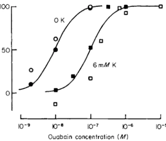
coside in inhibiting the preparation is greatest when the glycoside is applied in the absence of Κ ions and becomes progressively less as the Κ concentration is increased. Measurement of the dose-response curve at different external Κ concentrations shows that as the Κ concentration is raised the curve is shifted to the right (Fig. 1).
The basis of this antagonism has recently been examined using 3H- ouabain to measure the binding of glycoside to pumping sites in cul
tures of various mammalian cells including HeLa cells and a cell line derived from the human heart [26,26a]. HeLa cells are very sensitive to glycosides and at low glycoside concentrations the bulk of the ouabain bound seems to be associated with pumping sites and ouabain binding can be used to measure the number of pumping sites per cell. One strik
ing finding was the large number of pumping sites in HeLa cells. They have 105 to 106 pumping sites/cell. When compared with the eryth- rolyte which has only 100-300 sites/cell [27,28], there is a very consid
erable difference; but HeLa cells are not peculiar in their large pumping capacity, similar values having been found in kidney and brain tissue and it seems that it is the erythrocyte with its low permeability to cations which is the exception rather than the rule. The large number of
250 P . F . B A K E R 100 r
5 0 h
ΙΟ"9 ΙΟ" 8 ΙΟ" 7 Ό~(
Ouabain concentration {M)
10"
FIG. 1. Comparison on the same batch of HeLa cells of the effects of potassium ions on glycoside binding and inhibition of the Na pump. Glycoside binding was measured as described by Baker and Willis [26], and pumping was expressed as that fraction of the Κ influx inhibited by 10~5 Μ ouabain. Κ influx was measured over 5 minutes in 6 m M Κ following exposure to ouabain for 1 hour in the presence or absence of K. Ordinate: percent inhibition of pumping (open symbols) or percent of the maximum specific glycoside binding (closed symbols). Abscissa: ouabain concentration (M) on a log scale. The curves were drawn by eye. Temperature, 35° C. Note the stimulation of pumping in 6 m M K and 10" 8 Μ ouabain. (From Baker and Willis [26a].)
pumping sites per cell and high sensitivity to glycosides make HeLa cells an ideal preparation for studying the effects of Κ on glycoside binding.
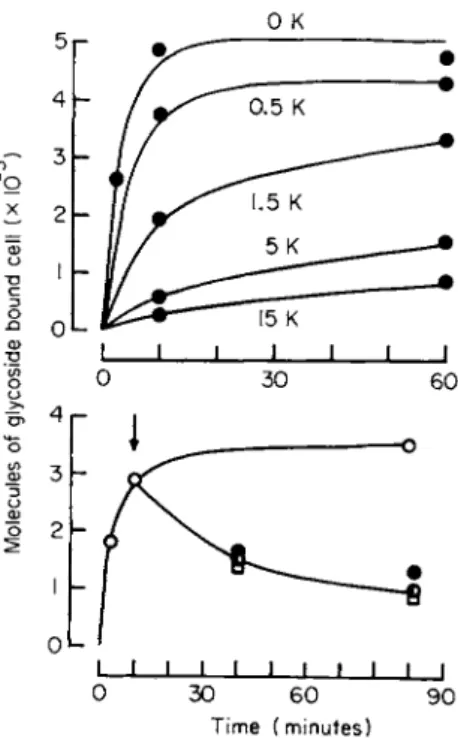
Examination of the kinetics of binding showed (1) that binding is first order with respect to glycoside concentrations; (2) that binding is reversible; and (3) that Κ ions markedly reduce the rate of binding but have no effect on the rate of release (Fig. 2). Quantitatively these effects can explain the observed shift in the dose-response curve.
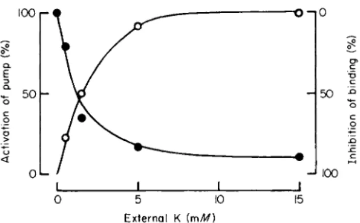
A particularly striking observation was the effectiveness of low concentrations of Κ ions in antagonizing binding. The Κ concentration required to effect half-maximal inhibition of binding was much the same as that required for half-maximal activation of pumping (Fig. 3).
Reduction of external Na, which increases the affinity of the pump for K, also reduced the Κ concentration required to effect half-maximal inhibition of binding. These results show that as the Na pump is activated the rate of glycoside binding is reduced. These results are consistent with competition between Κ (or K-like) ions and ouabain for some binding site at the cell surface. If the rate of pumping is propor
tional to the fraction (FEK) of the total number of K-binding sites (E) combined with K, then the rate of combination with ouabain behaves as
7. N a PUMP AND CELLULAR METABOLISM 251
0 30 60 90 Time (minutes)
FIG. 2. The influence of Κ ions on the rates of binding and release of 3H-ouabain from HeLa cells. For both graphs, ordinate: number of molecules bound per cell (divided by 105); abscissa: time (minutes). The upper graph shows the rate of binding from a glycoside concentration o f 2 x l O ~7M a t the Κ concentrations (mM) specified. The lower graph shows the rate of binding from 2 x 10" 7 Μ ouabain in K-free medium ( O ) and the rate of release of tracer under a variety of conditions. At the arrow the cells were washed free of extracellular tracer and reincubated in media which were either ouabain-free and K-free (C), ouabain-free but containing 15 mM Κ ( φ ) , or K-free containing 2 χ 1 0 "5 Μ cold ouabain ( • ) . Temperature, 35°C. The two graphs were obtained on different batches of cells. (From Baker and Willis [26a].)
if it were proportional to (1 — FEK). This might come about if ouabain can combine with Ε but is unable to combine with EK either because EK has a different conformation from Ε or because combination with Κ allows Ε to move to the inner face of the cell membrane where it is unavailable to react with externally applied ouabain. It should be stressed that it is not essential for ouabain and Κ to combine with the same site on E.
According to this view we can write the following reactions as occurring at the outer surface of the cell membrane:
Ε + Κ -> ΕΚ -> pumping E + 0->EO
252 P. F. BAKER
o n 0
c
ο c ο
•φ
-Q c
-J 100
0 5 10 15
External Κ [mM)
FIG. 3. Comparison on the same batch of HeLa cells of the effects of Κ ions on activation of the Na pump (expressed as a percent of the maximum ouabain-sensitive Κ influx, O ) and inhibition of glycoside binding (expressed as a percentage of the glycoside bound in a K-free medium during exposure to 2 χ 10~7 Μ ouabain for 5 minutes, # ) . Abscissa:
external Κ concentration (mM). Temperature; 35°C. (From Baker and Willis [26a].)
This is a case of competitive inhibition and ouabain (O) will reduce the affinity for Κ without affecting Km a x. Such simple competition has not been seen in all tissues. In the squid axon, Km a x is reduced without change in affinity for Κ [28a], and in a number of other tissues [9,29,30]
and transport ATPase preparations [31], ouabain reduces both Vmax and the affinity for K. These observations can be explained quite easily. The noncompetitive nature of inhibition in squid axons is probably explained by the relative irreversibility of glycoside binding in this tissue. It follows that equilibrium will not be reached within the useful life of the prep
aration and ouabain will reduce the number of pumping sites in an apparently noncompetitive manner. In those tissues where mixed inhibition is observed, it is possible to account quantitatively for the observed kinetics if it is assumed that Κ can also combine reversibly with EO although with lower affinity than with E. The product EOK may either be unable to participate in pumping or take part at a much slower rate. At equilibrium the rate of pumping is given by
where ρ is a constant of proportionality, LK and LQ are the equilibrium constants for the combination of Ε with Κ and O, respectively, LK' is the equilibrium constant for the combination of EO with K, and
Pumping rate = pFEK = ρ (2)
7. N a PUMP AND CELLULAR METABOLISM 253 [K] and [O] are the concentrations of potassium and ouabain, respec
tively. Provided LK' > LK, raising the glycoside concentration will decrease both Km a x and the apparent affinity for Κ in accord with observations.
To return to the clinical significance of K-ouabain antagonism, when Κ ions are used as an antidote to an overdose of glycoside, alle
viation is achieved in part by activating those pumping sites that are uncombined with glycoside and in part by effecting a reduction in the number of pumping sites combined with glycoside.
III. INTERACTIONS B E T W E EN THE Na PUMP A ND CELLULAR METABOLISM A ND FUNCTION
A. Introduction
This is a field in which new information is appearing daily. At the risk of oversimplification, there seem to be four primary ways in which the Na pump can influence cellular metabolsim and function. These are (1) by competition for ATP; (2) via the absolute levels of Na and Κ both inside the cell and immediately external to the cell membrane; (3) via the gradients of Na and Κ ions created by the Na pump; and (4) via the potential that can be generated by the pump. Each category en
compasses a number of specific cellular processes many more of which have probably still to be described. It is beyond the scope of this pre
sentation to deal exhaustively with all aspects of this topic; but I wish to point to some of the more interesting examples of interaction in each category.
B. Utilization of ATP
Competition for ATP is an obvious form of interaction between the pump and metabolism. It is particularly noticeable in cells with a large surface area/volume ratio where much of the metabolsim is directed toward supplying ATP for pumping [99a]. It is also important in certain epithelial tissues that effect the transcellular transport of large quanti
ties of Na ions (e.g., gut, kidney). In these cells the rate of pumping is relatively important in determining the ATP/ADP ratio and the amount of free inorganic phosphate in the cell, both of which may influence other metabolic events. For instance, the ADP level controls the rate of electron transport and oxygen consumption by the mitochondrion, and the Pf level is important in determining the rate of glycogenolysis.
254 P. F. BAKER
The breakdown of ATP, and more especially of the substrates used in the resynthesis of ATP are processes that yield energy in the form of heat, and the rate of pumping may be an important factor in determining the rate of heat production in a tissue. This may have important physio
logical implications. For instance the Na pump is implicated in the mechanism of thermogenesis in brown fat tissue, which is an important source of heat especially in the newborn animal. The mechanism of thermogenesis has been studied in some detail in brown fat tissue of the rat [32]. The stimuli that trigger increased heat production are the catecholamines adrenalin and noradrenalin. These drugs produce an immediate fall in resting potential, which seems to result from an in
crease in the permeability of the fat cell membrane to Na ions. The increased inward leak of Na stimulates the Na pump, which, in turn, utilizes ATP and leads to increased ATP production at the expense of fat. According to this view, adrenalin initiates an ion cycle in which activation of the Na pump provides a means of using up ATP and so allows a high rate of fat breakdown and heat production.
C. The Absolute Levels of Na and Κ
It can be difficult to distinguish between metabolic effects that depend on the absolute levels of Na and Κ and effects that depend solely on the ion gradients across the cell membrane. If the gradient is solely respon
sible, within certain limits it should be possible to compensate experi
mentally for a rise in Naf by increasing extracellular Na; but such experiments are often extremely difficult to perform satisfactorily. As a rough generalization, it seems likely that the absolute levels of Na and Κ will be important for soluble enzymes; but, wherever membranes are involved, either at the cell surface or at the surface of intracellular organelles, the ion gradients may also be relevant.
A number of enzymes and enzyme systems are activated by Κ ions (e.g., pyruvate kinase); but it is difficult to assess the extent to which changes in the absolute levels of Naf and K, influence these enzymes in vivo. It is particularly relevant that enzymes that are activated by Κ ions are often inhibited by Na; this means that a small fall in Kf and a gain in Naf will have a greater effect than a fall in Kf alone.
There is some evidence that changes in the intracellular levels of Na and Κ have very profound effects on the synthesis of macromolecules.
In a variety of cultured cells a fall in Kf, coupled presumably to a rise in Naf, reduces the incorporation of precursors into DNA, RNA, and protein [33,34]. Rather similar results have been obtained on brain slices where a rise in Naf and a fall in K, result in reduced incorporation
7. N a PUMP AND CELLULAR METABOLISM 255 into RNA and protein [35,36]. An interesting observation is that not all macromolecular synthesis in brain tissue is inhibited under these con
ditions: Jones and Banks [36] have reported increased incorporation into nuclear proteins following application of ouabain to brain slices.
A rise in Nat- and fall in K, also seems to inhibit cell division [36a,36b].
These observations are of considerable interest, but they should be treated with some caution. It is not fully certain that the observed effects result solely from changes in Na£ and Kf. They might be secondary to these changes. For instance, alteration in the ion gradients across the cell membrane may lead to a fall in the intracellular concentration of some essential metabolite or activator or to a rise in the intracellular concentration of an inhibitor (see Section III,D). Nevertheless, the results show that changes in the intracellular levels of Na and Κ can influence the synthesis of macromolecules, and this observation imme
diately suggests a number of important questions. Which macromole
cules are most sensitive to changes in intracellular ions? Can small changes in ions switch on or off the synthesis of individual proteins or even effect the transcription of individual genes as has been postulated to occur in insects [37,38]? Is there any feedback tending to stimulate production of the transport systems required to bring the ionic composi
tion back to normal ?
From a different standpoint, these changes in the synthesis of macro- molecules may be of importance in the therapeutic actions of Li ions.
As discussed earlier, it seems likely that Li is accumulated in nerve cells to the exclusion of K. It would seem worth examining whether such changes in internal ions produce any alterations in the synthesis of macromolecules.
D. The Gradients of Na and Κ Ions
The ion gradients created by the Na pump represent a store of poten
tial energy that is used by cells in a variety of ways, including the generation of transmembrane potentials and the transport of substances against their electrochemical gradient. A few important examples are discussed below.
7. Membrane Potentials
Each ion gradient represents a battery capable of generating a poten
tial difference across the cell membrane. Whether or not such a po
tential is generated depends on the other ion gradients present and the
256 P. F. BAKER
relative permeability of the membrane to these ions. Thus, if we are considering only the relative permeability to Na and Κ ions and the permeability to other ions is assumed to be extremely low, the potential difference will be given by
F - 5 8 1 o g1 0 [ K ] j + 6 [ N a ] j (3) where b is the relative permeability to Na and Κ ions (PHJPK) and [ ]
are activities. If the membrane is completely impermeable to Na ions, the potential will be determined solely by Κ and, as there is more Κ inside the cell than outside, the internal potential will be negative with respect to the outside. If, on the other hand, the membrane is imper
meable to Κ ions, the potential will be determined solely by Na and, because there is more Na outside the cell than inside, the internal potential will be positive. These two examples represent the extremes of potential that can be obtained using the Na and Κ gradients alone. By varying the permeability ratio b, all intermediate potentials can be obtained. Under physiological conditions the permeability to anions (usually chloride) must also be considered. In general, most animal cells are more permeable to Κ and CI than to Na and the internal potential of these cells is negative with respect to the external medium.
Although the ratio of permeabilities PNa:PK:Pci may be quite charac
teristic of a particular cell, it does not always remain constant. Thus the relative permeability may change as a function of membrane potential or following application of various chemicals such as transmitter substances or drugs. A particularly striking example of such changes in permeability is the action potential of excitable tissues. Conduction along nerve and muscle fibers is essentially electrical in nature; but without some boosting mechanism a current fed in at one end of a nerve would be attenuated long before it reached its destination. The mecha
nism by which the flow of current is boosted is known as the action potential. It results from three temporally distinct changes in permeabil
ity that occur in response to a sudden reduction in membrane potential (depolarization). These are (1) a rapid increase in the permeability to Na ions; (2) a less rapid blocking or inactivation of the increase in permeability to Na ions, and (3) a slow maintained increase in the permeability to Κ ions. The upshot of these permeability changes is that the internal potential swings from negative to positive and back again to negative, the period of positivity serving to boost the flow of electric current along the fiber [see 39,40].
A common feature of all these potential generating mechanisms is that they involve the movement of ions down their electrochemical gradients
7. N a PUMP AND CELLULAR METABOLISM 257 and if the potential is to remain constant the gradients must be maintain
ed by pumping. Thus after passage of an' action potential, the interior of the cell is richer in Na and poorer in K, and it follows that electrical activity is dependent on the operation of the Na pump—although it is important to realize that the Na pump and action potential mechanisms are two quite distinct processes (see Table I). The extent to which they are separable in practice depends on the surface area/volume of the cell.
In large nerve fibers, with a small surface area/volume, each action potential causes little change in Nat and many action potentials can be carried in the absence of a functioning Na pump; but in small fibers with a large surface area/volume, maintenance of the ion gradients requires continuous operation of the Na pump.
2. Control of Cell Volume
The maintenance by the Na pump of a stable low Naf and high K( coupled with the greater permeability of cells to Κ ions plays an impor
tant part in the control of cell volume. This has been discussed by Hodg
kin [41]. If it is assumed that the gradients of Na and Κ are influenced by the operation of the Na pump, whereas CI ions distribute themselves passively across the membrane; in the steady state the potential (V) will be given by
RT [K]„ + Z>[Na]„ _ [Cl](
v
-T
ln[K
]i+b[N
a]
i-
RT,Flnm
(4)(5) from which
[Cl]f = [K]0+&[Na]0 [C1L [KL+6[NaL
If it is assumed that Naf is kept at a constant low value by a neutral Na pump, the internal concentrations of K, CI, and the cell volume (v) can be determined by Eq. (5) in conjunction with
[KL + [Na]f + [Cl]f + ^ =C (osmotic balance) (6) and
[K]f + [Na]f - [ C 1 L + ^ = 0 (electroneutrality) (7) where Cis the total concentration of all particles in the external solution, A is the total quantity of indiffusible particles inside the cell, and ζ is the average valency of these particles. If 6 = 0.01, the solution of these
258 P. F. BAKER T A B L E I
SUMMARY OF THE EVIDENCE SHOWING THAT THE ACTION POTENTIAL AND N A+ PUMP ARE T W O SEPARATE AND INDEPENDENT PROCESSES
Type of experiment Direction of ion
movements
Ionic selectivity
Linkage between N a+
ion and K+ ion movements Effect of metabolic
inhibitors
Source of energy
External calcium
Maximum rate of movement of N a+ ions
Specific blocking agents Tetrodotoxin
(ΙΟ"7 M) Ouabain
Effect of low tempera
ture
Action potential Down electrochemical
gradients, i.e., N a+ ions into the axon and K+ ions out of it Does not distinguish
between N a+ ions and L i+ ions
Independent; can have N a+
ion movements in the absence of K+ ions and vice versa
Unaffected by poisoning with cyanide (2 m M ) or 2,4-dinitrophenol (0.2 m M )
Potential energy stored in the ion gradients; can occur in simple salt solutions in the complete absence of other energy sources
An increase reduces excitability; a decrease increases excitability 10,000 /Lt/xmoles/cm2 sec
shows no evidence of saturation
Blocks
No effect at 10" 3 Μ Slows rates of change of
N a+ ion and K+ ion conductances, but does not alter their maximum value
N a+ pump Against electrochemical
gradient, i.e., N a+ ions out of the axon Will not pump L i+ ions
Movements linked; N a+
ion extrusion stops in a K+ ion-free medium Inhibited in a fully
poisoned axon
Energy-rich phosphate compounds probably adenosine triphosphate
Very little effect
60 /Lt/xmoles/cm2 sec;
displays saturation kinetics
No effect Blocks at 10" 7 Μ Maximum velocity much
reduced
a The data refer to squid axons under near-physiological conditions.
7. N a PUMP AND CELLULAR METABOLISM 259 equations agrees with data obtained in muscle. Thus provided the permeability to Na is small compared with that to K, a neutral Na pump could maintain the cell in a steady state similar to that seen in vivo.
If the Na pump is electrogenic, another term would have to be added to Eq. (5) but it would not alter the general conclusion.
3. Active Transport Processes Drawing Energy from the Downhill Movements o/Na and/or Κ Ions
Providing a suitable coupling mechanism is available, the movement of Na or Κ or both down their respective electrochemical gradients can be used to drive other substances against an electrochemical gradient. A few examples are described below.
a. Transport of Amino Acids and Sugars. Ion-gradient coupling plays an important part in the uptake of amino acids into a variety of cells and also in the transport of sugars into various epithelial cells such as the kidney and small intestine. Both these topics are covered elsewhere in this volume (Chapters 2, 10, and 12) and will not be discussed further except to point out that coupling of amino acid or sugar transport to, say, the Na gradient provides a very flexible source of energy. To take an example, to produce a tenfold gradient of a neutral amino acid requires about 2 kcal of energy. ATP can provide about 10 kcal; thus if the phosphate bond energy of the ATP is to be utilized efficiently, 3 or 4 amino acid molecules would have to be transported per energy-rich phosphate bond split. As the Na pump transports 3 Na out of the cell in exchange for 2 K/energy-rich phosphate bond split, allowing each Na ion to reenter the cell with 1 molecule of neutral amino acid transports 3 molecules of amino acid/energy-rich phosphate bond split and provides an efficient utilization of cellular energy. To achieve an accu
mulation ratio much greater than 10:1, transport of 1 molecule of material could be coupled to the inward movement of 2 or even 3 Na ions or to one or more Na ions moving into the cell in exchange for one or more Κ ions moving out.
Although ion-gradient coupling avoids the necessity for a whole series of ATP-requiring mechanisms in the cell membrane, in no case is the mechanism of gradient coupling known. Recent work on the carrier
like behavior of various cyclic peptides suggests one realistic model for this kind of coupling. For instance, an Na carrier in the cell membrane might become linked to a glucose or amino acid carrier, and it is not difficult to imagine that the binding of Na to the Na carrier could lead to a conformational change in the carrier dimer, increasing the affinity
260 P. F. BAKER
of the uncombined carrier for substrate as is observed experimentally in systems transporting amino acids and sugars.
b. Transport of Calcium Ions. Another particularly interesting example of ion-gradient coupling is the maintenance of a low intra
cellular level of Ca. In some cells ATP is used directly to extrude Ca ions [42]; but in excitable tissues and possibly the gut and elsewhere, there is evidence that the inward movement of Na and possibly the outward movement of Κ provide energy for the expulsion of Ca ions from the cell [43-45]. APT may also be required.
Extrusion of Ca can take place against a very steep electrochemical gradient. In nerve and muscle cells the concentration of free ionized Ca is 10~8 to 10"6 M, whereas that in the external medium is about 10"3 M. If the inward movement of 2 Na ions is coupled to the extrusion of 1 Ca ion, the Ca gradient that can be achieved is given by
] £ a L =[ W ( g i
[Ca]0 [Na]0 2 ^ ;
If Naf/Na0 is 1/10, this would give a Caf of 10"5 Μ which is not low enough; but if 3 Na ions moved in in exchange for 1 Ca, with one Na either moving down the electrochemical gradient or exchanging for K, the intracellular Ca would be given by either
[Ca1i [Na3*3 VF/RT / Q \
[Ca], [Na],3* w
or
[CaL = [ N a £ [Kl
[Ca]0 [Na]0 3 [K], 1 }
both of which are capable of achieving the observed range of intra
cellular Ca concentrations.
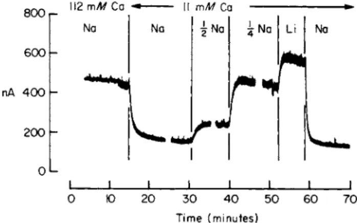
A notable feature of Na-dependent Ca transport is that reducing the external Na concentration not only reduces the Ca efflux but also increases the Ca influx. It must therefore lead to a rise in intracellular Ca. In large cells it is possible to monitor the free intracellular Ca concentration directly by introducing the Ca-sensitive protein aequorin into the cell [46,46a,46bj. Figure 4 shows that Ca, is dependent on Ca0 and that at a constant Ca0 replacement of Na0 by Li results in a rise in Caf [46c].
Ca influx is also increased by raising Naf. Of particular interest is the
7. N a PUMP AND CELLULAR METABOLISM 261
800 r- 600 h
112 mM Ca Ii mM Ca
nA 400 200
0 10 20 30 40 50 60 70 _L J
Time (minutes)
FIG. 4. Changes in the ionized Ca concentration inside a squid axon following alterations in the ionic composition of the external medium. The axon had been injected with the Ca- sensitive protein aequorin and the light emitted, presumably through reaction of the aequorin with Ca ions is expressed on the ordinate in nA. Abscissa: time (minutes).
Each break in the record represents a gap of about 10 minutes between solution changes.
(A reduction in N a0 was compensated by a rise in Li0.) Temperature 20°C. Data of Baker et al. [46c].
observation that the Ca influx increases at least as the square of the internal Na concentration and possibly as a higher power [43,47], Thus a small rise in internal Na results in a large increase in Ca influx.
There is also evidence that a rise in internal Na can release Ca from mitochrondria. These observations may have important physiological and pharmacological implications. For instance, the cardiac glycosides increase both the force of contraction of the heart and also the resting and stimulus-dependent secretions of various substances including nervous transmitters and hormones [for references, see 43]. Both actions require Na ions and the glycosides are only effective at con- centrations that effect partial or complete inhibition of the Na pump. It seems likely that glycosides first produce a rise in intracellular Na that leads, in turn, to a rise in intracellular Ca. Although this mechanism could explain the increased resting level of secretion, it might be argued that a small rise in the level of free intracellular Ca would have little effect on the contraction or secretion triggered by a large entry of Ca during the action potential [46b,48,49]. This objection might be overcome if both secretion and contraction are nonlinear functions of the internal calcium concentration. Dodge and Rahamimoff [50] have presented evidence that the release of acetylcholine at the neuromuscular
262 P. F. BAKER
junction is dependent on the fourth power of the Ca concentration and Katz and Miledi have shown that transmitter release at the squid giant synapse depends on at least the second power of the calcium concentra
tion [50a]. If a similar relation exists in other systems, it would follow that a small rise in the resting Ca level may increase considerably the effectiveness of the action potential at eliciting contraction or secretion.
In muscle it is also necessary to consider the Ca-binding properties of the sarcoplasmic reticulum and it is possible that raising Ca, leads to increased loading of the reticulum thus making more Ca available for release during contraction. This might provide a further means of amplifying small changes in Caf.
Apart from its role in initiating contraction and secretion, a rise in intracellular Ca in some tissues causes an increase in the permeability of the surface membrane to Κ ions [51-53] and in others affects various enzyme systems, including activating glycolysis by conversion of phos- phorylase b to phosphorylase a by phosphorylase b kinease [53a,53b]
and inhibition of the Na pump. This last effect probably requires a Ca concentration greater than would normally exist inside most cells, but the first two seem to occur under physiological conditions. It is possible that other enzymes will prove to be sensitive to Ca ions when tested at the Ca concentrations existing inside cells.
c. Transport of Mg and Η Ions. The intracellular concentrations of both these ions are less than would be expected for an ion distributed in accordance with the resting potential, but virtually nothing is known about the mechanisms that maintain this distribution. An obvious possibility is some form of ion-gradient coupling and this would seem well worth examining.
Bondani and Withrow [53c] have reported that ouabain produces a fall in intracellular pH which suggests that the Na pump may be involved in extruding Η ions either directly or by maintaining the gradi
ents of Na and Κ ions, and Baker and Crawford [53d] have shown that the extrusion of Mg ions from squid axons is dependent on the presence of Na ions in the external medium.
d. Transport of Cl. In many, but not all, cells chloride is distributed in accordance with the membrane potential. The squid axon is a tissue in which the intracellular Cl concentration is higher than would be pre
dicted from the resting potential and Keynes [53e] has shown that Cl is taken up by an energy-dependent mechanism. Chloride uptake is unaffected by ouabain and so seems not to be directly linked to the Na pump; although the possible involvement of ion gradients has not been examined. The uptake of iodide by the throid gland is inhibited by ouabain and appears to be linked to operation of the Na pump [53fj.
7. N a P U M P A N D C E L L U L A R M E T A B O L I S M 263
E. Electrogenic Pumping and the Control of Nervous Function
The Na pump contributes to the membrane potential in two ways (1) by maintaining the ion gradients and (2) by its own electrogenic behavior in driving a stream of Na ions outward across the cell mem
brane. In large cells where it is possible to stop the Na pump without also changing the ion gradients, it is possible to distinguish between these two sources of potential; but in small cells such a distinction is extremely difficult.
Moreton [54] has shown that the constant field equation can be modified to account for electrogenic pumping by the introduction of a
"sodium pump term," RTMa/VFpK[¥L]i9 where Ma denotes the rate of pumping which presumably is itself a function of KQ
M
aoz[\l(\+L
Kl[K])
nwhere LK is the apparent affinity for K, and η is the number of Κ ions required to activate the pump. The full equation is
PVFIRT= [ K ]0+ PN a[ N a ]Q RTMa
[KL+/7JKL ^FVp^K], ^ ; The presence of this sodium pump term is most noticeable at low K0, where it can produce a hyperpolarization greater than that obtained in the complete absence of external K. The term is most prominent in the physiological range of Κ concentrations, where increased pumping rates will tend to cause hyperpolarization. Such hyperpolarization will be most prominent if the passive permeability to ions is low, i.e., the membrane has a high electrical resistance. A further feature of impor
tance in the nervous system is that the extracellular space immediately external to the neurons is quite small, and, after nervous activity, Κ ions tend to accumulate just outside the cell membrane, tending to depolarize it. Pumping, on the other hand, will tend to lower K0 below the normal level and so hyperpolarize the membrane. The maintenance of a constant K0 may be a particularly important function of the Na pump in the central nervous system. It has been suggested that one role of glial cells may be to regulate K0 and disturbance of glial cell function may lead to a rise in K0 [54a]. This may have relevance to focal epilepsy, which has recently been ascribed to glial cell malfunction.
It is thus necessary to consider three effects of the Na pump on excit
ability—(1) maintenance of high Kf and low N ai 9 (2) effects on K0, and (3) electrogenic behavior.
In considering the interactions between the Na pump and the control of nervous activity it is convenient to make a rough division of the
264 P. F. BAKER
nervous system into three parts—(1) receptors—regions giving rise to action potentials; (2) axons—routes by which the action potential is conducted; and (3) terminals—regions effecting transmission of the nerve impulse from one cell to another. Observation has shown that depolarization and hyperpolarization, which from the standpoint of this discussion could result from altered rates of pumping, have very different effects on the behavior of these three regions of a nerve cell.
Thus progressive depolarization increases the rate of generation of action potentials at a receptor, but decreases the quantity of transmitter released per action potential at the nerve terminal. Hyperpolarization has the opposite effects. Over a wide range of potentials, conduction along the axon is little affected, but large depolarizations lead ulti
mately to conduction block through inactivation of the Na permeability mechanism.
At the present time there is relatively little information about the importance of these effects in the nervous system [see, for instance, 55].
Interaction between pumping rate and receptor potential may provide a mechanism for sensing oxygen (carotid body [56]) or glucose (hypothala
mus [57]). The most sensitive system would be very fine nerve terminals in which a high rate of pumping is necessary to maintain their resting potential. Any reduction in energy supply to the Na pump (i.e., lack of glucose or oxygen) would lead to a reduced rate of pumping, depolar
ization of the cell membrane, and an increased rate of action potential generation. The importance of cell size in constructing a sensory device of this kind is best illustrated by the following calculation. If it is assumed that (1) a cell has about 10 mM of energy-rich phosphate, (2) the Na pump transports 3Na/ ~ Ρ at a rate of 45 pmoles/cm2 second, and (3) all ~ P is used up by the pump and none by other processes, on stopping the Na pump the time taken to use up all the stores of ~ Ρ in a cylindrical nerve cell would be 4 seconds for a cell of diameter 0.2 μ, 400 seconds for a cell of diameter 20 μ, and 40,000 seconds for a cell of diameter 2000 μ. The carotid body responds to anoxia or cyanide with a lag of 2-3 seconds, and Mills and Jobsis [57a] have shown that cyto
chrome a3 is reduced in parallel with the rise in discharge.
Apart from exerting a direct control over action potential production, the rate of pumping might augment or reduce the effectiveness of some other stimulus. Thus a reduction in pumping rate should lower the threshold while an increase should raise it. An increased pumping rate might follow a period of depolarization and entry of Na, or it might be brought about by the direct action of a transmitter substance on the Na pump. Experimental evidence in favor of this latter suggestion has been advanced by a number of workers, but in no instance is the case strong
7. N a PUMP AND CELLULAR METABOLISM 265 and the available evidence now favors alternative explanations [58a].
It is possible that an increase in pumping rate may play some part in the slow adaptation of receptors.
Similar arguments apply to the nerve terminal except that here depolarization reduces the amount of transmitter released per action potential and hyperpolarization increases it. There is good evidence that increased pumping in the nerve terminal contributes to post-tetanic potentiation. During the tetanus, Na, is elevated to such an extent that following the tetanus the Na pump is activated and the resting potential raised above its normal level. An action potential invading the terminal at this time effects the release of more transmitter. Nakajima and Takahashi [58] have shown that post-tetanic hyperpolarization in stretch receptor neurons of the crayfish is abolished, with little change in membrane resistance, by removal of external K, by dinitrophenol, by substitution of Li for Na0, or by application of cardiac glycosides—all of which inhibit the Na pump. It should be stressed, however, that post-tetanic potentiation is a complex process and increased activity of the Na pump is only one factor contributing to it. Accumulation of Ca is also important.
F. General Remarks
It might be argued that the possible interactions between pumping and metabolism discussed above are of theoretical interest only because, in most cells, changes in pumping rate do not occur and hence the ion gradients, ATP level, etc., remain constant. This is clearly not the case in nerve where periods of increased activity lead to a rise in Na*, a fall in Ki 5 a rise in Pf, and a fall in the ATP/ADP ratio. Such changes can certainly affect the ion gradient-dependent uptake of various amino acids and transmitter substances, and it is possible that periods of prolonged nervous activity may influence the synthesis of macro
molecules in the cell body. Indeed, one wonders whether such changes in macromolecule production may play a part in long term alterations in central nervous system function such as must occur in learning. In other cells the permeability to Na and Κ can be altered by various hormones, and Na uptake can be increased during accumulation of amino acids. The pump may also be slowed by a shortage of ATP either because cellular ATP is being consumed by a process with a higher affinity for ATP than the Na pump or because of some general
ized reduction in ATP production, for instance in anoxia or hypogly
cemia. Apart from these physiological means of altering the pumping rate, many drugs,for instance,cardiac glycocides,can produce similar changes.
266 P . F . B A K E R
IV. C O N T R OL OF PUMPING CAPACITY
As the Na pump plays such an important part in the control of normal metabolism and function, it would seem likely that, in the long term, cells are capable of maintaining a constant low level of intracel
lular Na by matching their pumping capacity to the inward leak of Na ions. Examination of a variety of cells shows a rough corrrelation of this kind: cells with a large leak having more pumps than cells with a low leak [26]; but no one has demonstrated changes in the number of pumping sites in a cell in response to an experimental alteration in the leakage flux or to a reduction in the number of functional pumps, e.g., after the application of a low concentration of ouabain. Some cells may, however, be capable of such changes. In hereditary spherocytosis where erythrocytes have an increased leak, there is also an increase in pumping activity [59]. But a note of caution is necessary here because no measurements of glycoside binding have been made and there is therefore no clear evidence for a genuine increase in the number of pumping sites per cell. Another possibility is that there are a number of masked or inactive pumping sites in the cell membrane which can be activated when required. In this context the stimulation of potassium transport into low Κ sheep erythrocytes by external application of a specific antibody is particularly interesting [60]. It is, of course, possible that the erythrocyte with its small number of pumping sites is a special case and that the pumping capacity of most mammalian cells (105 to 106 pumps/cell) may represent the maximum number of pumps that can be accommodated in the cell membrane and any further control must be at the expense of the leakage pathway. Another possibility is that some control of pumping capacity can be achieved by altering the turnover rate of individual pumps. For instance, in the goldfish intestine there is evidence that the turnover rate of the Na pump depends on the relative amounts of saturated and unsaturated fatty acids in the cell membrane [61].
If cells do possess some feedback mechanism which under appro
priate conditions can lead to the synthesis of more pumps, this would provide a potentially valuable tool for elucidating the chemistry of the Na pump because it might allow parts of the pumping mechanism to be isolated before they are irreversibly bound to the membrane.
REFERENCES
1. P. F. Baker, Endeavour 25, 166 (1966).
2. I. M. Glynn, Brit. Med. Bull. 24, 165 (1968).
3. P. C. Caldwell, A. L. Hodgkin, R . D. Keyes, and Τ. I. Shaw, / . Physiol. (London) 152, 561 and 591 (1960).
7. N a PUMP AND CELLULAR METABOLISM 267
4. P. F. Baker, A. L. Hodgkin, and Τ. I. Shaw, / . Physiol (London) 164, 330 (1962).
5. F. J. Brinley and L. J. Mullins, / . Gen. Physiol. 52, 181 (1968).
6. G. Gardos, Acta Physiol. Acad. Sci. Hung, 6, 191 (1954).
7. L. J. Mullins and F. J. Brinley, J. Gen. Physiol. 53, 704 (1969).
8. P. F. Baker, / . Physiol. (London) 180, 383 (1965).
9. P. F. Baker and C. M. Connelly, / . Physiol. (London) 185, 270 (1966).
9a. R. Whittam, "Transport and Diffusion in Red Blood Cells." Arnold, London, 1964.
10. J. C. Skou, Prog. Biophys. 14, 131 (1964).
11. P. J. Garrahan and I. M. Glynn, / . Physiol. (London) 192, 237 (1967).
12. P. J. Garrahan and I. M. Glynn, / . Physiol. (London) 192, 189 (1967).
13. P. F. Baker, M. P. Blaustein, R. D. Keynes, J. Manil, Τ. I. Shaw, and R. A. Steinhardt, J. Physiol. (London) 200, 459 (1969).
13a. P. de Weer, Nature (London) 219, 730 (1968).
14. I. M. Glynn and V. L. Lew, J. Gen. Physiol. 54, 289s (1969).
14a. A. F. Rega, M. J. Pouchan, and P. J. Ganahan, Science 167, 55 (1970).
15. A. K. Sen and R. L. Post, J. Biol. Chem. 239, 345 (1964).
16. R. Whittam and Μ. E. Ager, Biochem. J. 97, 214 (1965).
17. P. J. Garrahan and I. M. Glynn, J. Physiol. (London) 192, 217 (1967).
18. E. J. Harris, / . Physiol. (London) 193, 455 (1967).
19. G. A. Kerkut and R. C. Thomas, Comp. Biochem. Physiol. 14, 167 (1965).
20. R. P. Kernan, Nature (London) 193, 986 (1962).
21. R. H. Adrian and C. L. Slayman, J. Physiol. 184, 970 (1966).
21a. S. B. Cross, R. D. Keynes, and R. Rybova, / . Physiol. (London) 181, 865 (1965).
22. R. C. Thomas, J. Physiol. (London) 201, 495 (1969).
23. J. C. Skou, Biochim. Biophys. Acta 23, 394 (1957).
24. J. D. Robinson, Biochemistry 6, 3250 (1967).
24a. J. D. Robinson, Nature (London) 220, 1325 (1968).
25. M. Fujita, K. Nagano, N. Mizuno, Y. Tashima, T. Nakao, and M. Nakao, Biochem. J.
106, 113 (1968).
25a. R. D. Keynes, / . Physiol. (London) 166, 16P (1962).
25b. Ε. E. Carmeliet, J. Gen Physiol. 47, 501 (1964).
26. P. F. Baker and J. S. Willis, Biochim. Biophys. Acta 183, 646 (1969).
26a. P. F. Baker and J. S. Willis, Nature (London) 226, 521 (1970).
27. J. F. Hoffman, J. Gen. Physiol. 54, 343s (1969).
28. J. C. Ellory and R. D. Keynes, Nature (London) 221, 776 (1969).
28a. P. F. Baker and J. S. Willis, / . Physiol, (in press).
29. H. J. Schatzmann, Biochim. Biophys. Acta 94, 89 (1965).
30. A. L. Abeles, / . Gen. Physiol. 54, 268 (1969).
31. H. Matsui and A. Schwartz, Biochim. Biophys. Acta 128, 380 (1966).
32. L. Girardier, J. Seydoux, and T. Clausen, J. Gen. Physiol. 52, 925 (1968).
33. M. Lubin, Nature (London) in, 451 (1967).
34. R. J. Kuchler, Biochim. Biophys. Acta 136, 475 (1967).
35. C. Prives and J. H. Quastel, Nature (London), 221, 1053 (1969).
36. C. T. Jones and P. Banks, Biochem. J. 114, 62P (1969).
36a. J. F. Lamb and D . McCall, / . Physiol. 206, 33P (1970).
36b. E. Robbins, J. Pederson, and P. Kleen, / . Cell. Biol. 44, 400 (1970).
37. H. Kroeger, Nature (London) 200, 1234 (1963).
38. M. Lezzi, Exp. Cell Res. 43, 571 (1966).
39. A. L. Hodgkin, " Conduction of the Nervous Impulse." Liverpool Univ. Press, Liver
pool, 1964.
268 P. F. BAKER 40. B. Katz, "Nerve, Muscle and Synapse." McGraw-Hill, New York, 1966.
41. A. L. Hodgkin, Proc. Roy. Soc, Ser. Β 148, 1 (1957).
42. Η. J. Schatzmann and F. J. Vincenzi, / . Physiol. (London) 201, 369 (1969).
43. P. F. Baker, M. P. Blaustein, A. L. Hodgkin, and R. A. Steinhardt, / . Physiol. (London) 200, 431 (1969).
44. M. P. Blaustein and A. L. Hodgkin, J. Physiol. (London) 200, 497 (1969).
45. H. Reuter and N. Seitz, J. Physiol. (London) 195, 451 (1968).
46. O. Shimomura, F. H. Johnson, and Y. Saiga,/. Cell. Comp. Physiol. 59, 223 (1962).
46a. Ε. B. Ridgeway and C. C. Ashley, Biochem. Biophys. Res. Commun. 29, 229 (1967).
46b. P. F. Baker, A. L. Hodgkin, and Ε. B. Ridgeway, J. Physiol. (London) 208,80P (1970).
46c. P. F. Baker, A. L. Hodgkin and Ε. B. Ridgeway,/. Physiol. (London) 218, 709 (1971).
47. P. F. Baker and M. P. Blaustein, Biochim. Biophys. Acta 150, 167 (1968).
48. A. L. Hodgkin and R. D. Keynes, / . Physiol. (London) 138, 253 (1957).
49. B. Katz and R. Miledi, / . Physiol. (London) 203, 459 (1969).
50. F. Dodge and R. Rahamimoff, / . Physiol. (London) 193, 419 (1967).
50a. B. Katz and R. Miledi, / . Physiol. (London) 207, 789 (1970).
51. R. Whittam, Nature (London) 219, 610 (1968).
52. V. L. Lew, 7. Physiol, 206, 35P (1970),
53. G. D. V. van Rossum, Nature (London) 225, 638 (1970).
53a. W. L. Meyer, Ε. H. Fischer, and E. G. Krebs, Biochemistry 3, 1033 (1964).
53b. E. Ozawa, K. Hosoi, and S. Ebashi, / . Biochem (Tokyo) 61, 531 (1967).
53c. A. Bondani and C. D. Withrow, Fed Proc. 24, 487 (1965).
53d. P. F. Baker and A. C. Crawford, / . Physiol. (London) 216, 385 (1971).
53e. R. D. Keynes, / . Physiol. (London) 169, 690 (1963).
53f. J. Wolff, Biochem. Biophys. Acta 38, 316 (1960).
54. R. B. Moreton, / . Exp. Biol. 51, 181 (1969).
54a. D. A. Pollen and M. C. Trachtanberg, Science, 167, 1232/1970).
55. D. A. Baylor and J. G. Nicholls,/. Physiol. (London) 203, 571 (1969).
56. T. J. Biscoe, Physiol. Rev. 51, 427 (1971).
57. R. L. Hinsworth, / . Physiol. London 206, 411 (1970).
57a. E. Mills and F. Jobsis, Nature, (London) 225, 1147 (1970).
58. S. Nakajima and K. Takahashi, / . Physiol. (London) 187, 105 (1966).
59. J. S. Wiley, Nature (London) 221, 1222 (1969).
60. J. C. Ellory and Ε. M. Tucker, Nature (London) 222, 477 (1969).