Although acini are the determining structures of tear fluid production, a secretory role of ducts in addition to their drainage function has been suspected for a long time [1,2].
Accumulating evidence suggests that the lacrimal gland (LG) duct system may modify the compositions of the primary acinar fluid [3,4]. Our previous experimental results provided direct evidence of the active role of the LG duct system in lacrimal fluid secretion [5]. Investigation of the underlying mechanisms revealed several components, but the regulation of ductal fluid secretion is still not fully understood. Previous reports demonstrated the contribution of vasoactive intestinal peptide (VIP) released by parasympathetic nerves in acinar protein secretion, but its impact on the fluid secretion of the LG ducts remained unknown [6]. VIP has been shown as a smooth-muscle-relaxant, vasodilator peptide in the lung [7], but it is also an important regulator of tear production in humans. This was illustrated by a case report of a patient who had a VIP-secreting metastatic pancreatic adenocarcinoma.
With an 80-fold elevation of serum VIP levels, this patient had tear overproduction indicating that VIP increases tear secretion in humans, most likely by stimulating LG fluid
secretion [8]. There are two known receptors for VIP termed VPAC1 and VPAC2 [9-11]. Both receptors bind VIP and pituitary adenylate cyclase-activating polypeptide (PACAP) to some degree. The VIP–receptor interaction activates the G protein Gsα that stimulates adenylyl cyclase, which increases the intracellular cAMP level. cAMP, in turn, activates protein kinase A that stimulates secretion by phosphorylating its target proteins. Hodges and coworkers suggested that most, but not all, of VIP-stimulated protein secretion is cAMP dependent. Using a myristoylated protein kinase A peptide inhibitor (PKI) based on the pseudosubstrate of protein kinase A, VIP-stimulated protein secretion was shown to be inhibited by about 70% [6].
Several hormones and neurotransmitters (including VIP) with the potential to elevate cytosolic cAMP level can acti- vate the cystic fibrosis transmembrane conductance regulator (CFTR) channel. Our previous findings strongly suggested the role of CFTR in ductal fluid secretion [12]. CFTR has been shown to play a critical role in exocrine glands, such as the pancreas, salivary, sweat glands, and airways epithelium [13-15]. Furthermore, there are several data about altered tear secretion seen in patients with cystic fibrosis implying the potential influence of CFTR [16,17]. Accumulating evidence from gene expression studies performed on rat and rabbit LGs
The regulatory role of vasoactive intestinal peptide in lacrimal gland ductal fluid secretion: A new piece of the puzzle in tear production
Orsolya Berczeli,1 Dóra Szarka,1 Gréta Elekes,1 Eszter Vizvári,1 László Szalay,1 János Almássy,2 László Tálosi,3 Chuanqing Ding,4 Edit Tóth-Molnár1
1Department of Ophthalmology, University of Szeged, Szeged, Hungary; 2Department of Physiology, Faculty of Medicine, University of Debrecen, Debrecen, Hungary; 3Department of Pharmacognosy, University of Szeged, Szeged, Hungary;
4Department of Pharmacology & Pharmaceutical Sciences, Ophthalmology, University of Southern California, Los Angeles, CA
Purpose: Vasoactive intestinal peptide (VIP) is an important regulator of lacrimal gland (LG) function although the effect of VIP on ductal fluid secretion is unknown. Therefore, the aim of the present study was to investigate the role of VIP in the regulation of fluid secretion of isolated LG ducts and to analyze the underlying intracellular mechanisms.
Methods: LGs from wild-type (WT) and cystic fibrosis transmembrane conductance regulator (CFTR) knockout (KO) mice were used. Immunofluorescence was applied to confirm the presence of VIP receptors termed VPAC1 and VPAC2 in LG duct cells. Ductal fluid secretion evoked by VIP (100 nM) was measured in isolated ducts using videomicroscopy.
Intracellular Ca2+ signaling underlying VIP stimulation was investigated with microfluorometry.
Results: VIP stimulation resulted in a robust and continuous fluid secretory response in isolated duct segments origi- nated from WT mice. In contrast, CFTR KO ducts exhibited only a weak pulse-like secretion. A small but statistically significant increase was detected in the intracellular Ca2+ level [Ca2+]i during VIP stimulation in the WT and in CFTR KO ducts. VIP-evoked changes in [Ca2+]i did not differ considerably between the WT and CFTR KO ducts.
Conclusions: These results suggest the importance of VIP in the regulation of ductal fluid secretion and the determining role of the adenylyl cyclase-cAMP-CFTR route in this process.
Correspondence to: Edit Tóth-Molnár Department of Ophthalmology University of Szeged 10-11 Korányi fasor Szeged, H-6720, Hungary; Phone: +36308864894; email: tme@tmedit.hu
demonstrated the predominant expression of CFTR in LG duct cells [3,18]. The main role of CFTR is regulated anion conductance in the apical membrane of many different epithe- lial cell types. Regulation of channel activity is predomi- nantly via cAMP/protein kinase A signaling [19,20]. The aim of the study was to determine the effect of VIP on ductal fluid secretion and to reveal the underlying mechanisms of VIP stimulation. Parts of the results in this manuscript have been presented in abstracts in the Annual Meeting of the Associa- tion for Research in Vision and Ophthalmology [21].
METHODS
Animals: CFTR knockout (KO) mice were used throughout the experiments [22,23]. The mice were congenic on the FVB/N background. Wild-type (WT) refers to the +/+ litter- mates of the CFTR KO mice. The mice used in this study were 14–16 weeks old and weighed 18–22 g. The gender ratio was 1:1 for all groups. Animals were narcotized intraperito- neally with ketamine (80 mg/kg) and xylazine (10 mg/kg) and euthanized with pentobarbital overdose (100 mg/kg).
All experiments were conducted in compliance with the ARVO Statement for the Use of Animals in Ophthalmic and Vision Research. The protocol was approved by the Ethical Committee for the Protection of Animals in Research of the University of Szeged (Szeged, Hungary) and conformed to the Directive 2010/63/EU of the European Parliament and to the Hungarian Government Decree 40/2013 (II:14.). Breeding of gene-modified mice was approved under the permission No.
XXII./2116/2018.
Solutions and chemicals: The compositions of solutions (for isolation, storage, and culture of isolated duct segments) used in the present studies are summarized in Table 1. Media supplements (Dulbecco's modified eagle medium [DMEM], McCoy, fetal calf serum [FCS], glutamine, and bovine serum albumin [BSA]), VIP, carbachol (carbamoylcholine chloride) and 1,2-bis(o-aminophenoxy)ethane-N,N,N′,N′-tetra-acetic acid (BAPTA-AM) were purchased from Sigma-Aldrich (Budapest, Hungary). The standard HCO3–/CO2 buffered solution was gassed with 95% O2/5% CO2 at 37 °C. FURA2 AM was purchased from Invitrogen (Waltham, MA).
Immunofluorescence: Immunofluorescence was used to confirm the presence of VPAC1 and VPAC2 receptors in the LG. A detailed description of the method was given previously by our laboratory [12]. Fifteen μm thick cryostat sections were rehydrated by washing in tris-buffered saline (TBS, 20 mM Tris-HCl pH:7.5, 150 mM NaCl) for 5 min, then fixed in 2% paraformaldehyde (PFA). After washing the sections in TBS three times for 5 min each, the samples were permeabilized with 0,1% Triton-X in TBS for 10-15 min.
The sections were blocked with 5% FCS in TBS for 1 h at room temperature followed by an overnight incubation with primary antibodies for for VPAC1 (1:1000) or VPAC2 (1:250, Abcam, Cambridge, UK) at 4 °C. The next day the samples were incubated with secondary antibody, Alexa-488 conju- gated goat anti-rabbit (1:1000, Abcam) for 1 hour and Hoechst (1:1000, Sigma-Aldrich) for 20 min at room temperature. The samples were examined under a Zeiss LSM 880 confocal laser scanning microscope (Oberkochen, Germany). Images were analyzed with ImageJ (NIH, Bethesda, MD).
Table 1. Composition of solutions.
Content of Solutions
Compound HCO3- /CO2- buffered solution Isolation
solution Storage
solution Culture solution
NaCl (mM) 115
KCl (mM) 5
MgCl2 (mM) 1
CaCl2 (mM) 1
D-Glucose (mM) 10
NaHCO3 (mM) 25
Dulbecco’s Modified Eagle Medium X X
Collagenase (U/ml) 100
BSA (mg/ml) 1 0.03
McCoy’s 5A Tissue Culture Medium X
Fetal calf serum, (vol/vol %) 10
Glutamine (mM) 2
Isolation and culture of lacrimal duct segments: Mouse LG interlobular ducts were isolated as previously described by our laboratory [4]. In brief, LG was dissected and transferred to a sterile flat-bottom glass flask containing storage solution (4 ºC). Isolation solution was injected into the LG tissue and the pieces were transferred to a glass flask containing 2 ml of isolation solution for incubation in a shaking water bath at 37 ºC Isolation solution was removed after incubating for 25 min and 5 ml of fresh storage solution (4 ºC) was added to the flask. LG tissues were then transferred onto a glass microscope slide, and interlobular ducts were micro-dissected under a stereomicroscope and then transferred to the culture solution in a Petri dish. Isolated ducts were cultured overnight in a 37 °C incubator gassed with 5% CO2.
Measurement of ductal fluid secretion: The videomicroscopic technique was used for the measurement of duct fluid secre- tion. The method was originally developed for the measure- ment of pancreatic duct fluid secretion and was modified by our laboratory for the investigation of LG duct secretion [5,24].
Measurement of intracellular Ca2+ concentration: Intracel- lular Ca2+ concentration [Ca2+]i was measured using Ca2+- sensitive fluorescent dye FURA2 AM (4–5 μM) as described [4]. Changes in [Ca2+]i were measured using an imaging system (Xcellence; Olympus, Budapest, Hungary). Four to five small areas (regions of interests [ROIs]) of five to ten cells were excited in each intact duct with light at 340 and 380 nm, and the 380/340 fluorescence emission ratio was measured at 510 nm. Results are expressed as the maximum value of the 380/340 fluorescence emission ratio [Fmax (380/340)].
One [Ca2+]i measurement was obtained per second.
Statistical analysis: A mixed analysis of variance (ANOVA) model was applied for the calculation of ductal fluid secre- tion. Effects of the stimulatory compound (VIP) were taken into account as fixed effects. The effect of the individual duct and the duct and effects of VIP interaction (we assumed that the value of the effect of the stimulatory compounds depends on the individual duct) were taken into account as random effects in the model. Statistical software (SigmaPlot Systat;
Software, Inc., London, England) was used to analyze the data, which were presented as means ± standard error of the mean (SEM). A p value of less than 0.05 was regarded as statistically significant.
RESULTS
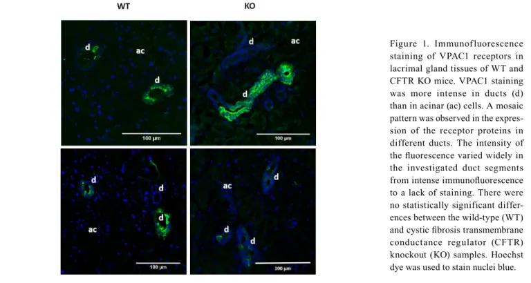
Immunofluorescence: The presence of VPAC1 was dominant in the duct cells as demonstrated in Figure 1. The receptor was localized on the basolateral surface of the duct cells.
Immunofluorescence staining of VPAC1 receptors revealed a
mosaic pattern among different duct segments in the expres- sion of the receptor proteins. The intensity of the fluorescence varied widely in the investigated duct segments from intense immunofluorescence to a lack of staining. No difference was detected in the immunoreactivity between the LG tissues from the WT and CFTR KO mice.
Intense VPAC2 staining was detected not only in the duct cells but also in the basolateral surface of the acinar cells (Figure 2). The intensity of the immunofluorescence staining was similar in the case of the WT and CFTR KO LGs.
VIP-induced fluid secretion in isolated LG ducts: The effect of VIP (100 nM) on ductal fluid secretion was investigated in WT and CFTR KO mouse LG ducts. A remarkable proportion of isolated ducts failed to react to the applied agent. These findings are in accordance with the results in the VPAC1 immunofluorescence studies in which the expression of the receptor protein showed a mosaic pattern, and the fluctua- tion of the presence of VPAC1 predicted a widely altering response of different duct segments to VIP stimulation. Obvi- ously, all data displayed in this section were obtained from the reacting (i.e., responding with a swelling reaction) ducts.
The concentration of VIP was 100 nM in these experi- ments. This value was determined based on data from the literature and on our preliminary experiments in which higher concentrations of VIP (200 and 500 nM) did not result in greater effects compared to the 100 nM concentration (results are not shown). The VIP stimulation (100 nM) resulted in a robust and continuous fluid secretory response in isolated LG duct segments derived from WT mice (the secretory rate in the first 10 min of stimulation was 213.1±37.30 pl/min/mm2, n = 7; Figure 3A). In contrast, the CFTR KO ducts exhibited only a weak pulse-like secretion in the first 5 min of stimula- tion (54.5±18.4 pl/min/mm2, n = 6), followed by a plateau (Figure 3B). The VIP-induced luminal volume changes in the WT and CFTR KO ducts observed during the videomicro- scopic experiments are shown in Figure 4.
VIP-induced Ca2+ signaling of isolated LG ducts: The CFTR KO ducts exhibited only a weak pulse-like secretion during VIP stimulation. We hypothesized in the background of this phenomenon that in addition to lacking the cAMP-CFTR pathway VIP stimulation may induce changes in [Ca2+]I, and this latter intracellular mechanism may also contribute to the fluid secretion of ducts. Therefore, in the next series of experiments possible effects of VIP stimulation on [Ca2+]
i were measured.
Intracellular Ca2+ homeostasis underlying VIP stimu- lation was measured in WT and CFTR KO duct segments.
VIP stimulation (100 nM) resulted in a small but statistically
significant increase in [Ca2+]i in the WT (11.6±0.70%, p = 0.001, n = 6) and CFTR KO ducts (11.1±0.50%, p = 0.002, n
= 6; Figure 5). Cholinergic agonist carbachol (100 µM) was used as positive control in these experiments. The selection of the 100 µM concentration for carbachol was based on our previous experiments where a definite dose–response relation was observed in the 1–100 µM concentration range [12].
VIP-induced fluid secretion in the presence of intracellular Ca2+-chelator BAPTA-AM in LG ducts isolated from CFTR KO mice: To analyze the role of Ca2+ signaling in VIP- induced ductal fluid secretion, the effect of VIP stimulation was investigated in the presence of intracellular Ca2+-chelator BAPTA-AM. In these fluid secretion experiments, isolated ducts were preincubated with BAPTA-AM (10 µM) for 30 min before application of VIP (100 nM). A total of eight ducts Figure 1. Immunofluorescence staining of VPAC1 receptors in lacrimal gland tissues of WT and CFTR KO mice. VPAC1 staining was more intense in ducts (d) than in acinar (ac) cells. A mosaic pattern was observed in the expres- sion of the receptor proteins in different ducts. The intensity of the fluorescence varied widely in the investigated duct segments from intense immunofluorescence to a lack of staining. There were no statistically significant differ- ences between the wild-type (WT) and cystic fibrosis transmembrane conductance regulator (CFTR) knockout (KO) samples. Hoechst dye was used to stain nuclei blue.
Figure 2. Immunofluorescence staining of VPAC2 receptors in lacrimal gland tissues of WT and CFTR mice. VPAC2 was observed on the basolateral surface of duct (d) and acinar (ac) cells. There were no significant differences between the wild-type (WT) and cystic fibrosis transmembrane conduc- tance regulator (CFTR) knockout (KO) samples. Hoechst dye was used to stain nuclei blue.
isolated from three CFTR KO animals were investigated in these series of experiments. Following the BAPTA-AM preincubation, VIP failed to elicit an increase in secretion in any of the ducts (Figure 3B). The secretory rates were similar to the control values (i.e., no secretion was detected either
in the BAPTA-AM+VIP or in the control ducts). A curve showing the secretory response of the BAPTA-AM+VIP- treated CFTR KO ducts appeared to be identical to the control curves; therefore, the results of the control experiments are not shown in Figure 3B.
Figure 3. Effect of VIP stimulation on ductal fluid secretion in isolated lacrimal gland ducts from WT and CFTR KO mice. A: The wild-type (WT) ducts were exposed either to 100 nM vasoactive intestinal peptide (VIP; filled rhombus) or to no agonist (empty triangle). B:
The cystic fibrosis transmembrane conductance regulator (CFTR) knockout (KO) ducts were exposed either to 100 nM VIP (filled square) or to 100 nM VIP following 10 µM 1,2-bis(o-aminophenoxy) ethane-N,N,N′,N′-tetra-acetic acid (BAPTA-AM) pretreatment (empty triangle). Changes in relative luminal volume (Vr) are shown. Data were obtained from six to eight ducts isolated from three different animals in each series and are presented as means ± standard error of the mean (SEM).
Figure 4. Representative photo series of secreting isolated lacrimal gland duct segments in response to VIP stimulation. The luminal spaces of the native images (first and third rows) are marked with blue (second and fourth rows). A strong secretory response could be shown in wild-type (WT) ducts to vasoactive intestinal peptide (VIP) stimulation as the luminal spaces were notably swollen after treatment. No remarkable changes could be detected in cystic fibrosis transmembrane conductance regu- lator (CFTR) knockout (KO) ducts following VIP stimulation.
DISCUSSION
In the last two decades, results of gene expression studies reaffirmed previous suggestions about the secretory role of LG duct epithelial cells, because greater expression of a wide range of transmembrane transporters could be demonstrated in the duct cells compared to acinar cells [3,25-27]. Although these results were important steps toward the clarification of duct cell function, they were unable to provide direct func- tional evidence. Our laboratory developed the first experi- mental model suitable for the investigation of lacrimal duct function which opened a new path in lacrimal duct research.
Our efforts are directed toward the clarification of the role of LG ducts in the secretory process of the gland using this isolated duct model [4,5]. These viable duct segments are appropriate models not only for the investigation of the func- tion of transmembrane transporters but also for the investiga- tion of the regulation of secretory machinery. Although the secretory process of the duct epithelium may play an impor- tant role in tear production, our knowledge about the regula- tion of lacrimal duct function is limited. Tear secretion is mediated mainly by the parasympathetic system [28,29]. The anatomically and functionally predominant parasympathetic nerves can be subdivided into two different branches based on the secreted neurotransmitters. Cholinergic nerve endings release the cholinergic agonist acetylcholine, while VIP-ergic nerve terminals produce VIP. Both neurotransmitters have major effects on the secretion of the LG [28]. The available
experimental evidence for the role of the parasympathetic system in LG fluid secretion is solely from intact glands or acini. We investigated the direct effect of VIP-ergic stimuli on isolated LG duct segments and explored the relation- ship between the CFTR chloride channel and the VIP-ergic signaling pathway.
Immunofluorescence was used to confirm the pres- ence and localization of VPAC1 and VPAC2 proteins on LG duct cells in WT and CFTR KO mice. A previous report by Hodges et al. showed the presence of these receptors with immunostaining on the basolateral surface of acinar and duct cells in rats [6]. An apparent difference was revealed in the distribution of VPAC1 and VPAC2 receptors in mouse LG in the present study. The presence of VPAC1 was proved predominantly in ducts with immunoreactivity. In addition to the characteristically ductal localization of VPAC1 receptors, a mosaic pattern in the expression of the receptor proteins was revealed. The intensity of the immunofluorescence varied on a wide scale from intense staining to almost complete unde- tectability in the analyzed duct segments. VPAC2 showed a more homogeneous distribution in LG tissue: These recep- tors were traceable in acinar and duct cells. VPAC1 and VPAC2 were observed most prominently on the basolateral membrane of mouse LG cells. Expression patterns of VPAC1 and VPAC2 receptors differ from the presence of CFTR in mouse LG. The CFTR protein was predominant in ducts in contrast to the VPAC2 receptors, while CFTR expression
Figure 5. Effects of VIP and carba- chol on [Ca2+]i in lacrimal gland duct cells from WT and CFTR KO mice. A: Representative record- ings of the microfluorescence experiments: Effects of vasoactive intestinal peptide (VIP; 100 nM) on [Ca2+]i in lacrimal gland duct cells from wild-type (WT) and cystic fibrosis transmembrane conduc- tance regulator (CFTR) knockout (KO) mice (panel A, left curve).
Carbachol (100 µM) was used for comparison, as a positive control, in these experiments (panel A, right curve). F380/340: 380/340 nm fluorescence emission ratio. B: Maximum values of the 380/340 nm fluorescence emission ratios. Fmax(380/340): maximum value of the 380/340 nm fluorescence emission ratio.
showed a reasonably homogeneous distribution in the duct system in contrast to the VPAC1 receptors [12,18,25]. A connection between CFTR and VPACs at the protein expres- sion level also seems implausible, as the lack of CFTR did not influence the expression of VPAC1 and VPAC2 proteins: No difference was detected between the immunoreactivity of the WT and CFTR KO LG tissues in the experiments.
To explore the fluid secretory effect of VIP, videomicro- scopic experiments were applied. VIP (100 nM) stimulation caused strong and continuous fluid secretion in the ducts from the WT mice. In contrast, as a result of VIP stimulation, the CFTR KO ducts exhibited only weak pulse-like secretion in the first 5 min, followed by a plateau. These findings are substantially different from the results we obtained previ- ously in carbachol stimulated experiments where identical ductal fluid secretory responses were observed in the WT and CFTR KO mouse LG ducts [12]. The observed reduction in response to VIP stimulation in CFTR KO ducts may be explained by the lack of CFTR rather than changes in VPAC1 and VPAC2, as no difference was detected in the density of the VIP receptors between the WT and CFTR KO ducts in the present immunofluorescence studies. The role of CFTR can be explained by the following chain of effects: Stimula- tion of VIP receptors increases the intracellular cAMP levels via adenylyl-cyclase, and then cAMP (as a potent activator) increases the activity of CFTR. Therefore, this transporter seems to be the determining component of the robust fluid secretion evoked by VIP in WT ducts. The weak secretory response observed in the CFTR KO ducts during VIP stimu- lation seems to confirm the described mechanism.
Ca2+ homeostasis underlying VIP stimulation and the potential role of CFTR protein was also investigated. The effect of VIP stimuli on the cytosolic Ca2+ signaling was measured with microfluorometry. VIP acts not only through the adenylyl cyclase-cAMP system, but also, to a smaller extent, VIP elevates [Ca2+]i . VIP-evoked elevations of [Ca2+]
i were also independent from the presence of CFTR and did not differ in the WT and CFTR KO ducts in a significant manner. Therefore, VIP stimulation had a similar effect on [Ca2+]i compared to the results of our previous experiments using carbachol stimulation. In those experiments, identical changes of [Ca2+]i were found in WT and CFTR KO mouse LG ducts [12]. However, the elevations in [Ca2+]i were much smaller in the case of VIP stimulation compared to the effect of carbachol. The role of these VIP-induced small changes of [Ca2+]i was further investigated in fluid secretion experiments where intracellular Ca2+-chelator BAPTA was applied. In a short series of preliminary experiments, WT ducts were incubated with BAPTA-AM (data not shown).
No effect of the BAPTA-AM pretreatment on the robust VIP stimulated swelling response of WT ducts was detectable.
The lack of demonstrability of the BAPTA-AM effect in the wild-type ducts can be explained by the sensitivity of the applied videomicroscopic method combined with the negli- gible effect of Ca2+-dependent mechanisms in addition to the robust cAMP-dependent effect. In the next series of experi- ments, ducts isolated from CFTR KO mice were incubated with BAPTA before VIP stimulation. The suspended Ca2+
signaling completely abolished the remaining VIP-induced secretory response of mouse LG ducts lacking the cAMP- CFTR pathway. These results strongly suggest the minor complementary role of Ca2+-regulated mechanisms in VIP- induced ductal fluid secretion. However, there is one detail that must be taken into consideration regarding these results:
According to the mosaic pattern of VPAC1 receptors in the different duct segments, a remarkable portion of isolated ducts (approximately 50%–65% of all ducts in to complete series of all experiments) failed to react to VIP. This result suggests caution in the interpretation of negative results (i.e., non responding ducts). However, we performed a relatively high number of experiments in these series: eight ducts isolated from three different animals were investigated. We believe that this number makes our conclusions plausible and minimizes the probability of a random series of all non- responding ducts.
Findings in our previous experiments studying effects of carbachol stimulation and the present results in immunos- taining studies and in fluid secretion together with [Ca2+]i experiments suggest two partially independent parasympa- thetic regulatory pathways. Briefly, carbachol stimulation acts solely through elevation of [Ca2+]I, and it does not involve the adenylyl cyclase-cAMP route. Consequently, CFTR is not involved in carbachol-stimulated fluid secretion as it is acti- vated by cAMP. For VIP stimuli, [Ca2+]i was slightly elevated without a significant difference between the values measured in the WT and CFTR KO ducts. However, the secretory response of the LG ducts showed a large significant differ- ence in the WT and CFTR KO ducts: the rate of fluid secre- tion of the CFTR KO ducts was far below the value obtained in the WT ducts. These data prove the determining role of the adenylyl cyclase-cAMP-CFTR route in VIP-stimulated fluid secretion of LG ducts. Results of the VIP experiments are in accordance with previous observations that VIP acts predominantly through elevation of the cytosolic cAMP level, and only a small part of the action of VIP is thought to be mediated by Ca2+ signaling [30,31]. The lack of functionally active CFTR seen in the CFTR KO mice influences the VIP- induced secretory response of LG ducts and thus, can modify
the total parasympathetic-evoked secretory contribution of the LG.
The observed VIP-induced significant fluid secretion suggests the importance of this neurotransmitter in the regu- lation of the duct system. LG duct cells have several different Cl- and K+ selective ion channels and transporters located on the basolateral and apical membranes. Elevated cytosolic cAMP levels and intracellular Ca2+ signaling can activate Cl- and K+ secretion through these channels and transporters.
VIP seems to have target structures in basolateral and apical membranes. Previously, we reported the strong stimulatory effect of cell-permeable cAMP-analogue 8-bromo cAMP and VIP on basolaterally located sodium-potassium-chloride cotransporter type 1 (NKCC1) [32]. The present results suggest the important role of VIP in the regulation of apically located CFTR. Therefore, VIP seems to be able to activate Cl- transport on the basolateral and apical sides of LG duct cells. In summary, actions of Cl- selective channels located on the basolateral and apical membranes of duct cells result in intraluminal flux of Cl-. Elevation of intraluminal Cl- concentration is the main determinant of the lumen-negative transepithelial voltage difference, which is the driving force of ductal fluid secretion. Consequently, these results strongly suggest the important role of VIP in the regulation of lacrimal duct fluid secretion.
Identifying the mechanism underlying the regulation of ductal fluid transport is likely to be pertinent for treating ocular diseases. This study contributes to this objective by demonstrating that VIP-induced increases in cAMP followed by stimulation of CFTR chloride channel activity in the ductal epithelial cells drives fluid from the interstitium into the ductal lumen. This finding is relevant because treatment of some desiccating ocular surface diseases may benefit from applying cAMP mobilizing agents that, in turn, optimize this secretory process.
ACKNOWLEDGMENTS
Funding: The work was supported by the NKFIH NN 115611 (E T-M), the EFOP-3.6.1–16–2016–00008 (E T-M), the SZTE ÁOK-KKA No 2018/Tóth-MolnárE (E T-M), the NEI/NIH EY017731 (CD) and The Webb Foundation grant (CD).
REFERENCES
1. Alexander JH, van Lennep EW, Young JA. Water and elec- trolyte secretion by the exorbital lacrimal gland of the rat studied by micropuncture and catheterization techniques.
Pflugers Arch 1972; 337:299-309. [PMID: 4674879].
2. Dartt DA, Moller M, Poulsen JH. Lacrimal gland electrolyte and water secretion in the rabbit: localization and role of (Na+-K+)-activated ATPase. J Physiol 1981; 321:557-69.
[PMID: 6461755].
3. Ubels JL, Hoffman HM, Srikanth S, Resau JH, Webb CP. Gene expression in rat lacrimal gland duct cells collected using laser capture microdissection: evidence for K+ secretion by the duct cells. Invest Ophthalmol Vis Sci 2006; 47:1876-85.
[PMID: 16638994].
4. Tóth-Molnár E, Venglovecz V, Ozsvári B, Rakonczay Z, Varró A, Papp JG, Tóth A, Lonovics J, Takáts T, Ignáth I, Iványi B, Hegyi P. New experimental method to study acid/base transporters and their regulation in lacrimal gland ductal epithelia. Invest Ophthalmol Vis Sci 2007; 48:3746-55.
[PMID: 17652747].
5. Katona M, Vizvári E, Németh L, Facskó A, Venglovecz V, Rakonczay Z, Hegyi P, Tóth-Molnár E. Experimental evidence of fluid secretion of lacrimal gland duct epithe- lium. Invest Ophthalmol Vis Sci 2014; 55:4360-7. [PMID:
24925876].
6. Hodges RR, Zoukhri D, Sergheraert C, Zieske JD, Dartt DA. Identification of vasoactive intestinal peptide receptor subtypes in the lacrimal gland and their signal-transducing components. Invest Ophthalmol Vis Sci 1997; 38:610-9.
[PMID: 9071214].
7. Said SI, Mutt V. A peptide fraction from lung tissue with prolonged peripheral vasodilator activity. Scand J Clin Lab Invest Suppl 1969; 107:51-6. [PMID: 5375727].
8. Gilbard JP, Dartt DA, Rood RP, Rossi SR, Gray KL, Donowitz M. Increased tear secretion in pancreatic cholera: a newly recognized symptom in an experiment of nature. Am J Med 1988; 85:552-4. [PMID: 2845780].
9. Laburthe M, Couvineau A, Tan V. Class II G protein-coupled receptors for VIP and PACAP: structure, models of activa- tion and pharmacology. Peptides 2007; 28:1631-9. [PMID:
17574305].
10. Laburthe M, Couvineau A, Marie JC. VPAC receptors for VIP and PACAP. Receptors Channels 2002; 8:137-53. [PMID:
12529932].
11. Laburthe M, Couvineau A. Molecular pharmacology and structure of VPAC Receptors for VIP and PACAP. Regul Pept 2002; 108:165-73. [PMID: 12220741].
12. Berczeli O, Vizvári E, Katona M, Török D, Szalay L, Rárosi F, Németh I, Rakonczay Z, Hegyi P, Ding C, Tóth-Molnár E. Novel Insight into the Role of CFTR in Lacrimal Gland Duct Function in Mice. Invest Ophthalmol Vis Sci 2018;
59:54-62. [PMID: 29305607].
13. Hong JH, Park S, Shcheynikov N, Muallem S. Mechanism and synergism in epithelial fluid and electrolyte secretion.
Pflugers Arch 2014; 466:1487-99. [PMID: 24240699].
14. Lee MG, Ohana E, Park HW, Yang D, Muallem S. Molecular mechanism of pancreatic and salivary gland fluid and HCO3 secretion. Physiol Rev 2012; 92:39-74. [PMID: 22298651].
15. Reddy MM, Quinton PM. PKA mediates constitutive activa- tion of CFTR in human sweat duct. J Membr Biol 2009;
231:65-78. [PMID: 19865788].
16. Castagna I, Roszkowska AM, Fama F, Sinicropi S, Ferreri G.
The eye in cystic fibrosis. Eur J Ophthalmol 2001; 11:9-14.
[PMID: 11284496].
17. Sheppard JD, Orenstein DM, Chao CC, Butala S, Kowalski RP.
The ocular surface in cystic fibrosis. Ophthalmology 1989;
96:1624-30. [PMID: 2616148].
18. Lu M, Ding C. CFTR-mediated Cl- transport in the acinar and duct cells of rabbit lacrimal gland. Curr Eye Res 2012;
37:671-7. [PMID: 22578307].
19. Frizzell RA, Hanrahan JW. Physiology of epithelial chloride and fluid secretion. Cold Spring Harb Perspect Med 2012;
2:a009563-[PMID: 22675668].
20. Hwang TC, Kirk KL. The CFTR ion channel: gating, regula- tion, and anion permeation. Cold Spring Harb Perspect Med 2013; 3:a009498-[PMID: 23284076].
21. Tóth-Molnár E, Berczeli O, Vizvári E, Rakonczay Z, Hegyi P, Ding C. Effect of vasoactive intestinal peptide on lacrimal gland ductal fluid secretion in wild type and CFTR knockout mice. Invest Ophthalmol Vis Sci 2017; 58:2242-.
22. Ratcliff R, Evans MJ, Cuthbert AW, MacVinish LJ, Foster D, Anderson JR, Colledge WH. Production of a severe cystic fibrosis mutation in mice by gene targeting. Nat Genet 1993;
4:35-41. [PMID: 7685652].
23. Seidler U, Singh A, Chen M, Cinar A, Bachmann O, Zheng W, Wang J, Yeruva S, Riederer B. Knockout mouse models for intestinal electrolyte transporters and regulatory PDZ adap- tors: new insights into cystic fibrosis, secretory diarrhoea and fructose-induced hypertension. Exp Physiol 2009; 94:175-9.
[PMID: 18931049].
24. Fernández-Salazar MP, Pascua P, Calvo JJ, Lopez MA, Case RM, Steward MC, San Roman JI. Basolateral anion transport
mechanisms underlying fluid secretion by mouse, rat and guinea-pig pancreatic ducts. J Physiol 2004; 556:415-28.
[PMID: 14978209].
25. Ding C, Parsa L, Nandoskar P, Zhao P, Wu K, Wang Y. Duct system of the rabbit lacrimal gland: structural characteristics and role in lacrimal secretion. Invest Ophthalmol Vis Sci 2010; 51:2960-7. [PMID: 20107177].
26. Ding C, Lu M, Huang J. Changes of the ocular surface and aquaporins in the lacrimal glands of rabbits during preg- nancy. Mol Vis 2011; 17:2847-55. [PMID: 22128232].
27. Thomson-Vest N, Shimizu Y, Hunne B, Furness JB. The distribution of calcium-activated, intermediate conductance potassium (IK) channels in epithelial cells. J Anat 2006;
208:219-29. [PMID: 16441566].
28. Dartt DA. Neural regulation of lacrimal gland secretory processes: relevance in dry eye diseases. Prog Retin Eye Res 2009; 28:155-77. [PMID: 19376264].
29. Dartt DA. Signal transduction and control of lacrimal gland protein secretion: a review. Curr Eye Res 1989; 8:619-36.
[PMID: 2545411].
30. Dérand R, Montoni A, Bulteau-Pignoux L. Activation of VPAC1 receptors by VIP and PACAP-27 in human bronchial epithelial cells induces CFTR-dependent chloride secretion.
Br J Pharmacol 2004; 141:698-708. [PMID: 14744818].
31. Dickson L, Aramori I, McCulloch J. A systematic comparison of intracellular cyclic AMP and calcium signalling highlights complexities in human VPAC/PAC receptor pharmacology.
Neuropharmacology 2006; 51:1086-98. [PMID: 16930633].
32. Vizvári E, Katona M, Orvos P, Berczeli O, Facskó A, Rárosi F, Venglovecz V, Rakonczay Z, Hegyi P, Ding C, Tóth-Molnár E. Characterization of Na+-K+-2Cl- Cotransporter Activity in Rabbit Lacrimal Gland Duct Cells. Invest Ophthalmol Vis Sci 2016; 57:3828-35. [PMID: 27438543].
Articles are provided courtesy of Emory University and the Zhongshan Ophthalmic Center, Sun Yat-sen University, P.R. China.
The print version of this article was created on 6 December 2020. This reflects all typographical corrections and errata to the article through that date. Details of any changes may be found in the online version of the article.


![Figure 5. Effects of VIP and carba- carba-chol on [Ca 2+ ] i in lacrimal gland duct cells from WT and CFTR KO mice](https://thumb-eu.123doks.com/thumbv2/9dokorg/968273.57620/6.918.79.844.665.1046/figure-effects-carba-carba-lacrimal-gland-cells-cftr.webp)
