This is an Accepted Article that has been peer-reviewed and approved for publication in The Journal of Physiology, but has yet to undergo copy-editing and proof correction. Please cite this article as an 'Accepted Article'; doi: 10.1113/JP282203.
Bile acid- and ethanol-mediated activation of Orai1 damages pancreatic ductal secretion in acute pancreatitis
Petra Pallagi * 1,2,3, Marietta Görög* 2,3, Noémi Papp 2,3, Tamara Madácsy 1,2,3, Árpád Varga1,2,3, Tim Crul 1,2,3, Viktória Szabó 1,2,3, Melinda Molnár 2,3, Krisztina Dudás 2,3, Anna Grassalkovich 2, Edit
Szederkényi 4, György Lázár 4, Viktória Venglovecz 5, Péter Hegyi 2,6,7, József Maléth 1,2,3
* These authors contributed equally.
1 HCEMM-SZTE Molecular Gastroenterology Research Group, University of Szeged, Szeged, Hungary
2 Department of Medicine, University of Szeged, Szeged, Hungary
3 ELKH-USZ Momentum Epithelial Cell Signaling and Secretion Research Group, University of Szeged, Szeged, Hungary
4 Department of Surgery, University of Szeged, Szeged,
5 Department of Pharmacology and Pharmacotherapy, University of Szeged, Szeged, Hungary
6 Hungary Centre for Translational Medicine, Semmelweis University, Budapest, Hungary
7 Institute for Translational Medicine and First Department Medicine, Medical School, University of Pécs, Pécs, Hungary
Short title: Orai1 mediated ductal damage in acute pancreatitis
Correspondence to József Maléth MD, Ph.D.
ORCID ID.: 0000-0001-5768-3090
HAS-USZ Momentum Epithelial Cell Signaling and Secretion Research Group First Department of Internal Medicine and Department of Public Health University of Szeged, H6720, Szeged; Hungary
Phone: +36 (62) 342-877; +36 70 41 66500
e-mail: jozsefmaleth1@gmail.com; maleth.jozsef@med.u-szeged-hu
Key words: Orai1 channel, acute pancreatitis, Ca2+ signaling, bile acid, ethanol, epithelial ion transport
Abbreviations: AP: acute pancreatitis, CDC: chenodeoxycholate, CFTR: cystic fibrosis transmembrane conductance regulator, FAEEs: fatty acid esters, ER: endoplasmic reticulum, PA:
palmitic acid, POA: palmitoleic acid, OC: organoid cultures, SOCE: store operated Ca2+ entry, SERCA: sarco/endoplasmic reticulum calcium ATPase, ERCP: endoscopic retrograde cholangiopancreaticography, NHERF1: Na+/H+ Exchanger Regulatory Factor 1, PM: plasma membrane, PMCA: plasma membrane calcium ATPase, Na-TC: sodium taurocholate
KEY POINTS SUMMARY
Sustained intracellular Ca2+ overload in pancreatic acinar and ductal cells is a hallmark of biliary and alcohol-induced acute pancreatitis, which leads to impaired ductal ion and fluid secretion.
Orai1 is a plasma membrane Ca2+ channel that mediates extracellular Ca2+ influx upon endoplasmic reticulum Ca2+ depletion.
Our results showed that Orai1 is expressed on the luminal plasma membrane of the ductal cells and selective Orai1 inhibition impaired Stim1-dependent extracellular Ca2+ influx evoked by bile acids or ethanol combined with non-oxidative ethanol metabolites.
The prevention of sustained extracellular Ca2+ influx protected ductal cell secretory functions in in vitro models and maintained exocrine pancreatic secretion in in vivo AP models.
Orai1 inhibition prevents the bile acid-, and alcohol-induced damage of the pancreatic ductal secretion and holds the potential of improving the outcome of acute pancreatitis.
ABSTRACT
Regardless of its etiology, sustained intracellular Ca2+ overload is a well-known hallmark of acute pancreatitis (AP). Toxic Ca2+ elevation induces pancreatic ductal cell damage characterized by impaired ion- and fluid secretion –essential to wash out the protein-rich fluid secreted by acinar cells while maintaining the alkaline intra-ductal pH under physiological conditions– and mitochondrial dysfunction. While prevention of ductal cell injury decreases the severity of AP, no specific drug target has yet been identified in the ductal cells. Although Orai1 –a store operated Ca2+ influx channel– is known to contribute to sustained Ca2+ overload in acinar cells, details concerning its expression and function in ductal cells are currently lacking. In this study, we demonstrate that functionally active Orai1 channels reside dominantly in the apical plasma membrane of pancreatic ductal cells. Selective CM5480-mediated Orai1 inhibition impairs Stim1-dependent extracellular Ca2+
influx evoked by bile acids or ethanol combined with non-oxidative ethanol metabolites. Furthermore,
prevention of sustained extracellular Ca2+ influx protects ductal cell secretory function in vitro and decrease pancreatic ductal cell death. Finally, Orai1-inhibition partially restores and maintains proper exocrine pancreatic secretion in in vivo AP models. In conclusion, our results indicate that Orai1 inhibition prevents AP-related ductal cell function impairment and holds the potential of improving disease outcome.
Petra Pallagi is a research fellow at the First Department of Internal Medicine at the University of Szeged. She earned her Ph.D. under the supervision of Peter Hegyi and Zoltan Rakonczay at the University of Szeged for her work on the role of pancreatic ductal secretion in acute pancreatitis pathogenesis. She also investigated how activated trypsin inhibits pancreatic ductal secretion. Her current work is focusing on the intracellular signaling and secretion in the exocrine pancreas.
Marietta Görög is a PhD candidate at the University of Szeged, Internal Medicine and
supervised by Dr. József Maléth és Dr Pallagi Petra. Marietta holds a MSc degree in Biology,
specialized in Molecular biology. Currently, her research focuses on understanding the role of
Ca
2+signaling in the physiology and pathophysiology of the pancreatic ductal cells. She
found that that Orai1 inhibition prevents acute pancreatitis-related ductal cell function
impairment.
Introduction
Acute pancreatitis (AP) is one of the most common inflammatory diseases of the gastrointestinal tract which requires hospitalization. It is primarily caused by impacted gallstones, heavy alcohol consumption, hypertrigliceridaemia or through iatrogenic side effects of medical treatments such as Asparaginase or endoscopic retrograde cholangiopancreaticography (ERCP) thus representing a major clinical challenge (Yadav & Lowenfels, 2013). Despite advances in basic and clinical research, specific treatments are still lacking resulting in a remarkably high mortality rate (~28%) in case of severe AP (~10% of all cases) (Párniczky et al., 2016). Previously, a crucial involvement of impaired ductal secretion in AP development was shown. Our group demonstrated ethanol+fatty acid-induced reductions in expression and activity of cystic fibrosis transmembrane conductance regulator (CFTR) in pancreatic ductal epithelial cells in a mouse model of alcohol-induced AP, ultimately leading to impaired ductal secretion and increased severity of the disease (Maléth et al., 2015). Also, mislocalization of CFTR in mice lacking the Na+/H+ exchanger regulatory factor-1 (NHERF1) –a scaffolding protein that targets membrane proteins to the apical membrane– resulted in decreased ductal secretion and more severe experimental AP (Pallagi et al., 2014). Notably, dysfunction of ductal epithelia equally damages acinar cells. Freedman et al. showed disturbed plasma membrane (PM) dynamics and apical endocytosis in pancreatic acini of CFTR knockout mice (Freedman et al., 2001). Importantly, pharmacological restoration of CFTR-mediated secretion with the corrector C18 and the potentiator VX-770 restored acinar cell function and decreased pancreatic inflammation in mouse models of chronic and autoimmune pancreatitis (Zeng et al., 2017). Proper establishment of an alkaline intraductal pH environment prevents premature intra-pancreatic –acidic pH-dependent–
autoactivation of trypsinogen (Pallagi et al., 2011).
Regardless of the etiology, different forms of AP are characterized by a sustained intracellular Ca2+
elevation (Pallagi et al., 2020). Biologically active compounds –such as bile acids or fatty acid ethyl esters– induce the release of Ca2+ from endoplasmic reticulum (ER) Ca2+ stores and subsequent extracellular Ca2+ influx through the Orai1 Ca2+ channel, a process referred to as store operated Ca2+
entry (SOCE). Although SOCE is part of the physiological Ca2+ signaling events in non-excitable
cells, it can significantly contribute to sustained intracellular Ca2+ overload under pathological conditions. In addition, ERCP-mediated intra-pancreatic pressure increase leads to extracellular Ca2+
influx through the activation of TRPV4 and the mechanoreceptor ion channel Piezo1 (Swain et al., 2020). Intracellular Ca2+ overload leads to premature activation of trypsinogen in pancreatic acinar cells (Krüger et al., 2000) as well as mitochondrial damage and cell necrosis in pancreatic ductal cells (Criddle et al., 2006). Based on our previous observations –indicating sustained intracellular Ca2+
elevation-mediated impairment of fluid - and HCO3--secretion (Maléth & Hegyi, 2014; Madácsy et al., 2018) as well as ductal cell mitochondrial malfunctioning, subsequent ATP depletion, and cell damage (Maléth et al., 2011)– we hypothesize that extracellular Ca2+ influx might contribute to disease development through establishment of a persistent alcohol- or fatty acid esters (FAEEs)- induced intracellular Ca2+ increase resulting in impaired mitochondrial ATP production. Subsequently, this leads to failing ATP-dependent Ca2+ extrusion by the plasma membrane Ca2+-ATPase (PMCA) and Ca2+ reuptake by the sarco/endoplasmic reticulum Ca2+-ATPase (SERCA) (Kim et al., 2002) thus maintaining the spatiotemporal localization of the Ca2+ signal and developing a global, sustained Ca2+
elevation. Selective inhibition of Orai1 by GSK-7975A or CM-128 (also known as CM4620 or Auxora developed by CalciMedica) markedly impaired bile acid-mediated extracellular Ca2+ influx and sustained Ca2+ overload in pancreatic acinar cells and decreased AP severity in experimental models (Wen et al., 2015). Moreover, selective inhibition of Orai1 by CM4620 abolished myeloperoxidase activity and inflammatory cytokine expression in pancreatic and lung tissues and prevented oxidative burst in neutrophils (Waldron et al., 2019). Based on these preclinical observations, CalciMedica successfully completed two Phase I clinical trials and then tested 7 patients with AP to assess the pharmacokinetic and pharmacodynamics profile of Auxora when administrated by intravenous infusion. Based on these a phase 2, open-label, dose-response clinical study evaluated the safety of Auxora in patients with AP, SIRS, and hypoxemia (Bruen et al., 2021). While low-dose Auxora treatment improved moderate AP to mild AP in 36,5% of the patients, very interestingly Auxora also improved the tolerance of solid foods, which might be explained by improved exocrine pancreatic secretion. Although the beneficial effect of selective Orai1 inhibition in AP is well-
Therefore, in this study, we aimed to analyze the effect of Orai1 inhibition on ductal secretion in isolated ductal fragments, pancreatic organoids, and in vivo AP models. Our results demonstrate that selective inhibition of Orai1 by CM5480, another CalciMedica selective Orai1 channel inhibitor, impairs Stim1-dependent extracellular Ca2+ influx evoked by bile acids or ethanol combined with non- oxidative ethanol metabolites. Furthermore, prevention of sustained extracellular Ca2+ influx protects ductal cell secretory function in vitro and decreases pancreatic ductal cell death. Finally, Orai1- inhibition partially restores and maintains exocrine pancreatic secretion in in vivo AP models. In conclusion, our results indicate that Orai1 inhibition prevents AP-related impairment of ductal cell function and holds the potential of improving disease outcome.
Materials and methods Ethics
Animals were used with adherence to the NIH guidelines and the EU directive 2010/63/EU. The study was approved by the National Scientific Ethical Committee on Animal Experimentation under license number XXI./1541/2020. The collection and use of human samples including cadaver donor pancreas and liver samples were executed in adherence with the EU standards and approved by the Regional Committee of Research Ethics of the Hungarian Medical Research Council under license number 37/2017-SZTE.
Animals
FVB/N mice (8-12 weeks, 20-25 g) were kept at constant room temperature of 22-24°C under a 12 h light–dark cycle with free access to food and water. For all experimental groups, the gender ratio was 1:1. Animals used in the experiments were breed in the animal facility of the Department of Public Health, University of Szeged. Animals received VRF1(P) standard rodent food (Manufacturer:
Special Diets Services, Cat.No.: 801900), and standard bedding (Manufacturer: JRS; REHOFIX
MK2000 corn cob) were purchased from Akronom (https://www.akronom.hu/products/corn-cob- bedding-rehofix.html). Interventions were done during the light cycle and animals were not fasted before the experiments.
Isolation of pancreatic ductal fragments
Pancreatic ductal fragments were isolated as described earlier (Maléth et al., 2015). Briefly, following pentobarbital-induced terminal anesthesia, the pancreas was surgically removed, placed into ice-cold DMEM/F12, and injected with a solution containing 100 U/ml collagenase, 0.1 mg/ml trypsin inhibitor, 1 mg/ml bovine serum albumin followed by shaking at 37ºC for 30 min. Next, small intra- and interlobular ducts were identified and isolated under stereomicroscope.
Mouse and human organoid cultures
Organoid cultures (OC) were established as previously described (Boj et al., 2015; Molnár et al., 2020; Breunig et al., 2021) with some modifications. Human pancreatic OC were generated from cadaver donor-derived pancreatic tissue samples (Ethical approval No.: 37/2017-SZTE). Mouse and human tissues were minced into small fragments and –depending on tissue stiffness– incubated for 30 min up to 1 hr in digestion solution on a vertical shaker at 37°C. Next, cells were collected, washed twice, and mixed with Matrigel in a 1/5 ratio. OC were maintained in feeding media, which was changed every other day. For passaging, OC were incubated with TrypLE™ Express Enzyme at 37ºC for 15 min on a vertical shaker, after which the resulting cells were washed and plated in Matrigel.
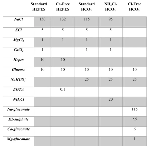
Details of materials and solutions used can be found in Tables 1-5.
Gene expression analysis and gene knockdown
Total mRNA from acini and ductal fragments was purified with NucleoSpin RNA XS kit according to the manufacturer’s instructions. One μg mRNA was used to synthetize cDNA with iScript cDNA Synthesis kit (Bio-Rad, Hercules, CA, USA; Cat. No.: 1708890). Conventional PCR amplification was performed with DreamTaq Hot Start DNA Polymerase and cDNA-specific Orai1 primers (forward: 5’ CTTCGCCATGGTAGCGAT 3’; reverse: 5’ TGTGGTGCAGGCACTAAAGA 3’) for 35 cycles. For gene knockdown studies, isolated mouse ductal fragments were transfected with 50 nM pre-designed siRNA for mouse Stim1 or siGLOGreen transfection indicator with Lipofectamine 2000 in feeding media for 24h.
Immunofluorescent labeling
Upon freezing in Shandon Cryomatrix, isolated pancreatic ducts were sectioned and stained as previously described (Molnár et al., 2020). Briefly, sections were fixed in 4% paraformaldehyde for 15 min, washed 3 times in Tris-buffered saline (TBS), and blocked for 1 h with 0.1% goat serum and 10% BSA in TBS. Next, sections were incubated with anti-Orai1 antibody for 16 h at 4°C followed by incubation with anti-rabbit antibody conjugated with Alexa Fluor 488 for 2 h at room temperature.
Nuclei were visualized through staining with 1 μg/ml Hoechst33342 for 15 minutes after which sections were mounted with Fluoromount. Images were captured with a Zeiss LSM880 confocal microscope using a 40X oil immersion objective (Zeiss, NA: 1.4).
Measurement of intracellular Ca2+, Cl-, pH, and mitochondrial potential by fluorescence microscopy
Intracellular Ca2+ concentration ([Ca2+]i), intracellular Cl- ([Cl]i), or intracellular pH (pHi) were measured as described earlier (Molnár et al., 2020) by loading the cells with Fura-2-AM (2 μmol/l), MQAE (2 μmol/l), or with BCECF-AM (1 μmol/l), respectively. Ducts or organoids were attached to a poly-L-lysine-coated coverslip and mounted on an Olympus IX71 fluorescence microscope
equipped with an MT-20 illumination system. Filter sets used for Fura2, MQAE, and BCECF measurements were described previously (Molnár et al., 2020). The signal was captured by a Hamamatsu ORCA Flash 4.0 V3 CMOS camera through a 20X oil immersion objective (Olympus;
NA: 0.8) with a temporal resolution of 1 sec. Ratiometric image analysis for pHi measurements was performed by Olympus excellence software. Fluorescent signals of Fura2 and MQAE loaded samples were normalized and represented as F1/F0 values.
Apoptosis/Necrosis detection assay
Mouse pancreatic ductal organoids were attached on a CELLview™ Slide pre-coated with Poly-L- Lysine. Organoids were treated with either 250 µM chenodeoxycholate (CDC) or 100 mM ethanol (EtOH) and 200 µM palmitic acid (PA) for 30 minutes; CM5480 groups were treated with 10 µM CM5480 simultaneously with CDC or EtOH+PA treatment prior to the assay. Apoptosis/Necrosis detection assay was carried out according the manufacturer’s protocol (Fanczal et al., 2020). Briefly, organoids were washed twice with assay buffer, resuspended in a mixture containing 100x diluted Apopxin Green, 200x diluted 7-AAD, and 200x diluted Cytocalcein 450 in assay buffer. Organoids were incubated in the concoction for 30 minutes at 37°C in a humidified incubator. Next, organoids were washed twice in fresh assay buffer and visualized in fresh assay buffer by a Zeiss LSM 880 confocal microscope at Ex/EM = 490/525 nm for Apopxin Green, Ex/Em = 550/650nm for 7-AAD, and Ex/Em = 405/450 nm for Cytocalcein Violet 450.
Cell Viability Assay
Mouse pancreatic ductal organoids were grown in Cultrex Ultrimatrix until passage number 3.
Organoid domes were transferred into 96-well Lumitrac microplate (Greiner, 655075) and treated in 100 µl cell culture media for 30 min at 37°C in a humidified incubator. CellTiter-Glo® 3D cell viability assay (Promega Corporation, G9681) was carried out according to the manufacturer’s
protocol. Briefly, a volume of CellTiter-Glo® 3D Reagent was added equal to the volume of each well. Contents were mixed for 5 minutes in CLARIOstar Plus plate reader (BMG Labtech). Plate was allowed to incubate at room temperature for an additional 25 minutes. Luminescence was recorded in CLARIOstar Plus plate reader. Total protein amount was determined using by Bradford assay in Spectrophotometer (Thermo Scientific™ NanoDrop™ One). Blank corrected data was normalized to the total protein amount.
In vivo acute pancreatitis models
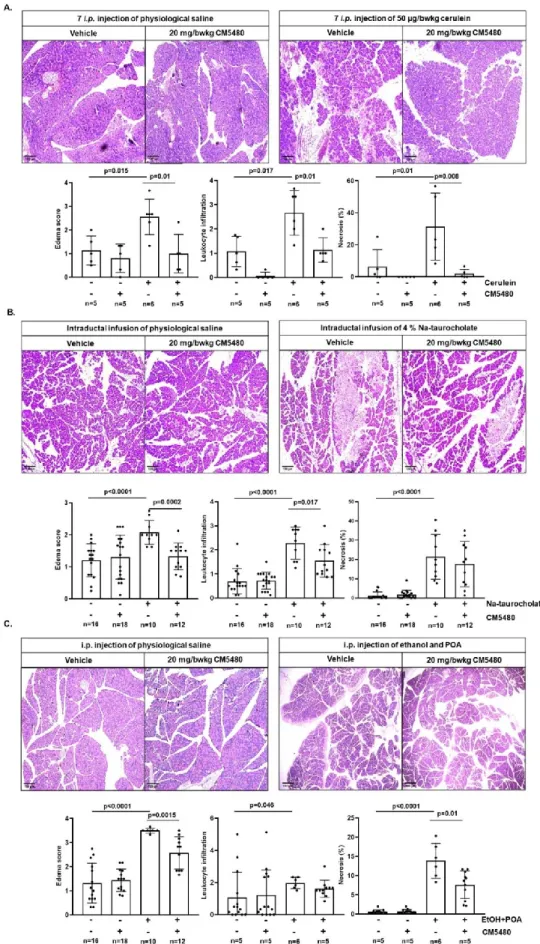
In the cerulein-induced AP model, AP mice received 7 hourly injections of cerulein (50 μg/kg, i.p.) whereas control animals received injections containing physiological saline (i.p.) solution. One hour after the first cerulein injection, CM5840 (20 mg/kg, i.p.) was administered. Twelve hours after the first cerulein injection, mice were sacrificed with pentobarbital (85 mg/kg, i.p.).
Biliary AP was induced by intraductal administration of 4% Na-taurocholate as previously described by Perides et al. (Perides et al., 2010). Briefly, mice were anesthetized with a ketamine and xylazine (respectively 125 mg/kg and 12.5 mg/kg, i.p.) cocktail followed by median laparotomy, the common biliopancreatic duct was cannulated across the duodenum with a 0.4 mm diameter needle connected to an infusion catheter, and the bile duct was occluded with a microvessel clip. Next, the mice received 2 µl/g of 4% Na-taurocholate (Na-TC) or physiological saline at a perfusion rate of 10 µL/min (TSE System GmbH). Following the infusion, the abdominal wall and skin were closed separately, and the mice were placed on a heating pad until waking while buprenorphine (0.075 mg/kg, i.p.) was administered to relieve the pain. One hour after the 4% Na-TC infusion, the animals received CM5480 (20 mg/kg, i.p.). Directly after recovery from anesthesia and 12 h after the operation the mice received 0.1 mg/kg buprenorphine. Twenty-four hours after the operation, the mice were anesthetized with pentobarbital (85 mg/kg, i.p.) and sacrificed through exsanguination through the heart.
The mouse model of acute alcohol-induced pancreatitis was originally developed by Huang et al (Huang et al., 2014). Mice received 2 hourly injections of ethanol (1.35 g/kg, i.p) mixed with palmitoleic acid (POA; 150 mg/kg). To prevent ethanol-induced peritoneal irritation, 200 µl physiological saline was injected before the ethanol/POA treatment. One hour after the first and directly before the second ethanol /POA injection, CM5480 (20 mg/kg, i.p.) was administered.
Control mice received 200 µl physiological saline (i.p.) instead of ethanol/POA. Twenty-four hours after the first ethanol/POA treatment, the mice were sacrificed under pentobarbital (85 mg/kg, i.p.) anesthesia.
For all experimental AP models, histological parameters were monitored to estimate the severity of induced pancreatitis. For histological scoring, pancreata were quickly removed, cleaned from fat and lymph nodes, and stored at 4°C in 4% formaldehyde. Paraffin-embedded pancreas samples were sliced in 4 μm thick sections and stained with hematoxylin–eosin. To estimate severity of induced pancreatitis, edema, inflammatory cell infiltration, and necrosis of the samples were scored by three independent investigators blinded to the protocol (0–5 points for edema and leukocyte infiltration, or
% of total area for necrosis (Fanczal et al., 2020)). Averages of the obtained scores are included in the manuscript.
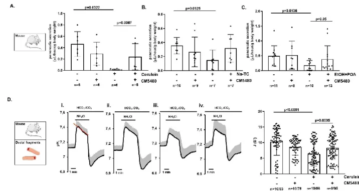
In vivo measurement of pancreatic fluid secretion
In all experimental AP models, pancreatic fluid was collected in vivo directly before sacrifice. Mice were anesthetized with ketamine/xylazine cocktail (respectively 125 mg/kg and 12.5 mg/kg, i.p.) and placed on a heated pad to maintain body temperature. The operation was performed as described in case of 4% Na-TC-induced AP. Following stimulation with secretin (0.75 Clinical Unit/kg, i.p.) for 30 min, the pancreatic juice was collected and the secretory rate was calculated as µl/body weight in g for 1 hr.
Statistics
Statistical analysis was performed with GraphPad Prism software. All data are expressed as means ± SD. Both parametric (Tukey’s multiple comparisons test) and nonparametric (Mann Whitney test and Kruskal-Wallis test –used for analysis of the organoid cell survival assay) tests were used based on the normality of data distribution. The specific statistical analysis is indicated in the figure legends for each experimental condition. A p value below 0.05 was considered statistically significant. The first n number defines the number of animals and the second n number is the number of independent experiments (ductal fragments/organoids) analyzed.
Results
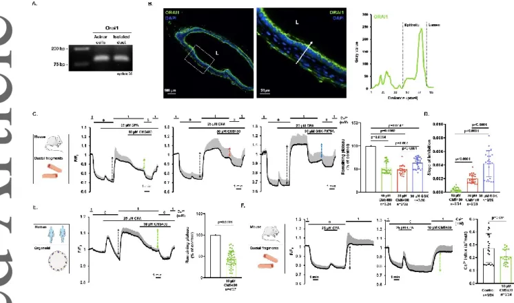
Orai1 is expressed on the apical plasma membrane of pancreatic ductal epithelia
First, we analyzed the expression of Orai1 in mouse primary pancreatic ductal epithelial cells. End- point PCR analysis of isolated acini and ductal fragments confirmed that Orai1 is expressed in pancreatic ductal cells (Figure 1.A.). Immunofluorescent labelling of cross sections of isolated mouse pancreatic ducts revealed that Orai1 is localized dominantly on the apical PM of the pancreatic ductal epithelia (Figure 1.B.). Of note, Orai1 was also detected in cells surrounding the ductal epithelia (most likely fibroblasts) and Orai1 expression on the basolateral membrane of the ductal cells cannot be excluded due to the resolution limit of the light microscopy. Next, to demonstrate Orai1 functionality in mouse pancreatic ductal cells, ER Ca2+ stores were depleted with 25 µM cyclopiazonic acid (CPA) in Ca2+-free extracellular media to activate SOCE. Under these conditions, re-addition of 1 mM extracellular Ca2+ resulted in a marked extracellular Ca2+ influx, which was impaired by 10 µM CM5480, a potent Orai1 inhibitor (Figure 1. C.). Although the maximal inhibition did not increase when using a higher concentration of CM5480, resulting in a similar decrease of the plateau phase (45.15±3.41% at 10 µM CM5480 vs. 52.38±2.45% at 30 µM CM5480, respectively), inhibition was achieved significantly faster at 30 µM CM5480 (Figure 1. D.). To
compare the effect of CM5480 to a known Orai1 inhibitor, we applied 30 µM GSK-7975A during the same protocol. We found that although the inhibitory effect of GSK-7975A was achieved significantly faster than CM5480, the maximal inhibition was higher during CM5480 administration.
Then, we applied the same protocol to human pancreatic organoids consisting of primary polarized ductal cells. In these organoids, 10 µM CM5480 remarkably decreased the extracellular Ca2+ influx (56.63±2.85%) indicating that Orai1 is active in the human pancreatic ducts (Figure 1. E.). As the plateau phase of the Ca2+ signal under the applied conditions is a mixture of Ca2+ influx and efflux, which may affect the characterization of the Orai1 mediated Ca2+ entry, we also applied another protocol. Similarly, in mouse pancreatic ducts, addition of CM5480 before the re-addition of extracellular Ca2+ resulted in a significant decrease of the extracellular Ca2+ influx (Figure 1. F.). Of note, despite the inhibition of the Orai1 channels, a significant proportion of the extracellular Ca2+
influx remained active in every case suggesting that other Ca2+ influx channels or transporters may contribute to the extracellular Ca2+ influx in ductal cells.
The inhibition of Orai1 abolishes toxin-induced extracellular Ca2+ influx in the pancreatic ductal epithelia
Next, we investigated the role of Orai1-mediated Ca2+ entry in the development of bile acids- or ethanol and ethanol metabolites-induced sustained intracellular Ca2+ elevation. In these experiments, isolated murine pancreatic ductal fragments were challenged with 250 µM CDC, known to induce a sustained elevation of the intracellular Ca2+ concentration (Venglovecz et al., 2008), or with a combination of 100 mM ethanol and 200 µM PA. Next, following reaching a stable plateau, the ductal fragments were challenged with 10 µM CM5480. Based on the ΔF value of the plateau, CM5480- mediated inhibition of Orai1 significantly decreased the extracellular CDC-induced Ca2+ influx.
However, as the plateau phase of the evoked Ca2+ signal was not clearly separated from the peak in case of ethanol/PA, we decided to apply CM5480 simultaneously with the EtOH+PA treatment.
Similar to CDC, CM5480-mediated inhibition of Orai1 significantly impaired the effect of
ethanol/PA-induced Ca2+ influx and also the area under curve (Figure 2. A-C.). These results suggest the potential of CM5480 or Orai1 inhibition to prevent Ca2+-overload-mediated functional and morphological damage of ductal cells associated with biliary- or ethanol-induced AP.
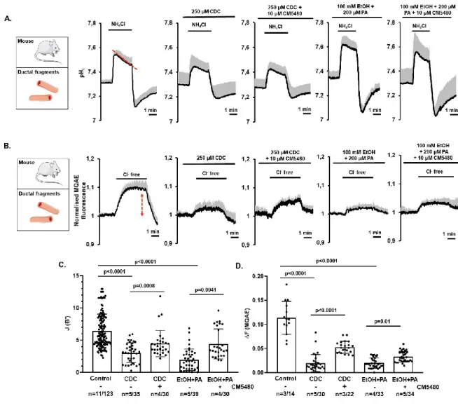
Inhibition of Orai1 prevents bile acid- and ethanol-induced decrease of HCO3- secretion and CFTR function in pancreatic ductal epithelia
HCO3- secretion –the primary function of pancreatic ductal epithelia– is significantly impaired by bile acid- and ethanol-mediated sustained Ca2+ elevation and mitochondrial damage (Maléth et al., 2015;
Molnár et al., 2020). To assess the potential protective effect of Orai1 inhibition on ductal HCO3- secretion, we treated isolated murine ductal fragments with CDC or ethanol/PA in the presence or absence of CM5480 and compared HCO3- efflux across the apical membrane (Maléth et al., 2015). In these experiments, exposure of the ductal cells to 20 mM NH4Cl in HCO3-/CO2-buffered solution triggered a rapid alkalization caused by the passive NH3 uptake of the cells, which was followed by a slow recovery of the alkaline pH - due to the SLC26 Cl-/HCO3- exchanger - and CFTR-mediated HCO3- (Molnár et al., 2020) efflux (i.e. secretion) from the ductal epithelia to resting pHi (Figure 3.A). Subsequent removal of NH4Cl rapidly decreased pHi below the resting value which later restored due to basolateral NHE1 and NBCe1 channel activity. To calculate the base flux values [J(B−)] of HCO3- extrusion (calculated as ΔpH/Δt (Molnár et al., 2020)), the initial recovery rates were measured over the first 30 s. By using this approach, we found that CM5480 significantly improved the apical HCO3- efflux in the CDC- or ethanol/PA-treated isolated ducts (Figure 3.C.).
Next, we used the intracellular Cl- ([Cl-]i)-sensitive fluorescent indicator MQAE to follow CFTR- driven Cl- extrusion in intact pancreatic ductal fragments (Figure 3. B.). Considering that MQAE- mediated fluorescence inversely correlates with [Cl-]I,an increased MQAE signal reports Cl- efflux.
Removal of extracellular Cl- from the HCO3-/CO2-buffered solution resulted in a [Cl-]i decrease, most likely due to CFTR-mediated Cl- efflux from the cytosol (Molnár et al., 2020). While treatment of ductal cells with 250 µM CDC or 100 mM ethanol/200 µM PA resulted in a significant drop of CFTR
activity, this was significantly improved by co-administration of 10 µM CM5480 (Figure 3. B, D.).
These results suggest that CM5480 treatment has the potential to prevent the AP-induced inhibition of the pancreatic ductal HCO3- secretion and partially restored CFTR activity.
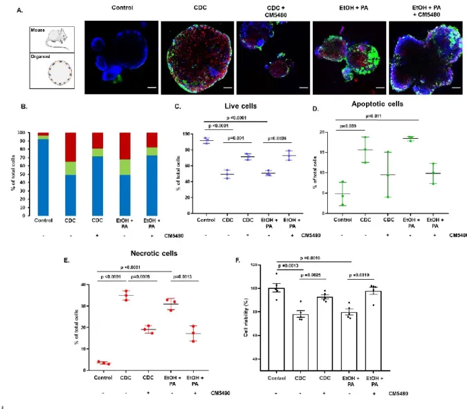
Inhibition of Orai1 prevents bile acid- and ethanol-induced cell death in pancreatic ductal epithelia
Considering that bile acid- and ethanol metabolite-induced sustained intracellular Ca2+ elevation is known to trigger cell death (Voronina et al., 2002; Criddle et al., 2006), we next sought to characterize the effects of Orai1 inhibition on pancreatic ductal cell survival. For this, we used mouse pancreatic ductal organoids as they are more suitable for reaching single cell resolution with confocal microscope allowing reliable counting of labelled cells. Also, as no other cell type is surrounding the epithelial monolayer in these organoids, we are confident only quantifying epithelial cell death (Figure 4.A). In the untreated control samples, minimal cell damage was observed (% of viable cells:
92.2 ± 3.4). Incubation of pancreatic ductal organoids with CDC or EtOH+PA for 30 min remarkably decreased the number of viable cells and significantly increased necrotic cell death (% of viable cells:
49.4 ± 5.3 and 50.6 ± 3.2, respectively) (Figure 4.B-E). Similarly, the apoptosis rate significantly increased upon CDC- or EtOH+PA administration. Although CM5480-mediated Orai1 inhibition decreased the percentage of apoptotic cells to some extent, this improvement failed to reach statistical significance (Figure 4.D). However, both in bile acid- or alcohol-treated organoids, CM5480- mediated Orai1 inhibition significantly decreased the percentage of necrotic cells (Figure 4.E).
Importantly, inhibition of Orai1 resulted in about 45% less pancreatic ductal cell death, suggesting an important role of Orai1-mediated extracellular Ca2+ influx in biliary- and alcoholic AP-mediated ductal cell death. The availability of ATP is crucial to determine cell fate during stress. To provide an insight into the changes of the intracellular ATP in the different conditions, we measured the cell viability using CellTiter-Glo® 3D cell viability assay (Figure 4.F). Our results showed that incubation
of pancreatic ductal organoids with CDC or EtOH+PA for 30 min caused significant reduction of the ATP dependent bioluminescence, which was almost completely restored by CM5480.
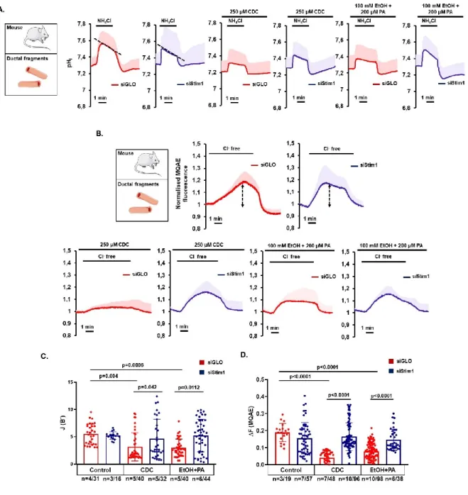
Bile acid- and alcohol-induced Orai1-mediated extracellular Ca2+ entry depends on the activation of Stim1
Exposure of ductal cells to bile acids and ethanol release Ca2+ from intracellular stores –most prominently from the ER (Maléth et al., 2011, 2015)– which induces a conformational change of Stim1 triggering Orai1-mediated Ca2+ influx. To assess the involvement of Stim1 in bile acid- and alcohol-induced ductal cell functional damage, we treated isolated ducts with specific siRNA to knock down Stim1 expression. Treatment of pancreatic ductal cells with siStim1 did not alter CFTR- mediated HCO3-- or Cl- secretion (Figure 5.A. and B). Whereas CDC or ethanol/PA significantly impaired HCO3- secretion or CFTR activity in siGLO-Green-treated pancreatic ductal cells, this was prevented in siStim1-treated cells (Figure 5.C). These results suggest that bile acids and ethanol induce Orai1-mediated Ca2+ influx in a Stim1-dependent manner.
The effect of CM5480 on experimental acute pancreatitis
Based on our obtained results, we wanted to confirm the protective effect of Orai1 inhibition on ductal secretion in vivo. To analyze the potential of CM5480 to reduce the severity of experimental AP, we used three independent experimental models. In the first experimental model, mice received 7 hourly injections of cerulein (50 µg/kg, i.p.) to induce pancreatitis (Figure 6.A). Similar to previous independent reports (Wen et al., 2015; Waldron et al., 2019), inhibition of Orai1 by CM5480 (20 mg/kg, i.p.) –administrated after the second cerulein injection– significantly reduced the histological inflammatory parameters (Figure 6. A.).
Next, AP was induced by intraductal infusion of 4% Na-TC. In these experiments, administration of CM5480 (20 mg/kg, i.p.) 1 hour after the Na-TC infusion improved the edema and leukocyte
infiltration, although tissue necrosis was only moderately decreased (Figure 6. B.). Despite the lack of a significant decrease in necrosis by CM5480, this experimental protocol allowed us to study the changes of in vivo fluid secretion (see further). Likely, a further decrease of necrosis might have been obtained in case of repetitive CM5480-administration.
Finally, in the third experimental AP model, mice received a mixture of ethanol and POA to mimic alcohol-induced AP (Figure 6.C.). In this model, administration of CM5480 (20 mg/kg, i.p.) directly before the second ethanol/POA injection significantly improved AP-mediated edema and necrosis but only moderately decreased leukocyte infiltration.
Inhibition of Orai1 preserves pancreatic ductal secretion in vivo during acute pancreatitis Finally, we wanted to provide evidence that inhibition of Orai1 prevents AP-mediated impairment of pancreatic ductal function in vivo as well. For this, we chose to use the same model systems as described above –as they successfully reproduced previously reported data– to analyze AP-mediated changes of fluid secretion in vivo. Upon establishment of experimental AP, secretin-induced fluid secretion was measured in vivo in anesthetized mice. While in vivo fluid secretion was significantly impaired in all three AP groups (i.e. cerulein-, Na-TC-, and ethanol/POA-treated animals), it was most pronounced in the cerulein-treated group (Figure 7. A.) although still more than 60% impaired in the two other AP groups. Importantly, whereas CM5480 treatment alone did not affect secretin-stimulated pancreatic secretion, it preserved the in vivo fluid secretion in the AP mice. In fact, in both cerulein- and ethanol/POA-treated animals, CM5480 significantly improved in vivo fluid secretion to levels comparable to the untreated –healthy– control group (Figure 7. A., C.). Moreover, in the Na-TC- treated group, CM5480 resulted in an almost twofold increased fluid secretion compared to the Na-TC group; however, the difference failed to reach statistical significance (Figure 7.B.). To further confirm these results, we isolated pancreatic ductal fragments from cerulein-induced AP mice and measured in vitro HCO3- secretion (Figure 7. D.). As expected, whereas cerulein significantly decreased HCO3- secretion in the ductal fragments, this secretory activity was preserved in ductal
fragments derived of cerulein-induced AP mice receiving CM5480. Importantly, these results confirmed that inhibition of Orai1 preserves the ductal ion and fluid secretion both in vitro and in vivo in different forms of AP.
Discussion
Regardless of its etiology, sustained elevation of intracellular Ca2+ is a hallmark in the development of AP-mediated cellular injury (Maléth & Hegyi, 2016; Pallagi et al., 2020). Although selective Orai1inhibitors –limiting the excessive extracellular Ca2+ influx– prevented acinar cell damage and decreased the severity of AP in multiple animal models (Wen et al., 2015; Waldron et al., 2019), precisely how Orai1 inhibition affects the pancreatic ductal cell functions is currently unknown. In this study, we first demonstrated that Orai1 resides in the apical PM of the pancreatic ductal cells where it mediates extracellular Ca2+ influx upon ER Ca2+ store depletion. Next, we provided evidence that bile acid- and ethanol-mediated SOCE activation contribute to sustained intracellular Ca2+
elevations leading to damaged ductal secretion and cell death. Finally, prevention of intracellular Ca2+
overload with selective Orai1 inhibitors preserved pancreatic ductal ion and fluid secretion and maintained exocrine pancreatic secretion during AP.
Expression of Orai1 in exocrine pancreatic acinar cells was previously described by two independent groups. Lur et al. demonstrated Stim1-translocation and Orai1-activation in the lateral and basal plasma membrane (Lur et al., 2009), whereas Hong et al. reported a more pronounced Orai1 expression in the apical membrane (Hong et al., 2011). In our experiments, Orai1 expression was observed on the apical membrane of ductal cells, but not on the basolateral membrane. Although the significance of this polarized expression pattern is currently unknown, it may be of importance in reuptake of intraluminal Ca2+ secreted by the acinar cells during digestive enzyme secretion.
Interestingly, CM5480-mediated functional inhibition of Orai1 did not completely abolish the ER store depletion-induced extracellular Ca2+ influx. In fact, in the current study, we achieved a maximal inhibition of around 50% –both in case of 10 µM and 30 µM CM5480– suggesting that additional
PM-residing Ca2+ channels or transporters contribute to SOCE in ductal cells. When compared to a known Orai1 inhibitor – GSK-7975A – CM5480 achieved significantly higher inhibition of the extracellular Ca2+ influx, however GSK-7975A reached the maximal inhibitory effect more rapidly.
Interestingly, genetic deletion of the TRPC3 Ca2+ channel resulted in a 50% reduction of receptor- stimulated SOCE in pancreatic acinar cells and prevented bile acid- and ethanol metabolite-induced sustained Ca2+ elevation and intracellular trypsin activation. These beneficial effects ultimately resulted in reduced cerulein-induced AP severity in vivo (Kim et al., 2009). Similar results were achieved with the specific TRPC3 inhibitor Pyr3 (Kim et al., 2011). However, the contribution of TRPC3 to SOCE in pancreatic ductal cells is currently unknown. On the other hand, the accessibility of the apical membrane in these ex vivo models may be limited as the ductal fragments are sealed and the epithelial cells in the organoids form a polarized and closed monolayer, which can limit the diffusion of the drugs to the apical membrane proteins.
To achieve strict control and tune the activity of each other, Ca2+-and cAMP/PKA signaling – a well- known key regulator of CFTR activity and HCO3- secretion– interact at multiple levels to facilitate maximal response (Ahuja et al., 2014). On the other hand, the most common biologically active molecules inducing AP –including bile acids, non-oxidative ethanol metabolites, and trypsin– induce toxic, sustained intracellular Ca2+ elevation in the exocrine pancreas (Voronina et al., 2002; Criddle et al., 2006) . Previous data indicated that the non-conjugated bile acid CDC dose-dependently impairs pancreatic HCO3- secretion (Venglovecz et al., 2008) via sustained Ca2+ elevation and subsequent mitochondrial damage in pancreatic ductal (Maléth et al., 2011) cells and isolated pancreatic acinar cells (Gerasimenko et al., 2006).
Considering the detrimental effect of heavy ethanol consumption –in combination with non-oxidative ethanol metabolites (such FAEE)– on acinar and ductal cells (Petersen et al., 2009; Maléth & Hegyi, 2014), our group previously demonstrated that EtOH+POA impaired the activity of the apical SLC26 Cl-/HCO3- exchanger and CFTR Cl- channel together with decreased HCO3- secretion in ductal cells (Maléth et al., 2015). Mechanistically, ethanol and POA induced a sustained Ca2+ elevation through IP- and ryanodine receptor-mediated Ca2+release from the ER combined with extracellular Ca2+
influx; a mechanism which was also described in pancreatic acinar cells (Criddle et al., 2004, 2006, 2007). The involvement of Orai1 in the development of AP was first highlighted by Gerasimenko et al. demonstrating that Orai1 inhibition decreased acinar cell necrosis in vitro (Gerasimenko et al., 2013). In fact, selective GSK-7975A-mediated inhibition of Orai1 inhibited SOCE in a concentration- dependent manner and reduced the sustained Ca2+ elevation, trypsin activation, and acinar necrosis upon FAEE exposure. Others found that GSK-7975A and CM_128 markedly impaired bile acid- induced extracellular Ca2+ influx and sustained Ca2+ overload in pancreatic acinar cells and significantly decreased pancreatic edema, inflammation, and necrosis in experimental models of AP (Wen et al., 2015). Waldron et al. showed that inhibition of SOCE by CM4620 prevented trypsinogen activation, acinar cell death, NF-κB and NFAT activation, and inflammatory responses in multiple in vitro and in vivo models (Waldron et al., 2019). Other reports described cerulein-mediated interaction between Stim1 and Orai1, subsequent activation of SOCE, and calcineurin-mediated activation of NFAT and transcription factor EB promoting transcription of chemokine and autophagy-associated genes (Zhu et al., 2018). In our study, inhibition of Orai1 by CM5480 in pancreatic ductal cells significantly decreased the bile acids and ethanol/PA-mediated extracellular Ca2+ influx in pancreatic ductal fragments and organoids. The CM5480-mediated inhibition of Orai1 was sufficient to significantly improve the in vitro HCO3- secretion and CFTR activity in pancreatic ductal cells.
Moreover, CM5480 preserved the intracellular ATP production, significantly improved the overall ductal cell survival and remarkably prevented CDC- and ethanol/PA-induced cell necrosis.
Interestingly, Orai1 inhibition prevented the decrease of ATP production, however it didn’t completely abolish necrotic cell death. Notably, our previous work showed that bile acids can damage the mitochondria independently from Ca2+ overload, as BAPTA was not able to prevent the mitochondrial damage (Maléth et al., 2011). As specific siRNA knockdown of the ER Ca2+ sensor protein Stim1 reproduced the effect of selective pharmacologic Orai1 inhibition, CDC- and ethanol/PA-induced activation of Orai1 seems to be Stim1 dependent.
The importance of pancreatic ductal fluid and HCO3- secretion in the physiological function of the exocrine pancreas is supported by several independent studies. Di Magno et al. demonstrated that
CFTR knockout mice –in which the exocrine pancreatic secretion was impaired– develop more severe cerulein-induced AP which is accompanied with increased pancreatic edema, neutrophil infiltration, and expression of inflammatory mediators (Dimagno et al., 2005). In addition, our group showed that genetic deletion of Na+/H+ exchanger regulatory factor-1 (NHERF-1) –a scaffolding protein that anchors CFTR to the apical PM– reduces pancreatic fluid and HCO3- secretion in mice (Pallagi et al., 2014). Compared to wild type littermates, NHERF-1 KO mice developed more severe experimental AP upon cerulein hyperstimulation or bile acid infusion to the main pancreatic duct. In addition, the alcohol-induced impairment of CFTR function and expression resulted in increased severity of experimental AP. In the current study, we confirmed previous reports by indicating that in vivo CM5480-mediated inhibition of Orai1 in mice markedly decreases the severity of AP in three different model systems with independent pathogenic triggers. Importantly, in all three AP models, we demonstrated improved secretin-stimulated in vivo pancreatic fluid secretion in CM5480-treated animals. Notably, at the tested dose, CM5480 had no effects on the secretin-stimulated in vivo pancreatic fluid secretion itself in the control groups. Based on these and on our in vitro results we can conclude that bile acids and ethanol directly damage ductal cell functions, which can be restored by the Orai1 inhibition. As both the injury of acinar and ductal cells can affect each other functions (Hegyi et al., 2011) the inhibition of Orai1 may have synergistic effects, which can improve the outcome of AP.
Restoration of pancreatic fluid secretion could have significant beneficial impact on the disease outcome. In a healthy pancreas, the digestive enzymes produced by the acini are washed out by HCO3- -rich fluid into the duodenum where it neutralizes the local pH. Previously, our group demonstrated pH-dependent autoactivation of trypsinogen together with elevated trypsinogen activity in acidic environment indicating the primordial role of HCO3- to prevent early autoactivation of trypsinogen (Pallagi et al., 2011). In addition, Zeng et al. reported that pharmacological correction of CFTR expression and activity rescues pancreatic acinar cell function and reduces autoimmune pancreatitis- induced inflammation, further highlighting the importance of proper ductal function in the disease outcome (Zeng et al., 2017). Recently, the role of Saraf (Jha et al., 2013) –an Orai1 channel regulator
protein– was reported in AP (Son et al., 2019). In contrast to constant expression levels of Stim1 and Orai1, expression levels of Saraf decreased during AP in both mice and human. In addition, whereas Saraf knockout mice developed more severe AP accompanied by increased Ca2+ influx in acinar cells, its overexpression reduced acinar Ca2+ influx and decreased AP severity.
Very recently, a phase 2, open-label, dose-response clinical study by CalciMedica evaluated the safety of Auxora in patients with AP, SIRS, and hypoxemia (Bruen et al., 2021). In this clinical study, the patients received low- or high-dose Auxora plus standard of care (SOC). Overall, no differences in the number of serious adverse events with Auxora compared to SOC alone were reported. Of patients with moderate AP receiving low-dose Auxora, 36.5% improved to mild AP. Very interestingly, patients receiving Auxora better tolerated solid foods, had less persistent SIRS, and had a reduced hospitalization rate compared to SOC. It is tempting to speculate that the increased tolerance towards solid food might be explained by improved exocrine pancreatic secretion as observed in our current study. Based on these results, further clinical studies are needed to clarify the utility of Orai1 inhibition in AP patients.
Taken together, we report that inhibition of Orai1 protects pancreatic ductal cells from sustained intracellular Ca2+ overload triggered by bile acids and ethanol in combination with non-oxidative ethanol metabolites. Importantly, this protection seems to be sufficient to maintain crucial ductal functions –such as fluid and HCO3- secretion– both in vitro and in vivo during AP. Considering that Auxora is currently in Phase 2b clinical trials to treat severe AP, our current results can further contribute to the development of specific pharmacological treatments for AP.
Data availability statement
The authors declare that all data supporting the results are included in the paper. There are no shared datasets in this manuscript.
Competing interests
The authors have no conflict of interest to declare. CalciMedica supplied CM5480 for the study.
Author contributions
PP and JM designed the research project; MG, PP, TM, ÁV, NP, TC, VV, AG, VSZ, MM, KD and JM contributed to acquisition, analysis and interpretation of data for the work. ESZ, LGY provided human pancreatic tissue samples. MG, PP, TM, ÁV, RZ, HP and JM drafted the work and all authors approved the final version of the manuscript, agree to be accountable for all aspects of the work in ensuring that questions related to the accuracy or integrity of any part of the work are appropriately investigated and resolved. All persons designated as authors qualify for authorship, and all those who qualify for authorship are listed.
Funding
The research was supported by funding from the Hungarian National Research, Development and Innovation Office (GINOP-2.3.2–15–2016–00048 to JM, PD116553 to PP), the Ministry of Human Capacities (EFOP 3.6.2-16-2017-00006 to JM), Bolyai Research Fellowship (BO/00569/17 to PP), the Hungarian Academy of Sciences (LP2017–18/2017 to JM), by the National Excellence Programme (20391-3/2018/FEKUSTRAT, TUDFO/47138-1/2019/ITM and TKP2020) to JM by the New National Excellence Program of the Ministry of Human Capacities (ÚNKP-20-3-SZTE-128 to ÁV and ÚNKP-20-3-SZTE-120 to TM) and EFOP 3.6.3-VEKOP-16-2017-00009 to ÁV, TM, BJ, VSZ. This work was supported by Albert Szent-Györgyi Research Grant (to MJ and PP) by the Faculty of Medicine, University of Szeged and BT was supported by the scholarship of the National Talent Program (NTP-NFTÖ-20-B-0332) The project has received funding from the EU’s Horizon 2020 research and innovation program under grant agreement No. 739593.
Figures and figure legends
Figure 1. Expression and functional activity of Orai1 in murine and human pancreatic ductal epithelial cells. A. Agarose gel image proves active Orai1 gene expression in isolated mouse acinar cells and ductal fragments. B. Representative confocal image and exported line profile analysis of an isolated mouse ductal fragment demonstrates the dominantly apical localization of Orai1 in polarized epithelial cells. L: lumen. C. Isolated pancreatic ducts were perfused with standard HEPES solution, which was changed to Ca2+-free HEPES solution. After Ca2+ store depletion via 25 μM cyclopiazonic acid and re-addition of 1 mM extracellular Ca2+, mouse pancreatic ductal fragments were challenged with 10 or 30 μM CM5480 or 30 μM GSK-7975A specific Orai1 inhibitor resulting in a reduced Ca2+
influx (average traces). The degree of inhibitory effect of 30 μM GSK-7975A was significantly higher, than 10 or 30 μM CM5480, however there was no significant different between 10 and 30 μM CM5480. D. The slope of inhibition significantly increased upon administration of 30 μM CM5480 or GSK-7975A compared to 10 μM, and GSK-7975A was more effective than 30 μM CM5480. E. Orai1 inhibition induced by 10 μM CM5480 showed the same decreasing characteristics in human pancreatic organoids. F. Average traces demonstrates that CM5480 used prior to Ca2+ re-addition, significantly reduced extracellular Ca2+ influx in mouse pancreatic ductal fragments. Statistical analysis was performed by Tukey’s multiple comparisons test (C and D), and Mann-Whitney test (E and F).
Figure 2. Pharmacological inhibition of Orai1 leads to reduced extracellular Ca2+ influx induced by bile acid or ethanol and fatty acid in pancreatic ductal epithelia. Isolated mouse pancreatic ductal fragments were challenged with 250 µM CDC or with a combination of 100 mM ethanol and 200 µM PA in standard HEPES buffered solution. A. Average traces and the bar charts demonstrate that the inhibition of Orai1 by 10 μM CM5480 significantly decreased the plateau phase of the CDC- induced intracellular Ca2+ signal. B-C. Orai1 inhibition also reduced the intracellular Ca2+ elevation and value of area under curve triggered by the combination of 100 mM ethanol and 200 μM PA.
Statistical analysis was performed by Mann-Whitney test.
Figure 3. The inhibition of Orai1 prevents bile acid- and ethanol-induced decrease of apical HCO3- secretion and CFTR function in pancreatic ductal epithelia. A. Mouse pancreatic ducts were perfused with 250 µM CDC or 100 mM ethanol and 200 µM PA in HCO3−/CO2-buffered extracellular solution and intracellular alkalization was achieved by 20 mM NH4Cl administration in the absence or presence of Orai1 inhibitor CM5480. Both pathogenic conditions significantly reduced the base flux, which was offset using the Orai1 inhibitor. B. Average traces of intracellular Cl− levels reflecting CFTR activity in mouse isolated ductal fragment using Cl− sensitive fluorescent dye (MQAE). Removal of extracellular Cl− induced a decrease in intracellular Cl− levels (reflected by an increase in fluorescent intensity) due to CFTR activity. Cl- removal from the extracellular solutions in the presence of 250 µM CDC or 100 mM ethanol and 200 µM PA inhibited CFTR activity which was improved by CM5480. Bar charts of the calculated base fluxes of HCO3− (C) and Cl- efflux (D).
CM5480 significantly increased both parameters in the presence of CDC, ethanol+PA. Statistical analysis was performed by Tukey’s multiple comparisons test.
Figure 4. Inhibition of Orai1 prevents bile acid- and ethanol-induced cell death in the pancreatic ductal epithelia. A. Representative confocal z-stack images of mouse pancreatic ductal organoids under different treatment conditions (blue: live cells labelled with CytoCalcein 450, green: necrotic cells labelled with Nuclear Green, and red: apoptotic cells labelled with Apopxin Deep Red). Scale bar: 50 μm. B. Bar chart representing the % of live, apoptotic, and necrotic cells in the organoids after incubation with 250 µM CDC or 100 mM EtOH and 200 µM PA in the presence and absence of 10 μM CM5480 for 30 min on 37ºC. Treatment of the organoids with CDC or with ethanol+PA markedly decreased the number of viable cells (C.), whereas the number of apoptotic (D.) and necrotic (E.) cells were significantly increased. 10 μM CM5480 markedly decreased the rate of apoptotic and significantly reduced necrotic cells and in parallel increased the number of detectable live cells. F. Bar charts summarizing the cell viability as measured by the ATP dependent bioluminescence using CellTiter-Glo® 3D cell viability assay. Treatment conditions were the same as above described. CM5480 treatment significantly increased the cell viability compared to CDC or EtOH+PA treated groups. Statistical analysis was performed by Kruskal-Wallis test.
Figure 5. siStim1 treatment prevents bile acid- and ethanol-induced decrease of apical HCO3- secretion and CFTR function in pancreatic ductal epithelia. Average traces and bar charts demonstrate the effects of 250 µM CDC or 100 mM ethanol and 200 µM PA on control (red traces) and siStim1-treated (blue traces) pancreatic ductal fragments. A. Pancreatic ducts were challenged with 20 mM NH4Cl in HCO3-/CO2 buffered extracellular solution and the apical Cl-/HCO3- exchange activity was determined. B. Extracellular Cl- was removed to measure CFTR activity in control and treated ductal fragments. C-D. Bar charts of the calculated base fluxes of HCO3- or maximal fluorescent intensity changes of MQAE show that administration of CDC or ethanol+PA significantly impaired pancreatic ductal HCO3- secretion and CFTR-mediated Cl- secretion in the siGLO-Green- treated control ducts. siStim1 treatment prevented the inhibition of these parameters. Statistical analysis was performed by Tukey’s multiple comparisons test.
Figure 6. Orai1 inhibition by CM5480 reduces the severity of acute pancreatitis. A.
Representative images of pancreatic histology in cerulein-induced pancreatitis. Mice were given 7
hourly i.p. injections of either physiological saline (PS, control group) or 50 µg/kg cerulein. CM5480 or vehicle was administered 1 hour after the first injection of cerulein or PS. Cerulein administration caused extensive pancreatic damage, which was significantly reduced by 20 mg/bwkg CM5480 treatment. B. Representative images of pancreatic histology in Na-taurocholate-induced pancreatitis.
Pancreatitis was induced by intraductal infusion of 4% Na-taurocholate (TC). CM5480 or vehicle was administered 1 hour after the Na-TC or PS infusion. 4% Na-TC-induced necrotizing pancreatitis in mice accompanied by elevated histological parameters. Orai1 inhibition by CM5480 significantly reduced the extent of edema and leukocyte infiltration; however, there was no significant difference in the extent of necrosis. C. Representative images of pancreatic histology in ethanol+ palmitoleic acid (POA)-induced pancreatitis. Mice were given 2 hourly i.p. injections of either PS or 1.75 g/kg ethanol and 750 mg/kg POA. CM5480 or vehicle was administered 1 hour after the first injection. Orai1 inhibition significantly improved the edema and necrosis induced by ethanol+POA, whereas no significant difference was observed in leukocyte infiltration. Scale bar: 100 µm. Statistical analysis was performed by Tukey’s multiple comparisons test.
Figure 7. Orai1 inhibition by CM5480 prevents pancreatic ductal secretion during acute pancreatitis. Acute pancreatitis (AP) was induced in mice as described above. Pancreatic juice was collected for 30 min in vivo under secretin-stimulated (0.75 CU/kg i.p.) conditions from anesthetized mice after induction of AP. A. Summary bar charts show that cerulein administration significantly reduced the volume of pancreatic juice, which was preserved by CM5480 treatment. B. CM5480 administration increased the reduced in vivo fluid secretion caused by Na-taurocholate, however the difference was not significant. C. The volume of pancreatic juice was significantly lower after ethanol+POA treatment; however, Orai1 inhibition significantly improved it. D. Averages traces and bar charts demonstrate the in vitro HCO3- secretion of pancreatic ductal fragments isolated from mouse pancreas after the induction of AP with cerulein. Pancreatic ducts were perfused with HCO3- /CO2 buffered extracellular solution and intracellular alkalization was achieved by 20 mM NH4Cl administration. Comparison of recovery from alkalosis shows that ductal HCO3- secretion was significantly reduced in the cerulein-treated group, which was restored by CM5480 treatment.
Statistical analysis was performed by Tukey’s multiple comparisons test.
References
Ahuja M, Jha A, Maléth J, Park S & Muallem S (2014). cAMP and Ca2+ signaling in secretory epithelia: crosstalk and synergism. Cell Calcium 55, 385–393.
Boj SF et al. (2015). Organoid models of human and mouse ductal pancreatic cancer. Cell 160, 324–
338.
Breunig M et al. (2021). Modeling plasticity and dysplasia of pancreatic ductal organoids derived from human pluripotent stem cells. Cell Stem Cell 28, 1105-1124.e19.
Bruen C, Miller J, Wilburn J, Mackey C, Bollen TL, Stauderman K & Hebbar S (2021). Auxora for the Treatment of Patients With Acute Pancreatitis and Accompanying Systemic Inflammatory Response Syndrome: Clinical Development of a Calcium Release-Activated Calcium Channel Inhibitor. Pancreas 50, 537–543.
Criddle DN, McLaughlin E, Murphy JA, Petersen OH & Sutton R (2007). The pancreas misled:
signals to pancreatitis. Pancreatol Off J Int Assoc Pancreatol IAP Al 7, 436–446.
Criddle DN, Murphy J, Fistetto G, Barrow S, Tepikin AV, Neoptolemos JP, Sutton R & Petersen OH (2006). Fatty acid ethyl esters cause pancreatic calcium toxicity via inositol trisphosphate receptors and loss of ATP synthesis. Gastroenterology 130, 781–793.
Criddle DN, Raraty MGT, Neoptolemos JP, Tepikin AV, Petersen OH & Sutton R (2004). Ethanol toxicity in pancreatic acinar cells: mediation by nonoxidative fatty acid metabolites. Proc Natl Acad Sci U S A 101, 10738–10743.
Dimagno MJ, Lee S-H, Hao Y, Zhou S-Y, McKenna BJ & Owyang C (2005). A proinflammatory, antiapoptotic phenotype underlies the susceptibility to acute pancreatitis in cystic fibrosis transmembrane regulator (-/-) mice. Gastroenterology 129, 665–681.
Fanczal J, Pallagi P, Görög M, Diszházi G, Almássy J, Madácsy T, Varga Á, Csernay-Biró P, Katona X, Tóth E, Molnár R, Rakonczay Z, Hegyi P & Maléth J (2020). TRPM2-mediated
extracellular Ca2+ entry promotes acinar cell necrosis in biliary acute pancreatitis. J Physiol 598, 1253–1270.
Freedman SD, Kern HF & Scheele GA (2001). Pancreatic acinar cell dysfunction in CFTR(-/-) mice is associated with impairments in luminal pH and endocytosis. Gastroenterology 121, 950–
957.
Gerasimenko JV, Flowerdew SE, Voronina SG, Sukhomlin TK, Tepikin AV, Petersen OH &
Gerasimenko OV (2006). Bile acids induce Ca2+ release from both the endoplasmic reticulum and acidic intracellular calcium stores through activation of inositol trisphosphate receptors and ryanodine receptors. J Biol Chem 281, 40154–40163.
Gerasimenko JV, Gryshchenko O, Ferdek PE, Stapleton E, Hébert TOG, Bychkova S, Peng S, Begg M, Gerasimenko OV & Petersen OH (2013). Ca2+ release-activated Ca2+ channel blockade as a potential tool in antipancreatitis therapy. Proc Natl Acad Sci U S A 110, 13186–13191.