Downloadedfromhttps://journals.lww.com/pancreasjournalbyBhDMf5ePHKav1zEoum1tQfN4a+kJLhEZgbsIHo4XMi0hCywCX1AWnYQp/IlQrHD33nx2+yvo/efAtqTYzNd3AfWKeFyjPzYecu/RKbwb34k=on08/09/2019
Downloadedfrom https://journals.lww.com/pancreasjournalby BhDMf5ePHKav1zEoum1tQfN4a+kJLhEZgbsIHo4XMi0hCywCX1AWnYQp/IlQrHD33nx2+yvo/efAtqTYzNd3AfWKeFyjPzYecu/RKbwb34k=on
08/09/2019
Retrospective Matched-Cohort Analysis of Acute Pancreatitis Induced by 5-Aminosalicylic Acid – Derived Drugs
Ágnes Meczker, MSc,* Alexandra Mikó, MD, PhD,* Noémi Gede, MSc,* Andrea Szentesi, PhD,* † Andrea Párniczky, MD, PhD,* ‡ Szilárd Gódi, MD,§ and Péter Hegyi, MD, PhD, DSc, MAE(Med),||¶#**
on behalf of Hungarian Pancreatic Study Group
Objectives: This study aimed to compare the clinical course of 5-aminosalicylic acid–derived, drug-induced acute pancreatitis (5-ASA– DIAP) to acute pancreatitis (AP) caused by other etiologies.
Methods: A cohort of patients with 5-ASA–DIAP was established through literature search. As a control AP (CAP) group, a cohort was generated from a registry. Data on the diagnostic procedure, symptoms, enzyme elevation, imaging, severity, and recovery parameters were collected. Causality was assessed using the Naranjo algorithm.
Results:Twenty-nine articles were included, which describe 36 patients with fifty-one 5-ASA–DIAP episodes (60.78% female, 39.22% male).
There were 88.2% mild, 3.92% moderate, and 7.84% severe cases of AP in the 5-ASA–DIAP group, and 70.6%, 25.5%, and 3.92% such cases in the CAP population, respectively. Symptoms improved significantly faster (mean ± SE, 2.5 ± 0.34 vs 3.74 ± 0.42 days;P= 0.018); however, pancreatic enzyme levels normalized significantly more slowly (6.27 ± 1.53 vs 3.63 ± 0.61 days,P= 0.008) in the 5-ASA–DIAP cohort compared with the CAP group. This study confirms that there are no diagnostic differences between 5-ASA–DIAP and AP of other etiologies.
Conclusions:Fewer moderate but more severe cases were found in the 5-ASA–DIAP group; therefore, 5-ASA–DIAP must be taken as seriously as AP of other etiologies.
Key Words:acute pancreatitis, 5-ASA, mesalazine, rechallenge, drug-induced, IBD
(Pancreas2019;48: 488–495)
A
cute pancreatitis (AP) is a serious disease with high mortality.The reported incidence is variable in different countries (10–100/100,000 people), and AP is a leading cause of acute hos- pitalization for gastrointestinal disorders.1The most common eti- ologies for AP are gallstones, biliary sludge or microlithiasis, alcohol, hypertriglyceridemia, post–endoscopic retrograde cholan- giopancreatography status, hypercalcemia, genetic mutations, infec- tions or toxins, trauma, anatomic malformation of the pancreas, and vascular disease.2–4The rest of the episodes are usually termed idiopathic. Drug-induced AP (DIAP) is a rare entity, accounting for approximately 2% to 5% of AP episodes worldwide.5–7How- ever, estimates vary because of the challenging diagnosis and the difficulties of causality assessment. Because it is considered un- ethical to intentionally rechallenge with the offending drug owing to the potentially life-threatening nature of AP, DIAP remains a speculative diagnosis made by exclusion in most cases.
5-Aminosalicylic acid (5-ASA; mesalazine or mesalamine)– derived drugs are aminosalicylate anti-inflammatory medications generally considered as safe and effective therapy for patients with inflammatory bowel disease (IBD). Pancreatitis as an adverse re- action to 5-ASA–derived drugs was first reported in 1970 by Block et al8and was followed by several others.9–11According to the literature, DIAP is usually considered self-limited and mild in clinical course, with easy management, an excellent prognosis, and low mortality.6Rapid symptomatic improvement usually oc- curs after discontinuation of the offending medication.12,13How- ever, there are huge variations in clinical course in cases of 5-ASA–induced AP. Our main aims were to investigate whether 5-ASA–DIAP has the characteristics noted previously and to compare its clinical course with that of AP caused by other, more common etiologies.
MATERIALS AND METHODS Systematic Search
Initially, our aim was to find all reported 5-ASA–DIAP cases with a systematic search. We searched literature data for retrospec- tive cohort analyses describing these cases in individual medical centers. We performed a systematic literature search according to the Preferred Reporting Items for Systematic Reviews and Meta-Analyses guideline. We applied the following PICO format:
P, patients with AP; I, 5-ASA–DIAP; C, AP caused by other etiol- ogies; and O, severity, mortality, length of hospitalization, imaging alterations, symptoms, and resolution characteristics of AP. The search was performed in April 2017 on PubMed, Embase, and Cochrane Library with the search terms“5-ASA AND pancreatitis” From the *Institute for Translational Medicine, University of Pécs, Medical
School, Pécs;†First Department of Medicine, University of Szeged, Szeged;
‡Heim Pál Children's Hospital, Budapest; §Division of Translational Medicine, First Department of Medicine, University of Pécs, Medical School; ||Institute for Translational Medicine, University of Pécs, Szentágothai Research Centre, Medical School; ¶Division of Translational Medicine, First Department of Med- icine, University of Pécs, Pécs; #First Department of Medicine, University of Szeged; and **Hungarian Academy of Sciences–University of Szeged, Mo- mentum Gastroenterology Multidisciplinary Research Group, Szeged, Hungary.
Received for publication September 28, 2018; accepted February 16, 2019.
Address correspondence to: Péter Hegyi, MD, PhD, DSc, MAE(Med), Centre for Translational Medicine, Institute for Translational Medicine and Department of Translational Medicine/First Department of Medicine, University of Pécs Medical School, Szigeti út 12., 6724 Pécs, Hungary (e‐mail: hegyi2009@gmail.com).
This study was supported by project grants (K116634 and KH125678 to P.H.); an Economic Development and Innovation Operative Programme grant (GINOP 2.3.2-15-2016-00048 to P.H.) and a Human Resources Development Operational Programme grant (EFOP-3.6.2-16-2017-00006 to P.H.) from the National Research, Development and Innovation Office; and a momentum grant from the Hungarian Academy of Sciences (LP2014-10/2014 to P.H.).
The authors declare no conflicts of interest.
Á.M. acquired the data and wrote the manuscript; A.M., A.S., A.P., and S.G.
contributed to the acquisition and interpretation of the data; N.G. performed the statistical evaluation of the data; and P.H. developed the strategy and made critical revisions of the manuscript related to important intellectual content.
Supplemental digital contents are available for this article. Direct URL citations appear in the printed text and are provided in the HTML and PDF versions of this article on the journal’s Web site (www.pancreasjournal.com).
Copyright © 2019 The Author(s). Published by Wolters Kluwer Health, Inc.
This is an open-access article distributed under the terms of the Creative Commons Attribution-Non Commercial-No Derivatives License 4.0 (CCBY-NC-ND), where it is permissible to download and share the work provided it is properly cited. The work cannot be changed in any way or used commercially without permission from the journal.
DOI: 10.1097/MPA.0000000000001297
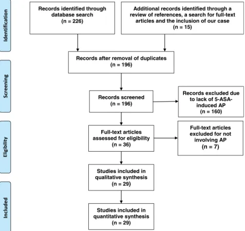
and limited to English language and human target (if applicable) re- gardless of the date of publication. We did not want to limit the search on cohort analyses; thus, case reports, case series, and other types of publications were included as well if they contained data appropriate to our questions. At this point of the work, we realized that no cohort analysis had been performed on this topic because of the small study sizes; only individual case reports and case series were found. The detailed result of the search process is presented in Figure 1. A retrospective cohort was established of the cases de- scribed. We identified 36 studies reporting 5-ASA–DIAP.8–10,12–43 In the analysis, we only included studies in which the authors had concluded that 5-ASA–derived medication is the most probable cause of the adverse drug reaction (ADR) and were concurrently reevaluated as a DIAP episode. In the end, cases from 29 articles were included in the qualitative and quantitative analysis.
Study Population
Based on the case reports found and a case previously re- ported by us,1136 patients with fifty-one 5-ASA–DIAP episodes were included in the qualitative and quantitative analysis. Each DIAP episode was considered as a discrete event, and the further characterization of the study population was based on the hospital- ization events instead of the patients.
Control Population
To characterize the clinical course of 5-ASA–DIAP as accu- rately as possible, we established an age- and sex-matched control
AP (CAP) population by using the Hungarian Pancreatic Study Group (HPSG) electronic registry database. In this database, more than 1400 cases of patients diagnosed as having AP of various etiol- ogies were prospectively enrolled over a 6-year period from January 1, 2012, from 17 Hungarian centers. For each 5-ASA–DIAP episode in the study population, a randomly selected control pair event was chosen from a patient with the same sex and age. If no event was ac- cessible in a patient of the same age, age matching was performed in the range of ±1 year. During the selection process, we excluded all possible drug-induced cases from the matched CAP population. A comparison for each variable was made between these 2 matched co- horts (see Supplemental Table 1, http://links.lww.com/MPA/A716, which demonstrates etiological diversity of the control age- and sex-matched population).
In addition, to see whether the epidemiological characteristics of the population with 5-ASA–DIAP differ from the population with AP caused by other etiologies, we compared age and sex dis- tribution to the whole cohort consisting of more than 1400 patients.
Definition of AP
We reevaluated all ADR events documented by the authors as AP. Each was considered as AP if it met the two-third rule of epigastric pain and/or pancreatic enzyme levels higher than triple the normal upper limit and/or morphological pancreatic abnor- malities seen on imaging according to the International Associ- ation of Pancreatology/American Pancreatic Association and HPSG evidence-based guidelines for the management of AP.44,45
FIGURE 1. Detailed procedure of the systematic literature search. Additional records were identified by reading the references of the articles resulting from the original search and during the process of the online search for the full-text copies. After removing duplicates and screening, we excluded records that were found not to describe AP cases of 5-ASA–induced etiology. Thirty-six articles were evaluated, and 7 were excluded from the analysis for not describing an episode of AP.
Pancreatic enzyme level elevation was considered as higher than tri- ple the upper normal limit if the authors had stated that there was biochemical evidence of AP, if the exact enzyme level and upper normal limit were described, or if the precise extent of elevation compared with the upper normal limit was provided.
Latency
Latency of 5-ASA–DIAP was defined by the time interval between the beginning of a drug therapy and the first symptoms of AP.
Rechallenge
Rechallenge with the offending drug was considered as positive in the presence or absence of a relapse of AP as long as an ADR occurred in the form of a pancreatic enzyme level increase and/or abdominal pain and/or nausea or vomiting.
Severity
To determine the severity of the clinical course of DIAP, we performed an evaluation using the data provided by the authors in accordance with the revised Atlanta classification system.46 Mild AP was defined by absence of organ failure, or local or systemic complications. Moderately severe AP was characterized by transient organ failure (with symptoms improving within 48 hours) and/or local (peripancreatic fluid collection, pseudocysts, acute necrotic collection, and walled-off necrosis) or systemic complications.
Severe AP was characterized by persistent organ failure (not im- proving in 48 hours) of one or multiple organs.
Causality Assessment
The causal relationship between drug intake and 5-ASA–DIAP was assessed with the Naranjo algorithm47to estimate the probability of ADRs. Each case was evaluated using the questionnaire and was assigned a value. The cases were thus categorized into the following groups: any adverse reaction was categorized as definite when scored
≥9, a score of 5 to 8 was rated as probable, 1 to 4 was considered pos- sible, and 0 was viewed as a doubtful correlation according to the algorithm.
Interpretation of the Data
The analysis of the variables studied was conducted with de- scriptive statistics (mean ± standard error of the mean) and relative frequency (Fisher exact test and the Mann–Whitney test). A 2-sided Pvalue of <0.05 was regarded as statistically significant. The
available-case analysis was used for missing data. Statistical anal- yses were performed using the Statistical Package for the Social Sciences (SPSS) for Windows version 22.0 (IBM, Armonk, NY).
The established study population of patients with 5-ASA–DIAP was created based on case reports. The case reporting system has some limitations in reporting quality; therefore, statistical conclu- sions drawn in this study based on this database must be handled with caution.
Results of the Cohort Analysis Demographic Data
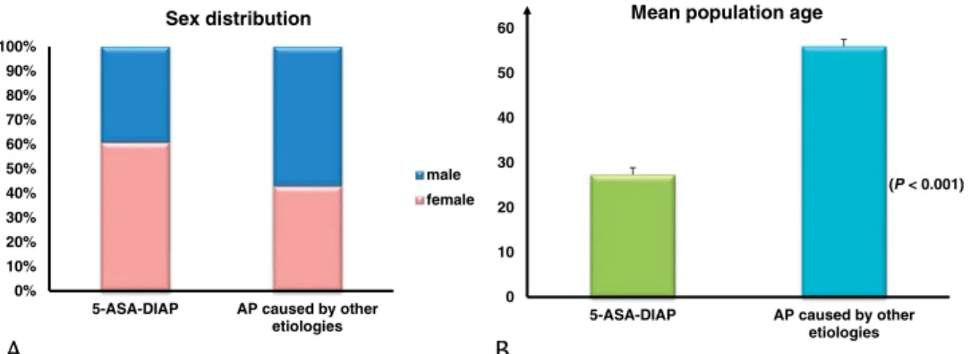
Of the fifty-one 5-ASA–DIAP cases identified, 60.78% were female (n = 31) and 39.22% were male (n = 20). Drug-induced AP due to intake of a 5-ASA–derived drug as treatment of IBD oc- curred at a significantly younger age compared with AP of other, more common etiologies according to the HPSG registry (27.31 ± 1.56 years vs 55.98 ± 0.45 years,P< 0.001; Fig. 2B).
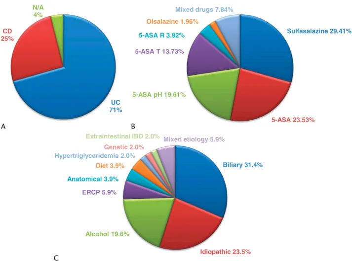
In the 5-ASA–DIAP population, 21.57% (n = 11) were pediatric cases (<18 years of age). There were 70.59% (n = 36) 5-ASA–DIAP cases that were caused by treatment of ulcerative colitis and 25.49% (n = 13) that resulted from medications taken for Crohn disease. There were no data in the reporting on the primary condi- tion in 2 cases.
Acute pancreatitis was described in the literature using mul- tiple types of 5-ASA–derived drugs: mesalazine (nonconjugated 5-ASA), sulfasalazine (5-ASA and a sulfapyridine moiety joined by an azo bond), olsalazine (two 5-ASA radicals linked by a diazo bond), and ethyl-cellulose–coated, time-dependent and resin- coated, pH-dependent 5-ASA formulations (prolonged-release preparations for supporting absorption in the distal gastrointesti- nal tract), the latter 2 available as oral or rectal formulations. In most cases, AP occurred due to intake of sulfasalazine in 29.41% (n = 15). A therapy with 5-ASA in otherwise unspecified formulation was responsible for AP in 23.53% (n = 12). pH-dependent and time-dependent coated forms resulted in AP in 19.61%
(n = 10) and 13.73% (n = 7), respectively. 5-Aminosalicylic acid applied as a rectal enema or suppository caused AP in 3.92% of the cases (n = 2), and olsalazine did so in 1.96% (n = 1). There were 7.84% (n = 4) AP cases that occurred due to intake of 2 dif- ferent 5-ASA formulations.
The etiological factors in the CAP cohort were the following:
biliary (31.4% [n = 16]); idiopathic (23.5% [n = 12]); alcohol con- sumption (19.6% [n = 10]); post–endoscopic retrograde cholan- giopancreatography (5.9% [n = 3]); anatomical disorders and dietary noncompliance (both, 3.9% [n = 2]), hypertriglyceridemia,
FIGURE 2.Epidemiological differences between the 5-ASA–DIAP population and the total HPSG cohort with AP caused by other etiologies. A, Differences of sex distribution showed a female dominance for 5-ASA–DIAP compared with most patients with AP of various etiologies being male. B, A comparison of the 5-ASA–DIAP population with the total cohort of patients with AP of various etiologies showed a significantly younger age group affected by 5-ASA–DIAP.
genetic predisposition, and extraintestinal manifestation of IBD (all 3, 2% [n = 1]); and multiple, mixed etiologies of the previ- ously mentioned (5.9% [n = 3]). A summary of the main charac- teristics of the 5-ASA–DIAP and CAP population is presented in Figure 3.
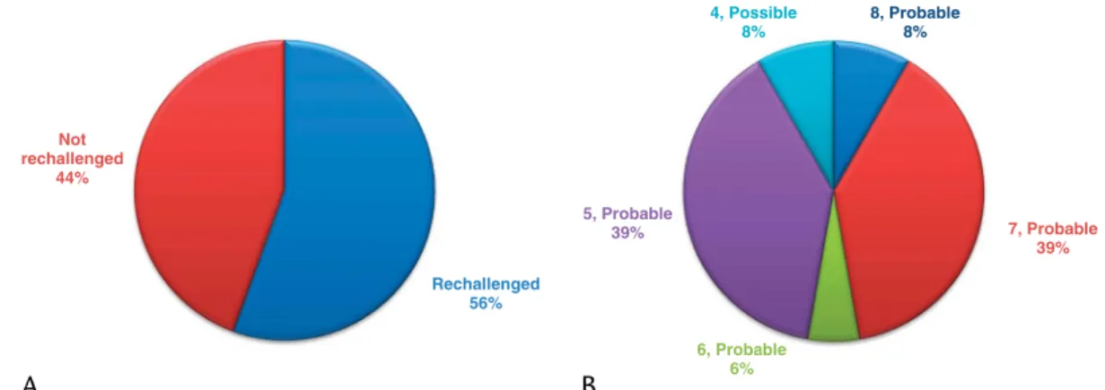
Causality Assessment and Evidence Level of Reporting There was no case found during the literature search with definite causality (Naranjo score≥9) between AP and drug admin- istration (eg, in which IBD is treated with a monotherapy of a 5-ASA–derived drug and all other etiologies of AP were concur- rently investigated and excluded). In some case reports, there were no data provided on the exclusion of other etiologies and the possi- bility of causality between drug intake and AP. There were 55.56%
(n = 20) of the patients who were rechallenged with a 5-ASA–derived drug (Fig. 4A). Of all DIAP cases, 91.67% (n = 33) were assessed as probable 5-ASA–induced AP, of which 8.33% received a score of 8, 38.89% were rated as 7, 5.56% scored 6, and 38.89%
received a 5 using the Naranjo algorithm for estimating the probability of ADRs. Three articles were evaluated as describing possible ADRs with a score of 4 (Fig. 4B).
Imaging Alterations and Symptoms
Imaging data in the form of either abdominal ultrasonography and/or computer tomography was available in 76.5% (n = 39) of the 5-ASA–DIAP population, of which 64.7% (n = 33) had imaging al- terations. In the CAP population, 90.2% (n = 46) underwent transabdominal imaging examinations and 68.6% (n = 35) were di- agnosed with imaging alterations (Fig. 5A). Acute pancreatitis caused by this drug type is usually described as manifesting in an edematous or enlarged pancreas. In the 5-ASA–DIAP population, a localized or diffuse edema or enlargement of the pancreas was demonstrated in 54.9% (n = 28), and the pancreas was identified as normal in 11.8% (n = 6) of the cases. In the CAP group, an edema or enlargement and normal structure of the pancreas were detected both in 23.5% of the cases (n = 12). In the 5-ASA–DIAP population, there were 3 cases of necrotizing pancreas (5.9%), 1 case of peripancreatic inflammation, and 1 case of pseudocyst formation with effusion.
In the CAP population, imaging characteristics were described in a more detailed manner; involvement of necrosis in AP and com- mon bile duct dilatation were both identified in 11.8% of that pop- ulation (n = 6), fluid collections (peripancreatic, free fluid, or ascites) in 37.3% (n = 19), and pseudocysts in 5.9% (n = 3).
FIGURE 3. Characterization of the 5-ASA–DIAP and CAP populations. A, Primary reasons for 5-ASA treatment in the study population. B, Types of 5-ASA–derived drug formulations reported to cause AP in the study population. C, Etiological factors contributing to AP in the matched-control population. 5-ASA pH, pH-dependent formulation; 5-ASA R, rectally administered formulations; 5-ASA T, time-dependent formulation; CD, Crohn disease; N/A, not applicable (condition not specified); UC, ulcerative colitis.
Abdominal pain was present in 90.2% (n = 46) and was coupled with nausea and/or vomiting in 19.6% (n = 10) of the 5-ASA–DIAP episodes compared with 98% (n = 50) of cases with abdominal pain being accompanied by nausea and/or vomiting in 74.5% (n = 38) in the CAP cohort. Pancreatic enzyme level eleva- tion occurred in the 5-ASA–DIAP population in 80.4% (n = 41) of
the episodes compared with 86.3% (n = 44) in the CAP group. In 45.1% (n = 23) of the 5-ASA–DIAP cases, all 3 diagnostic criteria were present compared with 58.8% (n = 30) in the CAP population.
A summary of the presence of abdominal pain of pancreatic local- ization, pancreatic enzyme level elevation, and imaging alterations as the 3 main diagnostic criteria of AP is presented in Figure 5C.
FIGURE 4. Evidence level of 5-ASA–DIAP case reports. A, The rate of case reports with and without rechallenge with 5-ASA–derived medication. B, Case reports categorized according to the quantified causal relationship between intake of 5-ASA and AP using the Naranjo ADR probability scale.
FIGURE 5. Clinical course of 5-ASA–DIAP compared with CAP caused by various etiologies. A, Proportional differences of imaging alterations (if performed) on admission in the 5-ASA–DIAP and CAP groups. B, Percentage of the severity groups in the 5-ASA–DIAP and CAP populations. A significant difference was found between the rates for moderately severe AP groups. C, A comparison of the clinical course of the 5-ASA–DIAP and CAP populations with regard to the presence of the following diagnostic criteria: P, pain (characteristic epigastric); E, enzyme level elevation (pancreatic); and I, imaging alteration. D, Significant differences were found by comparing the time interval for symptomatic improvement and enzyme level normalization (in days) between the 5-ASA–DIAP and CAP groups. Abdominal pain subsided faster, but pancreatic enzyme levels normalized more slowly in cases of 5-ASA–DIAP.
Severity and Mortality
In the 5-ASA–DIAP population, drugs caused clinically mild AP in 88.24% (n = 45) of the cases, a rate that is markedly higher than that of the CAP group (70.6% [n = 36]). In the 5-ASA–DIAP group, 4 episodes (7.84%) of severe AP were diagnosed compared with 3.9% (n = 2) in the CAP population. A significant difference (P< 0.05), however, was only found between the rates of moder- ately severe cases in the 5-ASA–DIAP population (3.92% [n = 2]) and the CAP group (25.49% [n = 13]; Fig. 5B). Overall mortality in the 5-ASA–DIAP population due to complications from clini- cally severe AP was 3.92% (n = 2, half of the severe cases). No deaths in the mild and moderately severe groups were registered.
In the CAP population, no lethal episode was found. Interestingly, 5-ASA–DIAP tended to be milder in most of the cases because of the sparse group of moderately severe cases compared with the CAP population of other etiologies (Fig. 4).
Resolution of AP
Length of hospitalization was usually not described by the authors of the studies identified; in 82.4% of the cases, no infor- mation on this variable was provided. Consequently, a comparison was not made because of lack of data. To characterize the timeline of the clinical course of 5-ASA–DIAP, we collected data on time intervals of symptomatic improvement and enzyme level normali- zation. Symptoms improved significantly faster for 5-ASA–DIAP after discontinuation of the drug compared with the CAP cases (in 2.5 ± 0.34 vs 3.74 ± 0.42 days,P= 0.018), thus confirming lit- erature data on rapid symptomatic recovery after dechallenge (Fig. 5D). However, a less investigated parameter, pancreatic enzyme level normalization, was found to occur significantly more slowly in the 5-ASA–DIAP population compared with the CAP cases (in 6.27 ± 1.53 days vs 3.63 ± 0.61 days,P= 0.008).
Consequently, we cannot conclude a rapid overall resolution of 5-ASA–DIAP. The significant differences in resolution period could be a consequence of DIAP being potentially an immune- mediated reaction with a slow overall fall-off of the disease.
Characterization of 5-ASA–DIAP Populuation Pathomechanism of 5-ASA–DIAP
Of the 29 publications included, the authors suggest an in- volvement of a hypersensitive reaction in the pathomechanism of 5-ASA–DIAP in 38.9% (n = 14). Idiosyncrasy is thought to have a role in 8.3% (n = 3), and one publication suggests a dose-dependent toxic mechanism. In 62.6% (n = 20), no data on the pathomechanism are provided (it is either not noted or de- scribed as unknown). According to the usual opinion found in the literature data, the mechanism is not dose dependent. This the- ory is partially supported by the data found because AP developed in patients with a low (0.8 g/d) and a relatively high (4 g/d) dosage of 5-ASA–derived drugs. The mean dosage administered was 2.43 ± 0.16 g/d if an average was calculated for all ADRs (evalu- ated as non-AP and AP). Average dosage taken during AP events (2.62 ± 0.19 g/d) was compared with average dosage taken during non-AP adverse events (2.09 ± 0.26 g/d), but the difference was shown not to be significant (P= 0.149). However, it is important to note that in some cases, AP occurred under long-term treatment (latency≥1 year) with 5-ASA–derived drugs just a few weeks or months after the dose was increased.32,42Al-Zayani15reported on such a case as well, in which the author presumed a dose- dependent toxic pathomechanism of 5-ASA–DIAP. The author doubled the dose of pH-dependent 5-ASA taken by the patient just 3 months before AP occurred after 1 year of constant, symptom- free treatment with low-dose medication.
Latency
Latency of 5-ASA–DIAP varies between a few days and a few weeks according to the literature. However, AP can develop even after 1 year of therapy with a constantly low dose of 5-ASA–derived medication.40After we excluded the data on long-term therapies (latency≥1 year), the average latency of 5-ASA–DIAP was found to be 43.82 ± 10.59 days from the start of the therapy to the onset of the first AP episode. There were 75% 5-ASA–DIAP episodes that occurred in less than 153 days, and 25% of them presented in less than 9 days from the first day of therapy. The interquartile range was found to be 145 days.
Rechallenge
In all of the identified cases, rechallenege with the 5-ASA– derived drug was proven as positive. Recurrence of epigastric pain and/or enzyme level elevation is usually very quick if 5-ASA is reintroduced. In analyzing the clinical course, no case was found, in which a rechallenge resulted in more severe ADR compared with the first. In 3 of the 4 severe AP cases in the 5-ASA–DIAP population,23,36a rechallenge was not performed and 2 patients died in the first episodes. In the fourth case,29a clinically mild re- lapse of AP occurred after readministration of the medication. Re- challenge performed with 5-ASA–derived drugs resulted in mild DIAP in all cases if ADR was evaluated as a relapse of AP.
DISCUSSION
The established study population with 5-ASA–DIAP was found significantly younger compared with the population of pa- tients with AP of other etiologies.
The study confirmed literature data in so far as DIAP caused by this drug type seems to be milder, with significantly fewer moderately severe episodes compared with AP caused by more common etiologies. This can be explained by the usually self-limited nature of DIAP with fewer local and systemic compli- cations. Both severe episodes and mortality occurred at (nonsig- nificantly) higher rates than expected compared with the CAP population. These differences in fact may be the result of the poor overall quality of the case reporting, a higher tendency to report severe cases of DIAP or underdiagnosis of DIAP cases due to various challenges.
5-Aminosalicylic acid–DIAP showed a significantly faster symptomatic improvement after permanent discontinuation of the offending drug; in parallel, however, a significantly slower enzy- matic normalization was found compared with the CAP population of other etiologies. Thus, the literature data on the markedly quick symptomatic improvement after drug discontinuation were sup- ported by these findings; however, a slower pancreatic enzyme level normalization means a longer resolution period of 5-ASA–DIAP in total compared with the CAP population.
It is important to note that IBD patients are special cases with regard to the evaluation of onset and presence of abdominal pain because pain due to this primary condition can easily mask ab- dominal pain of pancreatic localization as a diagnostic character- istic of AP.
The latency between the beginning of drug therapy and the first symptoms of AP was found to be mostly less than 153 days, a result that partly supports the literature data; however, in several cases, 5-ASA–DIAP occurred during long-term therapy as well.
In these cases, the dose of the medication was generally increased just a few weeks or months before the AP episode, suggesting a possible dose dependency factor in the pathomechanism, which is usually thought to be a dose-independent hypersensitivity reac- tion. The exact pathomechanism of 5-ASA–DIAP may thus have to be further investigated and confirmed.
If a rechallenge was performed, a rapid relapse usually oc- curred; however, rechallenge with a 5-ASA–derived drug did not represent a risk for a more severe AP episode compared with the first. These findings raise the question: does rechallenge pose a greater risk under a strict, clinically controlled environment to ensure the best maintenance therapy during remission for patients with IBD than a possible recurrence of mild AP? Interestingly, in a large cohort analysis,1the authors concluded that neither mortality nor severity was affected by recurrence of AP. If we consider re- challenge as a special type of recurrence of DIAP, our data are in full accordance with these findings.
No report was found with a highest level of causal corre- lation, which would represent the strongest evidence of any link between drug intake and AP. 5-Aminosalicylic acid– DIAP cases in which all potential etiologies are excluded and which are positively rechallenged in parallel remain sparse. The major problem during evaluation of an ADR is that there is no internationally agreed-upon standard method for assessing causality between drug intake and the adverse event. There are a variety of methods, of which any are used or no causality assessment tool is applied at all. However, there is a huge need to use a uniform method in attempting to quantify the causal relationship, not only to be able to esti- mate the real incidence of DIAP but also to characterize the clinical course.47
Of course the study has several limitations. First of all, this study is a cohort analysis based on case report data with limita- tions in reporting quality. Case reports are often written with insuf- ficient data, and this may cause imprecisions in our analysis, which we are aware of. Moreover, we cannot exclude the possibil- ity of publication bias. Investigators may not publish cases that are very mild or from the opposite end, which has fatal outcome in DIAP.
Our data highlight the importance of future studies on DIAP to understand the baseline characteristics of this disease.
In summary, this study confirms that there are no diagnostic differences between 5-ASA–DIAP and AP of other etiologies. Symp- toms improved faster; however, enzyme elevation persists longer in the case of 5-ASA–DIAP. Less moderate but more severe cases were found in the 5-ASA–DIAP group; therefore, 5-ASA–DIAP must be taken as seriously as AP of other etiologies.
REFERENCES
1. Párniczky A, Kui B, Szentesi A, et al. Prospective, multicentre, nationwide clinical data from 600 cases of acute pancreatitis.PLoS One. 2016;
11:e0165309.
2. Lankisch PG, Apte M, Banks PA. Acute pancreatitis.Lancet. 2015;386:
85–96.
3. Testoni PA. Acute recurrent pancreatitis: Etiopathogenesis, diagnosis and treatment.World J Gastroenterol. 2014;20:16891–16901.
4. Eland IA, van Puijenbroek EP, Sturkenboom MJ, et al. Drug-associated acute pancreatitis: twenty-one years of spontaneous reporting in the Netherlands.Am J Gastroenterol. 1999;94:2417–2422.
5. Vinklerová I, Procházka M, Procházka V, et al. Incidence, severity, and etiology of drug-induced acute pancreatitis.Dig Dis Sci. 2010;55:2977–2981.
6. Lankisch PG, Dröge M, Gottesleben F. Drug induced acute pancreatitis:
incidence and severity.Gut. 1995;37:565–567.
7. Wilmink T, Frick TW. Drug-induced pancreatitis.Drug Saf. 1996;14:
406–423.
8. Block MB, Genant HK, Kirsner JB. Pancreatitis as an adverse reaction to salicylazosulfapyridine.N Engl J Med. 1970;282:380–382.
9. Chiba M, Horie Y, Ishida H. A case of salicylazosulfapyridine (Salazopyrin)-induced acute pancreatitis with positive lymphocyte stimulation test (LST).Gastroenterol Jpn. 1987;22:228–233.
10. Erdkamp F, Houben M, Ackerman E, et al. Pancreatitis induced by mesalamine.Neth J Med. 1992;41:71–73.
11. Meczker Á, Mikó A, Hegyi P. 5-ASA induces mild acute pancreatitis. Case report and review of the literature.J Gastrointestin Liver Dis. 2018;27:
189–194.
12. Adachi E, Okazaki K, Matsushima Y, et al. Acute pancreatitis secondary to 5-aminosalicylic acid therapy in a patient with ulcerative colitis.
Int J Pancreatol. 1999;25:217–221.
13. Paul AC, Oommen SP, Angami S, et al. Acute pancreatitis in a child with idiopathic ulcerative colitis on long-term 5-aminosalicylic acid therapy.
Indian J Gastroenterol. 2000;19:195–196.
14. Abdullah AM, Scott RB, Martin SR. Acute pancreatitis secondary to 5-aminosalicylic acid in a child with ulcerative colitis.J Pediatr Gastroenterol Nutr. 1993;17:441–444.
15. Al-Zayani J. Acute pancreatitis associated with the use of 5-aminosaliclic acid and sulfasalazine in a patient with ulcerative colitis.J Bahrain Med Soc. 1997;9:55–59.
16. Anderson JR, Johnston GW, Kennedy TL. Drug-associated recurrent pancreatitis.Dig Surg. 1985;2:24–26.
17. Arai Y, Arihiro S, Ide D, et al. Acute pancreatitis due to pH-dependent mesalazine that occurred in the course of ulcerative colitis.Case Rep Gastroenterol. 2011;5:610–616.
18. Daniel F, Seksik P, Cacheux W, et al. Tolerance of 4-aminosalicylic acid enemas in patients with inflammatory bowel disease and 5-aminosalicylic-induced acute pancreatitis.Inflamm Bowel Dis. 2004;10:258–260.
19. Debongnie JC, Dekoninck X. Sulfasalazine, 5-ASA and acute pancreatitis in Crohn's disease.J Clin Gastroenterol. 1994;19:348–349.
20. Deprez P, Descamps C, Fiasse R. Pancreatitis induced by 5-aminosalicylic acid.Lancet. 1989;2:445–446.
21. Din S, Pang QYL, Chakraborti E, et al. Two episodes of acute pancreatitis triggered by consecutive administration of 5-aminosalicylic acid and azathioprine therapy for Crohn's disease.J Pharm Clin Sci. 2013;7:26–28.
22. Eto H, Kawabe K, Miyahara Y, et al. Drug-induced pancreatitis diagnosed by Mesalazine challenge test: case report.J Gastroenterol Hepatol. 2016;
31:255.
23. Faintuch J, Mott CB, Machado MC. Pancreatitis and pancreatic necrosis during sulfasalazine therapy.Int Surg. 1985;70:271–272.
24. Fernández J, Sala M, Panés J, et al. Acute pancreatitis after long-term 5-aminosalicylic acid therapy.Am J Gastroenterol. 1997;92:2302–2303.
25. Fiorentini MT, Fracchia M, Galatola G, et al. Acute pancreatitis during oral 5-aminosalicylic acid therapy.Dig Dis Sci. 1990;35:1180–1182.
26. Garau P, Orenstein SR, Neigut DA, et al. Pancreatitis associated with olsalazine and sulfasalazine in children with ulcerative colitis.J Pediatr Gastroenterol Nutr. 1994;18:481–485.
27. Inoue H, Shiraki K, Okano H, et al. Acute pancreatitis in patients with ulcerative colitis.Dig Dis Sci. 2005;50:1064–1067.
28. Isaacs KL, Murphy D. Pancreatitis after rectal administration of 5-aminosalicylic acid.J Clin Gastroenterol. 1990;12:198–199.
29. Kutsenko A, Herzog K, Cohen M, et al. A case of lialda®-induced pancreatitis.Am J Gastroenterol. 2014;109(Suppl):S295.abstr.
30. Manfredini R, Bariani L, Chierici F, et al. Acute pancreatitis associated with mesalamine: a case report and review of the literature.Adv Ther. 1996;13:
216–219.
31. Niu G, Zhang X. A case of drug-induced acute pancreatitis.Pancreatology. 2016;16(1 suppl):S46.abstr.
32. Ouakaa-Kchaou A, Gargouri D, Kochlef A, et al. Acute pancreatitis secondary to long-term 5-aminosalicylic acid therapy in a patient with ulcerative colitis: a case-report.Tunis Med. 2014;92:423.
33. Paerregaard A, Krasilnikoff PA. Pancreatitis in a child after rectal administration of 5-aminosalicylic acid.Inflamm Bowel Dis. 1997;3:20–21.
34. Poldermans D, van Blankenstein M. Pancreatitis induced by disodium azodisalicylate.Am J Gastroenterol. 1988;83:578–580.
35. Radke M, Bartolomaeus G, Muller M, et al. Acute pancreatitis in Crohn's disease due to 5-ASA therapy.J Pediatr Gastroenterol Nutr. 1993;16:
337–339.
36. Rubin R. Sulfasalazine-induced fulminant hepatic failure and necrotizing pancreatitis.Am J Gastroenterol. 1994;89:789–791.
37. Sachedina B, Saibil F, Cohen LB, et al. Acute pancreatitis due to 5-aminosalicylate.Ann Intern Med. 1989;110:490–492.
38. Suryapranata H, De Vries H. Pancreatitis associated with sulphasalazine.
Br Med J (Clin Res Ed). 1986;292:732.
39. Tanigawara Y, Kita T, Aoyama N, et al.N-acetyltransferase 2 genotype-related sulfapyridine acetylation and its adverse events.
Biol Pharm Bull. 2002;25:1058–1062.
40. Toubanakis C, Batziou E, Sipsas N, et al. Acute pancreatitis after long-term therapy with mesalazine, and hyperamylasaemia associated with azathioprine in a patient with ulcerative colitis.Eur J Gastroenterol Hepatol. 2003;15:933–934.
41. Tran K, Froguel E, Jian R, et al. Acute pancreatitis induced by mesalazine.
J Clin Gastroenterol. 1991;13:715–716.
42. Uribarri-Gonzalez L, Barreiro-de Acosta M, Dominguez-Muñoz JE. Acute pancreatitis secondary to topical and oral treatment with mesalamine in a patient with ulcerative colitis.Int J Hepatobiliary Pancreat Dis. 2016;6:
64–67.
43. Wada S, Kumagai H, Yokoyama K, et al. Mesalazine allergy in a boy with ulcerative colitis: clinical usefulness of mucosal biopsy criteria.Clin J Gastroenterol. 2016;9:302–305.
44. Working Group IAP/APA Acute Pancreatitis Guidelines. IAP/APA evidence-based guidelines for the management of acute pancreatitis.
Pancreatology. 2013;13:e1–e15.
45. Hritz I, Czakó L, Dubravcsik Z, et al. [Acute pancreatitis. Evidence-based practice guidelines, prepared by the Hungarian Pancreatic Study Group].
[Article in Hungarian].Orv Hetil. 2015;156:244–261.
46. Banks PA, Bollen TL, Dervenis C, et al. Classification of acute pancreatitis—2012: revision of the Atlanta classification and definitions by international consensus.Gut. 2013;62:102–111.
47. Naranjo CA, Busto U, Sellers EM, et al. A method for estimating the probability of adverse drug reactions.Clin Pharmacol Ther. 1981;30:
239–245.