Confocal Microscopic Examination of the Normal and Abnormal Cornea
PhD Thesis
László Imre
School of PhD Studies Semmelweis University Doctoral School of Clinical Medicine
Supervisor:
Prof. Ildikó Süveges, M.D., Ph.D., D.Sc.
Official reviewers:
Zsuzsa Récsán M.D. PhD Péter Vámosi M.D. PhD
Head of the Final Examination Committee:
Prof. Judit Fidy D.Sc, Dr.Med.Habil.
Members of the Final Examination Committee:
Judit Kapocsy M.D. PhD Tibor Milibák M.D. PhD
Budapest
2016
2
1 Introduction
The in vivo morphological analysis of corneal cell layers became possible when the confocal microscope already used in cell biology was modified for ophthalmological use, approximately 15-20 years ago. Currently this is the only available instrument which makes the in vivo examination of corneal cell layers possible, with its approx.
800-fold magnification and its high resolution of 1-2µm. This non-invasive method has led to significant enhancement of our knowledge of the living cornea both in its physiological and pathological states. One of the major advantages of this method is its capability to record images of acceptable quality despite moderate corneal oedema, therefore corneal cell layers can be analysed even in these cases. Nowadays confocal corneal microscopy is still mainly used as a research tool, except for the analysis of the endothelial cell layer and the early diagnosis of Acanthamoeba keratitis. In everyday clinical practice this method provides diagnostic help for the ophthalmologist in only a few cases. Our department has purchased a confocal corneal microscope in 1997 and since then I have been preoccupied by the potentials of studying the in vivo morphological characteristics of the cornea under high magnification.
Confocal microscopic characteristics of the normal cornea
In vivo confocal corneal microscopy is still not a widely known technique in our country. Thus, I considered appropriate to describe the details of this method in this dissertation and also to present the in vivo microscopic features of the healthy cornea.
Reliability and reproducibility of confocal corneal microscopy
It is widely accepted that confocal corneal microscopy is a well reproducible and reliable technique. When endothelial microscopy became widely used in clinical practice many publications dealt with its reliability and reproducibility. Surprisingly, however, there were no similar publications about confocal corneal microscopy. Thus, although clinical experience supports the reliability and reproducibility of this method, no publication confirmed this before our study.
Evaluation of corneal transplants
There is still relatively little data gathered about the long-term morphological changes that occur in the different cell layers of clear corneal transplants following penetrating
3
keratoplasties. The endothelial cell layer makes an exception, since endothelial microscopic studies revealed its changes and their time course a long time ago, such as reduced endothelial cell density, increased polymegethism and pleomorphism. Some of the changes occurring in the superficial epithelial cells were also shown by endothelial microscopy. Confocal corneal microscopic studies and publications focused mainly on the early diagnosis of rejection, on the regeneration of corneal nerves and on the characteristics of the donor corneas. Prior to our study there is no publication dealing with long-term changes occurring in each layer of clear corneal transplants.
Corneal characteristics in diabetes
Abnormal corneal wound healing and recurrent corneal erosions are well-known clinical phenomena among diabetic patients. Recurrent erosions may be seen after intraocular surgery but may also occur spontaneously. Endothelial microscopic studies also revealed the changes occurring in the endothelial cell layer. There is a controversy in the literature regarding the reduction of endothelial cell density, but the increased polymegethism and pleomorphism is generally observed. The changes occurring in the other cell layers of the cornea in diabetic patients are less well known. Confocal corneal microscopy also showed alterations of the subepithelial nerve plexus, reduction of basal epithelial cell density, an increased number of Langerhans cells, and a decrease in the number of keratocytes. These findings however are partly conflicting and some of them need further confirmation.
Long-term contact lens wear
Corneal changes associated with contact lens wear have been documented as well, such as epithelial oedema and microcyst formation, increased desquamation of superficial epithelial cells, epithelial thinning and decreased corneal thickness, as well as keratocyte density reduction in the anterior stroma. Corneal endothelial changes are the most widely known. Increased endothelial polymegethism and pleomorphism have been described associated with each lens type, although a significant decrease in endothelial cell density was usually not observed. Hyperreflective, tiny deposits, so called microdots have also been described in corneal epithelial and stromal layers.
4
Acanthamoeba keratitis
Early diagnosis of this disease, which traditionally relies on cytological examination of corneal scrapings and on culture techniques, would be very important because of its severity and poor prognosis. Acanthamoeba cysts have been detected in the cornea using confocal corneal microscopy, but further studies are warranted in order to reliably differentiate Acantamoeba keratitis from various corneal inflammatory disorders by in vivo microscopic techniques.
My goal with this work was to present the method of confocal corneal microscopy and the in vivo microscopic characteristics of the healthy cornea, based on my own experience and on international publications. I hope that my scientific results will contribute to a more wide and effective use of this promising method in the clinical practice, and that it will aid the practicing ophthalmologist in the diagnosis of different corneal pathologies.
2 Objectives
2.1 Evaluation of the reproducibility and reliability of the CCM
The purpose of our study was to evaluate the reliability and reproducibility of in vivo confocal corneal microscopy. To the best of our knowledge, there is no published data on this topic in the literature. For our examinations we chose to image the endothelial layer of the cornea, since technically it was the most straightforward to evaluate. The comparison with images obtained by endothelial microscopy was not the aim of this work; we were concerned only with the analysis of confocal images.
2.2 Evaluation of corneal transplants
Our goal was to study each cell layer of clear corneal grafts using confocal corneal microscopy in our patients who underwent keratoplasty. In light of the previously published data, we wanted to find out if all the layers of the cornea were affected by the postoperative subclinical morphological changes and to determine their change over time after a long follow-up period.
5
2.3 Evaluation of corneal characteristics in diabetic patients
The purpose of our study was to demonstrate the previously described, but partly contradictory morphological findings in the corneas of diabetic patients using confocal corneal microscopy, such as abnormalities occurring in the basal epithelial cells, in the nerve plexus of Bowman layer, in the anterior and posterior stromal keratocytes and in the endothelial cell layer compared with healthy corneas. We also wanted to find out if these abnormalities are consistent with the increased vulnerability and the epithelial wound healing disorders of the cornea in diabetic subjects.
2.4 Evaluation of corneal responses to long-term contact lens wear
Using confocal corneal microscopy we studied the changes of individual corneal layers caused by long-term contact lens wear in patients without any contact lens-related signs or symptoms. We also compared the abnormalities found between soft and rigid contact lens wearers.
2.5 Acanthamoeba keratitis
Our goal was to detect the organism in its cystic and trophozoite form in the corneas of patients suffering from Acanthamoeba keratitis, using in vivo confocal corneal microscopy; and to determine the microscopic morphology of the organism and of the related corneal changes, in order to enhance the prompt and accurate diagnosis of this disease.
3 Methods
3.1 Evaluation of the reproducibility and reliability of the CCM
We examined the right eye of 12 healthy subjects. Their mean age was 39 ± 19 (17-78) years. All eyes were healthy. Uncorrected distance visual acuity was 1.0 in all cases.
During the manual and automatic evaluation of the endothelial images we opted for a rectangular frame of 151x124µm (RoI – Region of Interest , fixed-frame analysis). We placed the frame within the image in a way to include the area of endothelial cells most suitable for evaluation. For the automatic evaluation we used the built-in Cell and Layer Analyser (CLA) software supplied with the instrument, without subsequent manual cell- border correction.
6
We compared the results of image analysis in two series of tests. In the first group, two images were selected from each subject and both images were analysed by the same examiner, by manual and automatic evaluation. In the second group, one image of the endothelium (which was in fact the second image from the first group) was evaluated separately by two observers, by manual and automatic testing method. We evaluated the following parameters: endothelial cell density, total number of cells within the region of interest, mean cell area and the coefficient of variation of cell form (CV). We compared the automatic testing method with the manual one.
Statistical analyses were performed with SPSS for Windows 7.5 and with Microsoft Excel 7.0. The differences between image parameters were assessed by means of the paired Student’s t-test and the difference was considered statistically significant if p<0.05 (95%). Correlations between the examinations were evaluated utilizing the Pearson correlation test. The reliability coefficient (reliab. coeff.) was calculated using the Cronbach α test.
3.2 Evaluation of corneal transplants
We examined 25 eyes of 25 patients who underwent keratoplasty at our Department between 1997-98. We performed our first examination in 1999 (follow-up period of 15 months). In order to exclude changes caused by complications, we enrolled only patients who had uneventful surgery and postoperative recovery. We repeated our investigations in 2003 (second examination, follow-up period of 66 months). Due to a variety of reasons, we were able to repeat our examinations in only 7 patients out of the 25. Therefore we will only discuss the results of these 7 patients.
At the time of the second examination our patients mean age was 59.9±20.5 (38 - 91) years. The mean follow-up period at the time of the first examination was 15±7.6 (7-24) months, at the time of the second examination it was 66±6.9 (55-74) months. The preoperative diagnosis were keratoconus (n=2), corneal dystrophy (n=2), pseudophakic bullous keratopathy (ACL) (n=2), and corneal ulcer (n=1). No other eye diseases adversely affecting the prognosis of perforating keratoplasty were observed. We performed the following operations: perforating keratoplasty in 5 cases, keratoplasty and ACL replacement in 1 case and keratoplasty, ACL removal and sulcus-fixated PCL implantation in 1 case. The average age of corneal donors was 54.2±9.3 (40-75) years.
The storage media was Medium 2 in 5 cases, Optisol-GS in 2 cases; the average storage time was 9±4 (2-20) days. The recipient corneal trephination was performed manually
7
in each case. The donor corneas were also trephined manually from the endothelial side, using viscoelastic material for protection. The recipient/donor graft’s size in keratoconus patients was 7.0/7.0mm, otherwise it was 7.0/7.5mm. We sutured the donor corneas using single continuous 10/0 nylon sutures. No intraoperative or postoperative complications and no re-epithelization disorders were observed. Examinations were performed using the Tomey Confoscan P4 in vivo confocal corneal microscope. We examined the center of the cornea in each case, where we evaluated the corneal epithelial layers, the Bowman membrane, the subepithelial nerves, the anterior and posterior stromal keratocytes and the endothelial layer. We performed our calculations using Microsoft Excel 7.0, but due to the small number of cases we haven’t calculated statistical significance.
3.3 Evaluation of corneal characteristics in diabetic patients
We included 15 diabetic patients in our study and we examined their right eye. Our patients had NIDDM in 11 cases and IDDM in 4 cases. Their mean age was 67±8 (56- 79) years. We defined the eligibility criteria as the presence of diabetic retinopathy that required laser treatment. The average duration of diabetes was 14.8±6.3 years. We defined the exclusion criteria: any prior or current corneal disease, contact lens wear, eyelid or lacrimal system disease, previous eye surgery, previous laser treatment, glaucoma, uveitis, any local therapy and systemic connective tissue diseases. Due to the relatively small number of cases and to the principally identical complex metabolic disorder present in both types of diabetes, the two diabetic groups were considered uniform, and therefore we haven’t evaluated the correlations between the various parameters (age, stage of retinopathy, etc.). The control group consisted of non-diabetic patients who underwent cataract surgery at our Department. Besides cataract, no other eye disease was noted among these patients. We examined 15 eyes of 15 patients, their mean age was 71±7 (61-78) years.
During statistical evaluation the density of basal epithelial cells, the density of anterior and posterior stromal keratocytes, the density of endothelial cells and the coefficient of variation (CV) of endothelial cells was compared between the two groups.
Morphological abnormalities of the subepithelial nerves were examined, but the nerve plexus density was not addressed. Statistical analyses were performed using Microsoft Excel 7.0 program. The differences between the parameters examined were analysed
8
utilizing the 2-sample t-test. The difference was considered statistically significant if p<0.05 (95% CI).
3.4 Evaluation of corneal responses to long-term contact lens wear
Our study included 10 rigid contact lens (PMMA) wearer patients (mean age 32.53.7 years) and 10 soft contact lens wearer patients (mean age 40.711.4 years), who presented for routine examination at our Department’s contact lens clinic and were selected randomly. These patients were long-term contact lens wearers who had no contact lens-related signs or symptoms and had no other eye diseases either. We examined the right eye of these patients. The control group included 10 healthy subjects, whose right eye was also examined (mean age 30.41.6 years). We examined the central cornea. We evaluated the density of basal epithelial cells, the subepithelial nerve plexus, the number of anterior and posterior stromal keratocytes, the density of endothelial cells, the mean cell area and the coefficient of variation of cell form. The basal epithelial cells and keratocytes were evaluated manually. After performing confocal corneal microscopy, we determined the central corneal thickness using ultrasound pachymetry. We performed the calculations and the statistical analysis using SPSS 9.0 for Windows and Microsoft Excel 7.0. We compared the measured parameters using the 2-sample Student’s t-test. The conditions of this test – equal variance of each variable – were not met in every case, therefore we completed the statistical analysis by the so called non-parametric procedure (Mann-Whitney U-test). We considered the difference statistically significant if p<0.05 (95% CI).
3.5 Acanthamoeba keratitis
We examined 6 eyes of 6 patients with clinically presumed Acanthamoeba keratitis.
Their mean age was 27±7 (20-47) years. The diagnosis of Acanthamoeba keratitis was established after an average time of 5±2 (1-8) weeks. 4/6 patients were soft contact lens wearers. The mean lens wearing time was between 30 days and 19 years. Mean best spectacle-corrected visual acuity was 0.46±0.44 (0.01-1.0) at the first visit to our Department. Examinations were performed using the Tomey Confoscan P4 confocal corneal microscope. The cytological examination of the corneal scrapings was carried out in 4 cases, in two cases however only confocal corneal microscopy was performed.
9
Cultures from superficial corneal scrapings for Acanthamoeba and bacteria were carried out in each case.
4 Results
4.1 Evaluation of the reproducibility and reliability of the CCM
Comparing the endothelial cell density between images evaluated by the same observer (first group), the results differed by 31 cells/mm2 (p=0.59, Pearson 0.89, reliab. coeff.
0.945) using automatic analysis; and by 9 cells/mm2 (p=0.89, Pearson 0.88 reliab. coeff.
0.937) using the manual method. The average difference between the cell numbers counted within the frame was 1.8 cells (p=0.24, Pearson 0.61, reliab. coeff.0.7560) when measured automatically; and 0.1 cells (p=0.95, Pearson 0.84, reliab. coeff. 0.914) when measured manually. The mean difference between the average cell area was 3.9µm2 (p=0.51, Pearson 0.91, reliab. coeff. 0.9513), the mean difference between the coefficient of variation was 0.02 (p=0.24, Pearson 0.83, reliab. coeff. 0.8526).
The mean endothelial cell density difference between images evaluated by two separate observers (second group) was 33 cells/mm2 (p=0.57, Pearson 0.92, reliab. coeff. 0.958) using automatic evaluation; and 158 cells/mm2 (p=0.0034, Pearson 0.96, reliab. coeff.
0.980) using the manual method. Concerning the cell numbers within the frame, there was no significant difference between the two observers using automatic evaluation (the mean difference was 1.7 cells, p=0.053, Pearson 0.93, reliab. coeff. 0.954). However, we found significant difference between the two observers using the manual method (the average difference was 2.45 cells, p=0,0028, Pearson 0.97, reliab. coeff. 0.985).
Thereafter we compared the automatic testing method with the manual one. We could only compare the endothelial cell density and the evaluated cell numbers. In each group the endothelial cell density was higher when calculated by the automatic method than that found by the manual analysis, the difference was statistically significant (p<0.001).
On the other hand, the number of evaluated cells behaved inversely: in each case we counted more cells by manual testing than by the automatic method. The difference was also statistically significant (p<0.001). The relationship between the automatic and the manual results was analysed by means of linear regression and can be described by the following formula: A=479.4+0.874xM (A representing automatic, M manual parameters).
10
4.2 Evaluation of corneal transplants
Our patients’ mean best spectacle corrected visual acuity at the time of the first examination was 0.53±0.32 (0.04-0.9); at the second examination it was 0.42±0.34 (0.02-1.0).
We observed variations in the size and the shape of superficial corneal epithelial cells at both visits. These cells retained their nuclei. We also noticed the increased reflectivity of intermediate cells. The average density of basal epithelial cells at the time of the first examination was 3896±542 (2840-4220) cells/mm2, at the second examination however, it decreased to 3200±642 (2620-3970) cells/mm2.
Increased reflectivity of the Bowman membrane was also observed in each case at both visits. Corneal nerves were noticed at the first examination in 2/7 cases (28.5%, 13 and 23 months after surgery), however they were present in every case at the second examination. We observed the irregularity of the corneal nerves. Increased extracellular reflectivity was noted in the corneal stroma at both visits. At the time of the first visit micro-dot deposits and fine linear structures were seen in the deep stromal layers in each case. At the time of the second examination the linear structures had disappeared.
Thick folds in the stroma and in the Descemet membrane were noted only at the first visit. The average density of keratocytes in the anterior stroma was found to be 750±82 (640-820) cells/mm2 15 months after surgery, 383±52 (264-522) cells/mm2 66 months after surgery. In the posterior stroma keratocyte density was found to be 604±78 (510- 704) cells/mm2 and 411±98 (326-612) cells/mm2, respectively. Based on the data from the Corneal Bank, the average preoperative (donor) endothelial cell density was 2811±117 (2640-3000) cells/mm2. The average postoperative endothelial cell density was 1719±578 (1069-2716) cells/mm2 (15 months) and 965±271 (565-1346) cells/mm2 (66 months). Increased polymegethism was observed in the endothelial cell layer, the coefficient of variation being 0.56±0.099 (0.41-0.66) 15 months postoperatively, and 0.58±0.089 (0.42-0.66) 66 months after surgery. At both examinations increased pleomorphism was also noted. Changes characteristic of cornea guttata were observed in the corneal transplants in 1/7 cases (14.3%) at the first visit and in 3/7 cases (42.8%) at the second visit.
11
4.3 Evaluation of corneal characteristics in diabetic patients
No significant difference was found in the basal epithelial cell density (control 4025±384 cells/mm2 vs. diabetes 3850±462 cells/mm2 p> 0.05). There was no significant difference in the keratocyte density of the anterior stroma (control 765±46 cells/mm2 vs. diabetes 750±74 cells/mm2), or of the posterior stroma (control 604±52 cells/mm2 vs. diabetes 580±65 cells/mm2). Higher endothelial cell density was observed in the control group (control 2750±412 cells/mm2 vs. diabetes 2615±532 cells/mm2 p>0.05). There was a significant difference in the coefficient of variation of the endothelial cells (control 0.35±0.06 vs. diabetes 0.46±0.09 p <0.05). Increased pleomorphism was also noted during the subjective analysis. Abnormal morphology of the subepithelial nerves in the Bowman layer was observed in 4 out of 15 cases in the diabetic group. No similar changes were noted in the control group.
4.4 Evaluation of corneal responses to long-term contact lens wear
Best corrected visual acuity in the control group was 1.00.0, in the PMMA group it was 0.880.2, in the soft contact lens group it was 0.80.4. The refractive error of the control group was +0.140.9D (-1.0 - +2.0), of the soft contact lens group it was – 9.02.6D (-5.5 -11.0), of the rigid contact lens group it was –3.37.5D (-11.0- +7.5). No significant difference was found between the mean age of the control group and the mean age of the contact lens wearer group or between the average lens wearing times.
The central corneal thickness was found to be significantly higher in both the soft and the rigid contact lens group, compared with the control group (pC-soft=0.011, pC-PMMA
=0.011, psoft-PMMA=0.134).
The basal epithelial cell density was reduced in both the soft and the rigid contact lens group, compared with the control group (pC-soft=0.008, pC-PMMA=0.0001, psoft-
PMMA=0.828). The anterior stromal keratocyte density behaved similarly (pC-ssoft=0.0001, pC-PMMA=0.008, psoft-PMMA=0.900). The posterior stromal keratocyte density was also reduced, the difference being statistically significant between the control-soft lens and the soft lens-PMMA group (pC-soft=0.003, pC-PMMA=0.251, psoft-PMMA=0.016). There was no significant difference between the groups in the average endothelial cell density and the mean cell area (pC-soft=0.492, pC-PMMA=0.426, psoft-PMMA=0.31). The coefficient of variation of the endothelial cells was found to be significantly higher in both contact
12
lens wearer groups, compared with the control group (pC-soft=0.031, pC-PMMA=0.043, psoft-PMMA=0.134). Abnormal subepithelial nerves were observed in 4/10 cases among the PMMA lens wearers and in 2/10 cases among the soft lens wearers. Micro-dot deposits were noted in the corneal stroma in 6/10 cases in the rigid contact lens group and in 4/10 cases in the soft contact lens group.
4.5 Acanthamoeba keratitis
Our patients’ visual acuity at the time of hospitalization was 0.46±0.44 (0.01-1.0) and at the time of the last examination it was 0.66±0.41 (0.15-1.0). The confocal corneal microscopic examination was positive in each case. We observed hyperreflective, ovoid structures, ranging between 10 and 25 µm in diameter, situated in milder cases in and below the epithelium and in more severe cases in the anterior stroma as well. These structures were often observed in groups, with increased extracellular reflectivity between them. Epithelial morphology was barely discernable. No subepithelial nerves were seen and a reduction of the number of keratocytes was also observed. In more severe cases the Acanthamoeba cysts occurred in „nest-like” groups, situated in hyporeflective stromal cavities. The double-walled structure of the cysts became apparent during the digital processing of the images obtained. Besides the cysts, in 2/6 cases we also observed the irregularly shaped trophozoite form, ranging between 25 and 30µm in length and between 8 and 9µm in diameter. The examination of corneal scrapings detected Acantamoeba cysts in 3/4 cases. In each case the cultures obtained were negative for Acanthamoeba and bacteria.
5 Conclusions and personal novel results
We presented for the first time in our country the technique of confocal corneal microscopy and we also described confocal microscopic characteristics of healthy corneas.
We were the first to report on the reproducibility and reliability of confocal corneal microscopy, concerning the evaluation of the endothelial cell layer. Our results demonstrate that in vivo confocal corneal microscopic studies of the endothelial cell layer yield well reproducible and reliable results either by automatic or manual analysis.
Automatic and manual evaluations correlate well with each other, and any resulting
13
differences may arise from technical reasons and are not clinically significant. However, the comparison of manually evaluated images analysed by separate observers should only be performed with caution.
We studied for the first time the confocal microscopic characteristics of each cell layer of clear corneal grafts following uncomplicated keratoplasties, after long-term follow- up periods. We observed a continuous decrease in cell density in almost every layer of the cornea. The clinical significance and the effects on corneal function and on mechanical properties of the decrease in epithelial and stromal cell density – except for endothelial cell density – are not yet known. We also demonstrated the morphological changes that occur in corneal nerves during their regeneration. We described for the first time the presence of micro-dot deposits in the corneal stroma following keratoplasty.
Our studies also showed the manifestations of cornea guttata in corneal transplants.
We demonstrated and confirmed the previously described confocal corneal microscopic changes in diabetic corneas. We observed the decrease of cell density in each cell layer compared with the control group, but the difference – except for the coefficient of variation of endothelial cell form – was not statistically significant. We found abnormal subepithelial nerves in 26.6% of diabetic patients. We believe that both, the changes of cell layers and the abnormal subepithelial nerves lead to the development of diabetic keratopathy and may also explain the frequent epithelial problems in diabetic patients.
Our studies also confirm the previously published confocal corneal microscopic changes related to long-term contact lens wear. We observed the decrease in cell density in each corneal layer – except the endothelial cell layer – regardless of the contact lens type. We also noticed increased endothelial polimegethism, abnormal subepithelial nerves and micro-dot deposits. The only statistically significant difference between soft and rigid contact lens wearers was noted in the posterior stromal keratocyte density.
We described for the first time in our country the confocal corneal microscopic characteristics of Acanthamoeba keratitis, we demonstrated the organism in its cystic and trophozoite form, thus creating the possibility of early in vivo diagnosis of this disease.
14
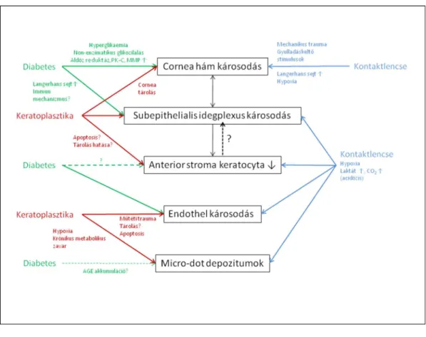
Figure 1. summarizes the presumed and partly proven mechanisms leading to confocal corneal microscopic changes seen following keratoplasty, in diabetes and after long- term contact lens wear.
Although the three groups are clinically significantly different, the in vivo microscopic changes seen in our studies suggest a similar pathomechanism. Actually, on the long run, acute and chronic corneal metabolic disorders occur in each group. The corneal epithelium, the subepithelial nerves and the anterior stromal keratocytes form a functional unit, therefore the changes occurring in one layer may cause further abnormalities in the other cell layers. In our opinion the similar abnormalities observed in the three groups can be regarded as in vivo microscopic morphological evidence of the disturbances occurring in corneal metabolism.
Figure 1.
The effects of diabetes, keratoplasty and contact lens wear on corneal cell layers
15
6 List of Publications
6.1 Papers related to the dissertation’s topic
1. Imre L. (1999) Első hazai tapasztalatok konfokális corneamikroszkópiával.
Szemészet, 136: 97-102.
2. Görög K, Imre L. (2000) Terápiás kontaktlencse alkalmazásával szerzett tapasztalataink. Szemészet, 137: 23-27.
3. Kerényi Á, Imre L, Süveges I. (2000) A hátsó polimorf dystrophia konfokális szaruhártya-mikroszkópos jellemzői. Szemészet, 137: 151-155.
4. Imre L, Nagymihály A. (2001) Reliability and reproducibility of corneal endothelial image analysis by in vivo confocal microscopy. Graefes Arch Clin Exp Ophthalmol, 239: 356-360. IF: 1,192
5. Imre L, Kerényi Á, Nagymihály A. (2003) In vivo konfokális corneamikroszkópia szövődménymentes keratoplastikák után. Szemészet, 140: 211-216.
6. Imre L, Papp A, Nagymihály A. (2003) Diabeteses betegek corneájának in vivo konfokális corneamikroszkópos vizsgálata. Szemészet, 140: 251-254.
7. Imre L, Görög K. (2004) Tartós kontaktlencse-viselés corneális hatásainak vizsgálata in vivo konfokális mikroszkóppal. Szemészet, 141: 459-463.
8. Imre L, Tóth J, Megyesi M, Lukáts O, Resch M. (2004) Az Acanthamoeba-keratitis in vivo diagnosztikája konfokális korneamikroszkóppal. Szemészet, 141: 359- 363.
9. Imre L, Resch M, Nagymihály A. (2005) In vivo confocal corneal microscopy after keratoplasty (Konfokale in-vivo-hornhautmikroskopie nach keratoplastik).
Ophthalmologe, 102: 140-147. IF: 1,559
10. Imre L, Resch M, Megyesi M, Németh J. (2007) In vitro microstructural analysis of commercial ophthalmic suspensions by HRT II Rostock Cornea Module (In- vitro-untersuchung der mikrostruktur von handelsüblichen ophthalmologischen suspensionen mittels HRT-II Rostock Cornea Modul). Ophthalmologe, 104:
697-704. IF: 0,791
11. Resch MD, Imre L, Tapasztó B, Németh J. (2008) Confocal microscopic evidence of increased Langerhans cell activity after corneal metal foreign body removal.
Eur J Ophthalmol, 18: 703-707. IF: 1,010
16
12. Imre L. A cornea konfokális biomikroszkópos vizsgálata. In Németh J. (szerk), Szemészeti diagnosztikus képalkotó eljárások. Semmelweis Kiadó, Budapest, 2011: 61-75
13. Füst Á, Lendvai Z, Imre L, Siló P. (2012) A kötőhártya in vivo konfokális cornea- mikroszkópiája okuláris pemphigoidban. Szemészet, 149: 166-169.
6.2 Papers not related to the dissertation’s topic
1. Kerényi Á, Süveges I, Imre L. (1994) Keratoplastika aphakiás és pseudophakiás bullosus keratopathia eseteiben. Szemészet, 131: 129-134.
2. Kerényi Á, Imre L, Süveges I. (1995) Betaloc a chorioretinopathia centralis serosa kezelésében. Szemészet, 132: 225-228.
3. Toth J, Bausz M, Imre L. (1996) Unilateral Malassezia furfur blepharitis after perforating keratoplasty. Br J Ophthalmol, 80: 488. IF: 1,328
4. Abdel-Salam GMH, Czeizel AE, Vogt G, Imre L. (2000) Microcephaly with chorioretinal dysplasia: Characteristic facial features. Am J Med Gen, 95: 513-515. IF:
2,749
5. Füst Á, Imre L, Nagy ZZ, Süveges I. (2001) Groenouw I cornea-dystrophia excimer lézerkezelése. Szemészet, 138: 149-153.
6. Imre L, Papp A, Süveges I. (2003) Elektrofiziologiai vizsgálatok retinopathia diabetica lézerkezelése kapcsán. Szemészet, 140: 245-249.
7. Papp A, Imre L, Süveges I. (2004) Rövid távú elektrofiziológiai változások panretinalis argonlézer-kezelés hatására nyulak retinájában. Szemészet, 141: 455-460.
8. Resch M, Korányi G, Füst Á, Szentmáry N, Imre L, Bausz M. (2007) Pterygium- műtét conjunctiva-limbus autograft fibrinragasztós rögzítésével (cut and paste technika).
Szemészet, 144: 115-118.
9. Imre L, Füst Á. (2009) Esztétikai céllal viselt kontaktlencse súlyos szövődménye.
LAM, 19: 523.
10. Resch MD, Resch BE, Csizmazia E, Imre L, Németh J, Révész P, Csányi E. (2010) Permeability of human amniotic membrane to ofloxacin in vitro. Invest Ophthalmol Vis Sci, 51: 1024-1027. IF: 3,466
17
11. Resch M, Csizmazia E, Resch BE, Imre L, Szabó-Révész P, Németh J, Csányi E.
(2011) Az amnionmembrán kettős farmakokinetikai hatásának in vitro vizsgálata.
Szemészet, 148: 95-100.
12. Resch MD, Resch BE, Csizmazia E, Imre L, Németh J, Szabó-Révész P, Csányi E.
(2011) Drug reservoir function of human amniotic membrane. J Ocul Pharmacol Ther, 27: 323-326. IF: 1,509
13. Füst A, Pállinger E, Stündl A, Kovács E, Imre L, Tóth S, Németh J. (2012) Both freshly prepared and frozen-stored amniotic membrane cells express the complement inhibitor CD59. ScientificWorldJournal, 2012: 815615. IF: 1,730
14. Imre L. (2012) Bakteriális keratitis. Szemészet, 149: 88-101.
15. Imre L. (2012) Első tapasztalataink a módosított Konstantinov-keratoprotézis implantációjával. Szemészet, 149: 188-193.
16. Füst A, Csuka D, Süveges I, Imre L, Bausz M, Nagymihály A, Csorvási A, Füst G, Németh J. (2013) Complement activation in the aqueous humor of pseudophakic bullous keratopathy patients. Ophthalmic Res, 49: 161-166. IF: 1,376
17. Süveges I, Füst A, Imre L. (2013) Herpes simplex keratitisekben végzett perforáló keratoplasztika posztoperatív terápiája. Orvosi Hetilap, 154: 2065-2070.
18. Füst Á, Süveges I, Tóth J, Imre L, Nagy ZZ. (2014) Súlyos bakteriális keratitis gyógyítása cross-linking kezeléssel - Esetismertetés. Szemészet, 151: 33-36.
19. Füst T, Csuka D, Imre L, Bausz M, Nagymihály A, Füst G, Csorvási T, Németh J, Varga L. (2014) The role of complement activation in the pathogenesis of Fuchs' dystrophy. Mol Immunol, 58: 177-181. IF: 2,973
20. Maka E, Imre L, Somogyvári Z, Németh J. (2015) Koraszülöttek ideghártya- elváltozása miatti lézerkezelés neonatális intenzív centrumokban. Csecsemő-Szem- Mentő Program Orvosi Hetilap, 156: 192-196.