New treatment modalities in superficial bladder cancer
PhD thesis
Dr. András Horváth
Semmelweis University
Clinical Medicine Doctoral School
Supervisor: Dr. Péter Nyirády PhD, D.Sc.
Official reviewers: Dr. Miklós Tóth, Ph.D.
Dr. Zoltán Bajory, Ph.D.
Head of the Final Examination Committee:
Prof. Janina Kulka, D.Sc.
Members of the Final Examination Committee:
Prof. Ilona Kovalszky, D.Sc.
Dr. András Kiss, Ph.D.
Budapest 2012
Contents
Contents ... 1
List of Abbreviations ... 3
1 Introduction ... 6
1.1 Bladder cancer ... 6
1.1.1 Incidence... 6
1.1.2 Risk factors ... 7
1.1.3 Staging ... 8
1.1.4 Grading ... 11
1.1.5 Histopathology ... 11
1.2 Non-muscle invasive (superficial) bladder cancer ... 14
1.2.1 Non-muscle invasive bladder cancer ... 14
1.2.2 Current therapeutical options for NMIBC ... 15
1.2.3 Intermediate risk NMBC ... 16
1.2.4 Current therapeutic options for intermediate risk NMIBC ... 16
1.3 Gene therapy ... 19
1.4 Gene therapy in bladder cancer ... 21
1.4.1 Route of delivery ... 22
1.4.2 Therapeutic genes used against bladder cancer ... 25
1.4.3 Vectors for bladder cancer gene therapy ... 39
1.5 Oncolytic viruses ... 49
1.5.1 Historical background of oncolytic viruses ... 49
1.5.2 Properties of oncolytic viruses ... 51
1.6 Herpes simplex virus ... 51
1.6.1 Herpes Simplex Virus-1 ... 52
1.6.2 Properties of Herpes Simplex virus 1, relevant to oncolytic virus therapy ... 53
1.6.3 HSV-1 mutations giving tumour selective replication ... 54
1.6.4 Herpes Simplex Virus against bladder cancer ... 58
1.6.5 OncoVexGALV/CD ... 59
1.7 Animal bladder tumour models ... 60
1.7.1 Assesment of tumour growth ... 63
2 Objectives ... 66
3 Materials and Methods ... 68
3.1 Cell culture ... 68
3.1.1 Cell lines ... 68
3.1.2 Cell Handling ... 69
3.1.3 Cell line storage ... 69
3.1.4 OncoVex GFP and OncoVex GALV/CD stocks ... 70
3.1.5 Viral titre assay (Plaque assay). ... 70
3.1.6 Fusion assay (GALV dose response assay) ... 71
3.1.7 Prodrug-activating assay ... 72
3.1.8 Fixing and staining protocol for cells or viral plaques... 72
3.1.9 MTS Assay ... 72
3.2 Cell cloning techniques ... 73
3.2.1 Linearisation of DNA. ... 73
3.2.2 Antibiotic killing curves ... 74
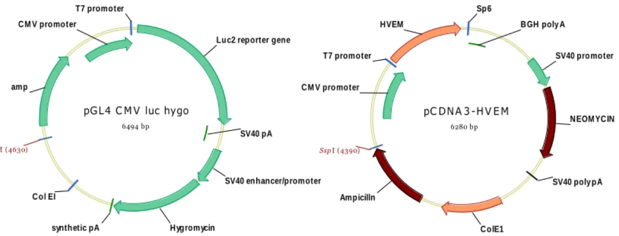
3.2.3 Transfection of cells to produce luciferase/HVEM expressing cell lines ... 75
3.2.4 Selection and scale up of transfected clones ... 75
3.2.5 Sub-cloning cell lines ... 76
3.2.6 Infection screening assay for AY-27 HVEM... 76
3.2.7 Luciferase activation screening assay. ... 77
3.3 Quantitative Reverse Transcrpition Polymerase Chain Reaction (QRT-PCR) ... 77
3.3.1 RNA extraction ... 77
3.3.2 cDNA formation and QRT-PCR ... 78
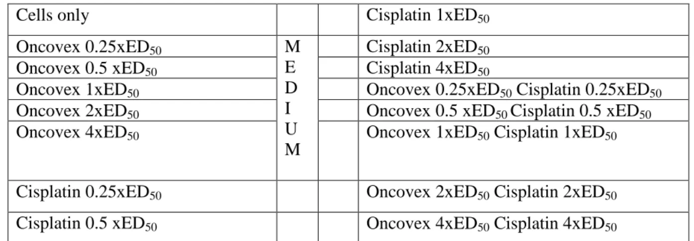
3.4 Synergy testing ... 79
3.4.1 Assessment of Synergy ... 79
3.4.2 In vitro synergy assay ... 80
3.5 Orthotopic rat bladder tumour model ... 81
3.5.1 Anaesthesia and analgesia of the rats ... 81
3.5.2 Bladder tumour implantation ... 82
3.5.3 Assessment of tumour growth ... 83
3.5.4 In vivo treatment of orthotopic rat bladder tumour model ... 84
3.6 Statistics ... 85
4 Results ... 86
4.1 Testing OncovexGALV/CD on bladder tumour cells in vitro with Fusion assay ... 86
4.2 Testing OncovexGALV/CD on bladder tumour cells in vitro with Prodrug assay ... 92
4.3 Testing OncovexGALV/CD on bladder tumour cells in vitro in combination with conventional chemotherapies ... 101
4.4 Set up a stable rat orthotopic bladder tumour model ... 111
4.4.1 Infection screening of wild type AY-27 rat bladder tumour cells ... 111
4.4.2 Transfection of AY-27 cells with the herpesvirus entry receptor (HVEM) and infection screening ... 113
4.4.3 Testing the efficacy of OncovexGALV/CD on AY-27 HVEM cell clone with Fusion and Prodrug assay ... 114
4.4.4 AY-27 and AY-27 HVEM flank tumour model ... 116
4.4.5 Rat orthotopic bladder tumour model ... 117
4.4.6 Detection of HVEM RNA by QRT-PCR as a marker for tumour cells ... 121
4.4.7 Transfection of AY-27 HVEM cells with a luciferase encoding plasmid for bioluminescence imaging ... 123
4.4.8 Bioluminescence imaging rat flank tumour model ... 124
4.5 Evaluation of the efficacy of OncovexGALV/CD on our previously developed rat orthotopic bladder tumour model in vivo ... 125
5 Discussion ... 131
6 Conclusion ... 143
7 Summary ... 145
8 References ... 147
9 List of publications ... 172
10 Acknowledgement ... 174
List of Abbreviations
AAV adeno-associated virus
ACV acyclovir
ALVAC canarypox virus
ATCC American Tissue Culture Collection
BAC bacterial artificial chromosome
BCG Bacille Calmette-Guerin
BCL-2 B-cell lymphoma 2
CAR coxsackie adenovirus receptor
CCD camera charge-coupled device camera
CD cytosine deaminase
CI combination index
CIS carcinoma in situ
CMV cytomegalovirus
Cox-2 cyclooxygenase 2
Cx26 connexin 26
dCK deoxycytidine kinase
DMEM Dulbecco‟s Modified Eagle‟s Medium
DMSO dimethyl sulfoxide
DNA deoxyribonucleic acid
dNTP deoxynucleotide triphosphate
DPD dihydropyrimidine dehydrogenase
dTMP deoxy thymidine monophosphate
dTTP deoxythymidine triphosphate
dUMP deoxy uridine monophosphate
EAU European Association of Urology
EBV Epstein Barr virus
ECACC European Collection of Cell Cultures
ED50 median effect dose 50
EMDA electromotive drug administration
EORTC European Organisation for Research and Treatment of Cancer
ERKs extracellular signal-regulated kinases Fa-CI fraction affected-combination index
FCS foetal calf serum
FGM full growth media
FMG fusogenic membrane glycoproteins
GADD34 growth arrest and DNA damage 34 protein
GAG glycosaminoglycan
GALV gibbon ape leukemia virus
GCV ganciclovir
GM-CSF granulocyte macrophage colony-stimulating factor
GPI glycosylphosphatidylinositol
HBSS Hanks Balanced Salt Solution(
hCNT human concentrative nucleoside transporters hENT human equilibrative nucleoside transporters HER-2 human epidermal growth factor receptor 2
HIV human immundefficiency virus
HRP horseradish peroxidase
HSV herpes simplex virus
HSV-TK herpes simplex virus tymidine kinase
HVEM herpesvirus entry mediator
IAA indole-3-acetic acid
ICP34.5 infected cell protein 34.5
IFN-γ interferon gamma
IL1 interleukin 1
ISUP International Society of Urological Pathology
LD50 lethal dose 50
MAb monoclonal antibody
MAP mitogen-activated protein kinases
MEK MAP/ERK kinase
MHC major histocompatibility complex
MMC mitomycin C
MOI multiplicity of interest
MTS assay mithocondrial tetrazolium salt assay
MuLV murine leukemia virus
NAT2 N-acetyltransferase 2
NK cell natural killer cell
NMIBC non muscle invasive bladder cancer
NSCLC non-small cell lung cancer
NYVAC attenuated vaccinia virus(
PDT photodynamic therapy
pfu plaque forming unit
PIG-U phosphatidylinositol glycan class U
PKR protein kinase R
PNP purine nucleoside phosphorylase
ProTα prothymosin alpha
PUNLMP papillary urothelial neoplasms of low malingnant potential QRT-PCR quantitative reverse transcription polymerase chain
reaction
RB retinoblastoma
RCR replication competent retroviral vector
RNA ribonucleic acid
RR ribonucleotide reductase
RSV promoter Rous sarcoma virus promoter
RT room temperature
SUP suppressor mutant virus
TCC transitional cell cancer
TK thymidine kinase
TNFα tumor necrosis factor alfa
TNM tumour - lymph nodes - metastasis system
TS thymidylate synthase
TURBT transurethral resection of bladder tumour
UDK uridine kinase
UDP uridine phosphorylase
UK United Kingdom
UMP uridine monophosphate
UPRT uracil phosphoribosyltransferase
VSV vesicular stomatitis virus
VZV varicella-zoster virus
WHO World Health Organisation
2‟d-5F- uridine 2‟ deoxy 5-fluoro uridine
5-FC 5-fluorocytosine
5-FdUMP 5-fluoro deoxyuridine monophosphate
5-FU 5-fluorouracil
5-FUTP 5-fluorouridine triphosphate
1 Introduction
1.1 Bladder cancer
1.1.1 Incidence
In Hungary approximately 2,600 new cases of bladder cancer are diagnosed every year making it the fifth commonest cancer in men and the eleventh commonest cancer in women. Bladder cancer is a heterogeneous disease with a variable natural history. At one end of the spectrum, low grade Ta tumors have a low progression rate and require initial endoscopic treatment and surveillance, but rarely present a threat to the patient.
At the other extreme, high grade tumors have a high malignant potential associated with significant progression and cancer death rates. The incidence of the disease increases by age and it occurs most commonly in people between 50 and 70 years of age. The average age at diagnosis is 65 years. Bladder cancer is 3 to 4 times more common among males than females. On the other hand, it has been suggested that the stage- adjusted survival of bladder cancer among women is worse than among men (Mungan et al. 2000).
The incidence is higher in Caucasians than in African Americans and these differences may be partly due to genes, as studies have shown that certain genetic polymorphisms linked to an increased risk of bladder cancer (for example, the N-acetyltransferase 2 (NAT2 ) slow acetylator polymorphism) are much more prevalent in Caucasian than non-white populations (Garcia-Closas et al. 2005). There is a positive social class gradient for bladder cancer in both sexes. Surveys of cancer incidence and mortality suggest that parous women have a lower risk of bladder cancer than nulliparous women, probably due to hormonal changes related to pregnancy, and that the risk may decrease with increasing parity (Cantor et al. 1992; Green et al. 1988; Miller et al. 1980; Plesko et al. 1985)
1.1.2 Risk factors
Cigarette smoking is the principal preventable risk factor for bladder cancer in both men and women. In Europe it is estimated that up to half the bladder cancer cases in men and a third in women are caused by cigarette smoking. Current smokers have around three times the risk of developing bladder cancer compared to non-smokers while ex-smokers have double the risk of non-smokers. Risk is positively associated with both increasing dose and duration of smoking (Brennan et al. 2000; Brennan et al. 2001; NICE 2002).
The causative agents amongst others are thought to be alpha- and beta- naphtylamine, which are secreted into the urine of smokers.
Bladder cancer was one of the first cancers shown to be industrially associated. In 1895 Rehn reported cases of bladder cancer in a German aniline dye factory (Rehn 1895).
Altogether it is estimated that between 5 and 10% of male bladder cancer cases in Europe are caused by occupational exposure (Kogevinas et al. 2003). This proportion may be higher in countries with less-regulated industrial processes. Workers in chemical, dye, rubber, petroleum, leather and printing industries are at increased risk.
Excess risks have been frequently observed among painters, which is thought to be due to exposure to possible carcinogenic constituents of paints like benzidine, polychlorinated biphenyls, formaldehyde, and asbestos and solvents like benzene, dioxane, and methylene chloride (Steenland and Palu 1999). The latency period between exposure and tumour development may be prolonged. A moderately increased risk is also found among leather workers and shoe makers, although the responsible agent is still un-known (Marrett et al. 1986). An excess risk of bladder cancer is also observed in aluminum, iron, and steelworkers, which may be the result of exposure to aromatic amines and polycyclic aromatic hydrocarbons in coal-tar pitch volatiles (Gaertner and Theriault 2002; Romundstad et al. 2000; Theriault et al. 1984). Several studies have been performed on chlorinated drinking water and bladder cancer, and all of these reported increased risks (King and Marrett 1996; Koivusalo et al. 1998). Other studies have evaluated the association between ingestion of arsenic in drinking water and the risk of bladder cancer.
Chronic urinary tract infection is associated with the development of bladder cancer, especially invasive squamous cell carcinoma (Kantor et al. 1984). Cyclophosphamide, an alkylating agent used in the treatment of malignant neoplasms, particularly
lymphoproliferative and myeloproliferative diseases, increases the risk of bladder cancer (mainly urothelial carcinoma) with a clear dose-response relationship (Fairchild et al. 1979; Travis et al. 1995). A chelating agent is now given with cyclophosphamide to reduce this risk. Treatment with radiotherapy to the pelvic area for cancers such as cervical cancer, prostate cancer, kidney cancer, fallopian tube cancer and testicular cancer can increase the risk of bladder cancer (Kaldor et al. 1995).
Ingestion of artificial sweeteners has been proposed to be a risk factor, but several, studies have failed to confirm any association (Elcock and Morgan 1993; Howe et al.
1977). Physical trauma to the urothelium induced by infection, instrumentation, and calculi increases the risk of malignancy (Hicks et al. 1982). Most studies show a two- six-fold increased risk of bladder cancer in first-degree relatives of bladder cancer patients, with a higher risk if the relative was diagnosed before the age of 45. (Randi et al. 2007).
Squamous cell carcinoma of the urinary bladder has been known to be associated with Schistosoma haematobium infection for many years. The epidemiologic association is based both on case-control studies and on the close correlation of bladder cancer incidence with the prevalence of S. haematobium infection within different geographic areas (Bedwani et al. 1998; Gelfand 1967; Lucas 1982).
1.1.3 Staging
The stage of a cancer describes its size and whether it has spread. The most appropriate treatment depends on the stage of the cancer. The most commonly used staging system for bladder cancer is the TNM system of the European Association of Urology (EAU), in which T is the size of the tumour, N is whether it has spread to the nearby lymph nodes, M is whether the cancer has spread to other parts of the body (metastases) (Figure 1.1).
Tumour size (T):
Superficial bladder cancer
CIS CIS (carcinoma in situ) is a high –grade (anaplastic) carcinoma confined to the urothelium, but with flat non-papillary configurations. CIS appears as reddened and velvety mucosa and is slightly elevated but sometimes not visible.
Ta These tumours are confined to the urothelium, have a papillary configuration of their exophytic part and do not penetrate from the urothelium into the lamina propria or detrusor muscle.
T1 tumours generate from the urothelium but penetrate the basement membrane which separates the urothelium from the deeper layers. T 1 tumours invade the lamina propria but not so deep that they reach the detrusor muscle.
Invasive bladder cancer
T2 The cancer has started to grow into the muscle of the bladder wall under the connective tissue layer.
T2a The tumour invades the superficial muscle.
T2b The tumour invades the deep muscle.
T3 The cancer invades the perivesical tissue.
T3a microscopically T3b macroscopically
Locally advanced bladder cancer
T4 The cancer has spread outside the bladder to any of the following: the prostate, uterus, vagina, pelvic or abdominal wall.
T4a The cancer has spread to the prostate, uterus or vagina.
T4b The cancer has spread to the pelvic or abdominal wall.
Figure 1.1 T stages of bladder cancer
Lymph nodes (N):
The N refers to whether the cancer cells have spread into the lymph nodes.
N0 There are no cancer cells in any lymph nodes.
N1 There are cancer cells in one lymph node smaller than 2cm across.
N2 There are cancer cells in one affected lymph node larger than 2cm, but smaller than 5cm, or more than one node affected, but all of them smaller than 5cm across.
N3 There are cancer cells in at least one affected lymph node larger than 5cm across.
Metastases (M):
M0 if the cancer cells have not spread.
M1 is when the cancer cells have spread to other parts of the body. If bladder cancer spreads, it‟s most likely to go to the bones, the lungs or the liver.
1.1.4 Grading
The grading system is also very important in the classification of bladder cancer. The grading system describes the anaplastic degree of the cancer cells.
Grade 1 is well differentiated tumour.
Grade 2 is moderately differentiated tumour.
Grade 3 is poorly differentiated tumour.
1.1.5 Histopathology
The normal urothelium is composed of 3-7 layers of transitional cell epithelium resting on a basement membrane composed of extracellular matrix (collagen, adhesive glycoproteins, glycosaminoglycans). The epithelial cells vary in appearance. The basal cells are actively proliferating cells resting on the basal membrane; the luminal cells, perhaps the most important feature of normal bladder epithelium, are larger umbrella- like cells that are bound together by tight junctions. Beyond the basement membrane is loose connective tissue, the lamina propria, in which occasionally smooth-muscle fibres can be identified. The fibres should be distinguished from deeper, more extensive muscle elements defining the true muscularis propria. The muscle wall of the bladder is composed of muscle bundles coursing in multiple directions. As these converge near the bladder neck, 3 layers can be recognized: inner and outer longitudinally oriented layers and a middle, circularly oriented layer.
According to the WHO/ISUP system all bladder tumour cases are devided into four groups.
1.1.5.1 WHO/ISUP system
1.1.5.1.1 Papilloma
The WHO/ISUP system has very restrictive histologic features for the diagnosis of papilloma, where normal appearing urothelium lines papillary fronds. Defined as such, it is a rare benign condition typically occurring as a small, isolated growth seen
primarily in younger patients. The majority of these lesions once excised will not recur (McKenney et al. 2003).
1.1.5.1.2 Papillary Urothelial Neoplasms of Low Malingnant Potential (PUNLMP) The category of PUNLMP was derived to describe lesions that do not have cytologic features of malignancy, yet have thickened urothelium as compared to papilloma. There is no or very little variation of nuclear features or the pattern of organization. Having a category of PUNLMP avoids labeling a patient as having cancer with its psychosocial and financial implications, although they are not diagnosed as having a benign lesion, whereby they might not be followed as closely. The current classification system allows for designation of a lesion (papillary urothelial neoplasm of low malignant potential), that biologically has a very low risk of progression, yet is not entirely benign.
1.1.5.1.3 Low and High grade papillary carcinoma
The WHO/ISUP system classifies papillary urothelial carcinoma into only 2 grades.
Low grade papillary urothelial carcinoma exhibits an overall orderly appearance but has minimal variability in architecture and/or cytologic features, which are easily recognizable at scanning magnification. High grade papillary urothelial carcinomas are characterized by a disorderly appearance due to marked architectural and cytologic abnormalities, recognizable at low magnification. It is important to remember that a single papillary urothelial neoplasm may contain a spectrum of cytologic and architectural abnormalities.
Bladder cancer can be classified by the histopathological type.
1.1.5.2 Histopathological type
1.1.5.2.1 Transitional cell carcinoma
Approximately 90% of all bladder cancers are transitional cell carcinomas. These tumours most commonly appear as papillary, exophytic lesions, less commonly they may be sessile or ulcerated. Whereas the former group are usually superficial in nature, sessile growths are often invasive.
1.1.5.2.2 Nontransitional cell carcinomas
1.1.5.2.2.1 Adenocarcinoma
Adenocarcinomas account for less than 2% of all blader cancer. Primary adenocarcinomas of the bladder may be preceded by cystitis and metaplasia.
Histologically adenocarcinomas are mucus secreting and may have glandular, colloid or signet-ring patterns. Whereas primary adenocarcinomas often arise along the floor of the bladder, adenocarcinomas arising from the urachus occur at the dome. Both tumour types are often localized at the time of diagnosis, but muscle invasion is usually present.
Five-year survival is usually less than 40%, despite aggressive surgical management (Abenoza et al. 1987; Kramer et al. 1979; Malek et al. 1983; Wright et al. 1988).
1.1.5.2.2.2 Squamous cell carcinoma
Squamous cell carcinoma account for between 5% and 10% of all bladder cancers and is often associated with a history of chronic infection, vesical calculi, or chronic catheter use. It may also be associated with bilharzial infection owing to Schistosoma haematobium, because squamous cell carcinoma accounts for approximately 60% of all bladder cancers in Egypt, parts of Africa and Middle East, where this infection is prevalent (El-Bolkainy et al. 1981). These tumours are often nodular and invasive at the time of diagnosis. Histologically they appear as poorly differentiated neoplasms composed of polygonal cells with characteristic intercellular bridges.
1.1.5.2.2.3 Undifferentiated carcinomas
Undifferentiated bladder carcinomas, which are rare (accounting for less than 2%), have no mature epithelial elements. A small cell type has been described that histologically resembles similar lesions of the lung (Mills et al. 1987).
1.1.5.2.2.4 Mixed carcinoma
Mixed carcinomas constitute 4-6% of all bladder cancers and are composed of transitional, glandular, squamous or undifferentiated patterns. The most common type comprises transitional and squamous cell elements (Murphy 1989). Most mixed carcinomas are large and infiltrating at the time of diagnosis.
1.1.5.2.2.5 Rare epithelial and nonepithelial cancers
Rare epithelial carcinomas identified in the bladder include villous adenomas, carcinoid tumours, carcinosarcomas and melanomas. Rare nonepithelial cancers of the urinary bladder include pheochromocytomas, lymphomas, choriocarcinomas and various mesenchymal tumours (hemangioma, osteogenic sarcoma and myosarcoma) (Murphy 1989). Cancers of the prostate, cervix and rectum may involve the bladder by direct extension. The most common tumours metastatic to the bladder include (in order of incidence) melanoma, lymphoma, stomach, breast, kidney and lung (Goldstein 1967;
Murphy 1989).
1.2 Non-muscle invasive (superficial) bladder cancer
1.2.1 Non-muscle invasive bladder cancer
Approximately 80% of all bladder cancer cases are non muscle invasive bladder cancer (NMIBC) defined as stage Ta-T1, grade 1-3 and carcinoma in situ (CIS). Historically these tumours were viewed as a homogeneous group, referred to as „superficial‟ bladder cancer and treatment was based at reducing recurrences. In the 1980‟s it was recognised that a subgroup of these patients with T1 G3 tumour and/or CIS had a significant rate of progression to muscle invasive disease and a poor long term survival (Cookson et al.
1997) which led to the concept of „high risk‟ NMIBC. Further studies have now shown that these „superficial‟ tumours in fact form a heterogenous group of tumours whose 1 year recurrence and progression rates vary from 15-61% and <1-17% respectively (Table 1.1) (Sylvester et al. 2006). As it is shown in this table low and intermediate risk patients have much less risk of recurrence and progression compared to the high risk group.
Based on the developing understanding of the pathological behaviour of NMIBC, the therapeutic options for the management of NMIBC have evolved over the last half century. Initially, treatment was transurethral resection (TURBT) alone until Jones and Swinney first described the use of intravesical chemotherapy using Thiotepa in 1961 (Jones and Swinney 1961). After TURBT the use of Bacille Calmette-Guerin as intravesical immunotherapy in NMIBC was first reported by Morales in 1976 (Morales
et al. 1976). and refined with the introduction of a maintenance regime by Lamm (Lamm et al. 2000). Radical cystectomy was advocated as a treatment for „high risk‟
NMIBC (Malkowicz et al. 1990). A single immediate instillation of intravesical chemotherapy has been shown to be of benefit (in terms of recurrence and progression) immediately following transurethral resection of bladder tumour (TURBT) (Sylvester et al. 2004).
Table 1.1 EORTC risk tables Sylvester 2006
% of total
Recurrence 1 year
Recurrence 5 year
Progression 1 year
Progression 5 year Low risk
Ta G1
50 15-24 31-46 ≤1 1-6
Intermediate risk Ta-1 G1-2
35 24-38 46-62 ≤1-5 1-17
High risk Ta-1 G3
15 24-61 46-78 1-17 6-45
The wide spectrum of pathological behaviour of NMIBC has led to the realisation that in some cases eg a solitary pTa TCC, the main therapeutic goal should be to prevent recurrences with minimal morbidity (Hall et al. 1994), whilst in other cases relatively high morbidity is acceptable in order to prevent progression to muscle invasive disease.
Out of this has been borne the concept of NMIBC risk groups. Whilst the exact definition of each risk category varies (EAU April 2008; Hall et al. 1994), two groups are clearly defined, i.e. low and high risk NMIBC.
1.2.2 Current therapeutical options for NMIBC
Current guidelines suggest that all patients with non muscle invasive bladder cancer should receive one immediate post operative instillation of chemotherapy within 6 hours after TURBT. Further treatment depends on the patients` risk of recurrence and progression to muscle invasive disease (Sylvester et al. 2006). Low risk group patients have single, small (<3cm), histologically Ta, low grade tumour. For these patients no further treatment is recommended prior to a subsequent recurrence (EAU April 2008;
Hall et al. 1994). Patients with Ta-T1, high grade tumour, or with carcinoma in situ, or with combination of these are in the high risk group. For this group a further
transurethral resection (re-TURBT) and 1-3 years of maintenance BCG (bacillus Calmette-Guérin) intravesical immunotherapy is recommended (EAU April 2008).
1.2.3 Intermediate risk NMBC
The remaining group with intermediate risk forms approximately 35% of all NMIBC and is by definition made up of patients excluded from the other risk categories.
(Patients with Ta-T1, low and intermediate grade, multifocal, >3cm tumours attend to this group). Intermediate risk group patients form a heterogeneous group of patients e.g. from solitary but recurrent G1pTa to multiple recurrent G2pTa. These patients have a significant risk of recurrence of between 24% at 1 year and 62% at 5 years, and a single instillation of intravesical chemotherapy is inadequate treatment. On the other hand, the relatively high morbidity of intravesical BCG, particularly during maintenance treatment, has meant that the use of BCG in this group has not been popular, at least in Europe. The current popular therapeutic option is weekly instillation of intravesical chemotherapy for 6 weeks (induction course) with or without maintenance therapy. However, the EORTC risk tables suggest a risk of progression as high as 17% at 5 years, which suggests that some patients within the intermediate group may have a significant risk of progression, and would therefore benefit from a more intensive treatment than is currently used even if this results in higher morbidity.
Unfortunately we currently cannot identify such patients individually, and so efforts are focused on new approaches to the therapy of these patients as a group, with the ultimate aim of reducing recurrences, preventing progression with acceptable morbidity.
1.2.4 Current therapeutic options for intermediate risk NMIBC
Four national and international urological association guidelines are available for NMIBC (BAUS 2008; EAU April 2008; Hall et al. 2007; Oosterlinck et al. 2005). All four guidelines recommend that all patients with intermediate risk (IR) NMIBC should receive one immediate instillation of chemotherapy after TURBT.
1.2.4.1 Induction course of intravesical chemotherapy
Whilst all the guidelines agree that further treatment is required, no clear consensus emerges as to the optimal dose, regimen and duration of treatment. The summary of product characteristics for mitomycin-C (Kyowa Hakko UK, Slough, UK) suggests that 20–40 mg of mitomycin-C in 20–40 mL of diluent is given weekly or three times a week for a total of 20 doses (Kyowa). Many urologists seem to prefer a 6-week course of weekly intravesical chemotherapy, often called an „induction‟ course, perhaps because this regimen is similar to the induction regimen for intravesical BCG.
1.2.4.2 Maintenance intravesical chemotherapy
Maintenance regimens of intravesical chemotherapy are based on the hypothesis that a prolonged course of intravesical chemotherapy will reduce the recurrence rate of bladder cancer by more than one immediate instillation or an induction course, with acceptable morbidity. It is not known whether maintenance chemotherapy is necessary if one dose of intravesical chemotherapy was given immediately after TURBT, and vice versa, i.e. that if a maintenance course of intravesical chemotherapy is given for in intermediate risk NMIBC, then an immediate instillation may not be necessary. If one immediate instillation is not given, then a prolonged course of intravesical chemotherapy of 1 year might be required to achieve the same magnitude of reduction in the recurrence rate (EAU April 2008). Given the simplicity and safety of an immediate instillation, it would seem sensible to pursue the former approach rather than the latter. Finally, there is a theoretical risk of carcinogenesis with prolonged use of any chemotherapeutic agent.
1.2.4.3 Optimizing intravesical chemotherapy
It was postulated that the variable response of patients to intravesical chemotherapy has two components; lack of sensitivity of the tumour to intravesical chemotherapy and inadequate drug delivery to the tumour (Au et al. 2001). The latter might be improved by optimizing intravesical chemotherapy. Au et al. (Au et al. 2001) assessed, in a randomized phase III trial, four approaches combined to optimize the efficacy of intravesical mitomycin C. Patients were randomized into either standard intervention
and received 20 mg mitomycin C in 20 mL of sterile water, or optimized intervention, and were asked to refrain from drinking for 8 h before treatment, given a total of 3.9 g of sodium bicarbonate orally, and had any residual urine drained from the bladder before receiving 40 mg mitomycin C in 20 mL of sterile water. The optimized arm showed a longer median (95% CI) time to recurrence of 29.1 (14.0–44.2) months and a greater 5-year recurrence-free fraction of 41.0 (30.9–51.1)% than the standard arm, of 11.8 (7.2–16.4) months and 24.6 (14.9–34.3)% respectively (P =0.005, log-rank test for time to recurrence). Despite the relative simplicity of the optimization schedule and the improved efficacy, optimized intravesical regimens have not been widely adopted.
Another simple way to increase the time of residence in the bladder is prolonged drug infusion into the bladder. Other methods have included triblock copolymers which release drug over time and in response to temperature.
1.2.4.4 BCG: Reducing side -effects
BCG therapy remains the reference standard non-surgical treatment for high-risk NMIBC and might therefore be, theoretically at least, the optimum therapy for intermediate risk NMIBC. In practice, BCG is associated with relatively high morbidity, with up to 25% of patients failing to complete a full maintenance course of BCG. To avoid this, recent studies have assessed various means of reducing the toxicity of BCG therapy whilst maintaining its therapeutic effects. The Spanish CUETO group have examined this issue. In a series of methodical trials they showed that 27 mg of BCG Connaught strain (one-third dose) had lower toxicity than 81 mg (full dose) but was as effective as a full dose for high-risk disease; 13.5 mg BCG was not as effective as 27 mg but produced the same toxicity, so 27 mg of BCG seems to be the minimum effective dose (Fernandez-Gomez et al. 2008). A further study suggested that monthly single instillations of 120 mg BCG for 1 year was as effective as 3-week courses of maintenance BCG at 3, 6, 12, 18 and 24 months, in terms of recurrence and progression rates, and with less toxicity (Ali-El-Dein 2007). The EORTC 30962 study assessed at the one-third vs full-dose BCG over 1 or 3 years of maintenance. The study closed in 2005 and the results are eagerly awaited. Finally, a randomized double-blind placebocontrolled multicentre trial showed that prophylaxis with 200 mg ofloxacin after
each BCG instillation reduced the incidence of BCG-induced side-effects (Colombel et al. 2006).
1.2.4.5 Device-assisted therapy
Another approach to improving the delivery of intravesical chemotherapy drugs to the tumour is with device-assisted therapy. The two main methods are with microwave hyperthermia to heat the bladder wall, or electromotive drug administration (EMDA).
The theoretical base of EMDA (Di Stasi et al. 2006) and thermochemotherapy (Gofrit et al. 2004; van der Heijden et al. 2004) is to increase the permeability of the bladder mucosa (with BCG induced inflammation and with hyperthermia in the bladder) before administering chemotherapy eg. Mitomycin. A very small electric current increases the permeation of charged and neutral compounds through the process of electromigration and elecro-osmosis. Microwave hyperthermia was designed to be used exclusively in the setting of high-risk NMIBC and patients in whom intravesical BCG failed. The use of EMDA-potentiated mitomycin C alone has been described but it appears to be particularly effective when used in conjunction with BCG (Di Stasi et al. 2006). In this randomized trial specifically in patients with stage pT1 disease (i.e. high-risk NMIBC) the combination of EMDA, mitomycin C and BCG when used sequentially showed a higher mean (95% CI) disease-free interval than BCG alone, at 69 (55–86) vs 21 (15–
54) months, respectively. That study was also unique in that it was the first study of intravesical therapy to show a reduction in disease specific mortality and overall mortality. The authors hypothesised that the mode of action in sequential BCG/EMDA mitomycin C is that BCG-induced inflammation increases the permeability of the bladder wall and improves the delivery of the mitomycin C to the tumour site. Thus sequential BCG/EMDA/mitomycin C might have a role in intermediate risk NMIBC in the future, particularly in patients thought to be at high risk of progression.
1.3 Gene therapy
Gene therapy is the treatment of disease by the introduction of normal genes into the patient to overcome the effects of those defective genes. Cancer gene therapy is the introduction of DNA or RNA which cause cellular changes that inhibit the uncontrolled
growth of tumour cells. Several viral and nonviral vectors have been developed to achieve the delivery of the transgene to the tumour cells. Gene transfer by viral vectors takes advantage of the natural ability of viruses to enter cells and express transgenes by the infected cells. The success of these strategies depends on the high level transfer of therapeutic genetic material to the cells of interest (ie. tumour, immune cell, endothelial cell).
This approach was first applied to cancer patients using non-replicating adenoviruses or retroviruses to deliver copies of the wild type tumour suppressor gene p53 to tumours with the aim of preventing uncontrolled cell division (Roth 1996; Spitz et al. 1996).
This technology uses our understanding of cancer at the molecular level and has been exploited to develop new strategies for killing cells selectively or arresting their growth (McCormick 2001). Gene therapy for the treatment of cancer was initiated with high levels of optimism and enthusiasm. Recently however, this perception has been tempered by the realisation that efficiency of gene delivery remains the most significant barrier to success (Vile et al. 2000).
The field of molecular cancer therapy embraces a range of technologies including direct attack on tumour cells using oncolytic viruses or prodrug activating systems, and viral fusiogenic envelopes. These fields are illustrated in Figure 1.2. However the categorisation of such therapies is somewhat artificial for two reasons. Firstly, one single therapy is unlikely to cure a particular cancer since evidence from conventional therapies suggests that a combination of therapies is much more likely to work in the clinic. Secondly, a particular therapy may have multiple modes of action. For example, the prodrug activating system of HSV-TK combined with ganciclovir promotes tumour reduction by directly killing tumour cells and also stimulates an immune response against the tumour (Vile et al. 1997).
Figure 1.2 An outline of the areas of cancer gene therapy being developed. The approaches highlighted in blue are discussed in this thesis.
1.4 Gene therapy in bladder cancer
In the following part we aim to review bladder cancer gene therapy by analyzing the stages which are key to its success: route of delivery, anti-tumour approaches and vectors used.
Cancer Vaccines
Viral Molecular Therapy
Prodrug Activating Therapy
Oncolytic Therapy Immuno-
therapy
Cytokine Therapy
Cell Death Therapy
Cellular Gene Therapy
Cancer Gene Therapy
Viral Fusogenic Envelopes
1.4.1 Route of delivery
1.4.1.1 Systemic administration
This route often fails in the treatment of bladder cancer due to loss of the therapeutic agent from metabolism in the liver and poor tumour penetration. There is a need for a sufficient level and an appropriate amount of time for the therapeutic agent to come into contact with the tumour cells in the bladder mucosa in order to achieve its effect which is not achieved via this route.
1.4.1.2 Intravesical administration
The bladder represents an ideal target for intravesical gene therapy in the management of bladder cancer. The vectors are in direct local tumour contact, eliminating the difficulties associated with systemic administration and ensuring a low risk of gene transfer to other organs. Easy external access is achieved through the urethra which avoids losses from the first pass metabolism in the liver and minimizes the clearance of the vector by the immune system allowing minimal systemic side effects.
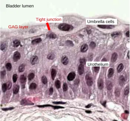
The inside of the bladder consists of 6-7 layers of cells which form a permeable transitional epithelium known as the urothelium (Figure1.3). The urothelium acts as a barrier and in healthy circumstances it is almost impermeable. The main part of this barrier is the superficial cell layer of the transitional epithelium made up of umbrella cells. The umbrella cells join to each other with tight junctions and constitute a watertight barrier (Hohlbrugger 1995). The apical surface of the umbrella cells is covered by uroplakins (UPIa, UPIb, UPII, UPIII), which make up the rigid plaques. In addition the umbrella cells are covered by a hydrophilic glycosaminoglycan (GAG) layer. The GAG layer physically blocks the adhesion of foreign substances such as drugs and viruses (Lande et al. 1995; Parsons 1994). Together these layers erect a tough bladder barrier that maintains a high electrochemical gradient between the urine and the blood. The impermeability of the urothelium is frequently exploited for instilling potentially toxic agents into the bladder to achieve localized pharmalogical effect.
Figure 1.3 Structure of bladder transitional epithelium on a H&E staining histology sample. The localisation of tight junction and GAG layer that is not visible under normal microscope is marked in red.
Several strategies have been developed to improve the intravesical delivery of drugs in the bladder. This includes helping them cross the permeability barrier of the urothelium by both physical and chemical enhancement methods and increasing residence time in the bladder. These approaches may also improve the efficacy of some gene therapy vectors.
1.4.1.2.1 Improving permeability using physical methods.
The permeability of bladder cells can be improved with physical approaches like electromotive drug administration (EMDA), thermotherapy or iontophoresis which was further discussed in section 1.2.4.5.
1.4.1.2.2 Improving permeability using chemical approaches.
Several chemical approaches that improve the permeability of the urothelium have been investigated in the literature. Protamine sulfate interacts with the GAG layer to increase the permeability of the urothelium (Cetinel et al. 2003; Tzan et al. 1994) whilst
Bladder lumen
Umbrella cells
Urothelium GAG layer
Tight junction
administration of dimethyl sulfoxide has been described to enhance the absorption of paclitaxel and pirarubicin (Chen et al. 2003; Hashimoto et al. 1992). The administration of 22% ethanol prior to the instillation of an adenoviral vector causes damage of the GAG layer and makes the treatment more effective (Engler et al. 1999). In addition a polyamide containing formulation, syn3, when used as a pre-treatment prior to intravesically administered adenovirus has been reported to enhance viral uptake (Yamashita et al. 2002). Recent studies in preclinical models with the syn3/adenoviral- based strategy have also shown an improved therapeutic effect (Benedict et al. 2004;
Connor et al. 2005). A phase I study of the Big CHAP polyamide and an adenovirus coadministered intravesically led to increased transduction compared to adenovirus alone (Kuball et al. 2002). Although the use of Syn3 appears to be non toxic to the bladder tissue the use of other transducing agents has been shown to have adverse effects on the urothelium (Benedict et al. 2004).
1.4.1.2.3 Increasing residence time in the bladder
Increasing the time of residence in the bladder is an obvious strategy to increase the efficacy of a drug or therapy by increasing the duration of direct contact between drug and abnormal urothelium and ensuring constant drug concentration. This method was further discussed in section 1.2.4.3.
1.4.1.2.4 Bioadhesion
This defines the interaction between a biological surface, such as the urothelium and a polymer such as algin, chitosan, fibrinogen, gelatin, polyethhylene glycol (Ozturk et al.
2004; Tyagi et al. 2006; Ye et al. 2001) . The efficiency of drug absorption is increased after bioadhesion properties are coupled to microspheres, liposomes or nanoparticles.
For example microspheres based on chitosan are strongly mucoadhesive and their instillation was able to increase the residence time and decrease the frequency of administration of acyclovir (Genta et al. 1997).
The residual urine in the bladder before instillation and the accumulated urine during instillation can cause dilution in the concentration of the administered therapeutic agent.
Restricted fluid intake before and during instillation and the complete emptying of bladder is required in any kind of intravesical treatment (Au et al. 2001). Significant
differences in gene expression are achieved by varying physical parameters during intravesical instillation. Increased gene expression associated with larger volume instillation may be responsible for some reported variability of gene transfer to the bladder. Significantly more gene expression was detected in bladders instilled with a higher volume of viral vectors (p <0.05). Likewise, higher instillation pressures resulted in higher transgene expression in distant organs (Siemens et al. 2001).
1.4.2 Therapeutic genes used against bladder cancer
1.4.2.1 Tumour suppressor genes
Mutations in tumour suppressor genes are prevalent in many cancers and mutation of the p53 gene is common in muscle-invasive bladder cancer (Smith et al. 2003).
Reintroduction of a functional copy of the p53 gene is one route taken by scientists.
Miyake et al (Miyake et al. 1998) introduced a functional copy of the p53 gene into the bladder cancer cell line (HT1376) in vitro and found it became resensitised to cisplatin.
Further in vivo studies used an adenoviral vector carrying the p53 gene which was injected directly into HT1372 tumours established in nude mice. Intraperitoneal administration of cisplatin then led to massive apoptotic destruction of the tumour.
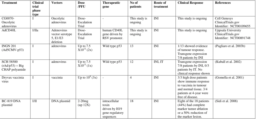
Adenoviral vectors carrying the p53 gene, INGN201 and SCH58500, have been tested in phase I clinical trials for bladder cancer (Table 1.2) (Barnard 2000).
Treatment Clinical trial phase type
Vectors Dose
/PFU
Therapeutic gene
No of patients
Route of treatment
Clinical Response References
CG0070- Oncolytic adenovirus
I Oncolytic
adenovirus
Dose- Escalation Trial
- This study is
ongoing
INI This study is ongoing Cell Genesys ClinicalTrials.gov Identifier: NCT00109655
AdCD40L I/IIa Adenovirus
vector serotype 5, E1/E3 deletion
Dose- Escalation Trial
human CD40L gene driven by RSV promoter.
This study is ongoing
INI This study is ongoing Uppsala University ClinicalTrials.gov Identifier: NCT00891748 INGN 201
(Ad5CMV-p53)
I adenovirus Up to 7.5
X1012 (2x)
Wild type p53 13 INI 1/13 showed evidence
of tumour response.
Transgene expression 7/8 patients by INI
(Pagliaro et al. 2003b)
SCH 58500 (rAd/p53) + Big CHAP polyamide
I adenovirus Up to 7.5
X1013 (1x)
Wild type p53 12 INI, IT Transgene expression 7/8 patients by INI, 0/3 patients by IT. No clinical response shown
(Kuball et al. 2002)
Dryvax vaccinia virus
I vaccinia Up to 108 (3x) - 4 INI 3/3 high dose patients
show immune response to vaccinia in tumour and normal tissue. 3/4 patients at 4-year were free of disease.
(Gomella et al. 2001)
BC-819 DNA plasmid
I/II DNA plasmid 2-20mg (up 12X)
intracellular toxin driven by H19 gene regulatory sequences
18 INI Eight of the 18 patients
(44%) had complete marker tumor ablation or a 50% reduction of the marker lesion.
(Sidi et al. 2008)
intravesical instillation (INI), intratumoral injection (IT)
Table 1.2 Clinical trials for bladder cancer with different gene therapy vectors
1.4.2.2 Suicide gene therapy
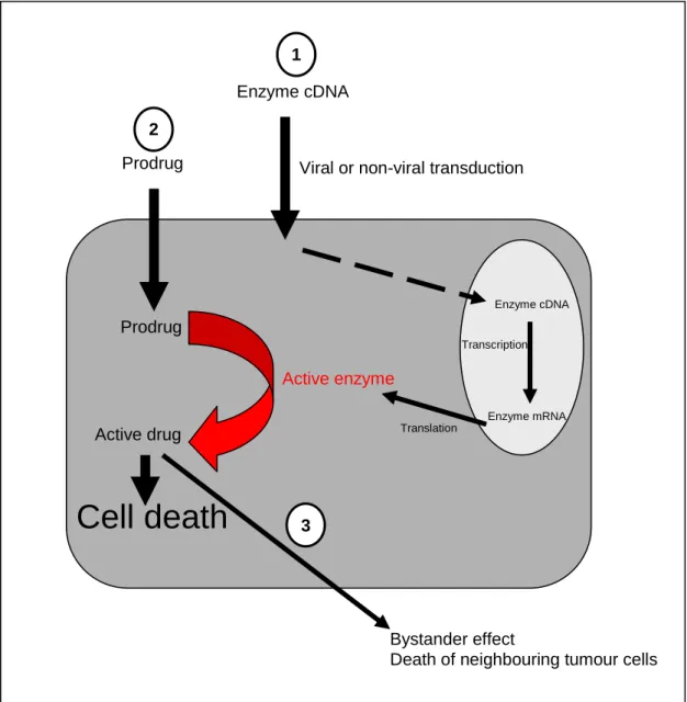
Prodrug activation therapy strives to deliver genes to cancer cells, which convert non- toxic prodrugs into active chemotherapeutic agents (Figure 1.4). The net gain is that a systemically administered prodrug can be converted into high local concentrations of an active anticancer drug in the tumour. To be clinically successful, both enzymes and prodrugs should meet certain requirements for this strategy (Niculescu-Duvaz et al.
1998; Xu and McLeod 2001; Yazawa et al. 2002). The prodrug activating genes should be either of non-human origin or a human protein that is absent or expressed only at low concentrations in normal tissues (Rigg and Sikora 1997; Weedon et al. 2000). To provide catalytic activity the prodrug activating genes need to be sufficiently highly expressed in tumour cells. The prodrug should be a good substrate for the activating enzyme in tumours, but not be activated by cellular enzymes in non-tumour cells. An ideal prodrug system will have a bystander effect; that is the ability to kill non- expressing neighbouring tumour cells by the export of toxic metabolites. In addition, the half-life of active drug should be long enough to induce a bystander effect, but short enough to avoid leakage of the drug into the systemic circulation. A number of suicide genes have been examined in different types of cancer including thymidine kinase, rat p450 CYP2B1, human intestinal carboxylesterase, yeast cytosine deaminase.
Figure 1.4 The three stages of activity of a prodrug activating system. In the first stage, a prodrug activating gene is transduced into and expressed in tumour cells. In the second stage a non-toxic prodrug is administered to the cells. The prodrug works as a substrate for the activating enzyme resulting in the production of toxic metabolites.
These metabolites inhibit DNA or RNA replication resulting in cell death. The final stage is the export of these metabolites to kill the surrounding tumour tissue (bystander effect).
1.4.2.2.1 Thymidine kinase
The thymidine kinase gene system is based on the metabolism of certain purine nucleosides. Thymidine kinase catalyses the phosphorylation of the purine nucleoside analogues Ganciclovir (GCV), Acyclovir (ACV) and 1-(2‟-deoxy-2‟-fluoro-β-D- arabinofuranosyl)-5-iodouracyl (FIAU) converting them to their corresponding
Enzyme cDNA
Viral or non-viral transduction
Enzyme cDNA
Enzyme mRNA Transcription
Translation
Active enzyme Prodrug
Prodrug
Active drug
Cell death
Bystander effect
Death of neighbouring tumour cells 2
1
3
nucleoside monophosphates, which are catalysed to nucleoside diphosphates by mammalian nucleoside monophosphate kinases and are subsequently converted to the tri phosphate form by nucleoside diphosphate kinase (Miller and Miller 1980). The phosphorylation of these substrates by thymidine kinase into a monophosphate form (eg GCV-MP) is the rate-limiting step in the generation of a cytotoxic drug (Field 1983;
Nishiyama and Rapp 1979; Oliver et al. 1985). These prodrug metabolites are able to stall DNA synthesis by inhibiting DNA polymerase and by incorporation into DNA causing chain termination (Davidson et al. 1981; Elion 1980) thus killing dividing cells.
It has been shown that therapeutic concentrations of these drugs are non-toxic to cells that lack thymidine kinase, and conversely expression of thymidine kinase is non toxic to cells in the absence of these drugs (Elion 1980; Field 1983). It was tested in retrovirus, adenovirus and has been shown effective in tumour killing in vitro and in vivo (Yazawa et al. 2002). Viral-mediated transfer of the herpes simplex virus thymidine kinase gene has been demonstrated by several investigators to confer sensitivity to nucleoside analogues such as ganciclovir in brain, prostate, liver, kidney, ovary, head and neck, lung, pancreas, and bladder cancers (Nasu et al. 2000). Oncolytic HSV-1 vectors with intact thymidine kinase should also activate these prodrugs such that infected cells are killed by both the activated prodrug active and oncolysis. This may of course be counterproductive as replication of the oncolytic HSV may also be inhibited. Most studies have shown very little or no synergy between oncolytic HSV vectors and TK mediated prodrug activation, due to the inhibition of virus replication, which occurs. Suicide gene therapy using adenoviral-mediated herpes simplex virus thymidine kinase and the prodrug ganciclovir in an orthotopic murine bladder cancer model demonstrated a greater than threefold reduction in tumor growth. Long-term survival results suggested a survival benefit for the treated animals compared to the control group. (Cheon et al. 2002; Sutton et al. 1997; Sutton et al. 2000). An adenovirus-mediated thymidine kinase suicide gene therapy was tested with different promoters in the treatment of experimental bladder cancer. Cytomegalovirus- immediate-early promoter and the Rous-sarcoma-virus promoters expressing thymidine kinase were both effective (Freund et al. 2000b). A retroviral vector was also tested with thymidine kinase gene plus ganciclovir on human bladder cancer cells and on a nude
mouse model and effective transduction and reduction of tumour volume was shown (He et al. 2004).
1.4.2.2.2 E. coli/yeast cytosine deaminase enzyme
A commonly utilised enzyme prodrug combination is the E-coli /yeast enzyme cytosine deaminase (CD) and 5-fluorocytosine (5-FC). CD, a pyrimidine salvage enzyme, is the only known route by which cytosine is metabolised through hydrolytic deamination to uracil and ammonia. CD also deaminates the anti-fungal compound 5-fluorocytosine (5- FC) into 5-fluorouracil (5-FU), a highly toxic compound, widely used as a cancer chemotherapeutic agent (Moolten 1994). This enzyme has been found in prokaryotes and lower eukaryotes, but appears to be absent in higher eukaryotes (Austin and Huber 1993; Kilstrup et al. 1989). Consequently, mammalian cells are resistant to 5-FC but not 5-FU (Mullen et al. 1992; Pirollo et al. 2008). The cytotoxicity of 5-FU is determined by its conversion into 5-fluoro deoxyuridine monophosphate (5-FdUMP) and 5- fluorouridine triphosphate (5-FUTP) (Figure 1.5). 5-FdUMP is an irreversible inhibitor of thymidylate synthase, and depletes replicating cells of the thymidine nucleotide precursor, deoxythymidine triphosphate (dTTP), during DNA synthesis, while 5-FUTP inhibits RNA maturation and processing. The rate limiting step is the formation of an intermediary metabolite, 5-fluorouridine monophosphate (5-FUMP) (Niculescu-Duvaz et al. 1998). It has been reported that the E. coli uracil phosphoribosyltransferase (UPRT) can markedly potentiate the anti-tumour effects of 5-FU by converting it directly into its intermediary 5-FUMP, thereby leading to more efficient generation of its active metabolites 5-FdUMP and 5-FUTP (Tiraby et al. 1998). A construct containing a fusion between the yeast Fcy 1 and Fur 1 genes encoding cytosine deaminase and uracil phosphoribosyltransferase, respectively, is commercially available (Invivogen) and an identical chimera (Fcy:Fur) has been described by (Erbs et al. 2000).
Fcy:Fur exhibited similar UPRTase activity to the natural yeast gene, but CDase activity was 100 fold elevated compared to the yeast CD gene alone.
The mechanism of 5-FU cytotoxicity is unclear, because it is converted to several metabolites which each have different biochemical actions (Nakamura et al. 2001).
However, much interest has been placed in a metabolite 5-fluorodeoxyuridylate that
inhibits thymidylate synthase, which has been suggested to inhibit cellular DNA synthesis more than viral DNA synthesis (Nakamura et al. 2001).
The CD/5-FC prodrug activating system has been studied as a potential gene therapy strategy in a number of solid tumours. Investigators have demonstrated the efficacy of the CD gene/5-FC prodrug activating system when treating colon carcinoma (Austin and Huber 1993), pancreatic cancer (Evoy et al. 1997), breast cancer (Anderson et al.
2000; Li et al. 1997), and peritoneal carcinomatosis (Bentires-Alj et al. 2000).
Adenovirus-mediated (Block et al. 2000; Ohwada et al. 1996; Topf et al. 1998) transfer of the CD gene showed enchanced local tumour control in colon carcinoma in the presence of prodrug 5-FC.
A murine leukaemia virus-based replication-competent retrovirus containing yeast cytosine deaminase gene plus 5-fluorocytosine dramatically inhibited the tumour growth in an orthotopic bladder animal model (Kikuchi et al. 2007b). An adenovirus- mediated CD gene therapy driven by human telomerase reverse transcriptase promoter was tested and found effective in combination with low-dose etoposide in bladder cancer cells (Shieh et al. 2006). A double suicide gene therapy using adenovirus mediated cytosine deaminase-thymidine kinase fusion gene with ganciclovir or 5- fluorocytosine in a murine subcutaneous bladder carcinoma model was effective by significantly inhibiting tumour growth. The treatment efficacy of combining ganciclovir and 5-fluorocytosine was superior to only ganciclovir or 5-fluorocytosine groups (Tan et al. 2006).
Expression of yeast CD from a replication competent HSV-1 oncolytic virus mediates intra-tumoral conversion of 5-FC to 5-FU, which results in enhanced tumour cell killing compared with the backbone virus alone (Nakamura et al. 2001; Simpson et al. 2006).
In vitro studies have shown increased killing in cell lines derived from lung cancer (A549, H460), pancreatic cancer (CAPAN-1, MIA PACA-2, BXPC-3) and colon cancer (HCT 116, HT-29, SW620) without inhibiting virus replication (Nakamura et al. 2001;
Simpson et al. 2006).
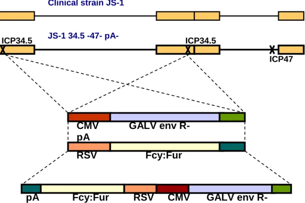
OncoVEXGALV/CD, described above, express both a highly potent version of CD (Fcy::Fur) and the fusogenic glycoprotein from gibbon ape leukaemia virus (GALV). In the presence of 5-fluorocytosine (5-FC), OncoVEXGALV/CD showed greatly improved tumour contol in vitro and in vivo (Simpson et al. 2006).
Figure 1.5 The metabolism of 5-fluoro-cytosine (5-FC) by cytosine deaminase (CD), and uracil phosphoribosyltransferase (UPRT) during DNA synthesis. CD converts 5-FC into 5-fluoro-uracil (5-FU). UPRT then converts 5-FU directly to 5-FUMP, which is then converted to its active metabolites 5-FdUMP and 5-FUTP. This bypasses the rate limiting mammalian cellular enzymes such as UDP (uridine phosphorylase), UDK (uridine kinase) and TK (thymidine kinase). This also reduces the degradation of 5-FU by the competitive enzyme, DPD (dihydropyrimidine dehydrogenase). UMP (uridine monophosphate), dUMP (deoxy uridine monophosphate), dTMP (deoxy thymidine monophosphate), 2‟d-5F- uridine, (2‟ deoxy 5-fluoro uridine), TS (thymidylate synthase)
1.4.2.2.3 Rat p450 enzyme and cyclophosphamide
5-fluorocytosine (5-FC)
5-fluorouracil (5-FU)
CD
UPRT
5-F-uridine
2’d-5F-uridine
5-FUMP 5-FUTP
UDP UDK
RNA synthesis
5-FdUMP
DPD
TK
5-F -alanine
(renally excreted metabolite of 5-FU)
Uracil Uridine UDP
UMP UDK
dUMP TS dTMP
(Thymidine precursor in DNA synthesis)
Expression of the rat p450 enzyme from hrR3 (rRp450), in the presence of cyclophosphamide, has also shown oncolysis and prodrug activation in vitro without inhibiting virus replication in both colon carcinoma and glioma cells (Aghi et al. 1999;
Chase et al. 1998; Ichikawa et al. 2001; Pawlik et al. 2002; Tyminski et al. 2005). In vivo studies on liver metastases and glioma models revealed a substantial decrease in the tumour burden in all animals treated with rRp450 (+cyclophosphamide) compared to controls (Aghi et al. 1999; Chase et al. 1998; Ichikawa et al. 2001; Pawlik et al.
2002).
1.4.2.2.4 Human intestinal carboxylesterase
Recently p450 has been co-expressed from an ICP34.5- and ICP6- mutant HSV with another prodrug activating gene, intestinal carboxylesterase (Tyminski et al. 2005). This new oncolytic herpes virus (MGH2) displays increased anti-tumour efficacy against human glioma cell lines (U251, T98G) when combined with the prodrugs, cyclophosphamide and CPT-11 (Tyminski et al. 2005). In vivo studies using MGH2 in glioma models have indicated an additive benefit of each of the pro-drugs (Tyminski et al. 2005).
1.4.2.2.5 Sweet almond beta-glucosidase
Amygdalin as a prodrug is a naturally occurring cyanogenic glycoside, which can be cleaved by sweet almond beta-glucosidase to yield free cyanide. If amygdalin is activated specifically at the tumour site, then malignant cells are killed without the systemic toxicity usually associated with chemotherapy. Beta-glucosidase was conjugated to a tumour-associated monoclonal antibody (MAb) (HMFG1) and the conjugate has been tested in vitro. The combination of amygdalin with HMFG1-beta- glucosidase enhanced the cytotoxic effect of amygdalin by 36-fold on HT1376 bladder cancer cells (Syrigos et al. 1998).
1.4.2.2.6 Escherichia coli purine nucleoside phosphorylase