M E T H O D O L O G Y A R T I C L E Open Access
Fast and accurate mutation detection in whole genome sequences of multiple isogenic samples with IsoMut
O. Pipek1, D. Ribli1, J. Molnár2, Á. Póti2, M. Krzystanek3, A. Bodor1, G. E. Tusnády2, Z. Szallasi3,4,5,6, I. Csabai1 and D. Szüts2*
Abstract
Background:Detection of somatic mutations is one of the main goals of next generation DNA sequencing. A wide range of experimental systems are available for the study of spontaneous or environmentally induced mutagenic processes. However, most of the routinely used mutation calling algorithms are not optimised for the simultaneous analysis of multiple samples, or for non-human experimental model systems with no reliable databases of common genetic variations. Most standard tools either require numerous in-house post filtering steps with scarce documentation or take an unpractically long time to run. To overcome these problems, we designed the streamlined IsoMut tool which can be readily adapted to experimental scenarios where the goal is the identification of experimentally induced mutations in multiple isogenic samples.
Methods:Using 30 isogenic samples, reliable cohorts of validated mutations were created for testing purposes. Optimal values of the filtering parameters of IsoMut were determined in a thorough and strict optimization procedure based on these test sets.
Results:We show that IsoMut, when tuned correctly, decreases the false positive rate compared to conventional tools in a 30 sample experimental setup; and detects not only single nucleotide variations, but short insertions and deletions as well. IsoMut can also be run more than a hundred times faster than the most precise state of art tool, due its straightforward and easily understandable filtering algorithm.
Conclusions:IsoMut has already been successfully applied in multiple recent studies to find unique, treatment induced mutations in sets of isogenic samples with very low false positive rates. These types of studies provide an important contribution to determining the mutagenic effect of environmental agents or genetic defects, and IsoMut turned out to be an invaluable tool in the analysis of such data.
Keywords:Next generation sequencing, Mutagenesis, Somatic mutation detection, Multiple isogenic samples, Low false positive rate, Demonstrative algorithm
Background
Next generation sequencing offers a powerful tool to investigate genetic aberrations in a comprehensive manner on a wide scale, ranging from point mutations [1] to large-scale genomic rearrangements [2]. How- ever, low complexity genomic regions, artefacts pro- duced by the NGS pipeline, and regions of homology
across diverse parts of the genome often make it diffi- cult to produce a reliable call on a given somatic single nucleotide variation (SNV) [3–6]. SNV identification is further hampered when no information is available on common variations among individuals (single nucleo- tide polymorphisms – SNPs). A well-annotated refer- ence genome, such as the human genome, and the use of appropriate controls, such as sequencing of matching germline DNA, can significantly reduce the effects of these problems. However, in many experimental setups such control reference genomes are not available. Also,
* Correspondence:szuts.david@ttk.mta.hu
2Institute of Enzymology, Research Centre for Natural Sciences, Hungarian Academy of Sciences, H-1117 Budapest, Hungary
Full list of author information is available at the end of the article
© The Author(s). 2017Open AccessThis article is distributed under the terms of the Creative Commons Attribution 4.0 International License (http://creativecommons.org/licenses/by/4.0/), which permits unrestricted use, distribution, and reproduction in any medium, provided you give appropriate credit to the original author(s) and the source, provide a link to the Creative Commons license, and indicate if changes were made. The Creative Commons Public Domain Dedication waiver (http://creativecommons.org/publicdomain/zero/1.0/) applies to the data made available in this article, unless otherwise stated.
even though NGS is a very effective way of genome analysis, it generates sequencing errors that may be falsely detected as mutations [7, 8].
While the human genome is relatively well-researched and extensive effort was put into retrieving information on variation among humans [9, 10] to reduce the detec- tion of false positive mutations, the case of less com- monly sequenced organisms and cell lines is different.
Also, the practice of repeat masking of the genome is usually either unavailable or less reliable for non-human organisms. As most publicly available mutation detec- tion software tools are optimised for human genomes and also for specific experimental scenarios such as can- cer genome analysis, it may be expected that they do not perform satisfactorily in many other experimental designs.
One of the most common ways of overcoming these difficulties and adjusting already existing software to the special needs of a given experiment is running the tool with default settings and applying in-house scripts with little or no documentation to remove false positive calls.
These methods are rarely tested or optimised and do not allow the straightforward reproduction of the results, which presents a great disadvantage when one attempts to compare scientific findings.
A specific, but relatively straightforward mutagenesis experimental set-up involves a population of essen- tially identical starting cells which over the course of the experiment individual cells are expected to acquire different treatment-induced sets of mutations. Such experiments are routinely used to survey the muta- genic effect of various drugs or environmental agents [11, 12], to detect mutations that contribute to the development of treatment resistance [13, 14], or to identify mutagenic processes dependent on various genetic backgrounds [15]. In the past, the read-out of mutagenesis assays was commonly obtained from the sequence of a single gene, but whole genome sequen- cing can provide a broader, unbiased mutation dataset.
During our work on chicken DT40 cells, we found that whole genome sequencing data from mutagenesis ex- periments could not be processed with sufficient reli- ability by routinely used mutation detection software, such as VarScan 2 [16] or MuTect [17], even when tuning the appropriate control parameters of the tools.
MuTect2, which was not yet available at the time of this study, is a more sophisticated version of MuTect and is able to detect indels (insertions and deletions) besides SNVs, however it would have taken unfeasibly long to run on our experimental data.
In this manuscript we describe a very fast method for accurate somatic mutation calling that is adequate when multiple, differently treated isogenic samples are investi- gated, by using information from many available samples
to rule out false positives. Samples were derived from single cell clones, and we made use of the assumption that mutations are independent in each sample. There- fore, our method identifies SNVs and short indels present in a single sample only, filtering out SNPs, se- quencing and alignment bias primarily on the basis that the false positive calls tend to be present at the same genomic location in multiple samples. This way, the need for a well-annotated reference genome or pre- existing databases of germline variants is eliminated.
IsoMut applies a very simple strategy for filtering by using fixed thresholds for most of the filtering parame- ters which are in clear connection with the actual se- quencing data, allowing the user to easily interpret the results without dwelling deep into statistical models. On the other hand, IsoMut also provides an additional filter- ing option which is based on the statistical Fisher’s exact test and can be used to finely tune the results to remove all false positive calls from control samples if such samples are available. We successfully used IsoMut to measure the mutagenic effect of common cancer chemotherapeutics [18] and to determine the effect of DNA repair gene de- fects on mutagenesis [19]. In this paper we present proof for the accuracy of mutation detection using IsoMut.
Methods Dataset
Our method was optimised using a dataset of whole genome sequences, obtained from a panel of cell line clones used for assessing the effect of various chemical agents on mutagenesis. The DT40 chicken lymphoblas- toma cell line [20] was used for the experiments; the wild type and BRCA1-/- cell clones used in this work were derived from a previous study [19]. Single cell clones were isolated and expanded before sample prep- aration. Instead of sequencing a mixture of genomes in a population, this arrangement allowed us to derive the se- quences of the individual cloned cells, as any mutation arising during the clonal expansion would only be present in a small proportion of the sequence reads and thus filtered out during the analysis. The experimental setup results in an expected number of 50–5000 muta- tions in each sample, similar to or lower than the num- ber of mutations found in cancer samples [21]. This relatively low overall mutation rate made it crucial to keep false positive mutations to an absolute minimum.
Altogether N= 30 samples were analysed, each of them identified by a unique ID (see Additional file 1 for detailed table). Samples differ from each other in both their genotype (‘WT’(wild type) or‘Mutant1’) and their treatment. Mutant1 samples carry a homozygous BRCA1 mutation that deletes exons 6–8 of the gene [19]. However, as the actual genotypes and treatments are irrelevant to the purposes of this paper, only general
names are used below. The genome sequences of sam- ples with same genotype and treatment are not identi- cal, as they arose from distinct cell clones. The only identical samples in the dataset are two pairs (S12, S15 and S27, S30), which were acquired by sequencing the same DNA preparation twice. The availability of repeat samples allowed us to control for false positive muta- tions occurring due to sequencing and alignment error.
Whole genome sequence data was obtained by Illumina paired end sequencing with read sizes of 125 and 150 bases in two sequencing batches. The different read lengths and variations in other sequencing parameters among sample groups are not unusual in real studies and present the substantial challenge of reliably comparing genetic information that may have been influenced by various kinds of instrumental and computational artefacts.
Sequencing data generated for this study have been submitted to the European Nucleotide Archive (ENA;
http://www.ebi.ac.uk/ena/) under study accession num- ber ERP014915.
Preparation of input files
As IsoMut uses BAM files as its input, and optimisation steps described below were carried out on pileup files generated from these, reads were first aligned to the chicken (Gallus gallus) reference sequence Galgal4.73, which was downloaded from Ensembl [22]. The align- ment was made using the Burrows-Wheeler Alignment Tool (BWA, version 0.7.5a-r405). The reference se- quence was indexed with the BWT-SW algorithm [23], which is recommended in the case of large genomes.
The alignments of paired-end reads were generated with the bwa-mem algorithm [24]. Duplicated reads were re- moved using the samblaster program [25]. Additionally the aligned reads were realigned near indels by the GATK IndelRealigner [26]. After the generation of BAM files, a joint and filtered pileup file of all investigated samples was created (using the samtools [27] mpileup command with options ‘-B’and ‘-Q 30’) for time man- agement purposes, as we needed to have access to this information repeatedly. Further details on the gener- ation of pileup files can be found in Additional file 2 and Additional file 3.
Mutation detection method based on multi-sample noise filtering
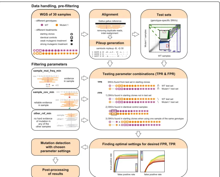
As the naïve approach of using commonly applied muta- tion detection tools with the suggested default settings failed to produce satisfactory results or could not detect small indels (for details see the Discussion section), we designed a filtering method that combines information from all available samples and gives robust SNV and indel calling with low false positive rate. A general over- view of the method can be seen on Fig. 1. The method
looks for heterozygous mutations with respect to the ref- erence genome, and filters out positions where other samples also differ from the reference. This approach ensures that ‘germline’ variations, present in multiple samples, are not called as false ‘somatic’mutations even in the absence of an available SNP database. The other common source of false positive mutation calls are align- ment errors. Typically they occur at the same genome positions in multiple samples, so with multi-sample fil- tering they are easily eliminated.
Results were evaluated using a set of validated genotype-specific SNVs (‘test sets’), the generation of which is described below. The calculated true positive and false positive rates (TPR/FPR) were used as indica- tors of the goodness of the filtering and optimisation was carried out based on these values.
Establishing SNV test sets
To measure false negatives and validate the SNV detection results, we established two different high-confidence refer- ence SNV sets from within our dataset. The test sets con- sist altogether of around 4000 positions, which is a sufficiently large number to calculate reliable estimates of TPR and FPR values.
The cell clone whole genome sequence panel used contains several isogenic samples of two different ge- notypes (WT and Mutant 1) that underwent various mutagenic treatments. Cell populations were grown separately for some time, accumulating mutations, be- fore the isolation of single clones for genome sequen- cing. Therefore, we expected to find two types of SNVs within our dataset. There should be treatment-induced, primarily heterozygous SNVs present in individual samples only. In addition, there would be SNVs arising from the genetic differences of the starting WT and Mutant 1 cell clones, which could be either heterozy- gous or homozygous. Heterozygous SNVs of the latter category were used as test set positions.
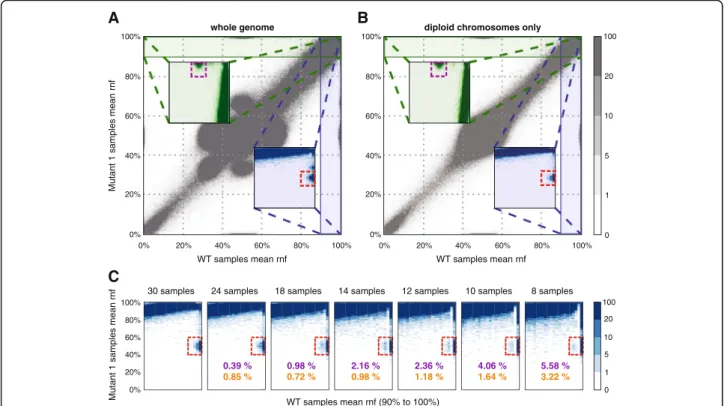
A plot of the mean reference nucleotide frequency (rnf) of all WT samples against the meanrnf of all Mu- tant 1 samples readily identifies heterozygous SNVs present in the Mutant 1 genotype at the [100, 50%] pos- ition. Mutations present in the WT samples but not in the Mutant 1 samples are clustered around [50, 100%]
(see Fig. 2a). Additional clusters around [100, 70] and [70, 100%] are due to genomic regions with ploidy > 2 in the experimental cell line. To verify this statement, we replotted the previous figure using only disomic chro- mosomes (Fig. 2b) and used the clusters on this latter graph as test cohorts, as the resulting clusters have very clear outlines and show no overlap with the rest of the data. Thus all optimisation procedures were limited to disomic chromosomes only, which proved to be suffi- cient for the relatively stable DT40 genome [20]. In cases
when ploidy varies greatly in the investigated genome, a ploidy-specific optimisation should be carried out. Also, it is impossible to separate loss of heterozygosity (LOH) events from real germline mutations during test set gen- eration. This is not a problem, as LOH regions can be included in the reference sets for testing purposes, in- creasing the number of positions which can be used for statistical analysis.
The described method for generating test sets is ap- plicable whenever two sample groups of related but separate genetic origin are available. Details of the very similar indel test set generation can be found in Additional file 2. In-depth workflow of the test set generation from pileup files for both SNVs and indels is in Additional file 4.
Testing of different filtering methods
Regardless of the SNV detection method selected, the above test sets can be used to determine their accuracy.
False positive rates (FPR) were calculated by running IsoMut on all samples, and counting independent muta- tions in pre-treatment starting cell clones and in the re- peatedly sequenced identical control samples, in which no independent mutations may be present. To obtain es- timates for the true positive rate (TPR), we ran the algo- rithm using the starting clone of one genotype and all clones of the second genotype. This way, the genotype- specific SNVs were detected as unique in the selected starting clone, and the proportion of the respective genotype-specific test set detected in this individual sample was used as TPR. Further details can be found in Additional file 2.
Defining filtering parameters to handle SNPs and alignment noise
IsoMut detects somatic mutations by considering all input samples and applying filtering criteria at each genomic position. To effectively discard germline mutations and
Fig. 1An overview of the testing and optimisation of the mutation detection method
false positives arising from alignment noise, three basic fil- tering parameters were introduced: a minimal threshold for the ratio of the most common type of non-reference reads in the investigated sample (sample_mut_freq_min), a minimal threshold for the ratio of reference reads in the noisiest non-selected (‘other’) sample (other_rnf_min) and a coverage limit for the selected sample (sample_cov_min).
For detailed verification of using such filters, see Additional file 5. These parameters were optimised for the desired values of TPR and FPR using the above described testing method.
This thorough optimisation procedure requires a very specific experimental setup and is often not feasible with the available set of sequencing data. Thus a more rapid and convenient method is desired to adopt IsoMut to specific needs. To provide a much simpler tuning option, IsoMut calculates theS score value of each can- didate mutation, which is inversely related to the prob- ability of falsely categorising a position as a unique mutation, thus high-confidence mutations have higher S values than unlikely ones. More precisely,Sis calculated as the negative logarithm of the probability p, that given the assumption that the distribution of bases in the two noisiest samples (containing the most non-reference reads) at the genomic position is the same, we would
observe the actual sequencing data. Thus a low p(high S) value suggests that it is unlikely that the two investi- gated samples have the same base-distribution, making it likely that the noisiest sample indeed has a unique mutation in the given position. The probability p is de- termined by the Fisher’s exact statistical test. For more details, see Additional file 2.
The availability of negative controls allows for a sim- ple, yet effective tuning of IsoMut by setting an S score threshold for more rapid results. In this case, a separate optimisation can be carried out on SNVs, insertions and deletions.
Post-processing
SNV and indel candidates were subjected to different post-processing steps, the details of which can be found in Additional file 2.
Results and discussion Optimal threshold values
We tested IsoMut on whole genome sequences of mutagen-treated cultured cells. In the tests described below, worst case scenarios were considered. One test for determining the FPR was the inclusion of only two samples of a given genotype, which makes the testing B
100%
80%
60%
40%
20%
0%
100
100%
80%
60%
40%
20%
0%
0 1 5 10 20
WT samples mean rnf
A
100%
80%
60%
40%
20%
0%
100%
80%
60%
40%
20%
0%
WT samples mean rnf
Mutant 1 samples mean rnf
diploid chromosomes only whole genome
C
30 samples 24 samples 18 samples 14 samples 12 samples 10 samples 8 samples
0.39 % 0.85 %
0.98 % 0.72 %
2.16 % 0.98 %
2.36 % 1.18 %
4.06 % 1.64 %
5.58 % 3.22 % 100%
80%
60%
40%
20%
0%
WT samples mean rnf (90% to 100%)
Mutant 1 samples mean rnf
0 1 5 10 20 100
Fig. 2Test set detection for WT and Mutant 1 samples.a,bPlots of mean reference nucleotide frequency values in the samples of the two geno-types;awhole genome,bdiploid chromosomes only. Insets are zoomed-in regions of the underlying plot. Dashed rectangles mark the clusters identified as test cohorts.cGenerating the same figures for different sample numbers. Percentages in purple show the ratio of lost test set positions, while percentages in orange represent the ratio of gained positions in the area in the dashed rectangle
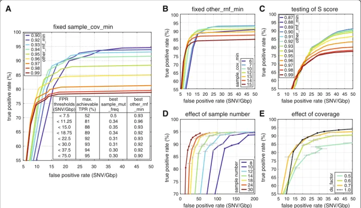
procedure as strict as possible. (For more details, see Additional file 2.) If samples are distributed more evenly among genotypes, IsoMut can achieve even better re- sults than the ones presented below, simply by adjusting the S score parameter. (For an example, see Additional file 6.) Filter threshold values were selected based on TPR and FPR requirements. We tested the effect on TPR and FPR of varying thesample_mut_freq_minfilter at different values of sample_cov_mincoverage require- ment and a fixed other_rnf_min= 0.93 (Fig. 3b). As the depth of the sequencing data limits coverage thresholds, we chose an intermediate, fixed sample_cov_min= 7 value for further optimising. At this value, we investi- gated how varying sample_mut_freq_min at different values of the other_rnf_min filtering parameter influ- ences the relation of TPR and FPR (Fig. 3a). Depending on the number of expected mutations in the investigated samples, different FPR values can be tolerated. The inset on Fig. 3a shows the corresponding maximal achievable TPRs and optimal filtering parameters to arbitrarily chosen FPR values. For the low FPR requirement of the
test sample set, a fixed parameter set of sample_mut_- freq_min= 0.31, other_rnf_min= 0.93, sample_cov_min
= 7 provided the best TPR of 92%. Further parameter settings with the respective TPR and FPR values can be found in Additional file 7.
Effects of sample number and sequencing depth
To assess the effects of having smaller datasets with fewer samples, different n sized subsets of the original 30 samples were investigated. Details on this technique can be found in Additional file 2. When using fewer samples, the outline of mutation clusters used for estab- lishing a test set gets progressively more blurred (Fig. 2c), but the number of lost and gained positions remains relatively small (less than 6 and 4% of the original set, respectively) even for only 10 samples. The dominant ef- fect of reducing the number of available samples is an increased FPR (Fig. 3d). For very strict parameter set- tings the number of true positives also increases when using smaller datasets. In case of the samples used for demonstration here, to keep FPRs below 50 per genome
false positive rate (SNV/Gbp)
true positive rate (%)
100 95 90 85 80 75 70 65 60 55
0.5 0.6 0.7 ds_factor 1.0
true positive rate (%)
100 95 90 85 80 75 70
false positive rate (SNV/Gbp)
50 100 150 200
0
108 12 14 18 24 sample number 30
0.87 0.88 0.89 0.90 0.91 0.92 0.93 0.94 0.95 0.96 0.97 0.98 0.99
other_rnf_min
true positive rate (%)
100 95 90 85 80 75 70 65 60 55
false positive rate (SNV/Gbp) 6
107 12 13 1415
sample_cov_min
true positive rate (%)
100 95 90 85 80 75 70 65 60 55
false positive rate (SNV/Gbp)
5 10 15 20 25 30 35 40 45 50 5 10 15 20 25 30 35 40 45 50
5 10 15 20 25 30 35 40 45 50 0.90
0.92 0.93 0.94 0.95 0.96 0.97 0.98 0.99
FPR thresholds (SNV/Gbp)
max.
achievable TPR (%)
best sample_mut
_freq
best other_rnf
_min
< 7.5 52 0.5 0.93
0.96
< 11.25 81 0.34
< 15.0 88 0.35 0.93
< 18.75 89 0.34 0.92
< 22.5 92 0.31 0.93
< 30.0 93 0.31 0.92
< 37.5 94 0.30 0.92
< 75.0 95 0.30 0.90
other_rnf_min
false positive rate (SNV/Gbp)
5 10 15 20 25 30 35 40 45 50
true positive rate (%)
100 95 90 85 80 75 70 65 60 55
A B C
D E
fixed sample_cov_min
fixed other_rnf_min testing of S score
effect of sample number effect of coverage
Fig. 3Quality components resulting from different parameter settings and different datasets.aEffects of varyingother_rnf_min(different curves) andsample_mut_freq_min(along the curves) with constantsample_cov_min= 7. The inset contains maximal achievable TPRs for given FPR thresholds with the optimal parameter settings.bEffects of changingsample_cov_min(different curves) andsample_mut_freq_min(along the curves) with fixedother_rnf_min= 0.93.cEffects of varyingother_rnf_min(different curves) and theSscore parameter (along the curves) with sample_cov_min= 5 andsample_mut_freq_min= 0.21.dEffects of varying the size of the dataset. Measurement points correspond to the parameter settings of the inset of (a). Mean values and standard deviation of three randomly chosen datasets are shown (see Additional file 2).
eEffects of decreased sample coverage. Measurement points correspond to the parameter settings of the inset of (a). Mean values and standard deviation of three randomly down-sampled measurements are shown (see Additional file 2)
(~1 GB) while maintaining a TPR of at least 85%, no fewer than 14 samples are required for analysis.
To demonstrate how a decreased coverage in one of the samples effects the results, we down-sampled the se- quence read data of the Mutant 1 starting clone using different down-sampling factors (ds_factors) of 0.7, 0.6 and 0.5 and recalculated TPRs and FPRs for the param- eter settings shown in the inset of Fig. 3a. Further details are included in Additional file 2. We found that having 70% of the original coverage had minimal impact on mutation detection, but further decreasing the sequen- cing depth produced lower TPR and higher FPR values.
As the Mutant 1 starting clone had a mean coverage of 21, we advise using samples with a mean coverage of at least 15.
IsoMut software implementation–guidelines for different experimental setups
We created an open-source C implementation of the somatic mutation detection steps of the above algorithm with a python wrapper for parallelisation (downloadable from https://github.com/genomicshu/isomut). The tool expects BAM files as its input and returns a list of de- tected mutations (both SNVs and indels) by applying predefined filtering parameters and a post-processing step different for SNVs and indels (see Additional file 2) in each genomic position. Thus an appropriate reference genome is necessary for running IsoMut for alignment purposes, but mutations are not detected based on dif- ferences of the samples and the reference genome, but on differences between investigated samples.
IsoMut can be applied whenever multiple isogenic samples are available and unique mutations are sought.
Negative control samples should be used when possible.
These can be either pre-experiment starting clones or DNA preparations sequenced multiple times, neither of which should contain experiment-induced unique muta- tions. With the availability of negative controls and a positive control test mutation set, best results are achieved by optimising the three IsoMut filtering param- eters as demonstrated above.
However, the availability of negative controls also al- lows for the tuning of the S score value for more rapid results, skipping the generation of positive test sets.
An example run of the IsoMut tool is shown in Additional file 6. In the following we present the main steps of the analysis. The generation of BAM alignment files is not included and should be carried out separately, prior to running IsoMut.
1. Downloading and compiling IsoMut.
2. Modifying user-specific data in the example
script (file names, paths, filtering parameter values).
3. Running IsoMut.
4. Tuning of the S score threshold value to minimise false positives in negative control samples.
The first three steps are necessary, the fourth one is optional and requires the availability of negative control sample(s). Whenever possible, this last fine-tuning step is strongly encouraged and yields better results than using the predefined filtering parameters only. For this procedure we suggest choosing less strict values for the sample_mut_freq_min and sample_cov_min filters, and further filtering the results based on the S score (see Additional file 6). The effects of tuning theSscore value and the other_rnf_min parameter with fixed sample_- mut_freq_min= 0.21 and sample_cov_min= 5 is shown in Fig. 3c. According to the figure, whenever a very low (< 2 · 10−8) FPR is desired, we suggest choosing a strict other_rnf_minvalue of 0.96 (or even larger for lower FPR).
When the FPR can exceed 30 per Gbp, less strict filtering is advised,other_rnf_mincan be decreased to around 0.9.
IsoMut default values are sample_mut_freq_min= 0.21, sample_cov_min= 5 andother_rnf_min= 0.93.
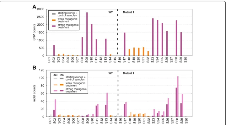
In the absence of negative controls, step (4) should be skipped and we advise using filtering values from the inset of Fig. 3a fitting the desired FPR. In this case SNVs and indels are detected with the same filtering thresh- olds. More details on the performance of our method in such cases can be found in Additional file 2. An example run without using an S score threshold, with parameter settings sample_mut_freq_min= 0.31, other_rnf_min= 0.93, sample_cov_min= 7 can be found on Fig. 4 for our dataset. This resulted in an average of only 6 mutations in starting clones or identical sample pairs (FPR ~ 6∙10−9), even though the DT40 genome differs from the chicken reference genome in 6.3 million SNPs [20].
On the other hand, differently treated samples have up to 2790 mutations, proving that the lack of these in untreated starting clones is not due to overly strict filtering.
Advantages of a straightforward filtering algorithm Although setting fixed thresholds for the above de- scribed simple filtering parameters might appear un- sophisticated, the approach has a general advantage over complex statistical models besides being just as effective.
In spite of recent developments of bioinformatics soft- ware and mutation calling algorithms, the unspoken consensus remains that ambiguous mutation calls are best verified by checking the raw sequencing data either in a genome viewer (for example IGV [28]) or a pileup file. The above filtering parameters are directly related to the number of different bases detected at each gen- omic position, making the evaluation of mutations very straightforward, without the need for decoding the meaning of differentp-values.
Performance comparison with standard tools
We developed IsoMut because we had found that stand- ard tools could not detect both SNVs and indels in the above described samples with the precision required for biological interpretation without heavy additional in-house filtering. Here we present a comparison with two very popular software tools, VarScan 2 [16] and MuTect [17].
VarScan 2 was run in its tumor-normal comparison mode for the two pairs of identical samples in our data- set (see Additional file 8). (Twice for both pairs, switch- ing the roles of ‘tumor’and‘normal’samples each time.) This way all mutations found by VarScan 2 are false pos- itives. Filtering parameters and additional filtering steps were applied according to the instructions provided in [29]. The analysis resulted in 368, 410, 1264 and 922 mutations in samples S12, S15, S27 and S30 respectively.
On the other hand, the numbers of false positives using IsoMut were 3, 1, 3 and 5 for the same samples. This difference in performance is probably due to the fact that VarScan 2 relies largely on filtering methods which have proved to be successful in case of human genomes, but are not available for our current dataset (dbSNP, re- peat masking).
MuTect is not capable of detecting indels (however, its recently released version, MuTect2 is, but was unavail- able for download at the time of this study), but we
selected it for testing because besides normal-tumor sample pairs, MuTect can also use a panel of normal samples. With default settings, MuTect did not perform efficiently for our dataset, but with our control samples we were able to optimise MuTect’s LOD parameter threshold (Additional file 9), and obtained good results.
Compared to MuTect IsoMut has similar characteris- tics at very low false positive rates (0.7/Mbp mutations detected at 0.5/Gbp FPR in our dataset), and it has higher sensitivity when we allow for higher false posi- tive rates (1/Mbp mutations detected by IsoMut and 0.75/Mbp mutations detected by MuTect at 1/Gbp FPR, Additional file 10). Additionally we found that IsoMut adapts significantly better to lower sample numbers (Additional file 10).
To get an estimate of the runtimes of different soft- ware, we ran IsoMut, MuTect, MuTect2 and VarScan 2 on the short chicken chromosome 28 (4.7 Mbp) using the 30 samples described above. We used a modest com- puter with a memory of 23 GB and 12 cores, with a per- formance achievable in a high-end desktop computer.
VarScan 2 was run in somatic mode by comparing each sample with its appropriate‘normal’pair, resulting in 30 comparisons. Both for MuTect and MuTect2, the gen- eral guidelines provided online were followed. First a unique panel of normal samples was created for each
0 500 1000 1500 2000 2500
A
3000starting clones + control samples weak mutagenic treatment strong mutagenic treatment
WT Mutant 1
S01 S02 S03 S04 S05 S06 S07 S08 S09 S10 S11S11 S12 S13 S14 S15 S16 S17 S18 S19 S20 S21 S22 S23 S24 S25 S26 S27 S28 S29 S30
0 20 40 60 80 100
B
120SNV countsindel counts
starting clones + control samples weak mutagenic treatment strong mutagenic treatment
del ins WT Mutant 1
S01 S02 S03 S04 S05 S06 S07 S08 S09 S10 S12 S13 S14 S15 S16 S17 S18 S19 S20 S21 S22 S23 S24 S25 S26 S27 S28 S29 S30
Fig. 4Results of running IsoMut without tuning theSscore value.aSNV counts for each sample, grouped by genotype. Colours indicate the treatment of the given sample.bIndel counts for each sample, grouped by genotype. Colours indicate the treatment of a given sample, darker bars representing insertions, lighter ones deletions
sample by combining the results of the artefact detection runs of all other samples. After this preliminary step, mutations were detected by comparing each sample with
its ‘normal’pair using the previously generated panel of
normals. For further details on the used pipelines and scripts see Additional file 11.
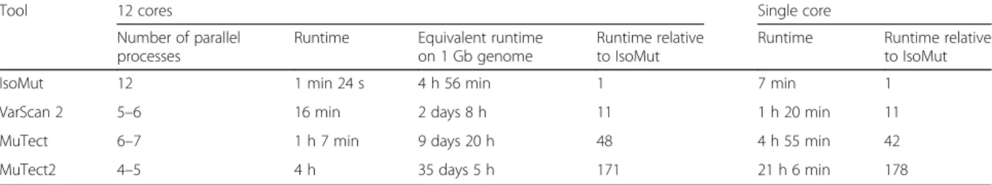
Using all resources of the above described computer, IsoMut turned out to be around 170 times faster than MuTect2, more than 40 times faster than MuTect and more than 10 times faster than VarScan 2 (see Table 1).
Extrapolating to the whole chicken genome, mutation analysis on the set of 30 samples using this 12-core com- puter would take 5 h with IsoMut, but over 35 days with Mutect2. The number of possible MuTect2, MuTect and VarScan 2 processes that can be run in parallel is limited by the finite memory of the computer, as all these soft- ware use java tools which require several java virtual machines when run in parallel. On the other hand, the parallelisation of IsoMut is only limited by the number cores on the computer and the runtime appears to be mainly I/O bound. The performance of the three java applications can be significantly improved by using a high-performance computer with a memory of 100–
200 GB. However, high-end computer clusters have limited availability, and IsoMut presents a great speed advantage when using modest resources. Even though it is not realistic to run any of the above tools on a sin- gle core without parallelisation, for a more straightfor- ward comparison the results of such a scenario are also presented in Table 1, demonstrating similar perform- ance advantages for IsoMut.
Conclusion
The described SNV identification method requires no prior knowledge of genomic nucleotide polymorphisms (SNPs). As these are expected to be present in all the isogenic samples, they are filtered out based on their dif- ference from the reference genome. The availability of a non-mutated reference sample is also not necessary if the mutated samples contain independently formed mutations.
Using the experimental dataset to establish reference test sets also presents a great advantage to currently
used alternative approaches, which usually use some in- dependent procedure to validate a small number of well-chosen SNVs [30, 31]. As this is usually done ex- perimentally at a great cost of time and money, it is de- sirable to generate test sets in a more efficient manner.
Using these test sets we demonstrated the optimisation of filtering parameters for diploid chromosomes. This way we were able to present filtering parameter settings suit- able for different desired FPRs that can be used on data- sets with no mutation-free control samples.
We designed IsoMut to be used in cases when mul- tiple isogenic samples are available and unique muta- tions are sought. It is easily adapted to cases when the independence of mutations in certain sample subsets is not guaranteed; in these cases all but one of these sam- ple subsets should be excluded from the analysis, while including several truly independent samples. Based on down-sampling an experimental dataset, we can recom- mend a minimum sample number of 14 and a minimum short-read sequence coverage of 15.
We strongly recommend sequencing negative control samples, and designed an adjustable approach that can be conveniently and quickly optimised for any specific dataset with such controls. This optimisation procedure can also be applied to non-diploid regions, where each level of ploidy should be treated separately.
Mutation analysis is widely used in the study of the DNA damaging effect of environmental substances and metabolism, DNA repair, cancer, and evolution. IsoMut can aid these studies by providing a solution for the ac- curate identification of SNVs and indels from pure iso- genic samples such as cell clones or animal progeny regardless of the species and the available data on gen- omic polymorphisms.
Additional files
Additional file 1:Table of samples. List of samples used in the study.
Half the samples had wild type (‘WT’) and the other half’Mutant 1′ genotype. Samples underwent different types of mutagenic treatments, which are also indicated in the table. (PDF 221 kb)
Additional file 2:Detailed methods. A detailed description of methods for testing and mutation detection. (PDF 598 kb)
Table 1Comparison of runtimes of different tools with all available resources
Tool 12 cores Single core
Number of parallel processes
Runtime Equivalent runtime on 1 Gb genome
Runtime relative to IsoMut
Runtime Runtime relative to IsoMut
IsoMut 12 1 min 24 s 4 h 56 min 1 7 min 1
VarScan 2 5–6 16 min 2 days 8 h 11 1 h 20 min 11
MuTect 6–7 1 h 7 min 9 days 20 h 48 4 h 55 min 42
MuTect2 4–5 4 h 35 days 5 h 171 21 h 6 min 178
Table of the runtime comparison of different mutation detection software using a computer with 23 GB memory and 12 cores or a single core only. The tools were run on the 4.735 Mb chicken chromosome 28 using the 30-sample dataset used throughout this study
Additional file 3:Generating pileup files. Scripts and pipeline for pileup file generation. (HTML 223 kb)
Additional file 4:Generating SNV test sets. Workflow for the generation of SNV test cohorts. (HTML 390 kb)
Additional file 5:Verification and description of filtering parameters. A detailed verification of the chosen filtering parameters. (PDF 391 kb) Additional file 6:Example run and tuning of IsoMut. An example run of IsoMut on a reduced dataset for easy testing. (HTML 1353 kb)
Additional file 7:Table of tested parameter settings. List of tested parameter settings with the resulting TPR and FPR values. (PDF 307 kb) Additional file 8:Running VarScan 2 on our dataset. Computational details and results of running VarScan 2 on the described dataset. (HTML 408 kb) Additional file 9:Running MuTect on our dataset with default settings.
Computational details and results of running MuTect on the described dataset without the tuning of the LOD parameter. (HTML 675 kb) Additional file 10:Comparison of IsoMut and MuTect. Comparison of false positives rates when running IsoMut versus running MuTect with a finely tuned LOD parameter. (HTML 373 kb)
Additional file 11:Runtime comparison of standard tools and IsoMut. A list of scripts and functions used to test the speed of standard mutation detection tools and IsoMut, using all resources of the available computer and using a single core only. (HTML 311 kb)
Abbreviations
ds_factor:Down-sampling factor; FPR: False positive rate; indel: Insertion and/
or deletion; LOH: Loss of heterozygosity; NGS: Next generation sequencing;
other_rnf_min: Minimal threshold for the ratio of reference reads in the noisiest non-selected sample;rnf: Reference nucleotide frequency;
sample_cov_min: Minimal threshold for the coverage of the selected sample;
sample_mut_freq_min: Minimal threshold for the ratio of the most common type of non-reference reads in the investigated sample; SNP: Single nucleotide polymorphism; SNV: Single nucleotide variation; TPR: True positive rate; WT: Wild type
Acknowledgements None.
Funding
This work was supported by Momentum Grant LP2011-015 of the Hungarian Academy of Sciences to DS and a Novo Nordisk Foundation Interdisciplinary Synergy Programme Grant no. NNF15OC0016584 to ZS and IC. ZS is sup- ported by the Breast Cancer Research Foundation, Basser Foundation and the Széchenyi Progam, Hungary (KTIA_NAP_13-2014-0021). GET is supported by Momentum Grant LP2012-035 of the Hungarian Academy of Sciences. GET and JM are also supported by the Hungarian Scientific Research Fund (OTKA K104586). IC, AB, DR and OP are supported by the European Commission H2020 program under contract number 643476 (www.compare-europe.eu).
Availability of data and materials
Sequencing data generated for this study have been submitted to the European Nucleotide Archive under study accession number ERP014915, downloadable from http://www.ebi.ac.uk/ena/data/view/PRJEB13358.
Authors’contributions
OP and DR developed the mutation detection algorithm; OP, DR, JM, AP and MK tested the algorithm and made comparisons to alternative tools; ZS, IC and DS conceived the study; DS, ZS, GET, AB and IC participated in the coordination of the study; OP and DS wrote the manuscript; all authors helped drafting the manuscript and read and approved the final version.
Competing interests
The authors declare that they have no competing interests.
Consent for publication Not applicable.
Ethics approval and consent to participate Not applicable.
Author details
1Department of Physics of Complex Systems, Eötvös Loránd University, H-1117 Budapest, Hungary.2Institute of Enzymology, Research Centre for Natural Sciences, Hungarian Academy of Sciences, H-1117 Budapest, Hungary.3Center for Biological Sequence Analysis, Department of Systems Biology, Technical University of Denmark, DK-2800 Lyngby, Denmark.
4Computational Health Informatics Program (CHIP), Boston Children’s Hospital, Boston, USA.5Harvard Medical School, Boston, MA 02215, USA.
6MTA-SE-NAP, Brain Metastasis Research Group, 2nd Department of Pathology, Semmelweis University, H-1091 Budapest, Hungary.
Received: 22 September 2016 Accepted: 20 January 2017
References
1. Duncavage EJ, et al. Targeted next generation sequencing of clinically significant gene mutations and translocations in leukemia. Mod Pathol.
2012;25:795–804.
2. Grossmann V, et al. Targeted next-generation sequencing detects point mutations, insertions, deletions and balanced chromosomal rearrangements as well as identifies novel leukemia-specific fusion genes in a single procedure. Leukemia. 2011;25:671–80.
3. Forster M, et al. From next-generation sequencing alignments to accurate comparison and validation of single-nucleotide variants: the pibase software. Nucleic Acids Res. 2013;41(1):e16.
4. Meacham F, et al. Identification and correction of systematic error in high- throughput sequence data. Bioinformatics. 2011;12:451.
5. Nakamura K, et al. Sequence specific error profile of Illumina sequencers.
Nucleic Acids Res. 2011;39(13):e90.
6. Nielsen R, et al. Genotype and SNP calling from next-generation sequencing data. Nat Rev Genet. 2011;12(6):443–51.
7. Kinde I, et al. Detection and quantification of rare mutations with massively parallel sequencing. Proc Natl Acad Sci U S A. 2011;108:9530–5.
8. Campbell PJ, et al. Subclonal phylogenetic structures in cancer revealed by ultra-deep sequencing. Proc Natl Acad Sci U S A. 2008;105:13081–6.
9. Sherry ST, et al. dbSNP: the NCBI database of genetic variation. Nucleic Acids Res. 2001;29(1):308–11.
10. The 1000 Genome Project Consortium, Abecasis GR, et al. An integrated map of genetic variation from 1,092 human genomes. Nature. 2012;491:56–65.
11. Johnson GE. Mammalian cell HPRT gene mutation assay: test methods.
Methods Mol Biol. 2012;817:55–67.
12. Mortelmans K, Zeiger E. The Ames Salmonella/microsome mutagenicity assay. Mutat Res. 2000;455:29–60.
13. Lazar V, et al. Bacterial evolution of antibiotic hypersensitivity. Mol Syst Biol.
2013;9:700.
14. Sakai W, et al. Secondary mutations as a mechanism of cisplatin resistance in BRCA2-mutated cancers. Nature. 2008;451:1116–20.
15. Lagerqvist A, et al. DNA repair and replication influence the number of mutations per adduct of polycyclic aromatic hydrocarbons in mammalian cells. DNA Repair (Amst). 2011;10:877–86.
16. Koboldt DC, et al. VarScan 2: somatic mutation and copy number alteration discovery in cancer by exome sequencing. Genome Res. 2012;22(3):568–76.
17. Cibulskis K, et al. Sensitive detection of somatic point mutations in impure and heterogeneous cancer samples. Nat Biotechnol. 2013;31:213–9.
18. Szikriszt B et al. A comprehensive survey of the mutagenic impact of common cancer cytotoxics. Genome Biol. 2016. in press
19. Zámborszky J et al. Loss of BRCA1 or BRCA2 markedly increases the rate of base substitution mutagenesis and has distinct effects on genomic deletions. Oncogene. 2016. in press
20. Molnár J, et al. The genome of the chicken DT40 bursal lymphoma cell line.
G3. 2014;4(11):2231–40.
21. Lawrence MS, et al. Mutational heterogeneity in cancer and the search for new cancer-associated genes. Nature. 2013;499:214–8.
22. Flicek P, et al. Ensembl 2014. Nucleic Acids Res. 2014;42(D1):D749–55.
23. Li H, Durbin R. Fast and accurate long-read alignment with burrows- wheeler transform. Bioinformatics. 2010;26:589–95.
24. Li H, Durbin R. Fast and accurate short read alignment with burrows- wheeler transform. Bioinformatics. 2009;25:1754–60.
25. Faust GG, Hall IM. SAMBLASTER: fast duplicate marking and structural variant read extraction. Bioinformatics. 2014;30:2503–5.
26. McKenna A, et al. The genome analysis toolkit: a MapReduce frame-work for analyzing nextgeneration DNA sequencing data. Genome Res. 2010;20(9):
1297–303.
27. Li H, et al. The sequence alignment/map (SAM) format and SAMtools.
Bioinformatics. 2009;25:2078–9.
28. Robinson JT, et al. Integrative genomics viewer. Nat Biotechnol. 2011;29:24–6.
29. Koboldt DC, et al. Using VarScan 2 for Germline variant calling and somatic mutation detection. Current Protoc Bioinformatics. 2013;44:15.4.1–5.4.17.
editoral board, Andreas D Baxevanis [et al].
30. Dahlman KB, et al. BRAF L597 mutations in melanoma are associated with sensitivity to MEK inhibitors. Cancer Discov. 2012;2(9):791–7.
31. Lam HYK, et al. Performance comparison of whole-genome sequencing platforms. Nat Biotechnol. 2012;30:78–82.
• We accept pre-submission inquiries
• Our selector tool helps you to find the most relevant journal
• We provide round the clock customer support
• Convenient online submission
• Thorough peer review
• Inclusion in PubMed and all major indexing services
• Maximum visibility for your research Submit your manuscript at
www.biomedcentral.com/submit
Submit your next manuscript to BioMed Central and we will help you at every step: