INTRODUCTION
Sudden cardiac death caused by ventricular arrhythmias is a major public health problem in modern industrialized countries (1), which emphasizes the importance of arrhythmia research.
Currently, there are clinical (1, 2) and experimental guidelines (3, 4) dealing with arrhythmia research and management, but these guidelines do not use unified arrhythmia definitions, and that is a notable problem when we try to objectively evaluate and compare results of arrhythmia investigations.
The Lambeth Conventions (LC I), a guidance for research on arrhythmias published in 1988 (3), had a substantial impact on the experimental arrhythmia research; the paper has been cited more than 1000 times since its publication 30 years ago according to the database of Web of Science. However, the advances in technology,
development of monitoring and pharmacologic solutions and of course the extensive research about arrhythmias had finally led to the realization that LC I was in need to be updated. Thus, a meeting was held in London in 2010 to update the guidance. The revised conventions were intended to be of practical value in terms of the design, execution, and analysis of experiments, with emphasis on the definition, classification, and quantification of ventricular and atrial arrhythmias (4). The revised Lambeth Conventions (LC II) were intended to be applied in preclinical and clinical research. Authors of LC II invited investigators to state whether or not they had used the conventions in their studies, and to test their validity by experiment.
Importantly, there are substantial changes in the definitions of the ventricular tachyarrhythmias between the original and the updated Lambeth Conventions (Table 1). The new A. REGEV1, H. TAKACS1, A.S. FARKAS1, F. RAROSI2,3, A. POLYAK1, H. PAPP1,
E. IVANY1, J.G. PAPP4,5, A. VARRO4,5, A. FARKAS1
APPLICATION OF VENTRICULAR TACHYARRHYTHMIA DEFINITIONS OF THE UPDATED LAMBETH CONVENTIONS PROVIDES INCOMPATIBILITY
WITH EARLIER RESULTS, MASKS ANTIFIBRILLATORY ACTIVITY AND REDUCES INTER-OBSERVER AGREEMENT
1Second Department of Medicine and Cardiology Centre, Faculty of Medicine, University of Szeged, Szeged, Hungary;
2Department of Medical Physics and Informatics, University of Szeged, Szeged, Hungary; 3Bolyai Institute, University of Szeged, Szeged, Hungary; 4Department of Pharmacology and Pharmacotherapy, Faculty of Medicine, University of Szeged, Szeged, Hungary; 5MTA-SZTE Research Group for Cardiovascular Pharmacology, Hungarian Academy of Sciences, Szeged, Hungary
The Lambeth Conventions (LC I), a landmark guidance document for arrhythmia research was updated and arrhythmia definitions were changed in the new Lambeth Conventions II (LC II). This study examined whether the arrhythmia definitions of LC I and LC II yield the same qualitative results and whether LC II improves inter-observer agreement.
Two independent investigators performed blinded arrhythmia analysis of the electrocardiograms of isolated, Langendorff rat hearts subjected to regional ischemia and perfused with Class I antiarrhythmics with 3 or 5 mM K+in the perfusate. Data obtained with arrhythmia definitions of LC I and LC II were compared within and between observers.
Applying ventricular fibrillation (VF) definition of LC II significantly increased VF incidence and reduced VF onset time irrespective of treatment by detecting ‘de novo’ VF episodes not found by LC I. LC II reduced the number of ventricular tachycardia (VT) episodes and simultaneously increased the number of VF episodes as compared with the respective values obtained according to LC I. Using VF definition of LC II masked the significant antifibrillatory effects of flecainide and the high K+concentration identified with the VF definition of LC I. When VF incidence was tested, a very strong inter-observer agreement was found according to LC I, whereas using VF definition of LC II reduced inter- observer agreement. It is concluded that LC II shifts some tachyarrhythmias from VT to VF class, and thus results obtained by arrhythmia definitions of LC I and LC II are not compatible; VF definition of LC II may change the conclusion of pharmacological, physiological and pathophysiological arrhythmia investigations and may reduce inter- observer agreement. Thus, VT and VF definitions of LC II should be amended in order to increase compatibility and inter-observer agreement.
K e y w o r d s : Lambeth Conventions, arrhythmia definitions, ventricular fibrillation, incompatibility of the results, inter-observer agreement
tachyarrhythmia definitions imply that some arrhythmias classified as ventricular tachycardia (VT) according to LC I are now classified as ventricular fibrillation (VF) according to LC II.
This suggests that the new definitions change the results and - more importantly - the conclusion of arrhythmia studies. Thus, the aim of the present study was to examine whether the arrhythmia definitions of LC I and LC II are compatible, and yield the same qualitative arrhythmia results. Also, it was tested whether arrhythmia definitions of LC I or LC II allow better inter-observer agreement. Thus, two independent investigators
reanalysed the electrocardiogram (ECG) recordings of experiments done earlier by Farkas and Curtis (5), and performed a retrospective, blinded analysis of the number and incidence of ventricular arrhythmias in isolated, Langendorff perfused rat hearts subjected to regional ischemia and treated with Class I antiarrhythmic agents. The arrhythmia data obtained by applying arrhythmia definitions of LC I and LC II were compared to test the compatibility between the arrhythmia definitions of LC I and LC II. Also, inter-observer agreement was determined to investigate whether arrhythmia definitions of LC I or LC II allow
Arrhythmia Definition according to
Lambeth Conventions I (3) Definition according to Lambeth Conventions II (4) Ventricular premature beat
(VPB) Isolated ventricular premature
beats are defined as discrete and identifiable premature QRS complexes (premature in relation to the P wave).
A VPB is defined as a ventricular electrical complex (complete electrical event:
QRS, RS, QRST or RST) that is different in shape (voltage and/or duration, i.e., height and/or width) from the preceding (non-VPB) ventricular complex, and is premature in relation to the preceding ventricular complex.
Bigeminy Bigeminy is characterised by the minimum sequence: P, QRS, VPB, P, QRS, VPB.
Bigeminy has the minimum sequence VPB, normal sinus beat, VPB (which may be repeated) in which the VPBs have the same shape and timing.
Salvo Two or three consecutive
VPBs do not constitute ventricular tachycardia but should be termed a salvo.
A run of two or three consecutive VPBs is defined as a salvo.
Ventricular tachycardia
(VT) A run of 4 or more
consecutive ventricular premature beats.
A sequence of a minimum of 4 consecutive ventricular complexes.
Monomorphic VT:
peak-peak interval, height and intrinsic shape are constant.
Polymorphic VT:
the peak-peak interval and/or height and/or intrinsic shape vary, and the variation of any or each of these is
progressive.
Ventricular fibrillation
(VF) A signal for which individual
QRS deflections can no longer be distinguished from one another (implying morphological instability) and for which a rate can no longer be measured.
A sequence of a minimum of 4 consecutive ventricular complexes without intervening diastolic pauses, in which intrinsic shape, peak-peak interval and height vary, and the variation between each is non- progressive. It is the non- progressive nature of the variation of all 3 variables that distinguishes VF from polymorphic VT and torsades de pointes.
Table 1.Comparison of the definitions of ventricular arrhythmias between the Lambeth Conventions I and II.
better agreement on the arrhythmia results between the two independent observers.
MATERIALS AND METHODS General experimental procedure
For detailed methodical description see the published study by Farkas and Curtis (5), from which the raw ECGs were obtained for arrhythmia analysis. Thus, here only a short description of the experiments is added as follows.
The animal-handling protocol was in accordance with the Guidance of the Operation on the Animals (Scientific Procedures) Act 1986, London, UK. Male rats were anesthetized with pentobarbitone (60 mg/kg intraperitoneally). Hearts were excised, and then perfused according to Langendorff with modified Krebs solution containing 118.5 mM NaCl, 25.0 mM NaHCO3, 1.2 mM MgSO4, 1.2 mM NaH2PO4, 1.4 mM CaCl2, 3 mM KCl (or 5 mM where indicated), and 11.1 mM glucose.
Perfusion solution was delivered at 37ºC and pH 7.4; perfusion pressure was maintained constant at 70 mm Hg. The left main coronary artery was occluded by a silk suture for 30 min. A unipolar ECG was recorded using a MacLab system (ADInstruments Ltd., Oxford, UK) (5-7).
Drug administration protocol, groups of hearts
In the first two sets of experiments, Krebs’ solution contained 3 mM K+. In the third and fourth sets of experiments, K+
concentration was elevated to 5 mM. Each set of experiments contained four groups of hearts: one control and three drug-treated groups, each treated with one concentration of a representative Class I antiarrhythmic (quinidine, lidocaine or flecainide). One lower and one higher concentration of quinidine (0.79 and 7.90 µM), lidocaine (3.88 and 12.93 µM), and flecainide (0.74 and 1.48 µM), representing the peak unbound plasma and total blood concentrations, respectively, at ‘therapeutic’ dosage, were evaluated in the first two sets of experiments and also in the last two sets of experiments. Each group contained 12 hearts in the first two sets of experiments (96 hearts in total; 3 mM K+in the Krebs), whereas every group in the third and fourth sets of experiments contained 6 hearts (48 hearts in total, 5 mM K+in the Krebs).
Experiments were done in a randomized and blinded manner.
Reanalysing ischaemic arrhythmias according to definitions of LC I and LC II, measurement of the number and incidence of tachyarrhythmias
The ECG recordings of the original investigation done by Farkas and Curtis (5) were reanalysed by two independent investigators using LabChart7 (ADInstruments Ltd., Oxford, UK). The two investigators were well trained and equally experienced in evaluating ECG. Ventricular arrhythmias were defined according to the LC I for the primary evaluation (Table 1). The number of VT and VF episodes, the incidence and the time to onset of VT and VF were determined from the ECG recorded during the 30-min-long ischaemia. Then the whole analysis was repeated according to the arrhythmia definitions of LC II (Table 1). All variables were measured in a blinded manner.
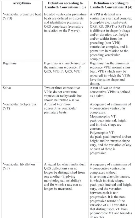
Fig. 1.Comparison of the percent incidences of ischaemic ventricular fibrillation (VF) obtained from the 1stset of experiments by two independent observers (Observer A and Observer B) using VF definition of Lambeth Conventions I (LC I) and Lambeth Conventions II (LC II). Langendorff perfused rat hearts were subjected to local ischaemia for 30 min. Hearts were perfused with the low concentration of quindine (0.79 µM), lidocaine (3.88 µM), flecainide (0.74) or solvent (Control), each group contained n = 12 hearts, Krebs solution contained 3 mM K+. *P < 0.05 versus Control.
Statistical analysis
Continuous variables were expressed as means ± SEM. Within group comparison of continuous data (e.g. the number of arrhythmia episodes, the onset times of VT and VF) were performed with the non-parametric Wilcoxon test for paired samples. Arrhythmia incidences were expressed as percent values, and were compared with Fisher’s exact test with the Bonferroni correction, that is, the P values of Fisher’s exact test were multiplied by the numbers of comparisons to allow multiple comparisons (8). A P value < 0.05 was taken as indicative of a statistically significant difference between values. The intra- observer agreement on arrhythmia incidence data obtained according to LC I and LC II was assessed with Cohen’s kappa statistic (9). Similarly, the inter-observer agreement on VT and VF incidence data among independent observers was calculated with Cohen’s kappa statistic (9).
RESULTS
Ischaemic ventricular fibrillation incidence in the first set of experiments (3 mM K+, low concentrations of drugs)
When the arrhythmia definitions of LC I were applied, both of the two independent investigators found that VF was frequently induced in the control group, and only flecainide was able to reduce VF incidence significantly at the applied low concentration (Fig. 1). These results are in a good accordance
with the arrhythmia results of the original investigation obtained by a different observer using the same arrhythmia definitions of LC I (5). This means that applying arrhythmia definitions of LC I allowed the two independent investigators to reproduce previous results.
When arrhythmia definitions of LC II were applied, both of the two independent investigators found that VF was induced in all control hearts, and none of the drugs affected significantly VF incidence at the applied low concentrations (Fig. 1). Thus, the significant antifibrillatory effect of the low concentration of flecainide was masked when arrhythmia definitions of LC II were used. This means that results about the antifibrillatory effect of flecainide qualitatively differ from those obtained by applying arrhythmia definitions of LC I.
Ischaemic ventricular fibrillation incidence in the second set of experiments (3 mM K+, high concentrations of drugs)
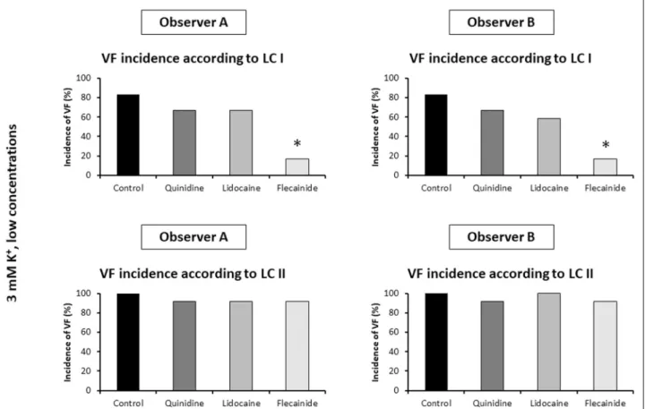
When the arrhythmia definitions of LC I were applied, both of the two independent investigators found that VF was frequently induced in the control group, and all three drugs reduced VF incidence significantly at the applied high concentrations (Fig. 2).
These results well accord with the arrhythmia results of the original investigation obtained by a different observer using the same arrhythmia definitions of LC I (5). This means that applying arrhythmia definitions of LC I allowed the two independent investigators to reproduce previous results.
When the arrhythmia definitions of LC II were applied, Observer B found that the high concentrations of all three drugs
Fig. 2.Comparison of the percent incidences of ischaemic ventricular fibrillation (VF) obtained from the 2ndset of experiments by two independent observers (Observer A and Observer B) using VF definition of Lambeth Conventions I (LC I) and Lambeth Conventions II (LC II). Langendorff perfused rat hearts were subjected to local ischaemia for 30 min. Hearts were perfused with the high concentration of quindine (7.90 µM), lidocaine (12.93 µM), flecainide (1.48 µM) or solvent (Control), each group contained n = 12 hearts, Krebs solution contained 3 mM K+. *P < 0.05 versus Control.
reduced the incidence of ischaemic VF as compared with control, which accords with the results obtained according to LC I (Fig. 2). However, Observer A found that the applied high concentration of flecainide did not significantly reduce the incidence of ischaemic VF, which qualitatively differs from the results obtained according to LC I, and it also contradicts the results of Observer B (Fig. 2).
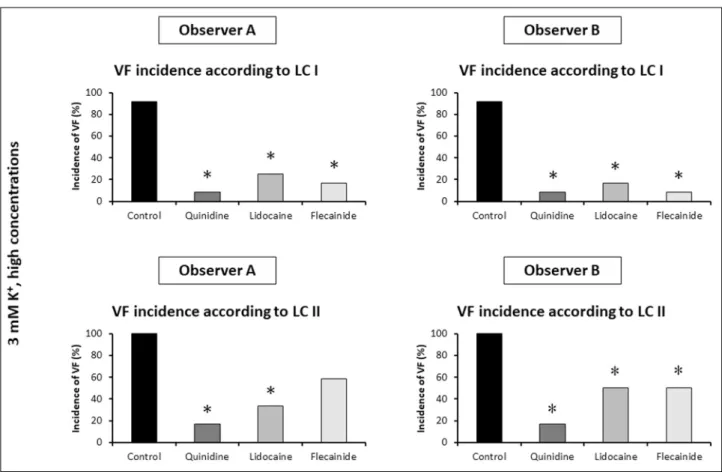
Ischaemic ventricular fibrillation incidence in the control hearts, the effect of K+concentration on ventricular fibrillation incidence
Ischaemic VF incidence was compared between the control group of hearts perfused with 3 mM K+(the hearts of the control groups of the 1st and 2ndsets of experiments) and the control hearts perfused with 5 mM K+(the hearts of the control groups
Fig. 3.Comparison of the percent incidences of ischaemic ventricular fibrillation (VF) between control rat hearts subjected to local ischaemia for 30 min and perfused with Krebs solution containing 3 or 5 mM K+(n = 24 and n = 12 hearts, respectively). Two independent observers (Observer A and Observer B) performed the blinded arrhythmia analysis using VF definition of Lambeth Conventions I (LC I) and Lambeth Conventions II (LC II). *P < 0.05 versus 3 mM K+.
Fig. 4.The total number of hearts with ischaemic ventricular fibrillation (VF) out of the 144 Langendorff perfused rat hearts subjected to local ischaemia for 30 min irrespective of the drug treatment and the K+content of the perfusate. VF was diagnosed by applying VF definitions of Lambeth Conventions I (LC I) and Lambeth Conventions II (LC II). Observer A and Observer B are the two independent observers. The number in the common part of the LC I and LC II classes shows the total number of hearts that experienced VF according to both LC I and LC II.
of the 3rdand 4thsets of experiments). Both investigators found that the elevation of the K+concentration from 3.0 mM to 5.0 mM significantly reduced the incidence of ischaemic VF, when arrhythmia definitions of LC I were applied (Fig. 3). These results well accord with the arrhythmia results of the original investigation obtained by a different observer using the same arrhythmia definitions of LC I (5). Again, this shows that
applying arrhythmia definitions of LC I allowed the two independent investigators to reproduce previous results.
However, the marked antiarrhythmic effect of the increased K+ concentration seen according to LC I was masked when arrhythmia definitions of LC II were applied in the present investigation, and these results did not differ qualitatively between the two investigators (Fig. 3).
Fig. 5.The effect of definitions of ventricular fibrillation (VF) of Lambeth Conventions I (LC I) and Lambeth Conventions II (LC II) on the onset time of the first episode of ischaemic VF in Langendorff perfused rat hearts subjected to local ischaemia for 30 min and experiencing VF according to both LC I and LC II. Parts (a) and (b): VF onset times in the 1stset of experiments, in which hearts were perfused with the low concentration of quindine (0.79 µM), lidocaine (3.88 µM), or solvent (Control), Krebs solution contained 3 mM K+. Part (c): VF onset times in the whole set of experiments irrespective of drug treatment and the K+content of the perfusate. Observer A and Observer B are the two independent observers. *P < 0.05 versus LC I.
Arrhythmia Observer Incidence (%) Kappa 95% Confidence interval
LC I LC II for kappa
VT A 85 84 0.974 0.922 – 1.000
B 88 76* 0.586 0.421 – 0.751
VF A 35 60* 0.529 0.411 – 0.646
B 33 60* 0.482 0.365 – 0.599
Table 2.The percent incidences of ischaemic ventricular tachycardia and ventricular fibrillation irrespective of the drug treatment and K+ content of the perfusate in n = 144 Langendorff perfused rat hearts subjected to local ischaemia for 30 min. Intra-observer agreement on the results obtained according to the arrhythmia definitions of Lambeth Conventions I and Lambeth Conventions II, results of the Cohen’s Kappa statistical analysis. Observers A and B are the two independent observers of the present investigation. LC I and LC II, VT and VF incidence data obtained according to the arrhythmia definitions of Lambeth Conventions I and Lambeth Conventions II, respectively.
VT, ventricular tachycardia; VF, ventricular fibrillation; Kappa, kappa coefficient obtained by Cohen’s Kappa statistical analysis; note that kappa value of 1.0 means perfect agreement, whereas the kappa value of 0.0 means absolutely no agreement; thus, the greater the kappa value in the range of 0.0 – 1.0, the greater the agreement. *P < 0.05 versus LC I.
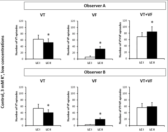
Fig. 6.The numbers of ventricular tachycardia (VT) and ventricular fibrillation (VF) episodes and the cumulative number of VT and VF episodes (VT + VF) in those eight hearts of the flecainide treated group of the first set of experiments, in which neither Observer A nor Observer B found VF according to Lambeth Conventions I (LC I), but both of them identified VF according to Lambeth Conventions II (LC II). Observer A and Observer B are the two independent observers. *P < 0.05 versus LC I. #P = 0.055 versus LC I. Note that VF episodes found by applying VF definition of LC II are ‘de novo’ VF episodes, and occurred only as a result of
‘arrhythmia shift’, and ‘arrhythmia fragmentation’ did not contribute to their occurrence.
Incidence
of Lambeth Convention Observers Kappa
95% Confidence interval for kappa
VT LC I A vs. O 0.918 0.826 – 1.000
B vs. O 0.615 0.424 – 0.805 A vs. B 0.652 0.471 – 0.833 LC II A vs. B 0.675 0.525 – 0.825
VF LC I A vs. O 0.908 0.836 – 0.980
B vs. O 0.938 0.877– 0.998 A vs. B 0.938 0.879 – 0.998 LC II A vs. B 0.884 0.806 – 0.962
Table 3.Inter-observer agreement on the incidences of ventricular tachycardia and ventricular fibrillation. Results of the Cohen’s Kappa statistical analysis. LC I and LC II: VF and VT incidence data obtained according to the arrhythmia definitions of Lambeth Conventions I and Lambeth Conventions II, respectively. Observers A and B are the two independent observers of the present investigation, Observer O is the observer of the original investigation published by Farkas and Curtis (5).
VT, ventricular tachycardia; VF, ventricular fibrillation; Kappa, kappa coefficient obtained by Cohen’s Kappa statistical analysis. VT and VF incidences were obtained irrespective of the drug treatment and K+content of the perfusate from n = 144 Langendorff perfused rat hearts subjected to local ischaemia for 30 min.
Ischaemic ventricular tachycardia and ventricular fibrillation incidences in the whole investigation, and intra-observer agreement
VT and VF incidences were calculated irrespective of the treatment and the K+content of the perfusate in the 144 hearts
of the whole investigation. Observer A did not find significant effect of LC II on VT incidence, and achieved good intra- observer agreement on VT incidence (Table 2). However, Observer B found that applying arrhythmia definitions of LC II significantly reduced VT incidence as compared with the respective value obtained according to LC I, which resulted in
Fig. 7.The numbers of ventricular tachycardia (VT) and ventricular fibrillation (VF) episodes and the cumulative number of VT and VF episodes (VT + VF) in the n = 12 control hearts of the first set of the experiments. LC I and LC II: data obtained by using arrhythmia definitions of Lambeth Conventions I and Lambeth Conventions II, respectively. Observer A and Observer B are the two independent observers. *P < 0.05 versus LC I. Note, that the extra VF episodes found by applying VF definition of LC II can be the result of ‘arrhythmia shift’. However, in theory, ‘arrhythmia fragmentation’ could also contribute to the increase in VF number according to LC II, as VF episodes found by LC I could be fragmented into shorter episodes due to containing diastolic pauses.
Observer A Observer B
Number of hearts % Number of hearts % VF found in the same heart by both LC I and LC II 51 100 47 100
VF onset time reduced by LC II 42 82 38 81
VF onset time not affected by LC II 9 18 9 19
VF onset time increased by LC II 0 0 0 0
Table 4.The effect of applying the new definition of ventricular fibrillation of Lambeth Conventions II on the onset time of ischaemic ventricular fibrillation in Langendorff perfused rat hearts. The onset time of ischaemic VF was examined in all hearts that experienced VF irrespective of K+content of the perfusate and drug treatment in the whole investigation (in all four sets of experiments utilising n = 144 isolated rat hearts).
LC I and LC II, Lambeth Conventions I and II, respectively. VF, ischaemic ventricular fibrillation. Observer A and Observer B, the two independent observers who performed blinded arrhythmia analysis of the experiments.
only moderate intra-observer agreement on VT incidence (Table 2).
Both observers found that VF was diagnosed in a significantly greater proportion of hearts, when the arrhythmia definitions of LC II were applied, as compared with the respective value measured according to the arrhythmia definitions of LC I (Table 2). Also, both observers found that when arrhythmia definitions of LC II were applied, VF was diagnosed in all hearts, in which VF was diagnosed according to LC I. However, there were many hearts, in which VF was diagnosed only according to LC II and VF was not found according to LC I (Fig. 4). Consequently, Cohen’s kappa analysis showed that intra-observer agreement on
VF incidence data obtained according to LC I and LC II was only moderate in case of both A and B Observers (Table 2).
The inter-observer agreement on ventricular tachycardia and ventricular fibrillation incidences among independent observers
The inter-observer agreement on VT and VF incidences among independent observers was calculated irrespective of the treatment and the K+concentration in the 144 hearts of the whole investigation. Applying VT definition of LC II did not remarkably improve the inter-observer agreement on VT incidence between Observers A and B; moderate inter-observer
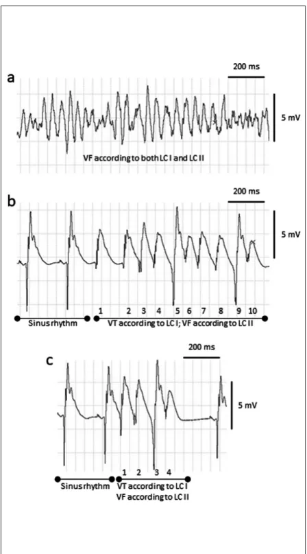
Fig. 8.Incompatibility between the classes of the ventricular fibrillation of the Lambeth Conventions I (LC I) and Lambeth Conventions II (LC II). Part (a): ventricular tachyarrhythmia diagnosed as ventricular fibrillation (VF) according to both LC I and LC II. Part (b): a ten-beat tachyarrhythmia episode of ‘de novo’ VF as this ventricular tachyarrhythmia is diagnosed as ventricular tachycardia (VT) according to LC I, while the same episode is diagnosed as VF according to LC II. LC II emphasised the importance of ‘diastolic pause’.
According to LC II, “if an arrhythmia is interrupted by an identifiable pause or quiescence (a ‘diastolic pause’, or a flat isoelectrical line) that is longer than the prevailing sinus rate (or idioventricular rate if the AV node is blocked) then it makes more sense to classify each arrhythmia present before and after the pause as separate events” (4). Based on the above recommendation of LC II, there is no relevant diastolic pause between the ventricular complexes of this tachyarrhythmia, thus this arrhythmia should be regarded as one single run of arrhythmia. Also note that the arrhythmia consists of sections with four consecutive beats (beats 3-4- 5-6 and beats 7-8-9-10), in which neither intrinsic shape, nor peek-peek interval, nor height of the ventricular complexes change progressively, thus each of these four-beat arrhythmia sections should be classified as VF according to LC II (see Table 1 for the arrhythmia definitions). Convention 14 of LC II states that “When rhythms segue from one to another without an identifiable interruption or quiescence, or vary between simultaneously recorded leads, then the arrhythmia should be classified as the most serious manifestation within segue or lead” (4). As this arrhythmia in part b should be regarded as one single run of arrhythmia and VF is the most serious arrhythmia manifestation within segue, the whole arrhythmia should be classified as VF according to LC II. On the other hand, QRS deflections can be distinguished from one another and the rate can be measured, thus the same arrhythmia should be classified as VT according to LC I. Part (c): a four- beat tachyarrhythmia episode of ‘de novo’ VF as this ventricular tachyarrhythmia is diagnosed as VT according to LC I, while the same episode is diagnosed as VF according to LC II. The ECG examples were recorded in three isolated, Langendorff perfused rat hearts subjected to regional ischaemia for 30 min.
agreement was found between them when VT incidence was tested either according to LC I or according to LC II (Table 3).
Importantly, when VF incidence obtained according to LC I was tested, very strong inter-observer agreements (high kappa values) were found among the two independent investigators (Observers A and B) and the investigator of the original investigation (Observer O), (5) (Table 3). When arrhythmia definitions of LC II were applied, a good agreement was found in VF incidence between the two independent investigators;
however, the kappa value was lower than that obtained according to LC I (Table 3).
The onset time of ischaemic ventricular fibrillation in the first set of experiments
When hearts were perfused with the low concentrations of the drugs and Krebs solution contained 3 mM K+(first set of experiments), VF incidences were high in the Control, Quinidine and Lidocaine groups of hearts irrespective of the applied VF definition (Fig. 1). This allowed us to examine the effect of the applied arrhythmia definitions on the onset time of the first episode of ischaemic VF in these three groups. Both observers found that VF onset time was significantly reduced in these three groups, when arrhythmia definitions of LC II were used as compared with the respective values obtained by applying arrhythmia definitions of LC I (Fig. 5aand 5b). This shows that some ischaemic arrhythmias, which were not identified as VF according to LC I, were identified as VF according to LC II, and these arrhythmias were found not only in drug-free hearts but in quinidine and lidocaine treated hearts, too. When VT definition of LC II was applied, VT onset times were not significantly affected in any of the groups as compared with the respective values obtained by applying VT definition of LC I (data not shown).
The onset time of ventricular fibrillation in all hearts that experienced ventricular fibrillation irrespective of K+content of the perfusate and drug treatment
When all those hearts were examined that experienced ischemic VF according to both LC I and LC II in the whole investigation, the two independent observers found that applying arrhythmia definitions of LC II significantly reduced the onset time of the first episode of VF as compared with the value obtained by using arrhythmia definitions of LC I (Fig. 5c).
Applying VF definition of LC II did not increase VF onset time in any of the hearts, but reduced this variable in approximately 80% of the hearts (Table 4). These results did not differ qualitatively between the two independent observers (Table 4).
The numbers of ventricular tachycardia and ventricular fibrillation episodes
In order to examine whether applying arrhythmia definitions of LC II causes an ‘arrhythmia shift’ from the VT class to the VF class, the numbers of VT and VF episodes were calculated in the control and the flecainide treated groups of the first set of experiments (n = 12 hearts in each group). These two groups were chosen, as the greatest and least number of arrhythmias occurred in the Control and Flecainide groups, respectively.
First, a statistical analysis was performed on the data of those eight hearts of the flecainide treated group of the first set of experiments, in which neither Observer A nor Observer B found VF according to LC I, but both of them identified VF according to LC II. In the flecainide treated 8 hearts, both observers found that applying VT definition of LC II reduced the number VT episodes as compared with the respective values obtained by
using VT definition of LC I (Fig. 6). On the other hand, applying VF definition of LC II significantly increased the number of VF episodes as compared with the respective zero value obtained by using VF definition of LC I (Fig. 6). Interestingly; Observer A found that arrhythmia analysis according to LC II significantly increased the cumulative number of VT and VF episodes as compared with the respective value obtained according to LC I in the flecainide treated 8 hearts. This suggests that when arrhythmia analysis was performed according to LC II, VT episodes were not only shifted to the VF class, but were also fragmented (i.e. broken up into shorter episodes due to containing diastolic pauses). Observer B did not find any significant effect of LC II on the cumulative number of VT and VF episodes (Fig. 6). Analysis of the number of the VT and VF episodes in the 12 hearts of the control group of the first set of experiments yielded qualitatively the same results despite identifying VF episodes not only by LC II but LC I, too (Fig. 7).
DISCUSSION
Present results show that arrhythmia analysis according to LC II qualitatively changed conclusions about pharmacological effects of flecainide. The change in tachyarrhythmia definitions also affected well documented pathophysiological effects, i.e.
the preventive effect of the high K+ concentration against ischaemic VF disappeared when the VF definition of LC II was applied. The reason behind these changes was that VF definition of LC II identified ‘de novo’ VF episodes not identified according to LC I, which is supported by the data showing that application of the VF definition of LC II increased VF incidence and reduced VF onset time. Importantly, a very strong inter- observer agreement was found among the two independent investigators, when VF incidence obtained according to LC I was tested. Applying VF definition of LC II resulted in less inter- observer agreement on VF incidence.
Arrhythmia shift from ventricular tachycardia to ventricular fibrillation category enlarges the ventricular fibrillation class of LC II and markedly increases sensitivity of ventricular fibrillation detection
VF definition of LC II diagnosed VF in all hearts, in which VF was identified according to LC I. Moreover, there were many other hearts, in which VF was detected only according to LC II and VF was not found according to LC I. Also, applying VF definition of LC II did not increase VF onset time in any of the hearts, however reduced this variable in the majority of the hearts. These results show that the VF class of LC II incorporated and enlarged the VF class defined by LC I. The reason behind this is that VF definition of LC II not only identified VF episodes detected by LC I, but also identified ‘de novo’ VF episodes not detected according to LC I. Our results clearly show that the enlarged VF category of LC II substantially increased the sensitivity of VF detection.
Comparing tachyarrhythmia definitions of LC I and LC II (Table 1) explains the mechanism of detection of ‘de novo’ VF episodes according to LC II. While arrhythmias containing 4 or more consecutive ventricular premature beats with non- progressive variation in peak-peak interval, height and intrinsic shape should be classified as VTs according to LC I, the same arrhythmias are now shifted to the VF category of LC II (Fig. 8, Table 1) (3, 4). These polymorphic VTs of LC I form the ‘de novo’
VF episodes detected by LC II. This ‘arrhythmia shift’ of LC II is also indicated by the data showing that applying arrhythmia definitions of LC II reduced the number of VT episodes and simultaneously increased the number of VF episodes.
Interestingly, Observer A found that when arrhythmia definitions of LC II were applied, the cumulative number of VT and VF episodes was greater than the respective value obtained according to LC I. The phenomenon of ‘arrhythmia fragmentation’ is most likely the reason behind this result. LC II introduced the concept of the importance of ‘diastolic pause’.
According to LC II, “if an arrhythmia is interrupted by an identifiable pause or quiescence (a ‘diastolic pause’, or a flat isoelectrical line) that is longer than the prevailing sinus rate (or idioventricular rate if the AV node is blocked) then it makes more sense to classify each arrhythmia present before and after the pause as separate events” (4). This concept enables an arrhythmia episode to be fragmented into many shorter arrhythmia episodes. Consequently, the number of VT and VF episodes measured according to LC I can be affected by
‘arrhythmia fragmentation’ when the same arrhythmias are evaluated according to LC II. However, the increase in VF incidence and the reduction in VF onset time caused by LC II can only be explained by the ‘arrhythmia shift’ phenomenon, and
‘arrhythmia fragmentation’ could not contribute to these results.
It can be concluded that the ‘arrhythmia shift’ caused by LC II is responsible for the only moderate intra-observer agreement on VF incidence values obtained according to LC I and LC II.
Applying the ventricular fibrillation definition of LC II may change the conclusions of pharmacological investigations
Applying VF definition of LC II qualitatively changed conclusions about the pharmacological effect of flecainide by questioning the significant antifibrillatory effect of the drug seen when the VF definition of LC I was applied. The effectiveness of flecainide against ischaemic VF is not well characterized as results of previous investigations are confounding (10-14). Thus, it is not possible to decide which VF definition can provide data that describes the real face of this drug. However, our results provide the first clear cut evidence that by changing the definition of VF the analysis of the same ECG recordings may yield a completely different conclusion.
The analysis of the VF onset times in the first set of experiments showed that application of the VF definition of LC II increases VF detection in all groups, not only in the flecainide- treated group, but even in the quinidine- and lidocaine-treated groups and also in the control group. These results imply that the change in VF definition may retrospectively change the results of previous pharmacological studies analysed according to LC I. As mentioned earlier, over 1000 studies have already based their conclusions on data obtained according to the definitions of LC I.
Changing the definition of VF may invalidate the conclusions of many of these previous investigations, and also make it impossible to compare future results that will have been obtained according to LC II to previous results obtained according to LC I.
Effect of K+on ventricular fibrillation incidence, application of the ventricular fibrillation definition of LC II may invalidate well-accepted physiological and pathophysiological concepts and studies based on these concepts
Hyperkalaemia can occur in various experimental and clinical settings (15-17). The physiological and pathophysiological effects of potassium in the development of arrhythmias have been intensively studied in earlier investigations. Arrhythmias following coronary occlusion may depend on factors such as serum potassium, i.e. higher potassium concentrations reduce the incidence of ischaemic VF in rats (18). Hyperkalaemia is antiarrhythmic during myocardial ischemia and infarction in man; serum potassium concentrations obtained on admission to hospital were inversely related to the
incidence of ventricular fibrillation (19). An association between low serum potassium concentrations and ventricular arrhythmias has also been observed by a number of investigators (20) and an increased frequency of ventricular fibrillation in patients with low serum potassium concentrations was also demonstrated (21). Furthermore, in the GISSI-2 trial, the incidence of VF among patients with a serum potassium < 3.6 mEq/L was nearly twice that seen in patients with a higher serum potassium levels (22). Only one of the above mentioned investigations (18) used the VF definition of LC I, still there was a good agreement between these studies on their results about the effect of potassium on ischaemic VF. Thus, it can be concluded that the effect of potassium on ischaemic VF is independent of the VF definition of LC I. However, based on the results of the above mentioned investigations and the results of several other previous experimental and clinical investigations, it is now widely accepted that the incidence of ischaemic VF inversely correlates with the potassium concentration (3, 4, 18, 23).
When we analysed the effect of K+ concentration on VF incidence in the control groups and applied the VF definition of LC I, results were in a good agreement with the above mentioned experimental and clinical data. This analysis showed that VF incidence was significantly lower in the control group perfused with Krebs solution containing 5 mM K+than that in the control group perfused with 3 mM K+. However when we applied the VF definition of LC II, the VF incidence was very high in the control groups irrespective of the K+concentration of the Krebs solution. This suggests that increased sensitivity of VF detection according to LC II coincided with markedly reduced specificity.
Consequently, application of VF definition of LC II substantially changed the conclusion about the physiological and pathophysiological effects of K+ concentration on VF development. Present results provide the first clear-cut evidence that applying VF definition of LC II may change the conclusion not only in pharmacological but also in physiological and pathophysiological arrhythmia investigations.
Importantly, application of the VF definition of LC II would totally invalidate the whole proarrhythmia investigation with 5 mM K+in the perfusate in 3rdand 4thset of experiments in the original investigation (5,6), as increased VF incidence in the control hearts perfused with 5 mM K+does not provide scope for examining profibrillatory effects of the test drugs. Present results show that application of the VF definition of LC II may invalidate a widely known and accepted concept of pathophysiology of arrhythmia development, and also invalidate earlier investigations based on this concept. This is a warning sign, which suggests that the tachyarrhythmia (VT and VF) definitions of LC II should be abandoned or at least amended in order to provide greater compatibility with the results of earlier investigations.
Excellent inter-observer agreement with ventricular fibrillation definition of LC I, smaller inter-observer agreement with ventricular fibrillation definition of LC II
Present investigation provides a strong evidence that VF results are well reproducible and the inter-observer agreement is high, when the VF definition of LC I is applied. This suggests that the VF definition of LC I is clear and it is not necessary to be changed in order to provide greater objectivity during arrhythmia evaluation, which was one of the main aims of introducing the new VF definition of LC II (4). This aim was clearly not fulfilled by the new VF definition of LC II, as applying VF definition of LC II resulted in less inter-observer agreement on VF incidence. Furthermore, VT definition of LC II did not remarkably improve inter-observer agreement on VT incidence. These suggest that applying VT and VF definitions of
LC II does not improve compatibility of the results of various research groups and investigators, which further emphasizes the need for an amendment of the VT and VF definitions of LC II.
Conclusions
Present investigation provides the first clear-cut evidence that arrhythmia results obtained by applying ventricular tachyarrhythmia definitions of LC I and LC II are not compatible in Langendorff perfused rat hearts. This calls for further research in order to test the validity of the arrhythmia definitions of LC II in other species and models, too. Nevertheless, present results indicate that applying VF definition of LC II may change the conclusion of previous pharmacological, physiological and pathophysiological arrhythmia investigations. The reason behind this is that VF class of LC II incorporated and enlarged the VF class defined by LC I. The enlarged VF category of LC II substantially increases the sensitivity of VF detection, but it coincides with markedly reduced specificity. Furthermore, present investigation provides an evidence that VF incidence results are well reproducible and the inter-observer agreement is high, when the VF definition of LC I is applied. However, applying VF definition of LC II may reduce inter-observer agreement on VF incidence. Thus, it is concluded that VT and VF definitions of LC II should be amended in order to allow better inter-observer agreement and greater compatibility with the results of earlier investigations obtained by applying arrhythmia definitions of LC I. However, any modified definitions should be subjected to research in order to test their validity.
Assaf Regev and Hedvig Takacs equally contributed and share first authorship.
Acknowledgements: Dr. Michael J. Curtis is thanked for designing and supervising the Langendorff rat heart experiments performed in his laboratory at the Cardiovascular Division, King’s College London, London, UK.
This work was supported by Hungarian Scientific Research Fund [OTKA PD 105882], the UNKP-17- 4 New National Excellence Program of The Ministry of Human Capacities, and EFOP-3.6.2-16-2017-00006, EFOP-3.6.1-16-2016-00008.
Conflict of interests: None declared.
REFERENCES
1. Priori SG, Blomstrom-Lundqvist C, Mazzanti A, et al.2015 ESC Guidelines for the management of patients with ventricular arrhythmias and the prevention of sudden cardiac death: The Task Force for the Management of Patients with Ventricular Arrhythmias and the Prevention of Sudden Cardiac Death of the European Society of Cardiology (ESC) Endorsed by: Association for European Paediatric and Congenital Cardiology (AEPC). Europace2015; 17: 1601-1687.
2. Al-Khatib SM, Stevenson WG, Ackerman MJ, et al. 2017 AHA/ACC/HRS Guideline for management of patients with ventricular arrhythmias and the prevention of sudden cardiac death: executive summary: a report of the American College of Cardiology/American Heart Association Task Force on Clinical Practice Guidelines and the Heart Rhythm Society.
Heart Rhythm 2018; 15: e190-e252. doi: 10.1016/
j.hrthm.2017.10.035
3. Walker MJ, Curtis MJ, Hearse DJ, et al. The Lambeth Conventions: guidelines for the study of arrhythmias in ischaemia infarction, and reperfusion. Cardiovasc Res1988;
22: 447-455.
4. Curtis MJ, Hancox JC, Farkas A, et al. The Lambeth Conventions (II): guidelines for the study of animal and human ventricular and supraventricular arrhythmias.
Pharmacol Ther2013; 139: 213-248.
5. Farkas A, Curtis MJ. Limited antifibrillatory effectiveness of clinically relevant concentrations of class I antiarrhythmics in isolated perfused rat hearts. J Cardiovasc Pharmacol 2002; 39: 412-424.
6. Farkas A, Curtis MJ. Does QT widening in the Langendorff- perfused rat heart represent the effect of repolarization delay or conduction slowing? J Cardiovasc Pharmacol2003; 42:
612-621.
7. Sarusi A, Rarosi F, Szucs M, et al. Absolute beat-to-beat variability and instability parameters of ECG intervals:
biomarkers for predicting ischaemia-induced ventricular fibrillation. Br J Pharmacol2014; 171: 1772-1782.
8. Altman DG. Practical Statistics for Medical Research.
London: Chapman & Hall; 1991.
9. Cohen J. A coefficient of agreement for nominal scales. Educ Psychol Meas1960; 20: 37-46.
10. Gout B, Nichols AJ, Feuerstein GZ, Bril A. Antifibrillatory effects of BRL-32872 in anesthetized Yucatan minipigs with regional myocardial ischemia. J Cardiovasc Pharmacol 1995; 26: 636-644.
11. Lederman SN, Wenger TL, Bolster DE, Strauss HC. Effects of flecainide on occlusion and reperfusion arrhythmias in dogs. J Cardiovasc Pharmacol1989; 13: 541-546.
12. Winslow E, Campbell JK, Barron E, Marshall RJ, Muir AW.
Effects of Org 7797 on early, late and inducible arrhythmias following coronary artery occlusion in rats and dogs. Br J Pharmacol1991; 104: 853-858.
13. Barrett TD, Hayes ES, Walker MJ. Lack of selectivity for ventricular and ischaemic tissue limits the antiarrhythmic actions of lidocaine, quinidine and flecainide against ischaemia-induced arrhythmias. Eur J Pharmacol 1995;
285: 229-238.
14. Barrett TD, Hayes ES, Yong SL, Zolotoy AB, Abraham S, Walker MJ. Ischaemia selectivity confers efficacy for suppression of ischaemia-induced arrhythmias in rats. Eur J Pharmacol2000; 398: 365-374.
15. Deska P, Nowicki M. Short-term changes of serum potassium concentration induced by physical exercise in patient with arterial hypertension treated with angiotensin- converting enzyme inhibitor alone or in combination with statin. J Physiol Pharmacol2017; 68: 133-138.
16. Wojcik B, Knapp M, Gorski J. Non-ischemic heart preconditioning. J Physiol Pharmacol2018; 69: 173-184.
17. Wojcik B, Miklosz A, Zabielski P, Chabowski A, Gorski J.
Effect of tachycardia on mRNA and protein expression of the principal components of the lipolytic system in the rat's heart ventricles. J Physiol Pharmacol2017; 68: 731-736.
18. Curtis MJ, Hearse DJ. Ischaemia-induced and reperfusion- induced arrhythmias differ in their sensitivity to potassium:
implications for mechanisms of initiation and maintenance of ventricular fibrillation. J Mol Cell Cardiol1989; 21: 21-40.
19. Nordrehaug JE, von der Lippe G. Hypokalaemia and ventricular fibrillation in acute myocardial infarction. Br Heart J1983; 50: 525-529.
20. Papp H, Sarusi A, Farkas AS, et al. Hyperventilation assists proarrhythmia development during delayed repolarization in clofilium-treated, anaesthetized, mechanically ventilated rabbits. J Physiol Pharmacol2016; 67: 731-737.
21. Solomon RJ. Ventricular arrhythmias in patients with myocardial infarction and ischaemia. The role of serum potassium. Drugs1986; 31 (Suppl 4): 112-120.
22. Volpi A, Cavalli A, Santoro L, Negri E. Incidence and prognosis of early primary ventricular fibrillation in acute
myocardial infarction--results of the Gruppo Italiano per lo Studio della Sopravvivenza nell'Infarto Miocardico (GISSI- 2) database. Am J Cardiol1998; 82: 265-271.
23. Cohn JN, Kowey PR, Whelton PK, Prisant LM. New guidelines for potassium replacement in clinical practice: a contemporary review by the National Council on Potassium in Clinical Practice. Arch Intern Med2000; 160: 2429-2436.
R e c e i v e d : October 17, 2018 A c c e p t e d : February 28, 2019
Author’s address: Dr. Andras Farkas, Second Department of Medicine and Cardiology Centre, Faculty of Medicine, University of Szeged, 8 Semmelweis Street, H-6725 Szeged, Hungary.
E-mail: farkas.andras@med.u-szeged.hu