European Journal of Pharmacology 896 (2021) 173924
Available online 3 February 2021
0014-2999/© 2021 The Author(s). Published by Elsevier B.V. This is an open access article under the CC BY license (http://creativecommons.org/licenses/by/4.0/).
The ontogeny of kisspeptin receptor in the uterine contractions in rats: Its possible role in the quiescence of non-pregnant and pregnant uteri
Annam ´ aria Schaffer
a, Judit Hajagos-T ´ oth
a, Eszter Ducza
b, Nikolett B ´ odi
c, M ´ aria Bagy ´ anszki
c, Zita Szalai
c, R obert G ´ ´ asp ´ ar
a,*aDepartment of Pharmacology and Pharmacotherapy, Interdisciplinary Excellence Centre, Faculty of Medicine, University of Szeged, Hungary
bDepartment of Pharmacodynamics and Biopharmacy, Faculty of Pharmacy, University of Szeged, Hungary
cDepartment of Physiology, Anatomy and Neuroscience, Institute of Biology, Faculty of Science and Informatics, University of Szeged, Hungary
A R T I C L E I N F O Keywords:
Uterus Rat Contractility Pregnancy Kisspeptin
A B S T R A C T
The objectives of this study were to investigate the effects of KISS1 94-121 fragment on the contractility of non- pregnant and pregnant rat uteri, and to determine the uterine and myometrial expressions of Kiss1r. Uterine muscle strips were obtained from non-pregnant Sprague-Dawley rats in oestrous phase and from pregnant rats on gestational days 5, 15, 18, 20 or 22. The in vitro contractility measurements were carried out in an isolated organ bath in the presence of KISS1 94-121. Experiments with 5-day pregnant tissues were also performed in the presence of kisspeptin-234 trifluoroacetate. The mRNA and protein expressions of Kiss1r were measured by RT- PCR and Western blot analysis, while localizations of receptors were defined by fluorescent immunohisto- chemistry. KISS1 94-121 induced a dose-dependent relaxation both in non-pregnant and pregnant intact and endometrium-denuded uteri. A gradual decrease was found in the uterine expressions of Kiss1r mRNA and protein towards the end of the gestational period, and it was further confirmed by the immunohistochemical results. The significant majority of Kiss1r is found in the myometrium, however the few endometrial Kiss1r also influences the uterine contractions. The relaxing effect of kisspeptin is continuously reduced towards the end of gestational period in parallel with the reduction of Kiss1r expression. Our results suggest a putative role of kisspeptin in the maintenance of uterine quiescence that may have significance in miscarriage or preterm contractions.
1. Introduction
Several neuropeptides regulate uterine smooth muscle contractions.
The most well-known of them is oxytocin, which plays a key role in inducing myometrial contractions at the time of labour (Gimpl and Fahrenholz, 2001). Towards parturition, the uterine oxytocin receptors are markedly upregulated, therefore the responsiveness of the myome- trium is intensified (Blanks and Thornton, 2003; Fuchs et al., 1984).
Despite the controversial studies reporting that maternal plasma con- centration may vary between individuals, oxytocin serum levels appear to gradually increase in most human pregnancies with peak concentra- tions at birth (De Geest et al., 1985).
The sensory neuropeptide calcitonin gene-related peptide (CGRP) has relaxant effects on different types of smooth muscles. CGRP has been shown to inhibit uterine contractions in both human and rat studies (Pennefather et al., 1990; Samuelson et al., 1985). CGRP caused
dose-dependent relaxation of spontaneous and electric field stimulated contractions. However, a loss of its action was observed at the end of gestation and after delivery (Anouar et al., 1998).
Nociceptin/orphanin FQ (N/OFQ), a product of the precursor pro- tein prepronociceptin (PNOC), acts as a natural ligand for the nociceptin receptor (NOP) (Meunier et al., 1995; Reinscheid et al., 1995). Besides the central effects of N/OFQ (e.g. pain transmission), its functional relevance as a neuroendocrine modulator of female reproduction has been reported as well (Foradori et al., 2007). The smooth muscle relaxant effect of N/OFQ on different types of smooth muscles has been well-documented (Meunier, 1997). N/OFQ relaxes rat and human myometria. Furthermore, both N/OFQ and its precursor peptide were identified in the preterm human myometrium, and in the pregnant rat uterus (De´ak et al., 2013; Klukovits et al., 2010). PNOC is the precursor for nocistatin, which also participates in the regulation of pain modu- lation, but exerts opposite effects than those of N/OFQ. Its action was
* Corresponding author.
E-mail address: gaspar.robert@med.u-szeged.hu (R. G´asp´ar).
Contents lists available at ScienceDirect
European Journal of Pharmacology
journal homepage: www.elsevier.com/locate/ejphar
https://doi.org/10.1016/j.ejphar.2021.173924
Received 1 December 2020; Received in revised form 20 January 2021; Accepted 29 January 2021
proved to be independent of the nociceptin receptor (Okuda-Ashitaka and Ito, 2000). The impacts of nocistatin on the function of rat and human uteri were also reported. The presence of nocistatin was detected in non-pregnant and pregnant rat uterine tissue, with elevated levels on the last day of gestation. Results of in vitro contractility studies showed that nocistatin exerts contraction-inhibitory effects in the human and rat myometrium, and that N/OFQ can enhance this action. Evidence also suggests that in rats the relaxant effect of nocistatin is mediated by the release of CGRP (De´ak, 2013; De´ak et al., 2013).
During the past decades, plenty of information has become available on kisspeptin and its potential roles in controlling reproductive biology.
The products of the KISS1/Kiss1 gene, widely referred to as kisspeptins, are derived from a larger inactive precursor peptide composed of 145 amino acids, which is proteolytically cleaved into shorter, biologically active fragments. The largest and most significant cleavage product consists of 54 amino acids, and it was first recognized for its anti- metastatic activities on human malignant melanoma cell lines in 1996 (Lee and Welch, 1997; Ohtaki et al., 2001). Through further proteolytic processing, other derivates are generated consisting of 10, 13 or 14 amino acids. All these fragments efficiently activate both rat and human kisspeptin receptor GPR54 (Kiss1r), which is coupled to Gq/11 leading to phospholipase C activation, hydrolysis of phosphatidylinositol-4, 5-bisphosphate into inositol-1,4,5-trisphospate (IP3) and diacylglycerol (DAG). Then, IP3 mediates Ca2+mobilization from intracellular stores, while DAG formation results in protein kinase C activation, which triggers the phosphorylation of different mitogen-activated protein ki- nases, like ERK1/2 and p38 (Kotani et al., 2001). Expressions of kiss- peptin and its receptor have been observed in reproductive tissues of various species. High levels of mRNA and protein expressions of kiss- peptin were detected in the syncytiotrophoblast cells of the human placenta (Kotani et al., 2001). Elements of the kisspeptin/GPR54 system were also identified in the ovaries and the uterus of both humans and rodents, as well as in other organs of the human female genital tract (Castellano et al., 2006; Cejudo Roman et al., 2012; Gayt´an et al., 2009;
Zhang et al., 2014). Kisspeptin acts as a potent upstream regulator of GnRH release, and therefore induces LH and FSH secretion, regulates pubertal development and fertility (Han et al., 2005; Roa et al., 2008).
The direct stimulatory effect on gonadotropin secretion is through the activation of GPR54 coexpressed in most GnRH neurons in the hypo- thalamus. However, possible indirect actions of kisspeptin via GABAergic/glutamatergic transmissions are also suspected (Pieleck- a-Fortuna et al., 2008). In late pregnancy, kisspeptin is able to increase the firing activity of oxytocin neurons, however, the exact mechanism and function of this action are still not clear (Scott and Brown, 2011).
Since among the neuropeptides there is no data available regarding the direct involvement of kisspeptin in uterine contractility, our aim was to investigate the effects of this neuropeptide on the contractions of non- pregnant and pregnant rat uteri. We also measured the uterine expres- sions of Kiss1r and defined their localization during pregnancy and in non-pregnant samples.
2. Materials and methods 2.1. Housing and handling
The animals were treated in accordance with the European Com- munities Council Directive (2010/63/EU) and the Hungarian Act for the Protection of Animals in Research (Article 32 of Act XXVIII). All ex- periments involving animal subjects were carried out with the approval of the National Scientific Ethical Committee on Animal Experimentation (registration number: IV./3071/2016.). The animals were housed in rooms with controlled temperature (22 ±3 ◦C), humidity (30%–70%), and light (12 h light/dark cycle), with tap water and standard rodent pellet (Animalab Hungary Ltd, V´ac, Hungary) available ad libitum.
2.2. Mating of the animals, selection of non-pregnant females
Sexually mature Sprague-Dawley rats (Animalab Hungary Ltd, Vac, ´ Hungary) were mated in a special mating cage in the early morning hours. Female (180-220 g) and male (220-260 g) rats were separated with a time-controlled metal door, which was opened before dawn (5.00 a.m.). About 3-4 h after possible mating, vaginal smears were taken from the female rats and checked under microscope at 1200x magnification.
Pregnancy was confirmed by the presence of spermatozoa in the sample or sperm plug in the vagina. The day of copulation was designated as first day of pregnancy. Non-pregnant fertile female rats in oestrous phase were also used. The oestrous cycle was detected on the day of the experiment by vaginal impedance measurement with Oestrus Cycle Monitor (Fine Science Tools, Foster City, CA), and rats with impedance values between 7-9 kΩ were chosen for the experiments.
2.3. In vitro contractility studies 2.3.1. Uterus preparation
On the day of the experiment, the animals were terminated by car- bon dioxide inhalation. Uteri were removed from non-pregnant rats in oestrous phase or from pregnant rats on gestational days 5, 15, 18, 20 or 22. Uterine tissues were then prepared for the in vitro contractility measurements. Briefly, two rings were sliced from the middle part of each horn (4 rings from 1 rat), including implantation sites in case of pregnancy, and then immediately placed in a Petri dish containing de Jongh solution (137 mM NaCl, 3 mM KCl, 1 mM CaCl2, 1 mM MgCl2, 12 mM NaHCO3, 4 mM Na2HPO4, and 6 mM glucose [pH 7.40]). Carbogen (95% oxygen and 5% carbon dioxide) was bubbled through the buffer and its temperature was maintained at 37 ◦C. The muscle rings previ- ously cleaned of fat and connective tissue were mounted vertically in the isolated organ bath under the same conditions described before. After mounting, the initial resting tension was set at about 1.5 g, and the strips were equilibrated for at least 45 min, with a buffer change every 15 min.
Uterine contractions were then elicited with 25 mM KCl. When the response became stable (about 5-7 min), rhythmic contractions were recorded for 5 min after each dose of kisspeptin.
2.3.2. Removal of the endometrium
The experiments with nonpregnant and pregnant rats were also carried out in the absence of the endometrium. After turning the uterine tissue inside out, the endometrial layer was removed by gentle scraping with a non-sharp blade to leave the muscle layers intact.
2.3.3. Kisspeptin studies
Cumulative concentrations of the KISS1 94-121 fragment (Sigma- Aldrich Kft., Budapest, Hungary) were added to each chamber of the organ bath (10 ml) in the concentration range of 10-12–10-7 M, and 5- min periods were recorded. The stock solution of kisspeptin was pre- pared with distilled water and it was stored at -20 ◦C. The working di- lutions were made just before the start of the experiment. The tension of the myometrial rings was measured with a strain gauge transducer (SG- 02; MDE GmbH., Walldorf, Germany) and recorded and analysed with the SPEL Advanced ISOSYS Data Acquisition System (MDE GmbH, Walldorf, Germany). The areas under curves of 5-min periods were evaluated, and the effects of KISS1 94-121 were expressed as a per- centage of the KCl-induced contractions. The concentration-response curves were plotted and the concentration for 50% of the maximum effect (EC50) and the maximum contraction-inhibiting value (Emax) were calculated. Experiments with 5-day pregnant tissues were also per- formed in the presence of kisspeptin-234 trifluoroacetate (Sigma- Aldrich Kft., Budapest, Hungary), an antagonist of Kiss1r.
2.4. RT-PCR and Western blot studies 2.4.1. Tissue isolation
After the termination of the rats, the uterine tissues from non- pregnant and pregnant animals (n = 6) were rapidly removed and placed into RNAlater Solution (Sigma-Aldrich, Budapest). The tissues were frozen in liquid nitrogen and stored at -75 ◦C until the extraction of total RNA.
2.4.2. Total RNA preparation
Total cellular RNA was isolated by extraction with guanidinium thiocyanate-acid-phenol-chloroform according to the procedure of Chomczynski and Sacchi (2006). After precipitation with isopropanol, the RNA was washed with 75% ethanol and then re-suspended in diethyl pyrocarbonate-treated water. RNA purity was controlled at an optical density of 260/280 nm with BioSpec Nano (Shimadzu, Kyoto, Japan); all samples exhibited an absorbance ratio in the range of 1.6-2.0. RNA quality and integrity were assessed by agarose gel electrophoresis.
2.4.3. Real-time quantitative reverse-transcriptase PCR
Reverse transcription and amplification of the PCR products were performed by using the TaqMan RNA-to-CT-Step One Kit (Life Tech- nologies, Budapest, Hungary) and an ABI StepOne Real-Time cycler.
Reverse-transcriptase PCR amplifications were performed as follows: at 48 ◦C for 15 min and at 95 ◦C for 10 min, followed by 40 cycles at 95 ◦C for 15 sec and at 60 ◦C for 1 min. The generation of specific PCR products was confirmed by melting curve analysis. The following primers were used: assay ID Rn00576940_m1 for Kiss1r water channel and Rn00667869_m1 for β-actin as endogenous control. All samples were run in triplicates. The fluorescence intensities of the probes were plotted against PCR cycle number. The amplification cycle displaying the first significant increase of the fluorescence signal was defined as the threshold cycle (CT).
2.4.4. Western blot analysis
25 μg of sample protein per well was subjected to electrophoresis on 4-12% NuPAGE Bis-Tris Gel in XCell SureLock Mini-Cell Units (Life Technologies, Hungary). Proteins were transferred from gels to nitro- cellulose membranes using the iBlot Gel Transfer System (Life Tech- nologies). The blots were incubated on shaker with Kiss1r and β-actin polyclonal antibodies (Bioss Antibodies, Massachusetts, 1:200) in blocking buffer. Antibody binding was detected with the Western Breeze® Chromogenic immunodetection kit (ThermoFisher Scientific, Hungary). Images were captured with the EDAS290 imaging system (Csertex Ltd., Budapest, Hungary), and the optical density of each immunoreactive band was determined with Kodak 1D Images analysis software.
2.5. Immunohistochemistry 2.5.1. Tissue collection
After the termination of the rats, the uterine and myometrial samples taken for immunohistochemical studies were embedded in cryomatrix medium (Shandon Cryomatrix, Thermo Scientific), snap-frozen by sub- merging in liquid nitrogen and then stored at -20 ◦C.
2.5.2. Fluorescent immunohistochemistry
The dissected tissue samples of the uterus were processed for fluo- rescent microscopy. For fluorescent immunohistochemistry, 5 μm-thick
cryosections were prepared from different days of gestation. After washing in TBS with 0.025% Triton X-100 and blocking in TBS con- taining 1% bovine serum albumin (Sigma-Aldrich, Budapest, Hungary) and 10% normal goat serum (Sigma-Aldrich, Budapest, Hungary) (2 h, room temperature), the samples were incubated overnight with anti- Kiss1r (rabbit; Alomone Labs, Jerusalem, Israel; final dilution 1:50) primary antibody at 4 ◦C. After washing in TBS, samples were incubated with anti-rabbit Alexa Fluor 488 (Life Technologies Corporation, Mo- lecular Probes, Inc., Eugene, OR; final dilution 1:200) secondary anti- body for 1 h at room temperature. Negative controls were performed by omitting the primary antibody when no immunoreactivity was observed. Sections were mounted in Fluoroshield TM with DAPI mounting medium (Sigma-Aldrich, Budapest, Hungary), observed and photographed with a Zeiss Imager Z.2 fluorescent microscope equipped with an Axiocam 506 mono camera.
2.6. Statistical analysis
The statistical analysis for each experiment was carried out with ANOVA using Tukey’s post hoc test by Prism 5.01 (GraphPad Software, San Diego, CA).
3. Results
3.1. In vitro contractility measurements
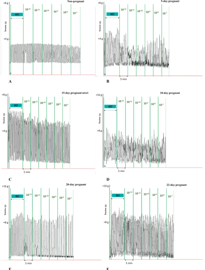
In non-pregnant and pregnant uteri, KCl elicited rhythmic contrac- tions and KISS1 94-121 modified these contractions (Fig. 1).
3.1.1. Effects of KISS1 94-121 on non-pregnant uterine and myometrial contractions
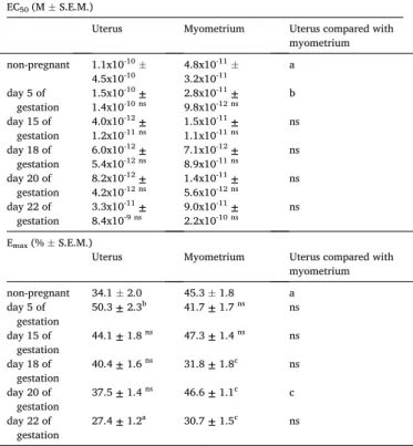
The KISS1 94-121 fragment relaxed the KCl-stimulated rhythmic contractions of non-pregnant uteri with or without endometrium in a concentration-dependent manner in the range of 10-12 to 10-7 M (Fig. 2A and 2B). In the non-pregnant and endometrium-denuded myometrium, the calculated maximal relaxation was more than 10% higher in com- parison to the intact uterus (Table 1). The EC50 value of KISS1 94-121 was shifted to the left in the denuded myometrium as compared with the uterus.
3.1.2. Effects of KISS1 94-121 on pregnant uterine and myometrial contractions
Contractility studies were performed on uterine and myometrial rings obtained from 5, 15, 18, 20 and 22-day pregnant rats. KISS1 94- 121 relaxed the pregnant uterine smooth muscles in a dose-dependent manner. The mean maximal relaxation of KISS1 94-121 gradually decreased up to the day of delivery (Fig. 2A, Table 1). The most potent uterorelaxant action was measured on gestational day 5. However, its relaxant effect was still detectable on the last day of pregnancy (day 22).
Although the EC50 values of KISS 94-124 numerically changed during the gestational period, these changes were not significant.
Contrary to the measurements with intact uteri, in the denuded myometrial samples we did not find a gradually declining tendency in the relaxing effect of KISS1 94-121 (Fig. 2B). A significant decrease in the maximum effect was found on the 18th and 22nd days of gestation, while a transient increase was found on day 20. The comparison of uterine and myometrial relaxations showed that KISS1 94-121 was more effective at the end of pregnancy when the endometrium was removed, although the difference between the Emax values was only significant on
Fig. 1. Representative records of KCl-induced uterine contractions and the cumulative effects of KISS1 94-121 on non-pregnant (A) and pregnant (B-F) uteri. Each case KISS1 94-121 elicited a relaxing effect.
day 20 of gestation. The 10-8 M concentration of KISS1 94-121 practi- cally elicits the maximum relaxing effect.
3.1.3. Kisspeptin studies in the presence of a Kiss1r antagonist
To confirm that KISS1 94-121 action is mediated via Kiss1r, contractility studies were performed in the presence of kisspeptin-234 Fig. 2. Effects of KISS1 94-121 in the range of 10-12 to 10-7 M on KCl-evoked
contractions of the non-pregnant and pregnant rat uterus (A) or endometrium-denuded myometrium (B). Cumulative concentrations of KISS1 94-121 caused smooth muscle relaxation in each case. The change in contrac- tion was calculated via the area under the curve and expressed in % ±S.E.M., n
=6 for each group.
Table 1
The EC50 and Emax values calculated from the dose-response curves demon- strated in Fig. 1A and 1B. Uterine and endometrium-denuded myometrial Emax
and EC50 were compared to each other. Also, day 5 was compared to non- pregnant values, and each investigated day of pregnancy was compared to the previous gestational day. Statistical analysis was carried out with one-way ANOVA with Tukey’s test for post-hoc analysis. ns: not significant, a: P <
0.05, b: P <0.01, c: P <0.001.
EC50 (M ±S.E.M.)
Uterus Myometrium Uterus compared with
myometrium non-pregnant 1.1x10⁻¹⁰ ±
4.5x10⁻¹⁰ 4.8x10⁻¹¹ ±
3.2x10⁻¹¹ a
day 5 of
gestation 1.5x10⁻¹⁰ ±
1.4x10⁻¹⁰ ns 2.8x10⁻¹¹ ±
9.8x10⁻¹² ns b day 15 of
gestation 4.0x10⁻¹² ±
1.2x10⁻¹¹ ns 1.5x10⁻¹¹ ±
1.1x10⁻¹¹ ns ns day 18 of
gestation 6.0x10⁻¹² ±
5.4x10⁻¹² ns 7.1x10⁻¹² ±
8.9x10⁻¹¹ ns ns day 20 of
gestation 8.2x10⁻¹² ±
4.2x10⁻¹² ns 1.4x10⁻¹¹ ±
5.6x10⁻¹² ns ns day 22 of
gestation 3.3x10⁻¹¹ ±
8.4x10⁻⁹ ns 9.0x10⁻¹¹ ±
2.2x10⁻¹⁰ ns ns Emax (% ±S.E.M.)
Uterus Myometrium Uterus compared with
myometrium non-pregnant 34.1 ±2.0 45.3 ±1.8 a day 5 of
gestation 50.3 ± 2.3b 41.7 ± 1.7 ns ns day 15 of
gestation 44.1 ± 1.8 ns 47.3 ± 1.4 ns ns day 18 of
gestation 40.4 ± 1.6 ns 31.8 ± 1.8c ns day 20 of
gestation 37.5 ± 1.4 ns 46.6 ± 1.1c c day 22 of
gestation 27.4 ± 1.2a 30.7 ± 1.5c ns
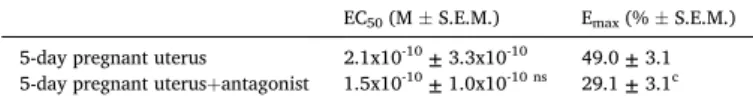
Fig. 3.Effects of KISS1 94-121 (10-12 to 10-7 M) in the presence of kisspeptin- 234 trifluoroacetate (Kissr1 antagonist) on KCl-evoked contractions on 5-day pregnant rat uterus. Kisspeptin-234 trifluoroacetate reduced the maximum ef- fect of KISS1 94-121. The change in contraction was calculated via the area under the curve and expressed in % ±S.E.M., n =6 for each group.
trifluoroacetate (Kiss1r antagonist) on 5-day pregnant uteri. The effect of KISS1 94-121 was significantly inhibited, its maximum effect was reduced by 40%. However, the reduction in the calculated maximal relaxation was significant only in the case of 5-day pregnant uterus (Fig. 3, Table 2).
3.2. RT-PCR and Western blot studies
3.2.1. Kiss1r mRNA expressions in non-pregnant and pregnant uterus and myometrium
Both the uterine (Fig. 4. full columns) and myometrial (Fig. 4. empty columns) mRNA expressions of Kiss1r were determined. Kiss1r mRNA was found both in non-pregnant and pregnant uterine tissues. The highest levels of Kiss1r mRNA were found in the non-pregnant and the 5- day pregnant uteri. Towards the end of pregnancy, Kiss1r mRNA levels were dramatically reduced, reaching the lowest value on gestational day 15. Although a slight elevation was found on gestational day 18, further alterations were not detected, and the Kiss1r mRNA level remained low till the day of delivery. The removal of the endometrium caused a pro- portional decrease in the amount of receptor mRNA on each investigated gestational day as well as in non-pregnant samples, although the change was significant only in the case of non-pregnant myometria.
3.2.2. Kiss1r protein expressions in non-pregnant and pregnant uterus and myometrium
The highest Kiss1r protein levels were measured in the non-pregnant tissues (Fig. 5). We also observed a high optical density on the 5th day of pregnancy both in uteri (full columns) and denuded myometria (empty columns). Kiss1r protein levels declined towards the end of the gesta- tional period, although the decrease was not significant after gestational Table 2
EC50 and Emax values calculated from the dose-response curves demonstrated in Fig. 2. The Emax and EC50 of KISS1 94-121 were compared to its action in the presence of the antagonist. Statistical analysis was carried out with one-way ANOVA with Tukey’s test for post-hoc analysis. ns: not significant, c: P <0.001.
EC50 (M ±S.E.M.) Emax (% ±S.E.M.) 5-day pregnant uterus 2.1x10⁻¹⁰ ± 3.3x10⁻¹⁰ 49.0 ± 3.1 5-day pregnant uterus+antagonist 1.5x10⁻¹⁰ ± 1.0x10⁻¹⁰ ns 29.1 ± 3.1c
Fig. 4. Changes in the uterine (full columns) and myometrial (empty columns) mRNA expressions of Kiss1r throughout gestation and in non-pregnant samples. The expression was significantly lower in late-pregnant rat uteri. Kiss1r expression in the uterus was compared to the myometrial expression on each gestational day as well as in non-pregnant samples (+). Also, day 5 was compared to the non-pregnant values, and each investigated day of pregnancy was compared to the previous Fig. 5.The uterine (full columns) and denuded myometrial (empty columns) protein expression of Kiss1r on different days of gestation and in non-pregnant samples along with representative blots. Kiss1r expressions show a declining trend throughout pregnancy as compared to the previous day in uteri (*) or denuded myometria (#). The results in the uterus were also compared to the myometrial expressions on each gestational day as well as in non-pregnant samples. Statistical analysis was carried out with one-way ANOVA with Tukey’s test for post-hoc analysis. np: non-pregnant uterus, np_m: non-pregnant (denuded) myometrium, ***: P <0.001, ##: P <0.01, ###: P <0.001, n =4- 7 for each group.
day 15. The removal of the endometrium caused a proportional decrease in Kiss1r levels in all myometria, but these changes were not significant.
3.3. Immunostaining of Kiss1r
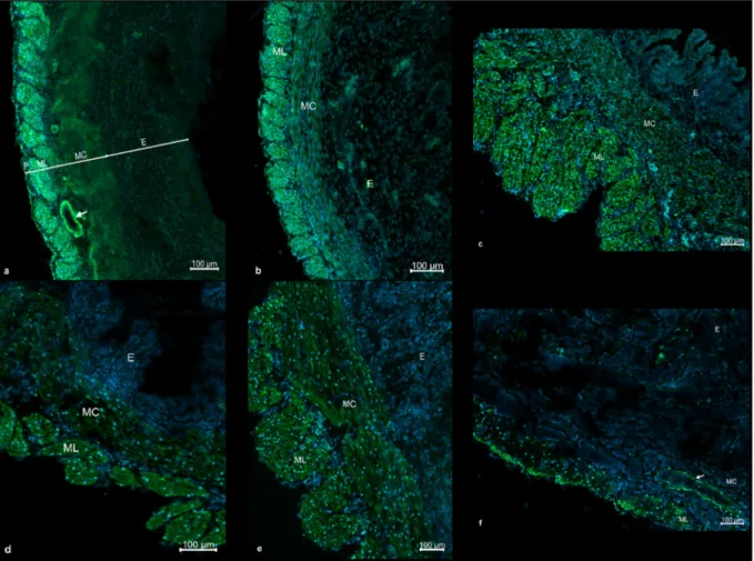
A strong immunoreactivity of Kiss1r was observed both in pregnant and non-pregnant rat uterine samples. Generally, the most intensive Kiss1r immunostaining was detected in the longitudinal layer of the myometrium, a less intense staining was found in the circular layer of the myometrium, while the weakest staining was found in the endo- metrial layer, although Kiss1r is also highly expressed in vascular smooth muscle cells. The highest intensities were found in the non- pregnant and the 5-day pregnant uteri. Gradually descending Kiss1r immunoreactivities were found towards the last day of pregnancy (day 22), both in myometrial and endometrial layers, but the reduction was especially spectacular in the endometrial layers. From day 18, immu- nostaining was primarily localized in the muscle layers of the uterus, while the endometrial activities became negligible. The staining in- tensity of 22-day pregnant uteri was marginal compared to the non- pregnant or 5-day pregnant samples (Fig. 6).
4. Discussion
Several neuropeptides have been shown to affect smooth muscle function, but only a few studies have investigated the local effects of these peptides in the uterus. Although kisspeptin and Kiss1r were identified in uterine tissue (Castellano et al., 2006; Cejudo Roman et al., 2012; Gayt´an et al., 2009; Zhang et al., 2014), no data has been available
about the role of kisspeptin in uterine contractions. Therefore, the main objective of this study was to determine the role of kisspeptin in uterine activity through the gestational period in rats.
In the contractility studies, KISS1 94-121 fragment was used to elicit kisspeptin action. This 28-amino-acid fragment has very high affinity to Kiss1r (Muir et al., 2001) with a much better stability than the other shorter fragments of kisspeptin (Kirby et al., 2010). Additionally, its water solubility is also good, therefore it was proper for the organ bath contractility measurements. We found that KISS1 94-121 inhibited the contractions both in non-pregnant and pregnant uteri in a dose-dependent manner. The uterine effect of KISS1 94-121 was also measured in the presence of a specific Kiss1r antagonist. The measure- ment was carried out on 5-day pregnant samples because the relaxing effect of KISS1 94-121 was the highest on that gestational day. We proved that the contraction inhibitory action of KISS1 94-121 can be diminished by the specific inhibitor of Kiss1r, therefore we can be sure that the relaxing effect is really mediated through Kiss1r.
The relaxing effect of KISS1 94-121 was the strongest in non- pregnant and early pregnant (day 5) uteri, while towards the end of gestation its inhibitory effect was gradually decreased, reaching the lowest value on the last day of pregnancy (day 22). A similar decrease was found in the expressions of Kiss1r mRNA and protein in non- pregnant and early pregnant samples towards the end of the gesta- tional period. The same phenomenon can be confirmed by the immu- nohistochemical results, which reveal the reduced intensity of immunostaining in all layers of the uterus as the end of the gestational period approached. Considering the fact that rat uterine tissue has a low connective tissue content (less than 6%) and the hormonal changes
Fig. 6.Representative fluorescent micrographs of cryosections from the uterus of non-pregnant rat (A), and on gestational days 5 (B), 15 (C), 18 (D), 20 (E) and 22 (F) after Kiss1r immunohistochemistry (green). DAPI was used to label the cell nuclei (blue). P: perimetrium, ML: longitudinal layer of myometrium, MC: circular
towards the end of pregnancy furtherly reduce its amount (Bienkiewicz ´ et al., 1996), it can be stated that the connective tissue mass is marginal as compared with that of smooth muscles. Since no data is available about the Kiss1r expression in any connective tissue (and its probability is very low), we suppose that the immunostaining revealed the Kiss1r expression in the smooth muscle of the non-pregnant and pregnant rat uteri. This is also supported by the high-resolution images where the stained Kiss1r are clearly located in both circular and longitudinal smooth muscle layers. Although our immunostaining study was not quantitative, the differences among the non-pregnant or 5-day pregnant and 22-day pregnant samples are spectacular. The most intense Kiss1r staining was seen on the non-pregnant tissues, whereas the lowest re- ceptor expression was found on the last gestational day, when staining was practically detected only in the myometrial layer. In general, the receptors were predominantly located in the longitudinal, then in the circular myometrial muscle layers, and the weakest expression was found in the endometrium. It was also proved by the removal of the endometrium that it did not significantly modify the expression of Kiss1r protein on any gestational day or even in non-pregnant uteri. These suggest that Kiss1r is mostly a myometrial receptor participating directly in the control of uterine smooth muscle contractions as a relaxing mechanism. This is corroborated by the findings that the highest ex- pressions of Kiss1r were found in non-pregnant and 5-day pregnant samples. The contractility of the non-pregnant uterus is usually low in rats (Ducza et al., 2009), while the 5th gestational day is considered as the time of embryo implantation in rats (Garcia et al., 2017), which requires relative quiescence in the uterine activity.
Surprisingly, KISS1 94-121 exerted a more potent uterorelaxant ef- fect in the non-pregnant and 20-day pregnant uteri without the endo- metrial layer. Additionally, significant reduction was found in mRNA level of Kiss1r after denuding the non-pregnant uteri. Furthermore, the removal ceased the gradual decreasing tendency in the relaxing effect of KISS1 94-121, suggesting the importance of the almost non-significant endometrial Kiss1r in the contractile response of the pregnant uterus (their expressions were so low that the Western blot study could not detect significant differences between the uterine and denuded myo- metrial pregnant samples on any gestational days and even in the non- pregnant samples as well). These findings might suggest that a currently unknown mechanism (maybe networking with other receptors or endometrial secretory responses) can be involved in the Kiss1r pathway modulating uterine contractility. The vital role of the endo- metrial layer in the control of uterine contractility was already demonstrated by the local production of different agents affecting smooth muscle function (Kothencz et al., 2018). A networking is already proved with kisspeptin neurons in the central nervous system (Arthur et al., 2008) and the reproductive organs (D’Occhio et al., 2020), but no data is available for the uterine smooth muscle in this respect.
In summary, our results suggest that Kiss1r mediates a relaxing effect in the non-pregnant and pregnant rat uterus. As the demand for more intensive contractions is increased towards the end of pregnancy, the expression of Kiss1r is gradually decreased, indicating its role in the maintenance of uterine quiescence during pregnancy. The modified re- sponses of denuded myometria suggest that endometrial Kiss1r might have some networking with other mechanisms for uterine relaxations.
These putative mechanisms must be revealed in further studies. The maintenance of the activation of Kiss1r during pregnancy might reduce the risk of miscarriage or premature contractions and could serve as a new drug target for obstetrics in the future.
Author statement
R´obert G´aspar: Conceptualization; Methodology; Writing - Review ´
& Editing: provided critical feedback; Supervision.
Annam´aria Schaffer: Investigation: Performed in vitro contractility
Judit Hajagos-Toth: Investigation: Performed in vitro contractility ´ studies; Formal analysis: aided in interpreting the results.
Eszter Ducza: Investigation: Performed RT-PCR and Western blot studies; Formal analysis of the acquired data; Writing - Review & Edit- ing: RT-PCR and Western blot studies.
Nikolett Bodi: ´ Investigation: Performed immunostaining; Formal analysis of immunohistochemical data; Writing - Review & Editing:
immunohistochemistry.
M´aria Bagy´anszki: Formal analysis of immunohistochemical data;
Supervision.
Zita Szalai: Investigation: Sample preparation for immunohistochemistry.
Acknowledgements
This work was supported by the Ministry of Human Capacities.
[Hungary grant 20391-3/2018/FEKUSTRAT]. We thank Cedars-Sinai Medical Center’s International Research and Innovation in Medicine Program, and the Association for Regional Cooperation in the Fields of Health, Science and Technology (RECOOP HST Association) for their support.
Appendix A. Supplementary data
Supplementary data to this article can be found online at https://doi.
org/10.1016/j.ejphar.2021.173924.
References
Anouar, A., Schirar, A., Germain, G., 1998. Relaxant effect of the calcitonin gene-related peptide (CGRP) on the nonpregnant and pregnant rat uterus. Comparison with vascular tissue. Naunyn-Schmiedeberg’s Arch. Pharmacol. 357, 446–453. https://
doi.org/10.1007/pl00005192.
Arthur, P., Taggart, M.J., Zielnik, B., Wong, S., Mitchell, B.F., 2008. Relationship between gene expression and function of uterotonic systems in the rat during gestation, uterine activation and both term and preterm labour. J. Physiol. 586, 6063–6076. https://doi.org/10.1113/jphysiol.2008.164004.
Bienkiewicz, A., Welfel, J., Ku´ ´s, E., 1996. The influence of estrogens, progesterone and their antagonists on the collagen content in the pregnant rat uterus. Horm. Metab.
Res. 28, 592–594. https://doi.org/10.1055/s-2007-979859.
Blanks, A.M., Thornton, S., 2003. The role of oxytocin in parturition. BJOG An Int. J.
Obstet. Gynaecol. 110, 46–51. https://doi.org/10.1016/S1470-0328(03)00024-7.
Castellano, J.M., Gaytan, M., Roa, J., Vigo, E., Navarro, V.M., Bellido, C., Dieguez, C., Aguilar, E., S´anchez-Criado, J.E., Pellicer, A., Pinilla, L., Gaytan, F., Tena- Sempere, M., 2006. Expression of KiSS-1 in rat ovary: putative local regulator of ovulation? Endocrinology 147, 4852–4862. https://doi.org/10.1210/en.2006-0117.
Cejudo Roman, A., Pinto, F.M., Dorta, I., Almeida, T.A., Hern´andez, M., Illanes, M., Tena- Sempere, M., Candenas, L., 2012. Analysis of the expression of neurokinin B, kisspeptin, and their cognate receptors NK3R and KISS1R in the human female genital tract. Fertil. Steril. 97, 1213–1219. https://doi.org/10.1016/j.
fertnstert.2012.02.021.
Chomczynski, P., Sacchi, N., 2006. The single-step method of RNA isolation by acid guanidinium thiocyanate-phenol-chloroform extraction: twenty-something years on.
Nat. Protoc. 1, 581–585. https://doi.org/10.1038/nprot.2006.83.
D’Occhio, M.J., Campanile, G., Baruselli, P.S., 2020. Peripheral action of kisspeptin at reproductive tissues—role in ovarian function and embryo implantation and relevance to assisted reproductive technology in livestock: a review. Biol. Reprod.
1–14. https://doi.org/10.1093/biolre/ioaa135.
De Geest, K., Thiery, M., Piron-Possuyt, G., Driessche, R. Vanden, 1985. Plasma oxytocin in human pregnancy and parturition. J. Perinat. Med. 13, 3–14. https://doi.org/
10.1515/jpme.1985.13.1.3.
De´ak, B.H., 2013. Uterus-relaxing effects of nociceptin and nocistatin: studies on preterm and term-pregnant human myometrium in vitro. Reprod. Syst. Sex. Disord. 1–5.
https://doi.org/10.4172/2161-038x.1000117, 02.
De´ak, B.H., Klukovits, A., Tekes, K., Ducza, E., Falkay, G., G´asp´ar, R., 2013. Nocistatin inhibits pregnant rat uterine contractions in vitro: roles of calcitonin gene-related peptide and calcium-dependent potassium channel. Eur. J. Pharmacol. 714, 96–104.
https://doi.org/10.1016/j.ejphar.2013.05.037.
Ducza, E., G´asp´ar, R., Mih´alyi, A., Korm´anyos, Z., Falkay, G., 2009. The roles of the α1- adrenergic receptor subtypes in rat embryonic implantation. Fertil. Steril. 91, 1224–1229. https://doi.org/10.1016/j.fertnstert.2008.01.081.
Foradori, C.D., Amstalden, M., Coolen, L.M., Singh, S.R., McManus, C.J., Handa, R.J., Goodman, R.L., Lehman, M.N., 2007. Orphanin FQ: evidence for a role in the control of the reproductive neuroendocrine system. Endocrinology 148, 4993–5001. https://
doi.org/10.1210/en.2007-0011.
Fuchs, A.R., Fuchs, F., Husslein, P., Soloff, M.S., 1984. Oxytocin receptors in the human uterus during pregnancy and parturition. Am. J. Obstet. Gynecol. 150, 734–741.
https://doi.org/10.1016/0002-9378(84)90677-X.
Garcia, J.P., Guerriero, K.A., Keen, K.L., Kenealy, B.P., Seminara, S.B., Terasawa, E., 2017. Kisspeptin and neurokinin b signaling network underlies the pubertal increase in gnrh release in female rhesus monkeys. Endocrinology 158, 3269–3280. https://
doi.org/10.1210/en.2017-00500.
Gayt´an, F., Gayt´an, M., Castellano, J.M., Romero, M., Roa, J., Aparicio, B., Garrido, N., S´anchez-Criado, J.E., Millar, R.P., Pellicer, A., Fraser, H.M., Tena-Sempere, M., 2009. KiSS-1 in the mammalian ovary: distribution of kisspeptin in human and marmoset and alterations in KiSS-1 mRNA levels in a rat model of ovulatory dysfunction. Am. J. Physiol. Endocrinol. Metab. 296, 520–531. https://doi.org/
10.1152/ajpendo.90895.2008.
Gimpl, G., Fahrenholz, F., 2001. The oxytocin receptor system: structure, function, and regulation. Physiol. Rev. 81, 629–683. https://doi.org/10.1152/
physrev.2001.81.2.629.
Han, S.K., Gottsch, M.L., Lee, K.J., Popa, S.M., Smith, J.T., Jakawich, S.K., Clifton, D.K., Steiner, R.A., Herbison, A.E., 2005. Activation of gonadotropin-releasing hormone neurons by kisspeptin as a neuroendocrine switch for the onset of puberty.
J. Neurosci. 25, 11349–11356. https://doi.org/10.1523/JNEUROSCI.3328-05.2005.
Kirby, H.R., Maguire, J.J., Colledge, W.H., Davenport, A.P., 2010. International union of basic and clinical pharmacology. LXXVII. Kisspeptin receptor nomenclature, distribution, and function. Pharmacol. Rev. 62, 565–578. https://doi.org/10.1124/
pr.110.002774.
Klukovits, A., Tekes, K., Gündüz Çinar, O., Benyhe, S., Borsodi, A., De¨ ´ak, B.H., Hajagos- T´oth, J., Verli, J., Falkay, G., G´asp´ar, R., 2010. Nociceptin inhibits uterine contractions in term-pregnant rats by signaling through multiple Pathways1. Biol.
Reprod. 83, 36–41. https://doi.org/10.1095/biolreprod.109.082222.
Kotani, M., Detheux, M., Vandenbogaerde, A., Communi, D., Vanderwinden, J.M., Le Poul, E., Br´ezillon, S., Tyldesley, R., Suarez-Huerta, N., Vandeput, F., Blanpain, C., Schiffmann, S.N., Vassart, G., Parmentier, M., 2001. The metastasis suppressor gene KiSS-1 encodes kisspeptins, the natural ligands of the orphan G protein-coupled receptor GPR54. J. Biol. Chem. 276, 34631–34636. https://doi.org/10.1074/jbc.
M104847200.
Kothencz, A., Hajagos-T´oth, J., Cs´anyi, A., G´asp´ar, R., 2018. Alpha-tocopherol succinate increases cyclooxygenase-2 activity: tissue-specific action in pregnant rat uterus in vitro. Life Sci. 192, 199–204. https://doi.org/10.1016/j.lfs.2017.11.048.
Lee, J.H., Welch, D.R., 1997. Suppression of metastasis in human breast carcinoma MDA- MB-435 cells after transfection with the metastasis suppressor gene, KiSS-1. Canc.
Res. 57, 2384–2387.
Meunier, J.-C., 1997. Nociceptin/orphanin FQ and the opioid receptor-like ORL1 receptor. Eur. J. Pharmacol. 340, 1–15. https://doi.org/10.1016/S0014-2999(97) 01411-8.
Meunier, J.C., Mollereau, C., Toll, L., Suaudeau, C., Moisand, C., Alvinerie, P., Butour, J.
L., Guillemot, J.C., Ferrara, P., Monsarrat, B., Mazarguil, H., Vassart, G., Parmentler, M., Costentin, J., 1995. Isolation and structure of the endogenous agonist of opioid receptor-like ORL 1 receptor. Nature. https://doi.org/10.1038/
377532a0.
Muir, A.I., Chamberlain, L., Elshourbagy, N.A., Michalovich, D., Moore, D.J., Calamari, A., Szekeres, P.G., Sarau, H.M., Chambers, J.K., Murdock, P., Steplewski, K., Shabon, U., Miller, J.E., Middleton, S.E., Darker, J.G., Larminie, C.G.
C., Wilson, S., Bergsma, D.J., Emson, P., Faull, R., Philpott, K.L., Harrison, D.C., 2001. AXOR12, a novel human G protein-coupled receptor, activated by the peptide KiSS-1. J. Biol. Chem. 276, 28969–28975. https://doi.org/10.1074/jbc.
M102743200.
Ohtaki, T., Shintani, Y., Honda, S., Matsumoto, H., Hori, A., Kanehashi, K., Terao, Y., Kumano, S., Takatsu, Y., Masuda, Y., Ishibashi, Y., Watanabe, T., Asada, M., Yamada, T., Suenaga, M., Kitada, C., Usuki, S., Kurokawa, T., Onda, H., Nishimura, O., Fujino, M., 2001. Metastasis suppressor gene KiSS-1 encodes peptide ligand of a G-protein-coupled receptor. Nature 411, 613–617. https://doi.org/
10.1038/35079135.
Okuda-Ashitaka, E., Ito, S., 2000. Nocistatin: a novel neuropeptide encoded by the gene for the nociceptin/orphanin FQ precursor. Peptides 21, 1101–1109. https://doi.org/
10.1016/S0196-9781(00)00247-3.
Pennefather, J.N., Reynoldson, N.A., Handberg, G.M., 1990. Inhibition of rat uterine contractions by rat and human CGRP. Peptides 11, 903–906. https://doi.org/
10.1016/0196-9781(90)90006-Q.
Pielecka-Fortuna, J., Chu, Z., Moenter, S.M., 2008. Kisspeptin acts directly and indirectly to increase gonadotropin-releasing hormone neuron activity and its effects are modulated by estradiol. Endocrinology 149, 1979–1986. https://doi.org/10.1210/
en.2007-1365.
Reinscheid, R.K., Nothacker, H.P., Bourson, A., Ardati, A., Henningsen, R.A., Bunzow, J.
R., Grandy, D.K., Langen, H., Monsma, F.J., Civelli, O., 1995. Orphanin FQ: a neuropeptide that activates an opioidlike G protein-coupled receptor. Science 270 (80), 792–794. https://doi.org/10.1126/science.270.5237.792.
Roa, J., Aguilar, E., Dieguez, C., Pinilla, L., Tena-Sempere, M., 2008. New frontiers in kisspeptin/GPR54 physiology as fundamental gatekeepers of reproductive function.
Front. Neuroendocrinol. 29, 48–69. https://doi.org/10.1016/j.yfrne.2007.07.002.
Samuelson, U.E., Dalsgaard, C.-J., Lundberg, J.M., H¨okfelt, T., 1985. Calcitonin gene- related peptide inhibits spontaneous contractions in human uterus and fallopian tube. Neurosci. Lett. 62, 225–230. https://doi.org/10.1016/0304-3940(85)90359-3.
Scott, V., Brown, C.H., 2011. Kisspeptin activation of supraoptic nucleus neurons in vivo.
Endocrinology 152, 3862–3870. https://doi.org/10.1210/en.2011-1181.
Zhang, P., Tang, M., Zhong, T., Lin, Y., Zong, T., Zhong, C., Zhang, B.P., Ren, M., Kuang, H. Bin, 2014. Expression and function of kisspeptin during mouse decidualization. PloS One 9, 1–8. https://doi.org/10.1371/journal.pone.0097647.