I
Ph.D. DISSERTATION
JÓZSEF CZIRÁKI DOCTORAL SCHOOL OF WOOD SCIENCES AND TECHNOLOGIES
SIMONYI KÁROLY FACULTY OF ENGINEERING, WOOD SCIENCES & APPLIED ARTS
UNIVERSITY OF SOPRON
by
CHARU AGARWAL
SOPRON, HUNGARY MAY 2019
II
ULTRASONIC EXTRACTION OF BIOACTIVE COMPOUNDS FROM CANNABIS SATIVA L. FOR THE GREEN REDUCTION OF
GRAPHENE OXIDE ON CELLULOSE FIBRES
A DISSERTATION
submitted in partial fulfilment of the requirements for the award of the degree of
DOCTOR OF PHILOSOPHY
by
CHARU AGARWAL
under the supervision of PROF. DR. LEVENTE CSÓKA
JÓZSEF CZIRÁKI DOCTORAL SCHOOL OF WOOD SCIENCES AND TECHNOLOGIES
SIMONYI KÁROLY FACULTY OF ENGINEERING, WOOD SCIENCES &
APPLIED ARTS UNIVERSITY OF SOPRON
SOPRON, HUNGARY MAY 2019
III Ultrasonic Extraction of Bioactive Compounds from Cannabis sativa L. for the
Green Reduction of Graphene Oxide on Cellulose Fibres Értekezés doktori (Ph.D.) fokozat elnyerése érdekében
a Soproni Egyetem Cziráki József Faanyagtudomány és Technológiák Doktori Iskolája Rosttechnikai és nanotechnológiai tudományok programja
Írta:
Charu Agarwal
Készült a Soproni Egyetem Cziráki József Faanyagtudomány és Technológiák Doktori Iskola Rosttechnikai és nanotechnológiai tudományok programja keretében Témavezető: Prof. Dr. Csóka Levente DSc.
Elfogadásra javaslom (igen / nem)
(aláírás) A jelölt a doktori szigorlaton …...93... % -ot ért el,
Sopron, 2018.06.07.
………...
a Szigorlati Bizottság elnöke Az értekezést bírálóként elfogadásra javaslom (igen /nem)
Első bíráló (Dr. …... …...) igen /nem
(aláírás) Második bíráló (Dr. …... …...) igen /nem
(aláírás) (Esetleg harmadik bíráló (Dr. …... …...) igen /nem
(aláírás) A jelölt az értekezés nyilvános vitáján…...% - ot ért el
Sopron,
………..
a Bírálóbizottság elnöke A doktori (PhD) oklevél minősítése…...
………..
Az EDHT elnöke
IV
UNIVERSITY OF SOPRON
DECLARATIONS
I hereby certify that the work presented in this dissertation titled “Ultrasonic Extraction of Bioactive Compounds from Cannabis sativa L. for the Green Reduction of Graphene Oxide on Cellulose Fibres" in partial fulfilment of the requirements for the award of the degree of Doctor of Philosophy in Material Science & Technology and submitted in the Simonyi Károly Faculty of Engineering, Wood Sciences & Applied Arts of the University of Sopron, is an authentic record of my own work carried out at Institute of Wood Based Products and Technologies during a period from September 2016 to May 2019 under the guidance and supervision of Prof. Dr. Levente Csóka (Head of József Cziráki Doctoral School of Wood Sciences and Technologies, University of Sopron).
The matter presented in this dissertation has not been submitted by me for the award of any other degree of this or any other Institute.
May, 2019 Charu Agarwal
This is to certify that the above statement made by the candidate is correct to the best of my knowledge.
May, 2019 Prof. Dr. Levente Csóka
V
This work is dedicated to my parents and
sister for always being there...
VI
©UNIVERSITY OF SOPRON, SOPRON-2019 ALL RIGHTS RESERVED
VII ACKNOWLEDGEMENTS
A dissertation is not only an accomplishment but also one of the achievements in life. The satisfaction and satiety of overcoming the hurdles faced during the research work cannot be attained without expressing sincere gratitude to all those who guided and supported me during the entire course of the PhD work.
I take this opportunity to bequeath my humble salutation and deep gratitude towards my supervisor, Prof. Dr. Levente Csóka, under whose invaluable guidance I have carried out this PhD study. I was very fortunate to have worked under his excellent guidance and thank him from the depth of my heart for having led me up the right path. With an eye of perfection, strong sense of dignity and integrity, he has set an example.
I am extremely grateful to the Tempus Public Foundation for providing financial assistance under the Stipendium Hungaricum scholarship programme. I also acknowledge the financial support received within framework of the programme “EFOP-3.6.1-16-2016- 00018 – Improving the role of research+development+innovation in the higher education through institutional developments assisting intelligent specialization in Sopron and Szombathely.”
I convey my sincere thanks to all the faculty members of University of Sopron, especially Dr. Tamás Hofmann for their kind support cooperation and extending all possible assistance to carry out this work. I also wish to thank Dr. Katalin Halász, Dr. Ádám Makk, and Dr. Éva Papp and administrative staff for their kindness and cooperation during the course of this study. I cannot forget to express sincere appreciation to all my lab colleagues and peers- Annamária, Boris, Chenar, Fatima, Shadabeh, Worakan, Yanin for their support and motivation towards this work.
I express my gratitude to RRCAT, India for extending the facilities for XRD and XPS measurements at the synchrotron radiation source. I am also highly obliged to all the authors whose work has provided a significant contribution in this work.
Finally, I also thank Lord Almighty and my family for preparing me for everything that I desire to be through the good times and bad. Without their moral support, this work would not have seen the dawn of the day.
Charu Agarwal
VIII TABLE OF CONTENTS
DECLARATIONS ... IV TABLE OF CONTENTS ... VIII LIST OF FIGURES ... XII LIST OF TABLES ... XV LIST OF ABBREVIATIONS ... XVI
ABSTRACT ... 18
CHAPTER I- INTRODUCTION ... 20
1.1. Chapter synopsis ... 21
1.2. Problem statement ... 21
1.3. Cannabis sativa L. & its extract ... 22
1.3.1. Bioactive compounds in Cannabis sativa L. ... 22
1.3.1.1. Terpenes in Cannabis ... 23
1.3.1.2. Flavonoids in Cannabis ... 24
1.3.2. Phytocannabinoids ... 24
1.3.2.1. Biosynthesis and pharmacology of cannabinoids ... 24
1.3.2.2. Chemotypes in Cannabis ... 26
1.3.2.3. Therapeutic potential of cannabinoids ... 26
1.3.3. Extraction of phytoconstituents... 27
1.3.3.1. Using conventional methods ... 27
1.3.3.2. Using modern methods ... 28
1.3.4. Plant extract as a green reducing agent ... 30
1.4. Cellulose as a substrate for functionalization of materials ... 31
1.4.1. Cellulose background ... 31
1.4.1.1. Structure ... 31
1.4.1.2. Polymorphs ... 33
1.4.1.3. Sources ... 33
1.4.1.4. Properties... 34
1.4.2. Cellulose chemistry ... 34
1.4.2.1. Functionalization via physical adsorption ... 35
1.4.2.2. Functionalization via chemical bonding ... 35
1.4.3. Functionalization of cellulose ... 36
1.4.4. Functionalization of cellulose with carbonaceous materials ... 38
IX
1.4.4.1. Carbon nanotubes ... 38
1.4.4.2. Graphene/reduced-GO ... 39
1.5. Green reduction of GO ... 43
1.6. Research rationale & objectives ... 44
1.7. Dissertation outline ... 45
1.8. Summary ... 46
References... 47
CHAPTER II- MATERIALS & METHODS ... 60
2.1. Chapter synopsis ... 61
2.2. Materials ... 61
2.2.1. Chemicals ... 61
2.2.2. Plant material ... 62
2.2.3. Instrumentation ... 62
2.2.3.1. Probe sonicator ... 62
2.2.3.2. Spectrophotometer ... 62
2.3. Methods ... 63
2.3.1. Ultrasonic extraction... 63
2.3.2. Control extraction without ultrasound treatment ... 64
2.3.3. Determination of total phenolic content (TPC) ... 64
2.3.4. Determination of total flavonoids (TF) ... 64
2.3.5. Determination of ferric reducing ability of plasma (FRAP) antioxidant capacity 64 2.3.6. Determination of extraction yield ... 65
2.3.7. Experimental design of & statistical analysis ... 65
2.3.8. Synthesis of GO ... 66
2.3.9. Green reduction of GO and preparation of composites ... 67
2.3.10. Handsheet-making ... 68
2.4. Characterization ... 69
2.4.1. High pressure liquid chromatography/mass spectrometry (HPLC-DAD-MS/MS) 69 2.4.2. Gas chromatography/mass spectrometry (GC-MS) ... 71
2.4.3. Fourier transform infrared spectroscopy (FTIR) ... 72
2.4.4. Scanning electron microscopy (SEM) ... 72
2.4.5. Synchrotron X-ray diffraction (XRD) ... 73
2.4.6. Synchrotron X-ray photoelectron spectroscopy (XPS) ... 74
2.5. Electrical measurements ... 75
2.6. Summary ... 75
References... 76
X CHAPTER III- ULTRASONIC EXTRACTION OF BIOACTIVE COMPOUNDS FROM
CANNABIS SATIVA L. OPTIMIZED BY RESPONSE SURFACE METHODOLOGY ... 79
3.1. Chapter synopsis ... 80
3.2. Extraction process and factor selection ... 80
3.3. Modelling and regression analysis ... 81
3.4. Influence of ultrasonic parameters (design factors) on the responses ... 84
3.4.1. Influence of design factors on TPC ... 84
3.4.2. Influence of design factors on TF ... 86
3.4.3. Influence of design factors on FRAP ... 88
3.4.4. Influence of design factors on yield... 90
3.5. Optimization of extraction conditions ... 92
3.6. Ultrasonic vs control extraction... 92
3.7. Summary and inferences ... 94
References... 94
CHAPTER IV- IDENTIFICATION OF CANNABINOIDS IN CANNABIS SATIVA L. & THEIR ENTOURAGE EFFECTS WITH OTHER BIOACTIVE CONSTITUENTS ... 98
4.1. Chapter synopsis ... 99
4.2. Synthesis, pharmacological and therapeutic effects of cannabinoids ... 99
4.3. Administration of cannabinoids ... 100
4.4. Entourage effects of cannabinoids ... 100
4.5. Identification of compounds in the Cannabis extract... 101
4.5.1. Identification of cannabinoids using HPLC-DAD-MS/MS ... 101
4.5.2. Identification of other bioactive compounds using GC-MS ... 108
4.6. Adverse events and safety concerns ... 109
4.7. Summary and inferences ... 109
References... 110
CHAPTER V- IN SITU GREEN SYNTHESIS AND FUNCTIONALIZATION OF REDUCED GRAPHENE OXIDE ON CELLULOSE FIBERS BY CANNABIS SATIVA L. EXTRACT... 113
5.1. Chapter synopsis ... 114
5.2. Morphology and structure analysis ... 114
5.2.1. SEM analysis ... 114
5.2.2. FTIR analysis ... 115
5.3. X-ray diffraction and photoelectron spectroscopy analysis ... 116
5.3.1. XRD analysis ... 116
5.3.2. XPS analysis ... 118
5.4. Electrical performance study ... 119
XI
5.5. Summary and inferences ... 122
References... 123
CHAPTER VI- CONCLUSIONS & FUTURE OUTLOOK... 127
6.1. Chapter synopsis ... 128
6.2. Main conclusions and achievements of the research ... 128
6.3. Scope for further research ... 129
LIST OF PUBLICATIONS ... 130
XII LIST OF FIGURES
Figure no. Figure caption Page
no.
Figure 1.1 Biosynthetic pathways for bioactive compounds in Cannabis 25 Figure 1.2 Therapeutic effects of cannabinoids for some diseases 27 Figure 1.3 Conventional and modern methods of extraction for
phytoconstituents
28 Figure 1.4 Formation and collapse of cavitation bubble 29 Figure 1.5 Diffusion in a plant tissue surrounded by a solvent layer during
ultrasonic extraction
30 Figure 1.6 Plants used for the green reduction of materials such as
nanoparticles
31 Figure 1.7 (a) Two glucose rings in cellulose chain connected by β(1-4)
glycosidic linkage; dotted line indicates the intra-chain hydrogen bonding, (b) amorphous and crystalline regions in a cellulose chain, (c) hydrogen bonding in cellulose, the thin dotted lines indicate the intrachain bonding, while the thick dotted line represents the interchain bonding
32
Figure 1.8 (a) Directional asymmetry of cellulose, (b) chemical reactions for modification of cellulose
35 Figure 1.9 Common functionalization chemistries of cellulose surfaces:
(clockwise from top-right) sulphuric acid treatment provides sulphate esters, carboxylic acid halides create ester linkages, acid anhydrides create ester linkages, epoxides create ether linkages, isocyanates create urethane linkages, TEMPO mediated hypochlorite oxidation creates carboxylic acids, halogenated acetic acids create carboxymethyl surfaces, and chlorosilanes create an oligomeric silylated layer
36
Figure 1.10 Applications of cellulose fibres modified with nanomaterials 37 Figure 1.11 (a) Schematic depicting transfer of graphene on to paper, (b)
Graphene paper strip in action as a gas sensor may glow an LED bulb
40
Figure 1.12 (a) Increase in resistance change with pressure for graphene- based pressure sensor for different number of layers of tissue paper, (b) Sensor application for detection of wrist pulse, (c) Pulse waveform of the sensor
41
Figure 1.13 a) SEM and b) TEM images of a cellulose fibre in a GCP membrane showing GNSs anchored on the fibre surface, c) cellulose-GNSs binding in GCP
42
Figure 1.14 (a) GNS/cellulose composite paper (black) against pure cellulose paper (white), (b) the bent composite paper, showing flexibility of
43
XIII the paper and (c) composite paper adhered to conducting copper
foil to make a flexible supercapacitor
Figure 1.15 Mechanism for the reduction of GO with plant extracts 44 Figure 2.1 Inflorescence of fibre-type Cannabis 62 Figure 2.2 Photographs of (a) Probe-type ultrasonicator used for the
extraction of phytoconstituents from Cannabis, (b) UV/vis spectrophotometer for the measurement of absorbance
63
Figure 2.3 Schematic illustration for the fabrication of RGO/cellulose composites
68 Figure 2.4 Photograph of laboratory sheet former with vacuum press-drying 68 Figure 2.5 RGO/cellulose composites with various loadings of RGO- (a) 0
m/m %, (b) 0.1 m/m %, (c) 1 m/m %, (d) 2 m/m %, (e) 5 m/m % and (f) 10 m/m %
69
Figure 2.6 Photograph of HPLC-DAD-MS/MS 70
Figure 2.7 Photograph of GC-MS 71
Figure 2.8 Photographs of (a) FTIR and (b) ATR probe 72
Figure 2.9 Photograph of SEM 73
Figure 2.10 Schematic layout of XRD beamline (BL-12) at Indus 2 synchrotron, RRCAT (India)
73 Figure 2.11 Schematic layout of XPS beamline (BL-14) at Indus 2
synchrotron, RRCAT (India)
74 Figure 2.12 Photographs of (a) Keithley resistivity text fixture and (b)
Keithley electrometer
75 Figure 3.1 Response surface plots depicting the influence of design factors
on TPC
85 Figure 3.2 Fig. 21 Response surface plots depicting the influence of design
factors on TF
87 Figure 3.3 Response surface plots depicting the influence of design factors
on FRAP
89 Figure 3.4 Response surface plots depicting the influence of design factors
on extraction yield
91 Figure 3.5 Comparison between ultrasonic (green) and control (blue)
extractions for the responses (The error bars indicate percentage error)
92
Figure 3.6 Qualitative HPLC chromatograms for ultrasonic (black represents optimal conditions & red represents central values) and control (blue) extracts of Cannabis
93
Figure 4.1 HPLC chromatogram showing various cannabinoids in the Cannabis extract
103
Figure 4.2 Mass spectrum of CBDVA 103
XIV
Figure 4.3 Mass spectrum of CBD 104
Figure 4.4 Mass spectrum of CBDA 104
Figure 4.5 Mass spectrum of CBGA 105
Figure 4.6 Mass spectrum of THCA-A 105
Figure 4.7 Mass spectrum of THC 106
Figure 4.8 Mass spectrum of THCVA 106
Figure 4.9 Mass spectrum of CBNA 107
Figure 4.10 Mass spectrum of CBLA 107
Figure 4.11 Mass spectrum of CBCA 108
Figure 4.12 GC-MS chromatogram of non-cannabinoid bioactive compounds in the Cannabis extract
109 Figure 5.1 SEM images of RGO/cellulose composites at various RGO
loadings: (a) 0 m/m %, (b) 0.1 m/m %, (c) 1 m/m %, (d) 2 m/m %, (e) 5 m/m % and (f) 10 m/m % (the arrows indicate the dispersion of RGO on the cellulose fibre surface)
115
Figure 5.2 FTIR spectra of (a) GO & RGO powders and (b) RGO/cellulose composites with various RGO loadings
116 Figure 5.3 XRD spectra of (a) GO & RGO powders and (b) RGO/cellulose
composites with various RGO loadings
117 Figure 5.4 XPS spectra of GO and RGO powders (a, b) C 1s region and (c,
d) O 1s region
119 Figure 5.5 Surface resistivity of RGO/cellulose composites with increasing
RGO loading from 0-10 m/m % at 40 V
120 Figure 5.6 Surface charging capacity of RGO/cellulose composites with
increasing RGO loading from 0.1-10 m/m % on log scale at 40 V 122
XV LIST OF TABLES
Table no. Table caption Page no.
Table 1.1 Constituents of Cannabis 23
Table 1.2 Classes of cannabinoids with their pharmacological attributes 25
Table 2.1 Design space factors and levels 66
Table 2.2 Different weight fractions of RGO functionalized on cellulose fibres
67 Table 3.1 Central composite design factors in coded and actual forms
along with the investigated responses of TPC, TF, FRAP and yield for various experimental run
81
Table 3.2 Regression coefficient estimates for second order polynomial model for TPC, TF, FRAP and yield along with their ANOVA parameter
83
Table 4.1 Major cannabinoids identified in the Cannabis extract using HPLC-DAD-MS/MS
102 Table 4.2 Bioactive compounds identified in the Cannabis extract using
GC-MS
108 Table 5.1 Surface charging capacity of RGO/cellulose composites with
different RGO loadings at 40 V
121
XVI LIST OF ABBREVIATIONS
11-OH-THC 11-hydroxy-Δ9-tetrahydrocannabinol
11-OH-THCA 11-hydroxy-Δ9-tetrahydrocannabinolic acid A 5-HT1A serotonin 1A receptor
ADXRD angle dispersive X-ray diffraction
AgNWs silver nanowires
ANOVA analysis of variance ATR attenuated total reflection
CB1 cannabinoid-one receptor
CB2 cannabinoid-two receptor
CBC cannabichromene
CBCA cannabichromenic acid
CBD cannabinoid
CBDA cannabidiolic acid
CBDV cannabidivarin
CBDVA cannabidivarinic acid
CBG cannabigerol
CBGA cannabigerolic acid CBLA cannabicyclolic acid
CBN cannabinol
CBNA cannabinolic acid
CBND cannabinodiol
CBNDA cannabinodiolic acid CNTs carbon nanotubes
DMAPP dimethylallyl diphosphate
FPP farnesyl diphosphate
FTIR Fourier transform infrared spectroscopy GC-MS gas chromatography/mass spectrometry
GPP geranyl diphosphate
HPLC-DAD-MS/MS high pressure liquid chromatography/mass spectrometry
17 IDA information dependent analysis
IPP isopentenyl diphosphate LFIAs lateral flow immunoassays MEP methylerythritol phosphate
MVA mevalonate
PMMA poly- (methyl methacrylate)
POC point-of-care
PPAR-γ peroxisome proliferator-activated receptor gamma SEM scanning electron microscopy
THC tetrahydrocannabinol
THCA-A tetrahydrocannabinolic acid A THCV tetrahydrocannabivarin THCVA tetrahydrocannabivarinic acid TPTZ 2,4,6-tri(2-pyridyl)-1,3,5-triazine TRPA1 transient receptor potential ankyrin-1
TRPV1 transient receptor potential cation channel vanilloid sub-family receptor 1
WHO World Health Organization XPS X-ray photoelectron spectroscopy XRD X-ray diffraction
18 ABSTRACT
Ultrasonication was used to extract bioactive compounds from Cannabis sativa L.
such as polyphenols, flavonoids and cannabinoids. The influence of three independent factors (time, input power and methanol concentration) was evaluated on the extraction of total phenols, flavonoids, ferric reducing ability of plasma (FRAP) assay and the overall yield. A face-centred central composite design was used for statistical modelling of the response data, followed by regression and analysis of variance in order to determine the significance of the model and factors. Both the solvent composition and the time significantly affected the extraction while the sonication power had no significant impact on the responses. The response predictions obtained at optimum extraction conditions of 15 min time, 130 W power and 80% methanol were 314.822 mg GAE/g DW of TPC, 28.173 mg QE/g DW of TF, 18.79 mM AAE/g DW of FRAP and 10.86% of yield. A good correlation was observed between the predicted and experimental values of the responses, which validated the mathematical model. On comparing the ultrasonic process with the control extraction, noticeably higher values were obtained for each of the responses.
Additionally, ultrasound treatment considerably improved the extraction of cannabinoids such as THC, CBD, CBGA, CBDA, THCVA, CBLA, CBNA, CBCA, etc.
The last decade has seen an enormous rise in the use of green reducing agents such as plant extracts for the chemical synthesis of several of materials in view of the limitations of the conventional reducing agents such as their toxicity and instability. This study reports the green reduction and simultaneous functionalization of graphene oxide on cellulose fibres using the aqueous extract from the inflorescences of Cannabis sativa L. The graphene oxide, synthesized using the modified Hummer’s method, was reduced in situ on the cellulose matrix in presence of the extract at elevated temperatures without external stabilizers in order to functionalize the fibres with reduced-graphene oxide (RGO). The cellulose fibres not only acted as a flexible, biodegradable and cost-effective matrix for the anchorage of RGO but also supported its in situ reduction on the fibre surface. Different weight fractions of RGO from 0.1 to 10 m/m % were used to fabricate RGO/cellulose composites by paper-making technique, which were characterized using Fourier transform infrared spectroscopy, scanning electron microscopy, X-ray diffraction and X-ray photoelectron spectroscopy techniques. The RGO sheets uniformly covered the surface of cellulose fibres and dispersed well within the fibre matrix. The surface resistivity at 40 V
19 decreased with increasing RGO content from 1.81 x 10¹¹ Ω for 0.1 m/m % RGO to 0.15 x 10¹¹ Ω for 10 m/m % RGO loading. The presence of air voids between the fibres hindered the physical contact between the RGO layers, thereby preventing the formation of an effective conductive network and significantly affecting the performance of the composites. Likewise, the surface charging capacity of the composites at 40 V dropped from 1.21 x 10⁻³ ΔmAh for 0.1 m/m % RGO to 0.05 x 10⁻³ΔmAh for 10 m/m % RGO content indicating a rise in conductivity with RGO loading. These composites show immense potential as sustainable materials for portable energy storage devices such as capacitors.
20
CHAPTER I-
INTRODUCTION
21 1.1. Chapter synopsis
This chapter presents an in-depth review of literature on the phytoconstituents in Cannabis sativa L., which include the cannabinoids as well as the other non-cannabinoid bioactive compounds such as the terpenes and flavonoids. Extraction of the phytoconstituents using both the conventional and modern techniques with emphasis on ultrasonication has also been discussed. Further, the potential of the phytoextracts as an eco-friendly reducing agent for the synthesis of materials has been reviewed. This is followed by a discussion on cellulose chemistry and the functionalization of cellulose with special focus on carbon materials such as carbon nanotubes and graphene along with the functionalization strategies. Finally, the chapter throws light on the reduction of graphene oxide (GO) using phytoextracts. The problem statement and the research objectives have been formulated based on the review. An outline for the dissertation has been presented in the end.
1.2. Problem statement
Nanomaterials have become an inseparable part in the present times, playing a dominant role in various sectors such as biomedicine, catalysis and biosensing [1-3]. The two major pathways for the synthesis of nanomaterials are the bottom-up and top down approaches [4]. Both approaches rely on physico-chemical methods, which employ chemical agents for the reduction and stabilization of the nanomaterials. Most of these chemical agents are toxic or corrosive in nature and lead to hazardous by-products [4-6].
This is especially undesirable in case of biomedical healthcare and diagnostic applications.
Moreover, they require an energy-intensive reaction environment and are expensive [4, 7].
Thus, there is an intense need for eco-friendly agents for the synthesis of nanomaterials having non-toxic reaction products.
In view of this, a number of eco-friendly agents have garnered the attention of the scientific community, which include entities of biological origin such as microorganisms, enzymes, plant extracts, etc. [8-10]. Of these, the plant extracts are particularly advantageous due to their wide availability and cost-effectiveness. Plant extracts have proven potential to act not only as natural reducing agents for the synthesis of nanomaterials but also as capping agents for the stabilization of the synthesized nanomaterials [11-13].
They need a much less energy-intensive environment and can react in ambient conditions of temperature and pressure [4]. Further, they can be used to synthesize nanomaterials in
22 large quantities free of chemical contaminants and hence, hold potential for industrial scale- up [4].
Having stated that, it is of utmost significance to select the appropriate extraction technique for the preparation of plant extracts. The conventional techniques used for extraction are laborious, time-consuming, energy-intensive and need large amounts of solvents [14]. These drawbacks can be overcome by employing the modern extraction techniques such as those assisted by ultrasound and microwaves [15, 16]. For instance, ultrasound allows the target compounds to dissolve in the solvent by disrupting the cell wall, thus enhancing the extraction yield in much lesser time [17, 18].
Another aspect to be considered is pertaining to the anchoring of the nanomaterials onto a matrix in order to prevent their dispersal and aid their recovery from the environment. Paper, polymer, glass, wood, ceramic and metal are some the commonly used matrices (or substrates) for embedding of the nanomaterials. Paper is a principally attractive choice for a substrate owing to its innumerable advantages such as biodegradability, biocompatibility, porosity, flexibility and hydrophilicity [1, 19].
1.3. Cannabis sativa L. & its extract
1.3.1. Bioactive compounds in Cannabis sativa L.
Plants have since long been exploited for their medicinal value apart from their nutritional value. Herodotus referred to the use of Leonurus cardiaca by those living to the north of the Danube river in as early as 5th century BC [14]. A number of bioactive compounds are responsible for imparting different medicinal properties to the plants. These compounds are synthesized as secondary metabolites, which aid in the overall survival in plants by allowing their interaction with the surrounding environment. In other words, the bioactive compounds exert pharmacological or toxicological effects in humans and animals. They have been broadly classified as: a) terpenes and terpenoids, b) alkaloids and c) phenolic compounds [14]. Of these, the phenolic compounds constitute one of the most widely distributed groups of secondary metabolites in the plant kingdom and are well known for their antioxidant behaviour. The natural phenolic antioxidants present in fruits such as guavas, grapes and strawberries have been greatly explored for their defensive effects and therapeutic potential [20-22]. Flavonoids represent another class of low molecular weight phenolic compounds, which are also very effective antioxidants and less toxic than their synthetic counterparts such as BHT and BHA [23].
23 Cannabis sativa L. (hereinafter: Cannabis) has been around since ages and cultivated as an annual crop plant for its commercial value. It belongs to the family of Cannabinaceae and has long been used in traditional Asian medicine, particularly in India [24]. Reportedly, the first successful attempt to extract active compounds from the flowers and leaves of Cannabis was made by Schlesinger in 1840 [25]. The phenolic compounds are comprised of phenolic acids such benzoic and hydroxycinnamic acids, flavonoids such as flavones and flavonols, lignans and stilbenes [26]. About 34 phenols and 23 flavonoids have been identified in Cannabis [27]. Table 1.1 lists the different constituents commonly found in Cannabis as taken from literature [27].
Table 1.1 Constituents of Cannabis
Sr. No. Cannabis constituents Number
1. Terpenoids 140
2. Hydrocarbons 50
3. Nitrogen compounds >70
4. Carbohydrates 34
5. Flavonoids 23
6. Fatty acids 33
7. Phenols 34
8. Alcohols 7
9. Aldehydes 12
10. Ketones 13
11. Acids 21
12. Esters and lactones - 1.3.1.1. Terpenes in Cannabis
Terpenes are basic hydrocarbons as against terpenoids, which contain functional groups of a number of chemical elements; however, these terms are often used interchangeably in the literature. Terpenes form the largest group of phytochemicals;
Cannabis contains up to 200 different terpenes. The most common primary terpenes found in Cannabis are β-caryophyllene, myrcene, α-pinene, humulene, linalool, limonene, terpinolene, terpineol, ocimene, valencene, and geraniol. Some of the common secondary terpenes in Cannabis include α-bisabolol, nerolidol, caryophyllene oxide, phytol, borneol, δ-3-carene, terpinene, camphene, sabinene, cineole (eucalyptol), phellandrene, guaiol,
24 isoborneol, cedrene, geranyl acetate, fenchol, camphor, menthol, isopulegol, cymene, citral, and citronellol [27, 28]. Preclinical studies using animal models have associated terpenes with a plethora of medicinal benefits including analgesia, anti-inflammatory, antidepressant, anxiolytic, anti-insomnia, cancer chemoprevention, antibacterial, antiviral, antifungal, anti-parasitic, and anti-hyperglycemic effects [28].
1.3.1.2. Flavonoids in Cannabis
Cannabis contains phenylpropanoid phenolic compounds, one of which is called the flavonoids. The flavonoids in plants act as antioxidants protecting them against the oxidative stress. They include apigenin, luteolin, quercetin, kaempferol, cannflavin A, cannflavin B (unique to Cannabis), β-sitosterol, vitexin, isovitexin, kaempferol, and orientin. The flavonoids have been associated with neuroprotective, anti-inflammatory and anti-cancer effects. For instance, apigenin exerts anxiolytic effects to inhibit TNF-α, which is involved in many inflammatory conditions. Cannflavin A and B also have potent anti- inflammatory effects, with cannflavin A shown to inhibit PGE-2 30 times more potently than aspirin. β-sitosterol was shown potential to reduce topical inflammation by 65% and chronic edema by 41% in skin models [28].
1.3.2. Phytocannabinoids
1.3.2.1. Biosynthesis and pharmacology of cannabinoids
Besides the common phenolic compounds, Cannabis has been widely explored for phytocannabinoids (or simply cannabinoids) which are unique to the plant. Cannabinoids represent a group of C21 terpenophenolic compounds and are usually concentrated in the female inflorescence, the plant being dioecious. Among the 483 compounds unique to Cannabis, about 66 have been identified as cannabinoids, the most important of them being
∆9-THC with psychoactive properties [27].
The cannabinoids are biosynthesized in acidic form as cannabinoid acids in the female inflorescences of Cannabis, which get decarboxylated into their respective neutral cannabinoids by heat, UV exposure and prolonged storage (Figure 1.1). The predominant cannabinoid acids are THCA, CBDA and CBNA followed by CBGA, CBCA, CBNDA, THCVA and CBDVA, which are converted to their free forms viz. THC, CBD and CBN, CBG, CBC, CBND, THCV and CBDV, respectively [28, 29]. The numerous health benefits linked to cannabinoids, which are an outcome of the interactions of the cannabinoids with the endocannabinoid system in humans, are already well documented
25 [29]. THC, the primary psychoactive component of Cannabis, works as a weak partial agonist on CB1 and CB2, with preferential binding to CB1, and is also an agonist on the PPAR-γ and TRPA1 receptors [30]. It is well-known for homeostatically regulating a myriad of physiological functions [27, 31]. On the contrary, CBD has much lower affinity for CB1 and CB2 receptors, with protean pharmacological effects on various other receptor systems including 5-HT1A, TRPV1, adenosine A2A. It acts as a non-competitive receptor antagonist that is responsible for its neutralizing actions on the side effects of THC [28].
Table 1.2 shows the various cannabinoid classes, subtypes and their pharmacological properties.
Figure 1.1 Biosynthetic pathways for bioactive compounds in Cannabis [29] – Published by the Frontiers publisher
Table 1.2 Classes of cannabinoids with their pharmacological attributes Sr.
No.
Cannabinoid class
Number of subtypes
Pharmacological attributes
1. CBG 6 Antibiotic, antifungal, anti-inflammatory, analgesic
2. CBC 5 Antibiotic, antifungal, anti-inflammatory, analgesic
26 3. CBD 7 Anxiolytic, antipsychotic, analgesic, anti-
inflammatory, antioxidant, antispasmodic 4. Δ9-THC 9 Euphoriant, analgesic, anti-inflammatory,
antioxidant, antiemetic
5. Δ8-THC 2 Similar to THC (less potent)
6. CBL 3 -
7. CBE 5 -
8. CBN and CBND
6+2 Sedative, antibiotic, anticonvulsant, anti- inflammatory, appetite-stimulant
9. CBT 9 -
10. Miscellaneous 11 - 1.3.2.2. Chemotypes in Cannabis
Apart from its utilization for medical purposes, Cannabis is the most extensively consumed drug of abuse in Europe. Based on the total THC content, it is categorized into three major chemotypes. Chemotype I or drug-type is predominant in THC with THC/CBD
>> 1.0, chemotype II or intermediate-type is mixed with THC/CBD ≈ 1.0 and chemotype III or fibre-type is predominant in CBD with THC/CBD << 1.0. Due to the presence of psychoactive compound, law governs the plant cultivation in many countries. The fibre- type Cannabis, commonly known as hemp, is native to France, Hungary and Russia and has low THC content with presence of other non-psychoactive cannabinoids such as CBD or CBG. The maximum allowable limit for THC for hemp cultivation in Europe is 0.3% of the dry weight [32].
1.3.2.3. Therapeutic potential of cannabinoids
Cannabinoids have shown immense therapeutic potential (Figure 1.2). The most common ones include palliative effects such as treatment of nausea and emesis in patients undergoing chemotherapy, stimulation of appetite in HIV-positive patients and spasticity associated to multiple sclerosis in adults [29]. Cannabinoids have shown effects in treating ailments like epilepsy, schizophrenia, diseases related to the gut and ocular pathologies such as glaucoma. They exhibit analgesic effects through non-receptor mechanism and cannabinoid receptors, which has been used to treat muscle pain. The anti-inflammatory and anti-oxidant properties of cannabinoids have been exploited for neuro-degenerative disorders like Parkinson’s disease, Huntington’s disease, Alzheimer’s disease, where they
27 were found to slow down the process of neurodegeneration [33]. Further, they have also shown promise as antitumor or anticancer agents [34, 35].
It is worth noting that the therapeutic potential and the observed biological effects of Cannabis vary among the different chemotypes (as discussed in section 1.3.2.2) as well as within the same chemotype depending on the chemical profile and the relative proportions of its constituents.
Figure 1.2 Therapeutic effects of cannabinoids for some diseases [Reproduced with permission from [36] ©2018 Elsevier publisher]
1.3.3. Extraction of phytoconstituents
Extraction is simply the separation of biologically active constituents of the plant by solvents using standard procedures with an aim to separate the soluble plant metabolites, leaving behind the insoluble cellular marc [15]. It can be achieved using conventional or modern techniques.
1.3.3.1. Using conventional methods
The isolation of these bioactive compounds and their separation from the plant matrix is made complicated due to their sensitivity to various process parameters such as temperature as well as the co-extraction of other undesirable components [23].
28 Conventional methods for extraction such as Soxhlet extraction, maceration and hydro- distillation have the major drawbacks of longer time needed for extraction and large amounts of solvent needed.
1.3.3.2. Using modern methods
In order to overcome these limitations, a number of modern techniques have been lately focussed on which include extractions assisted by ultrasound, microwave, enzymes, pulsed electric field, supercritical fluid extraction and pressurized liquid extraction (Figure 1.3) [14].
Figure 1.3 Conventional and modern methods of extraction for phytoconstituents [Reproduced with permission from [37] ©2017 JWS publisher]
Of these, ultrasonication has been widely employed for the extraction of bioactive compounds [21, 38, 39]. It is a simple technique with easy-to-operate equipment and relies on the phenomenon of bubble formation and their violent collapse called cavitation. As depicted in Figure 1.4, the phenomenon of cavitation occurs in the following steps:
i. Formation of bubble: Similar to the sound waves, ultrasound is propagated through the liquid medium via a series of compression and rarefaction waves. At high power, the rarefaction cycle may exceed the attractive forces of the liquid molecules leading to the formation of cavitation bubbles [40].
ii. Growth of bubble: The cavitation bubbles grow by a process known as rectified diffusion, where small amounts of vapour from the medium enter the bubble during its expansion phase and is not fully expelled during compression [40].
29 iii. Collapse of bubble: Eventually, the bubbles collapse in succeeding compression
cycles, which releases energy resulting in local hotspots with temperature of the order of 104 K and pressure as high as 103 bar [41, 42].
The energy released from implosion of cavitation bubbles brings about various physical effects such as turbulence due to circulation of solvent as well as chemical effects like free radical generation due to decomposition of water and pyrolysis of the trapped compounds. The shock waves generated because of cavitation are capable enough to break chemical bonds and cause cell lysis, thus assisting the process of extraction [18].
Figure 1.4 Formation and collapse of cavitation bubble [Reproduced with permission from [40] ©2017 Elsevier publisher]
Mechanism of ultrasonic extraction: The mechanism of ultrasonic extraction can be broadly described by the following two major phenomena occurring in the plant tissue/extraction solvent system [40, 43].
i. Swelling and hydration: The solvent forms a layer around the plant tissue as soon as it is dispersed in the extraction solvent. Gradually, the solvent seeps into the tissue to replenish the water loss, leading to the swelling of the tissue [40]. During swelling, some phytoconstituents that are soluble in the solvent migrate into the solution due to concentration gradient inside and outside the tissue.
ii. Mass transfer: Following the swelling of the plant tissue, mass transfer of soluble components into the solvent may take place by diffusion and osmotic processes in different ways (Figure 1.5) [40, 44]:
30 a. Type I diffusion of phytoconstituents towards the outer stagnant layer
b. Type II diffusion of phytoconstituents directly to the solvent
c. Type III diffusion of phytoconstituents from the stagnant layer towards the bulk solvent
d. Rinsing or washing out of the contents of broken cells
Advantages of ultrasonic extraction: Ultrasonication offers several advantages over the conventional techniques, which include improved mass transfer, cell disruption, milling and improved solvent penetration. Further, it also enhances the yield and provides better selectivity in the extraction of the phytoconstituents [16, 40]. Ultrasonication is environmental-friendly compared to the conventional techniques in terms of resources, time and energy. It avoids the use of large amounts of solvents and voluminous extraction vessels used for Soxhlet extraction and maceration. It far less environmental impact in terms of carbon footprint- only 200 g CO2/100 g of extracted material is released for ultrasonic extraction as against 6400 g CO2/100 g of extracted material for Soxhlet extraction and 3600 g CO2/100 g of extracted material for maceration [45].
Figure 1.5 Diffusion in a plant tissue surrounded by a solvent layer during ultrasonic extraction [Reproduced with permission from [40] ©2017 Elsevier publisher]
1.3.4. Plant extract as a green reducing agent
A number of plant extracts have been explored as green reducing agents for the synthesis of nanomaterials (Figure 1.6). The plant extracts are free from corrosion, carcinogenicity, and toxicity, and hence are termed as “green reducing agents” [46]. A broad range of biomolecules in the extracts including alkaloids, terpenes, phenols,
31 flavonoids, tannins, quinines etc. are known to facilitate the reduction process [6]. The bio- reduction is influenced by the reaction conditions such as time and temperature. For instance, chrysanthemum extract has been employed as a green reductant for the chemical reduction of GO [47]. The dried flowers of chrysanthemum contain alkanes, flavonoids, unsaturated fatty acids, polysaccharides, which have a high tendency to get oxidized in presence of reactive oxygen. Likewise, Another study utilized the aqueous leaf extract of Piper pedicellatum C.DC to synthesize Ag, Au and Ag–Au bimetallic nanoparticles [13].
The reduction of nanoparticles was possibly facilitated by the adsorption of flavonoids and phenolic acids on the surface of metal nanoparticles through π-electrons interaction.
Similarly, tea extract has been used to synthesize palladium nanoparticles at room temperature, where the polyphenols acted as a reducing agent as well as a capping agent for the ensuing nanoparticles in the range of 20–60 nm [11].
Figure 1.6 Plants used for the green reduction of materials such as nanoparticles [Reproduced with permission from [7] ©2013 Elsevier publisher]
1.4. Cellulose as a substrate for functionalization of materials 1.4.1. Cellulose background
1.4.1.1. Structure
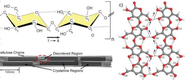
In view of the growing concerns on sustainable and economic development, natural materials such as cellulose have drawn significant attention for various scientific purposes in the recent years. Cellulose, the most abundantly available biopolymer on earth, was first
32 isolated from plants by Anselme Payen in 1838 [48, 49]. Cellulose is a linear homopolysaccharide consisting of β-D-glucopyranose units with an empirical formula of (C6H10O5)n, ‘n’ ranging from a couple of hundreds to 15,000 depending on the source of cellulose [49, 50]. The smallest repeating unit is cellulose consists of two anhydroglucose units (cellobiose) linked together by a covalently bonded oxygen connecting C1 of one unit with C4 of the adjoining unit, hence also referred to as β(1-4) glycosidic bonds as shown in Figure 1.7a [50, 51].
Natural cellulose from plants is synthesized in the form of chain bundles referred to as protofibrils, which are assembled to form microfibrils. The microfibrils contain regions, which are highly ordered (crystalline) and regions in-between that are not as ordered (amorphous) as depicted in Figure 1.7b. The ratio of the crystalline and amorphous regions depends on the origin and affects the density of cellulose [50, 51]. Wood is composed of 30 to 40 % cellulose, with almost an equal portion of the crystalline and amorphous phases [52]. The crystalline phase imparts mechanical strength, whereas the amorphous phase is associated with viscoelastic properties. The linearity in structure comes from the hydrogen bonding between hydroxyl groups and oxygen of the adjoining rings.
The inter- and intra-chain hydrogen bonding imparts stability and stiffness to cellulose (Figure 1.7c). The morphology of cellulose is closely related to its reactivity, the hydroxyl groups in the crystalline region may not be accessible due to close packing and strong bonding, while those in amorphous region may react easily [52, 53].
Figure 1.7 (a) Two glucose rings in cellulose chain connected by β(1-4) glycosidic linkage;
dotted line indicates the intra-chain hydrogen bonding, (b) amorphous and crystalline regions in a cellulose chain, (c) hydrogen bonding in cellulose, the thin dotted lines indicate
33 the intrachain bonding, while the thick dotted line represents the interchain bonding [Reproduced with permission from [50] ©2011 RSC publisher]
1.4.1.2. Polymorphs
Crystalline cellulose primarily has four polymorphs (I, II, III and IV). These polymorphs exhibit various conformations depending on the intra- and inter-molecular H- bonding. Of all, cellulose II is the most thermodynamically stable form with lowest energy [51]. Cellulose I is also called natural or native cellulose, by virtue of its predominate existence in most plants, trees and some sea animals. Cellulose II, reportedly found in bacteria cultivated in glycerol and in cold conditions, can be obtained from cellulose I by solubilisation, recrystallization (regeneration) and treating with sodium hydroxide (mercerization). Likewise, cellulose III can be obtained from either cellulose I (called cellulose IIII) or II (called cellulose IIIII) by treating it with liquid ammonia while cellulose IV (IVI or IVII) can be formed on further thermal treatment (heating with glycerol at 250
°C) [54].
1.4.1.3. Sources
Cellulose can be obtained from both plant as well as animal sources. Being the fundamental strengthening component of plant cell wall, cellulose forms an inexhaustible source of raw material. Wood (softwood or hardwood) is the most important source of cellulose. Cellulose roughly constitutes 45 % of the dry weight of wood. Cellulose is commonly extracted from wood for production of pulp and paper by removal of lignin via the Kraft process [55]. Apart from wood, cellulose also occurs in many non-woody plants such as bamboo, sisal, flax, hemp, jute, cotton and agricultural residues obtained from bagasse, wheat, rice, corn, coconut, banana, pineapple and sugar beet pulp [50, 56]. Like wood, they also need to be processed for cellulose.
Tunicates, a class of sea animals, have a mantle of cellulose embedded in a matrix of proteins. Some of the widely investigated species include Halocynthia roretzi and Metandroxarpa uedai. Several algae such as green, red and grey algae produce cellulose.
Micrasterias denticulate and Caldophora are some of the extensively studied species.
Finally, some bacterial strains such as Gluconacetobacter xylinus and Acetobacter xylinum secrete cellulose under special culturing conditions to form biofilms. They do so in order to possibly protect themselves from UV rays or to hinder fungi and other organisms [50].
34 1.4.1.4. Properties
The cellulose fibres are naturally hydrophilic due to the presence of hydroxyl groups, which allow capillary wicking of fluids required in many LFIAs [48]. The porosity of cellulose fibre network, as in paper, is also immensely useful for the incorporation of materials as well as their diffusion [57]. The biocompatibility, biodegradability and non- toxicity of cellulose make it ideal for a number of biomedical applications. All these attributes of cellulose have made possible for it to come a long way in making its place as an attractive biomaterial for a plethora of applications, ranging from simple wound-healing bandages to complex immunosensors.
1.4.2. Cellulose chemistry
The chemical functionality of native cellulose is because of its surface chemistry, which depends on the source of cellulose. Added functionality can be introduced on them by means of surface modification by physical adsorption of molecules or by chemical attachment of entities or by derivatisation by functional groups. The cellulose chain, with directional chemical asymmetry, consists of a hemiacetal hydroxyl group on the pyranose ring on the reducing end and a pendant hydroxyl group on the non-reducing end (Figure 1.8a).
Different sorts of interactions such as van der Waals, intra- and inter-chain hydrogen bonding between hydroxyl groups and oxygen atoms exist in cellulose. The abundance of hydroxyl groups on the surface of cellulose cause the surface to become very reactive which makes it possible to functionalize it with a number of moieties. Figure 1.8b depicts the major reactions for the chemical modification i.e. (1) esterification, (2) etherification, (3) replacement of the OH by amine or halogen groups, (4) replacement of hydrogen molecules by sodium, (5) oxidation or (6) addition compounds with acids, bases and salts [58]. These modifications only affect the terminal groups of cellulose without breaking down the chain. It is also to be noted that the reactivity of each of the three hydroxyl groups varies with its position in the glucose ring and is not the same due to steric effects arising by virtue of the structure [59].
35 Figure 1.8 (a) Directional asymmetry of cellulose, (b) chemical reactions for modification of cellulose [Reproduced with permission from [58] ©2018 RSC publisher]
1.4.2.1. Functionalization via physical adsorption
Cellulose may be functionalized by adsorbing the molecules on its surface by electrostatic forces of attraction, as in case of polyelectrolytes used as dry and wet strength additives in paper industry. The layer-by-layer deposition is the most commonly used technique for employing physical adsorption [60]. Non-ionic dispersants such as xyloglucan that have a strong, specific adsorption towards cellulose have also been used [61].
1.4.2.2. Functionalization via chemical bonding
Alternatively, cellulose may be functionalized by direct chemical bonding or covalent attachment of molecules. Since cellulose has ample surface hydroxyl groups, species reacting with alcohols such as epoxides, isocyanates, acid halides and acid anhydrides are commonly used for chemical attachment. These reactions can further be used to form a number of alternate surface chemistries including ammonium, amine, alkyl, hydroxyalkyl, ester, acid, etc. [50].
Thus, the structure of cellulose allows its surface to be chemically modified by different processes such as oxidation, amination, esterification, which play an instrumental role in the immobilization of materials by imparting new properties to cellulose without destroying its appealing intrinsic properties [48], as depicted in Figure 1.9.
36 Figure 1.9 Common functionalization chemistries of cellulose surfaces: (clockwise from top-right) sulphuric acid treatment provides sulphate esters, carboxylic acid halides create ester linkages, acid anhydrides create ester linkages, epoxides create ether linkages, isocyanates create urethane linkages, TEMPO mediated hypochlorite oxidation creates carboxylic acids, halogenated acetic acids create carboxymethyl surfaces, and chlorosilanes create an oligomeric silylated layer [Reproduced with permission from [50] ©2011 RSC publisher]
1.4.3. Functionalization of cellulose
Cellulose has gained considerable attention of researchers as a versatile biomaterial, in the quest for sustainable and economical materials in a myriad of fields. The fact that cellulose fibres have reactive surface makes it easy to modify them with a range of functional groups to conjugate various species such as biomolecules, nanoparticles for targeted applications such as bio-sensing, catalysis, biomedical, packaging, etc. [1].
Cellulose is abundantly available, biodegradable, biocompatible, flexible, and readily modifiable, which make it an excellent choice for composites [56].
The functionalization of materials onto substrates has mitigated their recovery issues, leading to environmental remediation [2]. The immobilization of an entity simply refers to its attachment to a surface leading to reduction or loss of its mobility. It can occur
37 by different approaches- physical, chemical, biological or a combination of these processes [48, 62]. Cellulose fibre substrates, such as paper, provide an ideal platform for immobilization by favouring the growth of nanostructures due to their inherently oriented and organized network of fibres [63]. Within the fast growing field of nanotechnology, metal, metal oxide, quantum dots and carbon nanomaterials are gaining increasing interest of researchers due to their overwhelming characteristics [2, 3, 64]. Cellulosic substrates have been widely explored for embedding these nanomaterials for building simple, portable and disposable devices in theranostics, electronics, microfluidics, environmental and POC applications (Figure 1.10) [48, 57]. Paper, being a sheet of randomly interwoven cellulose fibres, possesses all the aforementioned intriguing features of cellulose, which make an excellent platform for anchoring of materials. It also easily complies with the requirements of WHO for diagnostic devices, which should be ASSURED- Affordable, Sensitive, Specific, User-friendly, Rapid and robust, Equipment free and Deliverable to end-users [48].
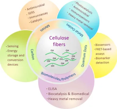
Cellulose has been functionalized with several materials mainly including metals, metal oxides, quantum dots, carbon and biomolecules. Considering the vast potential and demand of natural and renewable resources in virtually every sector, functionalized cellulosic materials show promise to overcome many hurdles and challenges posed during material synthesis and application.
Figure 1.10 Applications of cellulose fibres modified with nanomaterials
38 1.4.4. Functionalization of cellulose with carbonaceous materials
Various nanomaterials based on carbon such as fullerenes, graphene and CNTs have attracted significant attention in the past three decades. In view of their extraordinary mechanical, optical, electrical, thermal and chemical properties, they have been extensively explored for electronics, optoelectronics, photovoltaics and sensing applications [3]. Since the discovery of CNTs by Iijima in 1991, considerable efforts have been devoted to uncover their underlying potential. CNTs are seamless cylinders of single or few layered graphene with an aspect ratio ranging from 102 to 107. Apart from having a high surface area, CNTs have shown remarkable mechanical properties, good electrical conductivity, high thermal conductivity and stability as well as unique mesoporosity. These attributes have made them very attractive for various piezoelectric and thermoelectric energy-harvesting devices, cells, batteries and sensors [65]. Graphene is a two-dimensional, single atom thick building block for other carbon materials, comprising of planar sheets of sp2-bonded carbon atoms that are closely packed in a honeycomb crystal lattice. It has many similarities to CNTs in structure and property, which make it a promising candidate for use in similar areas as CNTs, including supercapacitors, solar cells, lithium-ion batteries, fuel cells, actuators and transistors [65, 66].
1.4.4.1. Carbon nanotubes
Carbon nanomaterials have been widely used to sense a variety of analytes including gases, solvents and biomolecules. Cellulose-based sensors using CNTs have been developed for the detection of glucose [67], ammonia [68, 69], tumour markers [70-73], chemical vapours [74], environmental toxins [75], water [76-78] and ions [79]. Different approaches used to anchor CNTs to cellulosic substrates include simple paper-making technique [78, 80], coating [81], mechanical drawing [69] and ink-jet printing [74]. A humidity sensor was fabricated by conformally coating single-walled CNTs functionalized with carboxylic acid on paper surface [81]. The roughness and porosity of paper were advantageous as they increased the contact area with the ambient air and promoted the adhesion to CNTs. The SEM images confirmed firm entanglement of the CNTs with the cellulose fibres. A shift in conductance of CNT network entangled on the fibres was used for humidity sensing. Since cellulose is an insulator, no current flowed in the bare paper up to 80% RH. Current, however, began to flow at higher RH due to ionic conduction from the dissociation of water. On the other hand, the CNT-coated paper showed linear decrease
39 in conductance with increasing RH up to 75% RH, after which there was marginal rise in conductance. On comparing to a control sensor on glass substrate, cellulose facilitated the charge transport on the paper substrate thus enhancing its sensitivity. Further, the wettability of paper allowed it to soak the CNT suspension leading to better adhesion [81].
In a different study, ink-jet printed films of CNTs on 100% acid-free paper demonstrated the use of cellulose as substrates for sensing chemically aggressive vapours [74]. It was shown that the instability of cellulose towards highly oxidizing vapours is not intrinsic to it but instead, it is an outcome of the surface finishes used during paper manufacture. The detection of NO2 and Cl2 vapours up to 250 and 500 ppb, respectively, was made possible by acid-free paper in ambient conditions by recording the resistance changes. Unlike as in case of PET-based substrate, the paper sensor exhibited spontaneous signal recovery and repeated use over multiple cycle without loss of functionality. It suggested that cellulosic substrates can meaningfully mitigate the aggressive behaviour of vapours such as Cl2 toward thin organic films by reducing the residence time of vapours [74].
Paper-based supercapacitors were prepared by depositing single-walled CNTs on a paper substrate via Meyer rod coating and ink-jet printing. The paper was treated with polyvinylidene fluoride prior to printing of CNTs in order to prevent short circuit and yet allow it to function as an electrolyte membrane and separator [82]. Earlier, they had deposited a combination of single-walled CNTs and AgNWs on paper using printable solution processing technique. The values of specific capacitance, energy, power and life were found to be better than those devices using plastic substrates due to the intrinsic properties of paper such as high solvent absorption and strong binding with nanomaterials.
Superior mechanical properties resulted from the porous nature of paper, which relaxed the bending strain. Additionally, the conductive paper also showed potential as a current collector in Li-ion batteries to replace the metallic counterparts [83]. Similar other works on CNT-coated paper supercapacitors have been reported [84-86].
1.4.4.2. Graphene/reduced-GO
Like CNTs, graphene [87-90], reduced-GO [91, 92], graphene quantum dots [93, 94] and carbon nanodots [95, 96] anchored to cellulosic substrates have also shown extraordinary sensing potential. The exceptional physical, mechanical, thermal, chemical and electrical properties of graphene have made it a versatile choice of research for material
40 scientists the world over. It has been widely used to develop a wide range of functional materials for a plethora of applications from electronics to antibacterial materials [97]. The top-down approach, with GO as the precursor, is the most-promising and a widely used technique for the preparation of graphene-based materials. GO is typically highly reactive due to the presence of a number of functional groups such as –OH, –COOH, epoxy and alkoxy. Hence, reducing the functionalities on the surface of GO is desirable for many biological applications as well as in the electronics sector, in order to regain the electrical activity by recovery of conjugated network of graphitic lattice [98, 99].
Graphene stands out as a material for sensing owing to it single atom thickness, which makes it extremely sensitive to changes in its environment. Graphene layers were directly transferred on paper for resistive sensing of NO2 with remarkably low limit of detection at 300 ppt [100]. As shown in Figure 1.11, graphene on copper foil was spin coated with a layer of PMMA and etched to foil. The PMMA-graphene film was then dredged on to paper and PMMA was dissolved with acetone. The normal paper yielded a patchy coverage while complete transfer occurred on smooth glossy paper [100].
Figure 1.11 (a) Schematic depicting transfer of graphene on to paper, (b) Graphene paper strip in action as a gas sensor may glow an LED bulb [Reproduced with permission from ref. [100] ©2015 ACS publisher]
A pressure sensor was developed by soaking tissue paper in GO solution and subsequently reducing thermally into reduced-GO (rGO paper) [89]. Optimization between sensor sensitivity and working range was achieved in the range of 0-20 kPa and sensitivity up to 17.2 kPa-1. Significant differences in the sensor performance was observed with number of tissue layers (Figure 1.12a). With increasing number of layers, the sensitivity rose 17.2 kPa-1 in the range of 0-2 kPa while it fell to 0.1 kPa-1 in the range of 2-20 kPa.
This was attributed to the presence of air gaps between the layers of tissue paper resulting
41 in their poor contact, which led to a large origin resistance when no pressure was applied on the sensor. The sensor showed good response for detection of pulse, respiration and other human movements (Figure 1.12 b&c) [89].
Figure 1.12 (a) Increase in resistance change with pressure for graphene-based pressure sensor for different number of layers of tissue paper, (b) Sensor application for detection of wrist pulse, (c) Pulse waveform of the sensor [Reproduced with permission from ref.
[89] ©2017 ACS publisher]
Likewise, rGO on paper with AuPd alloy nanoparticles has been used for real time and in situ analysis of hydrogen sulphide released from cancer cells [101]. rGO-paper electrode modified with ZnO nanorods was used as an electrochemical sensor platform for various antigens [102]. Flower-like rGO-modified paper biosensor showed a good linear response for the detection of Pb2+ from 0.005 to 2000 nM [103]. Long graphene nanoribbons (150 nm wide) synthesized via nanoscale cutting of graphite were cleaved to produce graphene quantum dots (100-200 nm) for humidity and pressure sensors [104].
Electrochemical immunoassays with graphene-based sensing have been used to detect biomolecules such as DNA and biomarker [105-107].
Apart from sensing, graphene-based materials have found a plethora of applications in energy storage and energy conversion devices. Supercapacitors fabricated from graphene/cellulose composites as flexible electrodes have demonstrated good capacitance, low resistance and high strength [66, 108]. The graphene-cellulose paper (GCP) was prepared by simply filtering a suspension of graphene nanosheets (GNSs) through a filter paper under vacuum. Electrostatic attraction between the functional groups on cellulose fibres and the negatively charged graphene caused the GNSs to strongly bind to fibres and

![Figure 1.2 Therapeutic effects of cannabinoids for some diseases [Reproduced with permission from [36] ©2018 Elsevier publisher]](https://thumb-eu.123doks.com/thumbv2/9dokorg/523030.993/27.892.203.714.317.748/figure-therapeutic-cannabinoids-diseases-reproduced-permission-elsevier-publisher.webp)
![Figure 1.3 Conventional and modern methods of extraction for phytoconstituents [Reproduced with permission from [37] ©2017 JWS publisher]](https://thumb-eu.123doks.com/thumbv2/9dokorg/523030.993/28.892.137.803.391.658/figure-conventional-methods-extraction-phytoconstituents-reproduced-permission-publisher.webp)
![Figure 1.5 Diffusion in a plant tissue surrounded by a solvent layer during ultrasonic extraction [Reproduced with permission from [40] ©2017 Elsevier publisher]](https://thumb-eu.123doks.com/thumbv2/9dokorg/523030.993/30.892.309.621.581.898/diffusion-surrounded-ultrasonic-extraction-reproduced-permission-elsevier-publisher.webp)
![Figure 1.6 Plants used for the green reduction of materials such as nanoparticles [Reproduced with permission from [7] ©2013 Elsevier publisher]](https://thumb-eu.123doks.com/thumbv2/9dokorg/523030.993/31.892.138.809.486.851/figure-reduction-materials-nanoparticles-reproduced-permission-elsevier-publisher.webp)