Essentials of extracellular vesicles: posters on basic and clinical aspects of extracellular vesicles
Rienk Nieuwlanda,b, Juan Manuel Falcon-Perezc,d, Carolina Soekmadji e, Eric Boilardf,g, Dave Carterh and Edit I. Buzas i,j
aLaboratory of Experimental Clinical Chemistry, Academic Medical Centre (AMC) of the University of Amsterdam, Amsterdam, Netherlands;
bVesicle Observation Centre, Academic Medical Center, University of Amsterdam, Amsterdam, The Netherlands;cExosomes Laboratory, CIC bioGUNE, CIBER, Bilbao, Spain;dExosomes Laboratory, IKERBASQUE Basque Foundation for science, Bilbao, Spain;eDepartment of Cell and Molecular Biology, QIMR Berghofer Medical Research Institute, Brisbane, Queensland, Australia;fDepartment of Infectious Diseases and Immunity, Centre de Recherche du CHU de Québec-Université Laval, Quebec City, Quebec, Canada;gCanadian National Transplantation Research Program, University of Alberta, Edmonton, Alberta, Canada;hDepartment of Biological and Medical Sciences, Faculty of Health and Life Sciences, Oxford Brookes University, Oxford, UK;iDepartment of Genetics, Cell- and Immunobiology, Semmelweis University, Budapest, Hungary;jMTA-SE Immune-Proteogenomics Extracellular Vesicle Research Group
ABSTRACT
The past decade has witnessed an exponential development in the field of extracellular vesicles.
Sporadic observations have reached a critical level and the scientific community increasingly recognizes the potential biomedical significance of these subcellular structures present in all body fluids as significant components of the cellular secretome. The Educational Committee of the International Society for Extracellular Vesicles prepared two posters (“Basic aspects of extra- cellular vesicles” and “Clinical aspects of extracellular vesicles”) to provide essential pieces of information on extracellular vesicles at glance for anyone not familiar with the field.
ARTICLE HISTORY Received 20 June 2018 Revised 31 October 2018 Accepted 6 November 2018 KEYWORDS
Extracellular vesicles (EVs);
biomarker; therapy;
biogenesis; molecular composition; biorepository
Basic and clinical aspects of extracellular vesicles
Since 2010, the scientific and clinical interest in extracel- lular vesicles (EVs) is growing exponentially. EVs are small, lipid membrane enclosed subcellular structures carrying biomolecules which are released by cells into their environment [1–3]. The term“EVs”is an umbrella term for all types of vesicles, including“exosomes”and
“microparticles” or “microvesicles” [4]. This term is endorsed by the International Society on Extracellular Vesicles (ISEV;www.isev.org), a global scientific society founded in 2011), because at present no biochemical or (bio)physical distinction can be made between types of EVs generated by different biogenesis pathways [2].
The two posters by ISEV on“Basic aspects of extra- cellular vesicles”and“Clinical aspects of extracellular vesicles” provide a brief introduction about EVs to students and (bio) medical healthcare professionals.
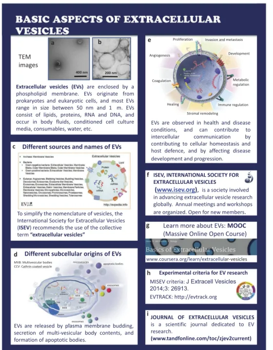
Basic aspects of extracellular vesicles (Figure 1) All cells release EVs into their environment, and (bio)fluids such as conditioned culture medium and body fluids
contain EVs. When EVs are visualized by electron micro- scopy (A and Bare EM microphotographs provided by Rienk Nieuwland and Agnes Kittel, respectively), EVs appear as small, usually spherical particles surrounded by a phospholipid bilayer membrane. This membrane often contains proteins from the parental cell, thus enabling the identification of the cell of origin. In addition, EVs contain biomolecules originating from the parental cells, including RNA and DNA, metabolites and lipids [5].
Initially, EVs were often named after the cell type of origin, or after the organ in which they were discov- ered, such as dexosomes or prostasomes. Because this nomenclature was not only confusing but also resulted in development of parallel research fields lacking inter- action, ISEV endorses the term “extracellular vesicles”
as an umbrella term [Chttp://evpedia.info,6].
Detailed analysis of the biochemical composition of EVs has shown that the overall composition of EVs differs from that of the releasing parental cells. This implies that cargo sorting mechanisms exits that affect the composition of EVs. Moreover, the biochemical composition depends on the“status”(resting, activated, etc.) of the parental cell.
EVs can be released directly by budding from the plasma
CONTACT Edit I. Buzas edit.buzas@gmail.com; buzas.edit@med.semmelweis-univ.hu Department of Genetics, Cell- and Immunobiology, Semmelweis University, Budapest, Hungary
*These authors contributed equally to this work JOURNAL OF EXTRACELLULAR VESICLES
2018, VOL. 7, 1548234
https://doi.org/10.1080/20013078.2018.1548234
© 2018 The Author(s). Published by Informa UK Limited, trading as Taylor & Francis Group on behalf of The International Society for Extracellular Vesicles.
This is an Open Access article distributed under the terms of the Creative Commons Attribution-NonCommercial License (http://creativecommons.org/licenses/by-nc/4.0/), which permits unrestricted non-commercial use, distribution, and reproduction in any medium, provided the original work is properly cited.
membrane, by secretion of prestored EVs in multi- vesicular bodies, by formation of apoptotic bodies as well as released from enveloped virus-infected cells (D). Due to all these variables, EVs are highly heterogeneous in size and composition [7–9].
EVs are not merely “innocent bystanders” but play important roles in physiology and pathology (E). In physiology, EVs contribute to homeostasis and promote host-defence mechanisms including haemostasis and inflammation [3], whereas in pathology EVs may con- tribute to disease development and progression, for example in cancer [10,11]. Because EVs are capable of
delivering their proteins, lipid and RNA cargo to target (recipient) cells, EVs can regulate gene expression and consequently the phenotype and biological functions of the target cell. Thus, by exchanging information between cells, EVs contribute to intercellular communication.
ISEV (F) developed a massive open online course on EVs that is freely available [Massive Online Open Course (MOOC) (G), http://coursera.org/learn/extracelllular- vesicles,12], published minimal requirements for studies on EVs [MISEV,13] (H). ISEV endorses a knowledge base that has been launched to monitor the quality of pre- analytical data reporting [http://evtrack.org,14] (H). The
Extracellular vesicles (EVs) are enclosed by a phospholipid membrane. EVs originate from prokaryotes and eukaryotic cells, and most EVs range in size between 50 nm and 1 m. EVs consist of lipids, proteins, RNA and DNA, and occur in body fluids, conditioned cell culture media, consumables, water, etc.
ISEV, INTERNATIONAL SOCIETY FOR EXTRACELLULAR VESICLES
(www.isev.org), is a society involved in advancing extracellular vesicle research globally. Annual meetings and workshops are organized. Open for new members.
JOURNAL OF EXTRACELLULAR VESICLES is a scientific journal dedicated to EV research.
(www.tandfonline.com/toc/zjev2current) EVs are released by plasma membrane budding,
secretion of multi-vesicular body contents, and formation of apoptotic bodies.
EVs are observed in health and disease conditions, and can contribute to intercellular communication by contributing to cellular homeostasis and host defence, and by affecting disease development and progression.
TEM images
Learn more about EVs: MOOC (Massive Online Open Course)
www.coursera.org/learn/extracellular-vesicles
Experimental criteria for EV research MISEV criteria: J Extracell Vesicles 2014;3: 26913.
EVTRACK: http://evtrack.org To simplify the nomenclature of vesicles, the
International Society for Extracellular Vesicles (ISEV) recommends the use of the collective term “extracellular vesicles”
400 nm 200 nm
Different subcellular origins of EVs Different sources and names of EVs
Proliferation
Coagulation
Angiogenesis Development
Stromal remodeling
Healing Immune regulation
Metabolic regulation Invasion and metastasis
a b
c
d
e
f
g
h
i
microvesicles apoptotic bodies
exosomes
exosomes MVB: Multivesicular bodies
CCV: Cathrin coated vesicle
MVB
MVB
Figure 1.Basic aspects of extracellular vesicles.
2 R. NIEUWLAND ET AL.
official journal of ISEV is the Journal of Extracellular Vesicles (JEV) (I).
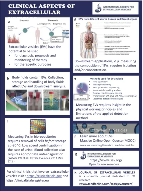
Clinical aspects of extracellular vesicles (Figure 2) In general, EVs have a huge potential for diagnosis of disease, prognosis and monitoring of therapy [15].
Because EVs can be considered as naturally occurring autologous nanocarriers”, EVs can be loaded with drugs for local drug delivery (A).
At present, standard operation procedures are being developed for routine collection, storage and handling of biofluids and body fluids such as urine, blood and breast milk to enable the comparison of measurements between patients and controls and to allow exchange of data between institutes and instru- ments, etc. (B).
In most clinical studies, EV-containing biofluids will be collected in biorepositories. The pre-analytical con- ditions may differ between biofluids, but removal of cells and cell fragments is essential to circumvent con- tamination due fragmentation of cells during freeze thawing (C).
Because all human (and animal) body fluids contain EVs, and because the cellular origin, composition and functions of EVs change in disease, the detailed analy- sis of EVs is thought to behold clinically relevant information. To get access to this information, how- ever, most downstream applications require either iso- lation and/or concentration of EVs (D).
Detection of (single) EVs and detailed insight into their biochemical composition by downstream methods such as proteomics requires a thorough understanding of the bio- chemical and (bio)physical limitations and possibilities of
https://www.isev.org/
Open for new members.
Extracellular vesicles (EVs) have the potential to be used
• for diagnosis, prognosis and monitoring of therapy
• for therapeutic purposes Downstream-applications, e.g. measuring the composition of EVs, requires isolation and/or concentration.
Measuring EVs requires insight in the physical working principles and limitations of the applied detection method.
Learn more about EVs:
Massive Online Open Course (MOOC)
www.coursera.org/learn/extracellular-vesicles
Body fluids contain EVs. Collection, storage and handling of body fluids affect EVs and downstream analysis.
Measuring EVs in biorepositories requires removal of cells beforestorage at -80 °C. Low speed centrifugation in the case of urine. Blood collection also requires appropriate anti-coagulation.
For clinical trials that involve extracellular vesicles visit : https://clinicaltrials.govand https:/clinicaltrialsregister.eu
delivery
Autologous EVs Exogenous EVs Biomarker
identification &
analysis
Biologics (RNA, protein, compound)
(Witwer KW et al.J Extracell Vesicles. 2013 May 27;2.)
EVs from different source tissues in different organs
JOURNAL OF EXTRACELLULAR VESICLES is a scientific journal dedicated to EV research.
(www.tandfonline.com/toc/zjev2current)
Flow cytometry
Methods used for EV analysis
• Flow cytometry
• Mass spectrometry
• Next generation sequencing
• Nanoparticle tracking analysis
• Tunable resistive pulse sensing
• Transmission EM, cryo EM, AFM, scanning EM
• Label free detection techniques
a
b
c
d
e
f
g
h
Figure 2.Clinical aspects of extracellular vesicles
JOURNAL OF EXTRACELLULAR VESICLES 3
the various isolation, detection and characterization meth- ods (E). In addition, the method of collection, handling and storage conditions of EV can alter the molecular composition of EV [16,17].
For more information, please visit the MOOC on EVs (F), the Journal of Extracellular Vesicles (JEV) (G) or contact the International Society for Extracellular Vesicles (H).
Disclosure statement
No potential conflict of interest was reported by the authors.
ORCID
Carolina Soekmadji http://orcid.org/0000-0002-6920-6627 Edit I. Buzas http://orcid.org/0000-0002-3744-206X
References
[1] Tkach M, Théry C. Communication by extracellular vesicles: where we are and where we need to go. Cell.
2016 Mar 10;164(6):1226–1232.
[2] van Niel G, D’Angelo G, Raposo G. Shedding light on the cell biology of extracellular vesicles. Nat Rev Mol Cell Biol.2018Apr;19(4):213–228.
[3] Yáñez-Mó M, Siljander PR, Andreu Z, et al. Biological properties of extracellular vesicles and their physiologi- cal functions. J Extracell Vesicles.2015May;4(1):27066.
[4] György B, Szabó TG, Pásztói M, et al. Membrane vesicles, current state-of-the-art: emerging role of extracellular vesicles. Cell Mol Life Sci.2011Aug;68(16):2667–2688.
[5] van der Pol E, Böing AN, Harrison P, et al.
Classification, functions, and clinical relevance of extra- cellular vesicles. Pharmacol Rev.2012Jul;64(3):676–705.
[6] Kim DK, Lee J, Kim SR, et al. EVpedia: a community web portal for extracellular vesicles research.
Bioinformatics. 2015Mar 15;31(6):933–939.
[7] Kowal J, Arras G, Colombo M, et al. Proteomic com- parison defines novel markers to characterize heteroge- neous populations of extracellular vesicle subtypes. Proc Natl Acad Sci U S A.2016Feb 23;113(8):E968–77.
[8] Soekmadji C, Riches JD, Russell PJ, et al. Modulation of paracrine signaling by CD9 positive small extracellular vesicles mediates cellular growth of androgen deprived prostate cancer. Australian prostate cancer collaboration bioresource. Oncotarget.2017;8(32):52237–52255.
[9] Verweij FJ, Bebelman MP, Jimenez CR, et al.
Quantifying exosome secretion from single cells reveals a modulatory role for GPCR signaling. J Cell Biol.
2018;217(3):1129–1142.
[10] Choi D, Lee TH, Spinelli C, et al. Extracellular vesicle com- munication pathways as regulatory targets of oncogenic transformation. Semin Cell Dev Biol.2017Jul;67:11–22.
[11] Sato S, Weaver AM. Extracellular vesicles: important collaborators in cancer progression. Essays Biochem.
2018Apr 17;62:149–163.
[12] Lässer C, Théry C, Buzás EI, et al. The international society for extracellular vesicles launches the first mas- sive open online course on extracellular vesicles.
J Extracell Vesicles.2016Dec 16;5:34299.
[13] Lötvall J, Hill AF, Hochberg F, et al. Minimal experi- mental requirements for definition of extracellular vesi- cles and their functions: a position statement from the international society for extracellular vesicles. J Extracell Vesicles.2014;3:26913.
[14] Van deun J, Hendrix A. Is your article EV-TRACKed?
EV-TRACK consortium. J Extracell Vesicles.2017 Nov 10;6(1):1379835.
[15] Roy S, Hochberg FH, Jones PS. Extracellular vesicles:
the growth as diagnostics and therapeutics; a survey.
J Extracell Vesicles.2018Feb 26;7(1):1438720.
[16] Coumans FAW, Brisson AR, Buzas EI, et al.
Methodological guideli120nes to study extracellular vesicles. Circ Res.2017May 11;120:1632–1648.
[17] Witwer KW, Soekmadji C, Hill AF, et al. Updating the MISEV minimal requirements for extracellular vesicle studies: building bridges to reproducibility. J Extracell Vesicles.2017;6(1):1396823.
4 R. NIEUWLAND ET AL.