Glucuronidetransportintheendoplasmicreticulum
PhD thesisoutline
Dr. KatalinRévész
DoctoralSchool of MolecularMedicine Semmelweis University
Supervisor: Dr. Miklós Csala Ph.D.
Officialreviewers: Dr. Katalin MonostoryPh.D.
Dr. Attila Ambrus Ph.D.
Head of theFinalExaminationCommittee:
Dr. László TretterPh.D.
Members of theFinalExaminationCommittee:
Dr. Katalin JemnitzPh.D.
Dr. Erzsébet KomoroviczPh.D.
Budapest
2014
1 Introduction
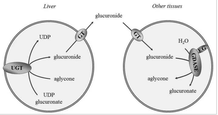
During the process of biotransformation, a wide array of endo- and xenobiotics are converted to polar compounds by conjugation reactions, which facilitates their efficient elimination from the body. Glucuronidation (conjugation with glucuronide) is a major reaction in the second phase of biotransformation.The reaction is catalysed in the lumen of the endoplasmic reticulum (ER) by theUDP-glucuronosyltransferase enzymes (UGTs), which transfer a glucuronosyl moiety from UDP-glucuronate to a nucleophilic functional group of the aglycone (Fig. 1). The producedβ-D- glucuronidesare more water-soluble, hence they are ready for elimination via urine or bile. Generally, conjugation inactivates biologically active compounds and reduces or abolishes toxicity.
Substrates of UGT enzymes can be endogenous compounds (e. g.
bilirubin, retinoids, thyroid hormones, steroids and catecholamines) or xenobiotics, such as drugs (e. g. chemotherapeutic agents or the analgesic morphine). Also a wide variety of carcinogenic compounds is detoxified through conjugation with glucuronate. Therefore low activity of UGT enzymes increases the risk of malignant transformation of cells due to slow elimination of chemical carcinogens. Accordingly, UGTs play an important role in the prevention of malignant tumours.
Glucuronides, produced especially in the liver, can reach peripheral cells with the systemic circulation, and here can be hydrolysed in the lumen of the ER by β-glucuronidase (Fig.1). This enzymatic hydrolysis yields the original, biologically active or toxic aglycone, which therefore
2
can achieve its effect locally. β-Glucuronidasereactivates carcinogenic compounds, too. This explains the correlation found between glucuronidase activity and the occurrence of certain tumours. Accordingly, cancer-preventing glucarate salts, natural components of fruits and vegetables, were reported to act by inhibiting β-glucuronidase activity.
Both glucuronidation and deglucuronidationarelocalized in the lumen of the ER. Due to this subcellular localization, activity of these enzymes requires inward transport of substrates and outward transport of products across the ER membrane. Lipophilic substrates of UGTs can enter the organelle through passive diffusion. However, the hydroxyl and
Figure 1. Glucuronidation and deglucuronidation EG: egasyn, GDASE: β-glucuronidase, GT:
glucuronidetransporter,UGT: UDP-glucuronosyl-transferase
3
dissociated carboxyl groups of the glucuronic acid moiety provide UDP- glucuronateand all glucuronides with remarkable polarity and hydrophilicity. Biological membranes are impermeable to such bulky polar and charged compounds unless equipped with suitable transporter proteins.
Functional studies prove that transport of UDP-glucuronate and glucuronidesacross the ER membrane is protein mediated. However, in spite of the evident importance of the transport in the process of glucuronidation and deglucuronidation, the proteins involved still remain to be identified.
Importance of transport processes is indicatedby latency of luminal enzymes, e. g. UGTs and β-glucuronidase. The relatively slow transport across the integrant ER membrane limits the activity of luminal enzymes.
However, enzymes have free access to their substratesafter membrane permeabilization, thus a remarkably increased enzyme activity can be measured. Difference between the activities, expressed as percentage of the activity observed after membrane permeabilization, is called latency.
90 % latency of UGTs is attributed to the UDP glucuronate uptake, and glucuronide transport is responsible for the 40% latency of β- glucuronidase.
Therefore, glucuronidetransport across the ER membrane is rate- limiting for deglucuronidation. Decrease of glucuronide uptake to the ER thus might prevent cancer, as direct inhibition of β-glucuronidase enzyme.
General anion transport inhibitors and thiol alkylating agents were shown to block glucuronide transport, but specific inhibitor of the process has not yet been reported.
4
The presence of multiple glucuronide transporters has been demonstrated in the ER. Comparison of the transport of various glucuronides revealed an inverse correlation between the size of the compounds and the rate of their transport across the ER membrane. The bulky, rigid morphine-3-glucuronide (M3G) crosses the membrane at a two orders of magnitude slower rate than other glucuronides. The strikingly slow transport suggests that newly formed M3G can be retained in the ER lumen, which leads to the build-up of an intracellular pool. This assumption is supported by earlier findings indicative of separate intracellular M3G pools is perfused rat liver. Elimination of M3G, major metabolite of the widely used narcotic analgesic morphine, is especially interesting, because this compound is devoid of analgesic activity, but it has been implicated in the non-opioid mediated side effects (e. g.
hyperalgesia and convulsion) of morphine treatment.
5 Objectives
Despite its important role in biotransformation, glucuronide transport has not been exactly characterized yet, neither at functional, nor at molecular level.Either its specific inhibitor have not been reported.
Our aim was to investigate glucuronide transport. We tested the assumption that newly formed M3G accumulates in the ER lumen due to the extremely slow release of this metabolite. Moreover, we looked for possible inhibitors of glucuronide transport. Such substances might reduce reactivation on chemical carcinogens through the indirect inhibition of β- glucuronidase enzyme, thus might decrease the risk of malignancies.
Green tea flavanols (catechins) have been shown to prevent cancer. We hypothesized that inhibition of deglucuronidation might contribute to the anti-cancer properties of tea flavanols. Therefore, we investigated the effect of green tea catechins on the activity of β-gluduronidase enzyme and glucuronide transporter.
6 Methods
Microsomes, separated from other organelles by differential centrifugation,are a widely used tool for investigating the function of the ER by in vitroresearch. In our experiments intact and permeabilizedrat liver microsomal vesicles and mouse hepatoma Hepa 1c1c7 cells were used. For membrane permeabilization, microsomes and cells were pretreated with the pore-forming reagent alamethicin.
Morphine UGT activity was measured in intact and permeabilized rat liver microsomes. Vesicles were incubated in the excess of UDP- glucuronate and the presence of morphine. Total and vesicle-associated M3G was detected using LC-MS.
β-glucuronidase activity of rat liver microsomes and mouse hepatoma cells was determined as the rate of 4-methylumbelliferone (MUMB) production by enzymatic hydrolysis of methylumbelliferyl- glucuronide (MUGA). All the studied green tea flavanols were added to the incubation medium 1 minute prior to substrate. MUMB content of the samples was measured by HPLC.
The rate of glucuronide transport across the microsomal membrane was determined using rapid filtration method. After creating inward or outward concentration gradient, microsomes were incubated, and then samples were withdrawn from the incubation mixture, filtered immediately through cellulose acetate/nitrate filter membranes and washed quickly.
Glucuronides were extracted from the filters with 50% acetonitrile, and were measured by HPLC (in case of MUGA) or LC-MS (in case of M3G).
7
The membrane bound fraction of glucuronides was determined by performing the same procedure with alamethicin-treated microsomes.
Luminal glucuronide content was calculated from the difference between the amounts associated to intact and permeabilized vesicles. Glucuronide transport was measured in the presence of saccharolactone to inhibit β- glucuronidase.
Time course of MUGA uptake was determinedalsointhe presence of epigallocatechin-gallate (EGCG), the major green tea flavanol. The concentration-dependence of the effect was studied at various EGCG concentrations (0-400 µM).
Components of samples were separated with HPLC, using gradient elution mode. Fluorescence of MUMB was detected at EX: 325 nm and EM: 455 nm wavelengths. Absorbance of MUGA was measured at a wavelength of 317 nm. For detecting M3G, the eluate was infused into a mass spectrometer. M3G was detected with electrospray using multiple reaction monitoring in positive ionization mode.
Experiments were performed at least in triplicate. Data are presented as means ± S. D. GraphPad Prism® software was used for parameter estimation and line or curve fittings. Differences with p<0.01 were considered significant.
8 Results
Accumulation of newly synthesized M3G in intact microsomes
Investigation of M3G uptake and release revealed a bidirectional, low affinity transport mechanism, which corresponds to earlier data.
The observed 90% latency of morphine UGT activity, which was irrespective of the applied morphine concentration, is also in accordance with earlier results.
After these preliminary measurements, microsome associated amount of M3G was determined during morphine glucuronidation (Fig. 2).
Glucuronide content of permeabilized vesicles increased in a linear manner, proportional to time and the amount of the synthetized M3G.
However, in intact microsomes the amount of vesicleassociated M3G increased nearly 30 times faster in short incubations compared with permeabilizedmicrosomes, despite the 10 times lower rate of glucuronide synthesis. Glucuronide content of intact vesicles reached a plateau after about 8-10 minute period of quick elevation, and the high level was sustained until the end of experiment.
We compared the results measured during in situ morphine glucuronidation with those data, which were detected in equilibrium, when luminal glucuronide concentration equals the extravesicular level due to long (100 minute) incubation. In the latter case, we incubated microsomes in the presence of M3G, and after reaching equilibrium, vesicle associated amount of M3G was determined. Glucuronide content of vesicles was
9
found to be proportional to total amount of M3G of the samples, both in intact and permeabilizedmicrosomes (Fig. 2).
Our results reveal a remarkably accumulation of newly synthesized M3G above the equilibrium in intact vesicles (Fig. 2).Rate of M3G efflux (characterized by low affinity)is adjusted to that of formation through the build-up of a high glucuronide concentration in the ER lumen. Our results suggest that a dynamic luminal M3G pool develops in the ER of hepatocytes, separated from the cytosol. Our observation explains earlier findings, which revealed the existence of different M3G pools in rat liver.
A remarkable part of the intracellular M3G content was unaffected after selective permeabilization of the apical plasma membranes in isolated
Figure 2: Accumulation of newly synthetized M3G in intact microsomes
10
perfused rat livers. It was concluded that M3G is contained by separate compartments within the hepatocytes.
Effect of green tea flavanols on microsomal glucuronide transport Latency of MUGA glucuronidase activity was about 40% in rat liver microsomes and mouse hepatoma cells.
Most of the investigated green tea catechins remarkably hindered the hydrolysis of MUGA, when the microsomal barrier was intact. At the same time, flavanols (with the exception of gallocatechin-gallate) were ineffective in permeabilizedmicrosomes. In other words, catechins result in dissociation of activities of intact and permeabilizedmicrosomes: the most potent flavanols, EGCG and gallocatechin-gallateraised the originally modest latency of glucuronidase to above 80%. The phenomenon observed in liver microsomes was also tested in mouse hepatoma cells. In accordance with our microsomal findings, EGCG inhibited glucuronidase activity in intact cells, while failed to affect the enzyme in alamethicin- treated cells.
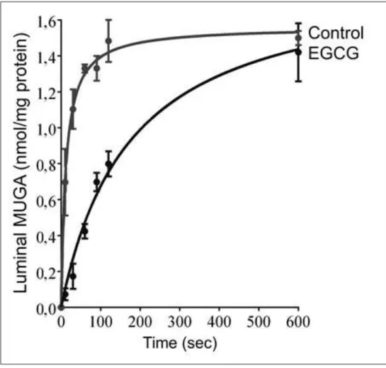
Flavanols were shown to be ineffective, when glucuronidaseenzyme can freely access the substrates due to elimination of microsomal membrane barrier. This phenomenon indicates that flavanols do not inhibit the enzyme directly. Increase of latency suggests that flavanols hinder glucuronide transport across the ER membrane. We investigated this assumption in transport measurements directly. Time course of MUGA uptake was detected by rapid filtration in control and EGCG-treated rat liver microsomal vesicles (Fig. 3).
11
Significantly slower uptake was measured in the presence of EGCG:
although the vesicles took up the same amount of MUGA in 10 minutes, they got only half-way to equilibration in 2 minutes (Fig. 3). Initial transport rate was reduced by 86%. Glucuronide transport was hindered in a concentration-dependent manner by EGCG. The inhibition was 80% at 50 µM EGCG; and nearly 90% inhibition was achieved at higher flavanol concentrations. Our findings indicate that green tea flavanols reduce deglucuronidation in the ER at the glucuronide transport stage.
Figure 3: Effect of EGCG on microsomal glucuronide
uptakeroszómaglukuronidfelvételére
12 Conclusions
1. We found evidence that newly synthetized M3G accumulates above the equilibriumin intact microsomesdue to the slow release.
2. Our observation on accumulation of newly synthetized M3G are in accordance with earlier findings, which revealed the existence of different pools of M3G in rat liver.At the same time, the build-up of a high luminal glucuronide concentration suggests the possibility that in certain cases glucuronide efflux can limit the rate of glucuronideformation.
3. Green tea flavanols that possess gallo and/or gallate moieties significantly inhibited microsomal glucuronidase activity in intact microsomes, while only gallocatechin-gallate reduced the enzyme activity after membrane permeabilization. The same effect was shown in EGCG- treated mouse hepatoma cells. This phenomenon – inhibition of deglucuronidation when membrane barrier is intact – might contribute to the known anti-cancer properties of green tea.
4. Direct transport measurements prove that EGCG reduces glucuronidase activity by inhibiting glucuronide entry into the ER. This catechin inhibits MUGA transport across the ER membrane in a concentration-dependent manner. We identified thus an efficient, nontoxic and – compared to general anion transport inhibitors and thiol alkylating agents – much more specific inhibitor. EGCG might also become a useful experimental tool for further investigations of glucuronide transporter proteins.
13 Bibliography
Publications related to the theme of the Ph.D. thesis
1. Révész K, Tüttő A, Margittai É, Bánhegyi G, Magyar JÉ, Mandl J,
Csala M.(2007)
Glucuronidetransportacrosstheendoplasmicreticulummembrane is inhibitedbyepigallocatechingallate and othergreen tea polyphenols.
International Journal of Biochemistry and CellBiology, 39: 922-930 IF: 4,009
2. Révész K, Tóth B, Staines AG, Coughtrie MW, Mandl J, Csala M.
(2013) Luminalaccumulation of newlysynthesized morphine-3- glucuronide inratlivermicrosomalvesicles. Biofactors, 39(3): 271-278 IF: 3,088
Publicationsnotrelatedtothetheme of the PhD thesis
1. Révész K, TüttőA, Konta L.Effect of green tea flavonolsonthe(2007) function of theendoplasmicreticulum. Orvosi Hetilap, 40: 1903-1907 2. Révész K, TüttőA, Szelényi P, Konta L.(2011) Tea flavan-3-ols
asmodulatingfactorsinendoplasmicreticulumfunction. Nutrition Research, 31(10): 731-740 IF: 1,974
3. Konta L, Száraz P, Magyar JÉ, Révész K, Bánhegyi G, Mandl J,
Csala M.(2011) Inhibition of
glycoproteinsynthesisintheendoplasmicreticulumas a
14
novelanticancermechanism of (-)-epigallocatechin-3-gallate.
Biofactors, 37(6): 468-476 IF: 4,933
4. Szelényi P, Révész K, Konta L, TüttőA, Mandl J, Kereszturi É, Csala M.(2013) Inhibition of microsomalcortisolproductionby (-)- epigallocatechin-3-gallate through a redox shift intheendoplasmicreticulum - A potentialnewtargetfortreatingobesity- relateddiseases. Biofactors, 39(5): 534-541 IF: 3,088