.. ..
.. ..
.. ..
.. ..
.. ..
.. ..
.. ..
.. ..
.. ..
.. ..
.. ..
..
COVID-19 pandemic and cardiac imaging:
EACVI recommendations on precautions, indications, prioritization, and protection for patients and healthcare personnel
Helge Skulstad
1,2, Bernard Cosyns
3, Bogdan A. Popescu
4, Maurizio Galderisi
5, Giovanni Di Salvo
6, Erwan Donal
7, Steffen Petersen
8,9, Alessia Gimelli
10,
Kristina H. Haugaa
1,2, Denisa Muraru
11, Ana G. Almeida
12,
Jeanette Schulz-Menger
13,14,15, Marc R. Dweck
16, Gianluca Pontone
17,
Leyla Elif Sade
18, Bernhard Gerber
19, Pal Maurovich-Horvat
20, Tara Bharucha
21, Matteo Cameli
22, Julien Magne
23,24, Mark Westwood
9, Gerald Maurer
25, and Thor Edvardsen
1,2*
1Department of Cardiology, Oslo University Hospital, Rikshospitalet, Oslo, Norway;2Faculty of Medicine, University of Oslo, Oslo, Norway;3Centrum voor Hart en Vaatziekten (CHVZ), Universitair Ziekenhuis Brussel, Vrij Universiteit van Brussel, Brussels, Belgium;4Department of Cardiology, University of Medicine and Pharmacy ‘Carol Davila’- Euroecolab, Emergency Institute for Cardiovascular Diseases ‘Prof. Dr. C. C. Iliescu’, Bucharest, Romania;5Department of Advanced Biomedical Sciences, Federico II University Hospital, Naples, Italy;6University Hospital Padua, Paediatric Cardiology, Padua, Italy;7University of Rennes, CHU Rennes, Inserm, LTSI–UMR 1099, Rennes, France;8William Harvey Research Institute, NIHR Barts Biomedical Research Centre, Queen Mary University of London, London, UK;9Barts Heart Centre, St Bartholomew’s Hospital, Barts Health NHS Trust, London, UK;10Fondazione Toscana G. Monasterio, Pisa, Italy;11Department of Medicine and Surgery, University of Milano-Bicocca, Milan, Italy;12Cardiology, University Hospital Santa Maria /CHLN, CCUL, Faculty of Medicine of Lisbon University, Lisbon, Portugal;13Charite´ Medical Faculty of the Humboldt University Berlin, ECRC, Berlin, Germany;14DZHK, Partner site, Berlin, Germany;15Helios Clinics Berlin-Buch, Cardiology, Berlin, Germany;16Centre for Cardiovascular Science, University of Edinburgh, UK;17Department of Cardiovascular Imaging, Centro Cardiologico Monzino IRCCS, Milan, Italy;18Department of Cardiology, University of Baskent, Ankara, Turkey;19Division of Cardiology, Department of Cardiovascular Diseases, Cliniques Universitaires St. Luc, Poˆle de Recherche Cardiovasculaire, Institut de Recherche Expe´rimentale et Clinique, Universite´ Catholique de Louvain, Brussels, Belgium;20Medical Imaging Centre, Semmelweis University, Budapest, Hungary;21Department of Congenital Cardiology, University Hospital Southampton NHS Foundation Trust, Southampton, UK;22Department of Medical Biotechnologies, Division of Cardiology, University of Siena, Siena, Italy;23CHU Limoges, Hoˆpital Dupuytren, Service Cardiologie, Limoges, France;24INSERM U1094, Univ. Limoges, CHU Limoges, IRD, U1094, GEIST, 2, Limoges, France; and25Division of Cardiology, Department of Internal Medicine II, Medical University of Vienna, Vienna, Austria
Received 25 March 2020; editorial decision 25 March 2020; accepted 25 March 2020
Introduction
The coronavirus disease 2019 (COVID-19) pandemic has created new and unpredictable challenges for modern medicine and health- care systems. Preliminary reports have demonstrated that older age, previous cardiovascular disease, diabetes, and hypertension are risk factors for increased mortality.1Data on the cardiac affinity of the virus and its potential to harm the cardiovascular system and the mechanisms by which this occurs are sparse.2,3A systemic infection generally increases demand on the heart, and can exacerbate under- lying cardiac conditions. When the lungs are heavily involved, as seen in COVID-19 patients, this may have a major impact on cardiac func- tion, particularly that of the right ventricle. Finally, COVID-19 may have direct effects on the heart, as may some drugs being used in its treatment.
The severe acute respiratory syndrome coronavirus-2 (SARS- CoV-2) is responsible for COVID-19 and is transmitted by droplets from person to person.4Echocardiographers in particular, and car- diac imagers more generally, are in close contact with patients and therefore likely to have a high risk of being infected. To decrease the risk of patient to patient, patient to imager, and imager to patient con- tamination, the indication for any cardiac imaging test should be care- fully considered, and only those tests considered essential to patient care performed.
Cardiologists and cardiology departments are heavily affected by this rapidly changing situation.5 The COVID-19 pandemic also increases the burden on cardiac imaging services generally. However, given its wide availability and key role as a bedside test, echocardiog- raphy is the most affected cardiac imaging modality. Common chal- lenges faced by all cardiac imaging modalities during the pandemic
* Corresponding author. Department of Cardiology, Oslo University Hospital, Rikshospitalet, Sognsvannsveien 20, 0372 Oslo, Norway/PO Box 4950 Nydalen, NO-0424 Oslo, Norway. Tel:þ47 2 3071176, Email: thor.edvardsen@medisin.uio.no
Published on behalf of the European Society of Cardiology. All rights reserved.VCThe Author(s) 2020. For permissions, please email: journals.permissions@oup.com.
doi:10.1093/ehjci/jeaa072
.. ..
.. ..
.. ..
.. ..
.. ..
.. ..
..
include limited expert staff availability [sickness or redeployment in prioritized areas, such as intensive care units (ICUs)] and the risk of peri-procedural transmission of SARS-CoV-2 between patients and staff. The scope of these EACVI recommendations is to summarize how these challenges may be addressed during the pandemic. In particular, we focus upon bold prioritization and provide specific indications and recommendations on how to per- form an echocardiogram during the pandemic whilst safeguarding both patient and staff.
Some of our recommendations relating to the appropriate use of imaging modalities in the COVID-19 pandemic must be considered only as expert advice due to the lack of evidence-based scientific data and the rapidly changing global situation.
General considerations
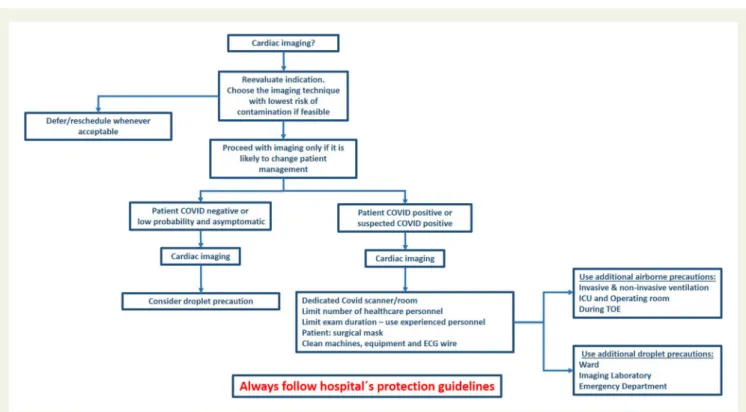
For all modalities, the main question is: ‘Will undertaking this study substantially change patient management or be lifesaving?’ If the an- swer is yes, use the imaging modality with the best capability to meet the request, but also consider the safety of medical staff regarding ex- posure. It is very important that every cardiac imaging study is per- formed appropriately to minimize the risk of further dissemination of the disease (Key point 1andFigure 1).
This question needs to be considered in detail for each echo- cardiogram requested given the risk of cross-infection and the consumption of personal protective equipment. However, com- puted tomography (CT) and cardiac magnetic resonance (CMR) may also have a significant potential for contamination of person- nel and patients, especially linked to transportation, but also via direct contamination during the scan. In parallel with echocardi- ography and other imaging modalities, CMR and CT should only be performed if the expected information is critical for clinical management and can be justified in the face of the following con- siderations: (i) risk of transportation of critically ill or high-risk patients; (ii) time duration of CMR; (iii) possible/significant risk of infection for professionals (technicians, physicians, nurses, and other personnel); (iv) possible/significant risk of contamination
of equipment and facilities, leading to the need for full disinfec- tion; and (v) whether the test is necessary to confirm the diagno- sis, or whether this can be achieved based simply upon the clinical probability. In many countries, imaging exams for elective non-urgent patients have been cancelled or postponed.
However, cardiac imaging is still being widely requested for inpatients or those presenting to the Emergency Department (Key point 2).
Indications
Patients with suspected or confirmed COVID-19 and no previous history of cardiac disease
The chest radiograph is the most commonly used imaging test in COVID-19 patients, but CT is frequently used to confirm COVID-19 pneumonia. Whilst this might conceivably provide some possible synergies and opportunities to gain infor- mation about the cardiovascular system, this requires bespoke protocols that are not widely employed. Dedicated coronary CT angiography is therefore usually required. One emerging clinical issue is that numerous patients with pneumonia caused by COVID-19 experience elevated troponins with and without signs of obstructive coronary artery disease. In this situation, coronary CT angiography can be of great help in excluding or confirming an acute coronary syndrome if the clinical picture is uncertain, substituting for an invasive coronary angiogram and the associated exposure of all the members of the cardiac catheterization laboratory team.6 Coronary CT angiography is also increasingly used to assess patients with chronic coronary syndromes, and can be considered in the COVID-19 pandemic in patients with severe symptoms. Another important and emerging role for CT in the pandemic is as a replacement for transoesophageal echocardiography (TOE) to rule out the presence of thrombus in the left atrial appendage before direct current (DC) cardioversion, thereby limiting operator exposure (Key point 3).
Key point 1
Important considerations in patients with suspected or confirmed COVID-19
• Cardiac imaging should be performed if appropriate and only if it is likely to substantially change patient management or be lifesaving
• Use the imaging modality with the best capability to meet the request, but consider also the safety of medical staff regarding exposure
• Elective non-urgent and routine follow-up exams may be post- poned or even cancelled
Key point 2
Risks of contamination in patients with suspected or confirmed COVID-19 include
• Possible/significant risk of infection for professionals (techni- cians, physicians, nurses, other personnel)
• Possible/significant risk of contamination of equipment and facilities
• Risk of widespread contamination due to transportation of critically ill or high-risk patients—the echo machine should be brought to the patient
• Prolonged duration of a cardiac imaging study will increase the likelihood of contamination
.. ..
.. ..
.. ..
.. ..
.. ..
.. ..
.. ..
.. ..
.. ..
.. ..
.. ..
.. ..
.. ..
.. ..
.. ..
.. ..
.. ..
.. ..
.. ..
.. ..
.. ..
.. ..
.. ..
.. ..
.
Echocardiography should not routinely be performed in patients with typical signs of COVID-19 disease. Indeed, it should be restricted to those patients in whom it is likely to result in a change in management. Nevertheless, many COVID-19 patients will develop a range of different cardiovascular manifestations which will require a bedside echocardiographic study.7Moreover, there are reasons to believe that the need for echocardiography might expand further as we understand more about COVID-19, with early reports indicating that patients with established cardiovascu- lar disease and cardiovascular risk factors have worse prognosis than others, and are more likely to be admitted to hospital and need respiratory support.1Dyspnoea is a typical finding in patients with cardiac disease, and echocardiography may be indicated in the diagnostic work-up, particularly in patients with subacute onset of dyspnoea, oedema, or cardiac murmurs and elevated cardiac biomarkers. Conversely, a nor- mal pro-BNP test can frequently be used to exclude the need for an echocardiogram in patients with dyspnoea or oedema.
In the ICU, echocardiography has sometimes been used to routinely monitor the progress of certain patients.7This should not be routinely performed in the COVID-19 pandemic. Instead, echocardiography should be restricted to patients with cardiovas- cular instability or signs of right ventricular dysfunction or pulmon- ary hypertension. Lung ultrasound to detect COVID-19 pneumonia is also useful.8Thickening of the pleurae, the appear- ance of B-lines, and lung consolidation indicate pneumonia, with pleural effusions rarely reported. Due to its bedside availability, scanning of the lungs by ultrasound can be performed as a quick diagnostic tool.
Key point 3
Advice for cardiac imaging
• Echocardiography should not routinely be performed in patients with COVID-19 disease
• A range of different cardiovascular manifestations can be found in COVID-19 which may require cardiac imaging, including a bedside echocardiographic study
• A focused cardiac ultrasound study (FoCUS) is recommended to reduce the duration of exposure
• The risk of contamination of equipment and personnel is very high during TOE—consider repeat TTE, CT scan, or CMR as alternatives
• Chest CT is frequently used to confirm COVID-19 pneumonia and might provide possible synergies and opportunities of car- diac imaging
• Coronary CT angiography can exclude or confirm an acute coronary syndrome in COVID-19 pneumonia where elevated troponins are common
• LV function can be assessed by LV angiogram in patients with acute coronary syndromes during the invasive revascularization procedure
• Positive troponins and myocardial dysfunction or severe ar- rhythmia suggestive of Tako-tsubo or myocarditis may be an indication for acute CMR if of vital importance for treatment, and patient can be safely transferred for imaging
• Indications for foetal echocardiography remain the same as outside the COVID-19 pandemic
Figure 1Suggested considerations and precautions before and during cardiac imaging.
.. ..
.. ..
.. ..
.. ..
.. ..
.. ..
.. ..
.. ..
.. ..
.. ..
.. ..
.. ..
.. ..
.. ..
.. ..
.. ..
.. ..
.. ..
.. ..
.. ..
.. ..
.. ..
.. ..
.. ..
.. ..
.. ..
.. ..
.. ..
.. ..
.. ..
.. ..
.. ..
.. ..
.. ..
.. ..
.. ..
.. ..
.. ..
.. ..
.. ..
.. ..
.. ..
.. ..
Transthoracic echocardiography protocol
.
If transthoracic echocardiography (TTE) is required to change patient management, we recommend a focused cardiac ultrasound study (FoCUS) as described below. The aim is to reduce the time of expos- ure with the patient and to decrease the risk of contamination.9 Hand-held or smaller lap-top-based scanners may have an advantage as they are easier to cover, clean, and disinfect than larger machines with higher capability.10At a minimum, such a focused echocardio- graphic study in patients with verified or suspected COVID-19 should include the following.
Left ventricle: systolic global function (ejection fraction), signs of regional dysfunction, end-diastolic cavity dimension.
Right ventricle: global function [right ventricular fractional area change (RVFAC) or tricuspid annular plane systolic excursion (TAPSE)], end-diastolic cavity dimension, tricuspid regurgitation pres- sure gradient (TRP) (if possible).
Valves: grosssigns of valvar disease, but only in cases of critical clinical importance should an in-depth evaluation be considered.
Pericardium: thickening or effusion.
ECG monitoring during imaging can be omitted and measurements should be performed offline to reduce exposure and contamination.
If a curtailed echocardiogram is performed because of the COVID- 19 situation, this should be stated in the report.
Patients with confirmed COVID-19 and known or acute cardiac disease
Due to increased metabolic and haemodynamic demands, infection and concomitant fever act as a cardiac stress test potentially aggravat- ing the effects of pre-existing valve disease, heart muscle disease, cor- onary artery disease, and congenital heart disease. Similar and additional effects can be caused by reduced oxygenation of the blood due to respiratory failure. This may help explain why co-existing car- diovascular disease is a negative predictor in COVID-19 patients.1 Clinical decision-making may be complicated in these critically ill patients, and cardiac imaging may be decisive. However, unnecessary examinations should be avoided to reduce risk of contamination of personnel and misuse of resources. Indeed, careful consideration should be given to whether echocardiography and cardiac imaging will change management, including whether patients would be candi- dates for more advanced treatment strategies on the basis of the results of an echocardiogram. If not, the scan may be futile.
Patients without symptoms or signs of COVID-19 but with known or acute cardiac disease
Clinical priorities and procedures will change during this pandemic.
Echocardiography should therefore generally be reserved for patients with symptomatic heart disease (NYHA III–IV) in this phase.
Routine follow-up echocardiographic studies of patients with non-severe symptoms or those not eligible for invasive or surgical treatment should be postponed or cancelled. This includes patients with stable congenital heart disease. Patients with acute heart failure and patients with valvular heart disease with severe symptoms such as chest pain, syncope, and dyspnoea during daily activity should be prioritized and receive adequate treatment. Their prognosis without treatment is probably worse than that of most COVID-19 patients.
Acute endocarditis, with no relationship to COVID-19, will also continue to appear, and the number of patients may even increase as visits to the dentist are cancelled. Endocarditis has a high mortality, and these patients should continue to have a high priority for echo- cardiography and treatment according to state of the art recommen- dations.11If the patient has concomitant COVID-19, an individualized approach is necessary.
In ST-segment elevation myocardial infarction (STEMI) we recom- mend an LV angiogram for LV function in patients having an invasive revascularization procedure. Echocardiography can then be avoided in these patients, with the exception of those that become haemo- dynamically unstable or develop potential post-STEMI complications.
In unstable non-STEMI patients with positive troponins and clinical signs of heart failure, echocardiography may be of importance to jus- tify a faster invasive revascularization procedure. Patients with mur- murs should undergo echocardiography to rule out valve disease as the cause of their presentation with chest pain.
Transoesophageal echocardiography
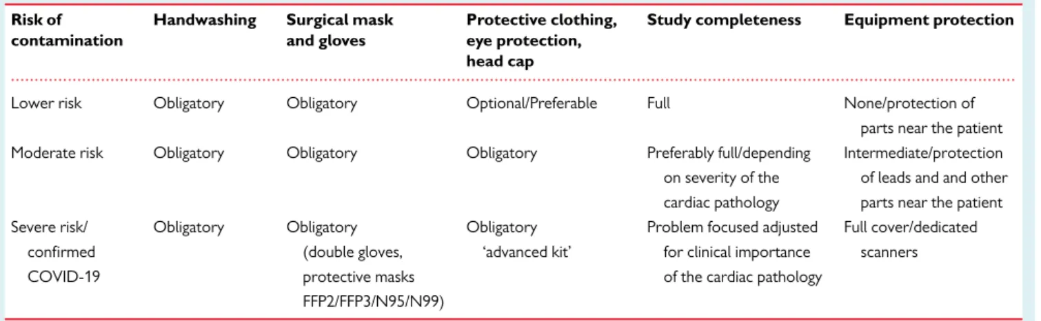
TOE might be stressful to our patients and should be avoided in most patients with ongoing COVID-19. The risk of contamination of equip- ment and personnel is also very high during the procedure due to droplets and aerosols containing virus. The incremental role of TOE over TTE should be carefully considered. This procedure should therefore be reserved for patients where the suspected findings are of crucial importance to confirm or exclude a diagnosis or to guide treatment.
Indication for other
echocardiographic methods
Indications for stress echocardiography, as well as for other stress imaging techniques, seem very limited in the COVID-19 pandemic, and should be avoided in patients with acute infection. Coronary CT angiography should be the preferred method if patients are being investigated for chronic coronary syndromes.6 Ultrasonic contrast agent may be useful in some patients, but should not be used in circu- latory unstable or critically ill patients.12
Foetal echocardiography
It is currently unclear whether maternal infection affects the foetus, by causing either structural heart disease or foetal myocarditis, and therefore routine foetal echocardiography in infected mothers is not recommended for the indication of COVID-19 infection alone.
However, the indications for foetal echocardiography remain the same during the pandemic, in that pregnancies judged to be at high risk for foetal cardiac disease need to continue to be referred accord- ing to local guidelines, and assessed within the appropriate time frame.13Counselling may be undertaken using video conferencing or other technology in order to reduce time in physical proximity to the
.. ..
.. ..
.. ..
.. ..
.. ..
.. ..
.. ..
.. ..
.. ..
.. ..
.. ..
.. ..
.. ..
.. ..
.. ..
.. ..
.. ..
.. ..
.. ..
.. ..
.. ..
.. ..
.. ..
.. ..
.. ..
.. ..
.. ..
.. ..
.. ..
patient, and the minimum number of healthcare professionals should attend scanning and counselling.
Indication for other imaging methods
Given the acute nature of the disease, and restrictions of hospital facilities for chronic patients, there are probably few defined indica- tions for coronary CT angiography, CMR, or nuclear cardiology in patients with COVID-19 infection during the acute phase.
Similar to the situation for echocardiography, patients scheduled for CMR, CT, and nuclear cardiology, with non-severe symptoms or not eligible for invasive or surgical treatment, should be postponed.
Routine follow-up scans should be re-scheduled. A possible indica- tion for CMR is the suspicion of COVID-19 myocarditis, but the clin- ical implication of detecting myocarditis in these patients is not determined. We suggest that positive troponins and myocardial dys- function or severe arrhythmia not explained by other methods may be an indication for acute CMR if of crucial importance for the treat- ment and the patient is stable enough to be scanned.
Cleaning, disinfection, and protection in patients with suspected or confirmed COVID-19
Equipment
All equipment used in close contact with patients has the potential to carry droplets containing the virus. This includes ECG leads which should therefore be avoided when performing echocardiographic studies in COVID-19 patients.
Moderately warm water and a mild detergent constitute the basis of equipment cleaning in all cardiac imaging machines, including the echocardiographic probe. An ordinary water-soluble disinfectant should also be added, but not on the membrane. A non-alcoholic
disinfectant should be used on the echocardiographic probe (please confirm with recommendations for each vendor). Dedicated wipes may also be used in this process.
Protecting the echocardiographic machines with custom-made covers may be possible if available, but, if not, other protective equip- ment can be fashioned using local entrepreneurs and ingenuity. It is, however, important not to cover the screen in a way that reduces the view for the echocardiographer. This may reduce the quality of the study and increase scan duration. Similarly, the keyboard should be fully operative during the investigation. To facilitate the cleaning of the scanner, all additional 3-D and single-Doppler probes should be removed before the scanning starts, if they are not needed for proper diagnosis. ECG leads should also be removed, although this may make the recordings of loops more difficult and potentially more time con- suming. In high-volume centres, one can consider dedicating separate scanners to be used exclusively for COVID-19-positive patients, that remain within designated COVID-19 areas. No additional disinfection procedure is necessary for a TOE probe as every location should have proper routines for cleaning and disinfection of these probes. A protective coat on the TOE probe might also be considered.
The positioning of the patient vs. the echocardiographer and the scanner may be of importance. Patients placed in the left lateral pos- ition with the scanner positioned on the right side of the bench will result in the longest possible distance between the faces of the pa- tient and the echocardiographer. The contamination of the scanner by airborne droplets from the patient will also probably be mini- mized. However, the preferred patient position is different among echocardiographers, and these recommendations should not be a hindrance to performing high-quality fast echocardiograms. A surgical mask on the patient will also reduce contamination by air droplets.
Facilities
The echocardiographic study will usually be performed in the ICU or in emergency rooms in critically ill patients. Less critical patients are usually examined in their ward rooms. Dedicated room(s) may be prepared in the echocardiographic lab, where unnecessary equip- ment can be removed to make the cleaning of the room as easy as possible. However, there is less risk of virus spread if the
...
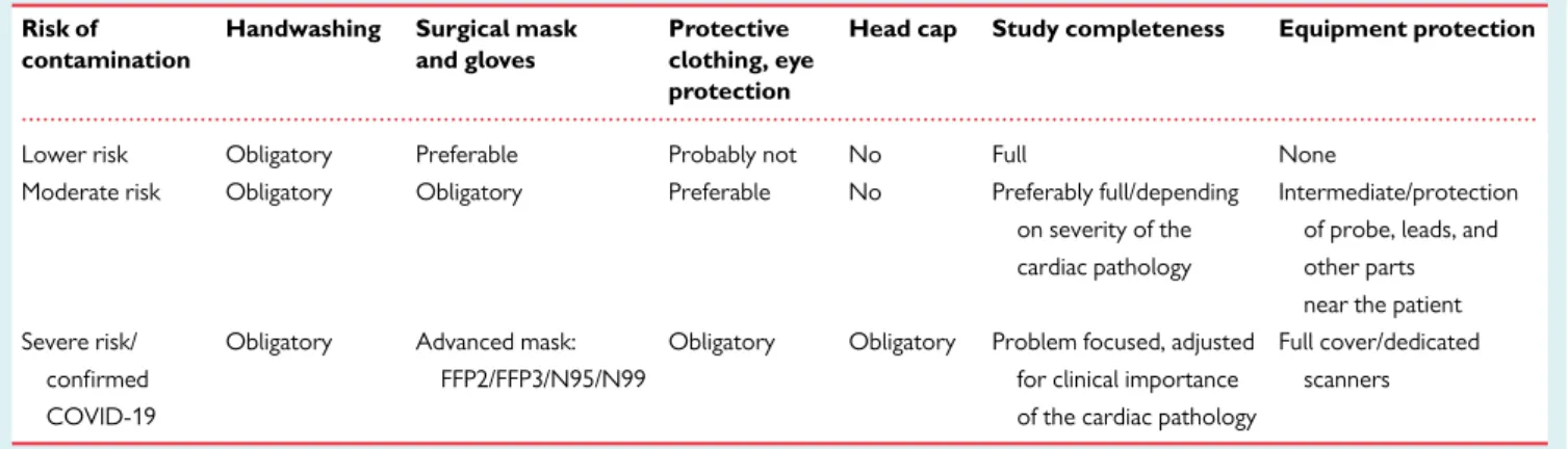
Table 1 Recommendations during TTE and foetal echo
Risk of contamination
Handwashing Surgical mask and gloves
Protective clothing, eye protection
Head cap Study completeness Equipment protection
Lower risk Obligatory Preferable Probably not No Full None
Moderate risk Obligatory Obligatory Preferable No Preferably full/depending
on severity of the cardiac pathology
Intermediate/protection of probe, leads, and other parts near the patient Severe risk/
confirmed COVID-19
Obligatory Advanced mask:
FFP2/FFP3/N95/N99
Obligatory Obligatory Problem focused, adjusted for clinical importance of the cardiac pathology
Full cover/dedicated scanners
Lower risk, patients with no symptoms, no increased risk behaviour, a recent negative virus test, or in areas with low risk of COVID-19. Moderate risk, patients with non-specif- ic/unclear symptoms or patients without symptoms in an area with moderate or high risk of COVID-19. Severe risk, patients with typical symptoms or confirmed COVID-19.
FFP2, Filtering Facepiece Particulate class 2 (FFP2 corresponds to US N95, FFP3 corresponds to US N99).
.. ..
.. ..
.. ..
.. ..
.. ..
.. ..
.. ..
.. ..
.. ..
.. ..
.. ..
.. ..
.. ..
.. ..
.. ..
.. ..
.. ..
.. ..
.. ..
.. ..
.. ..
.. ..
.. ..
.. ..
.. ..
.. ..
.. ..
.. ..
.. ..
echocardiographer brings the echocardiographic machine to the pa- tient, and the patient can remain in their isolation. Local factors must be considered with dedicated COVID-19 areas respected, ensuring COVID-19 ‘clean’ and ‘dirty’ areas are not mixed.
Reading and conference rooms where echocardiograms are pre- sented on small PC screens may prevent the recommended 2 m dis- tance between the experts. Thus, larger rooms, with projectors that can present the images on large screens are recommended. Virtual communication technology that allows several colleagues to simul- taneously visualise images on geographically remote screens is the preferred solution for multidisciplinary team meetings.
Healthcare personnel
All advice regarding personal protective equipment (PPE) should follow the internal rules in each institution. This will vary accord- ing to the local nature of the pandemic and the availability of PPE. We here offer some general advice. Repeated and thorough handwashing is the basis of virus protection for everyone, includ- ing patients and health professionals. In addition to handwashing after every examination, disinfecting agents should be used on the hands.
When examining a patient with confirmed or suspected COVID-19, protective clothing, gloves, headcovers, specific face- masks, and eye shields must be used (Table 1). Patients should wear a surgical mask during imaging (Table2). During TOE, medic- al protective masks must be used due to risk of aerosols and air- borne spread (Table3). How to adequately dress and undress is
described in several publications and is not further described here.
When performing an echocardiogram in patients without con- firmed COVID-19, surgical facemasks should be used in regions where the risk of virus spread is high or uncertain. Non-sterile gloves should also be used and renewed between every patient since the persistence of the virus on plastic is long. If the patient has low risk of infection due to low risk in the respective regions or has no symp- toms, this may be omitted, especially if there is shortage of face masks or gloves in the hospital.
Precautions in other imaging modalities
Precautions for all the other imaging modalities are similar. Imagers and technologists should wear protective clothing, gloves, and face- masks while undergoing scanning, and patients should wear a surgical mask during imaging. The scanner, coils, and ECG cables are manda- tory for cardiac CMR and CT scans but need to be thoroughly disin- fected after imaging (Table1). Cleaning of the scanners after imaging patients with COVID-19 infection is also obligatory (see above). An important strategy to reduce contamination is to reserve one scan- ner for known infected patients and another for low-risk and non- infected patients.
Conclusion
The COVID-19 pandemic has forced us to reconsider how best to perform cardiac imaging in the right patients at the right time and how to minimize the risk of cross-infection for imagers and patients alike. These recommendations are suggested as tools to guide good clinical practice during what is a turbulent period in our practice, and one that is rapidly changing both the premises and demands for car- diac imaging. We expect our understanding of how best to image patients during the COVID-19 pandemic to change rapidly and will adapt our guidance accordingly.
Conflict of interest:none declared.
...
Table 3 Recommendations during TOE
Risk of contamination
Handwashing Surgical mask and gloves
Protective clothing, eye protection, head cap
Study completeness Equipment protection
Lower risk Obligatory Obligatory Optional/Preferable Full None/protection of
parts near the patient
Moderate risk Obligatory Obligatory Obligatory Preferably full/depending
on severity of the cardiac pathology
Intermediate/protection of leads and and other parts near the patient Severe risk/
confirmed COVID-19
Obligatory Obligatory (double gloves, protective masks FFP2/FFP3/N95/N99)
Obligatory
‘advanced kit’
Problem focused adjusted for clinical importance of the cardiac pathology
Full cover/dedicated scanners
For definition of risks of contamination, seeTable1. Washing and disinfection of the TOE probe and its leads are not further described as standard procedures should include sufficient virus protection.
...
Table 2 Recommendations for patients during all imaging modalities
Risk of infection Surgical mask
Lower risk Preferable
Moderate risk Obligatory
Severe risk/confirmed COVID-19 Obligatory
.. ..
.. ..
.. ..
.. ..
.. ..
.. ..
.. ..
.. ..
.. ..
.. ..
.. ..
.. ..
.. ..
.. ..
.. ..
.. ..
.. ..
.. ..
.. ..
References ..
1. Wu Z, McGoogan JM. Characteristics of and important lessons from the corona- virus disease 2019 (COVID-19) outbreak in China: summary of a report of 72314 cases from the Chinese Center for Disease Control and Prevention.
JAMA2020;doi:10.1001/jama.2020.2648.
2. Zheng Y-Y, Ma Y-T, Zhang J-Y, Xie X. COVID-19 and the cardiovascular system.
Nat Rev Cardiol2020;doi:10.1038/s41569-020-0360-5.
3. Xiong T-Y, Redwood S, Prendergast B, Chen M. Coronaviruses and the cardio- vascular system: acute and long-term implications.Eur Heart J2020;doi:10.1093/
eurheartj/ehaa231.
4. Chan JF-W, Yuan S, Kok K-H, To KK-W, Chu H, Yang J, Xing F, Liu J, Yip CC-Y, Poon RW-S, Tsoi H-W, Lo SK-F, Chan K-H, Poon VK-M, Chan W-M, Ip JD, Cai J-P, Cheng VC-C, Chen H, Hui CK-M, Yuen K-Y. A familial cluster of pneumonia associated with the 2019 novel coronavirus indicating person-to-person trans- mission: a study of a family cluster.Lancet2020;395:514–523.
5. Elkind MS, Harrington RA, Benjamin IJ. Role of the American Heart Association in the global COVID-19 pandemic.Circulation2020;doi:10.1161/CIRCULATION AHA.120.046749.
6. Knuuti J, Wijns W, Saraste A, Capodanno D, Barbato E, Funck-Brentano C, Prescott E, Storey RF, Deaton C, Cuisset T, Agewall S, Dickstein K, Edvardsen T, Escaned J, Gersh BJ, Svitil P, Gilard M, Hasdai D, Hatala R, Mahfoud F, Masip J, Muneretto C, Valgimigli M, Achenbach S, Bax JJ, ESC Scientific Document Group.
2019 ESC Guidelines for the diagnosis and management of chronic coronary syn- dromes.Eur Heart J2020;41:407–477.
7. Lancellotti P, Price S, Edvardsen T, Cosyns B, Neskovic AN, Dulgheru R, Flachskampf FA, Hassager C, Pasquet A, Gargani L, Galderisi M, Cardim N, Haugaa KH, Ancion A, Zamorano J-L, Donal E, Bueno H, Habib G. The use of echocardiography in acute cardiovascular care: recommendations of the European Association of Cardiovascular Imaging and the Acute Cardiovascular Care Association.Eur Heart J Cardiovasc Imaging2015;16:119–146.
8. Peng Q-Y, Wang X-T, Zhang L-N, Chinese Critical Care Ultrasound Study Group (CCUSG). Findings of lung ultrasonography of novel corona virus pneu- monia during the 2019–2020 epidemic. Intensive Care Med 2020;doi:
10.1007/s00134–020–05996–6
9. Neskovic AN, Skinner H, Price S, Via G, De Hert S, Stankovic I, Galderisi M, Donal E, Muraru D, Sloth E, Gargani L, Cardim N, Stefanidis A, Cameli M, Habib G, Cosyns B, Lancellotti P, Edvardsen T, Popescu BA, Reviewers: This document was reviewed by members of the 2016–2018 EACVI Scientific Documents Committee. Focus cardiac ultrasound core curriculum and core syllabus of the European Association of Cardiovascular Imaging.Eur Heart J Cardiovasc Imaging 2018;19:475–481.
10. Cardim N, Dalen H, Voigt J-U, Ionescu A, Price S, Neskovic AN, Edvardsen T, Galderisi M, Sicari R, Donal E, Stefanidis A, Delgado V, Zamorano J, Popescu BA.
The use of handheld ultrasound devices: a position statement of the European Association of Cardiovascular Imaging (2018 update). Eur Heart J Cardiovasc Imaging2019;20:245–252.
11. Habib G, Lancellotti P, Antunes MJ, Bongiorni MG, Casalta J-P, Del Zotti F, Dulgheru R, El Khoury G, Erba PA, Iung B, Miro JM, Mulder BJ, Plonska-Gosciniak E, Price S, Roos-Hesselink J, Snygg-Martin U, Thuny F, Tornos Mas P, Vilacosta I, Zamorano JL, ESC Scientific Document Group. 2015 ESC Guidelines for the management of infective endocarditis: The Task Force for the Management of Infective Endocarditis of the European Society of Cardiology (ESC). Endorsed by:
European Association for Cardio-Thoracic Surgery (EACTS), the European Association of Nuclear Medicine (EANM).Eur Heart J2015;36:3075–3128.
12. Neglia D, Liga R, Caselli C, Carpeggiani C, Lorenzoni V, Sicari R, Lombardi M, Gaemperli O, Kaufmann PA, Scholte AJHA, Underwood SR, Knuuti J, EVINCI Study Investigators. Anatomical and functional coronary imaging to predict long- term outcome in patients with suspected coronary artery disease: the EVINCI- outcome study.Eur Heart J Cardiovasc Imaging2019;doi:10.1093/ehjci/jez248 13. Donofrio MT, Moon-Grady AJ, Hornberger LK, Copel JA, Sklansky MS,
Abuhamad A, Cuneo BF, Huhta JC, Jonas RA, Krishnan A, Lacey S, Lee W, Michelfelder EC, Rempel GR, Silverman NH, Spray TL, Strasburger JF, Tworetzky W, Rychik J, American Heart Association Adults With Congenital Heart Disease Joint Committee of the Council on Cardiovascular Disease in the Young and Council on Clinical Cardiology, Council on Cardiovascular Surgery and Anesthesia, and Council on Cardiovascular and Stroke Nursing. Diagnosis and treatment of fetal cardiac disease: a scientific statement from the American Heart Association.Circulation2014;129:2183–2242.