— 1 0 —
GEORG BENZ
Entomological Laboratory, Swiss Federal Institute of Technology, Zürich, Switzerland
I. Introduction 299
II. General Physiopathology 300
Α. Physiology of Infection 300
Β. Physiopathology of Alimentation 307 C. Physiopathology of Respiration 308 D. Physiopathology of the Circulatory System . 310 Ε. Physiopathology of the Hemocytes 314 F. Physiopathology of the Adipose Tissue . . . . 315
G. Physiopathology of Excretion 316
H. Physiopathology of the Nervous System Behavior
and of 317 III. Special Physiopathology and Histochemistry . . . . 320
Α. Mycoses 320
Β. Bacterioses 322
C. Rickettsioses 324
D. Vi roses 325
IV. Conclusion 331
References 331
I . INTRODUCTION
T h e science of disturbed function in organisms is generally called
"pathophysiology." However, since infectious diseases result from the presence of viruses or living microorganisms in the host, the physiolog
ical aspects of the host-parasite relationship, and the pathological proc
esses involved, depend on the physiology of both the parasitic microbe (although, strictly speaking, viral agents do not have a physiology of their own) and the host. Therefore, we prefer the term "physiopathol
ogy," thus intimating that it includes not only pathophysiology, but also the study of the processes leading to pathological conditions.
Physiopatkohgy and Histochemistry
299
T h e factors involved in infection, immunity, host specificity, patho
genicity, etc., may be understood, only if we know the physiological background of the pathogen and the insect host. However, the scope of this chapter does not allow us to enter into the details of insect physi
ology. T h e reader is therefore referred to such textbooks as those of Roeder (1953) and Wigglesworth (1950).
Although organic substances are but the results of biochemical proc
esses and do not represent biological functions, some biochemical facts are necessary for the understanding of the physiological processes. Be
sides, functions in organisms are linked with their structures. Thus, many physiopathological processes may be investigated with the aid of histopathological and cytopathological methods. Some biochemical and histochemical data are therefore included in this chapter.
Some viroses are probably the diseases studied best with regard to physiopathology (reviewed by Bergold, 1959). No survey of the physio- pathology of other insect diseases has been published until now; the reason for this may be our limited knowledge in this field. T h e facts presented in this chapter provide but a rather arbitrary picture of insect physiopathology. In order to gain a general view, most facts have been compiled according to the physiological system affected, i.e., with
out regard to the systematic order of the causative microbes. A few specific diseases of which the physiopathology and the histochemistry have been studied more thoroughly, are treated in the last section.
I I . GENERAL PHYSIOPATHOLOGY
A. Physiology of Infection
Insect pathogens may invade the insect organism by different routes:
through the integument, the tracheal system, the gut, the rectum, and by trans-ovum transmission. Physiological data are available on the first three modes of infection only.
1. Infection via the Integument
T h e structure of the epidermis makes the intact integument im
pervious to protozoa, bacteria, rickettsiae, and viruses. However, this does not apply to certain fungi and possibly to a Micrococcus, as sug
gested by Tauber (1940), and Spirochaeta pieridis Paillot (Paillot, 1940). Nothing is known, however, about the physiology of bacterial infection via the integument. A possible route of infection are the ducts of the dermal glands. In the two cases mentioned, one has to take into account a possible infection via the tracheae as well. T h e effectiveness of the insect integument as a barrier, even to most fungi, is demonstrated by the phenomenon of wound infection as studied by Burnside (1930),
10. PHYSIOPATHOLOGY AND HISTOCHEMISTRY 301 Vago (1956b, 1958), Hurpin and Vago (1958), Jolly (1959), and Müller-Kögler and Huger (1960). It has been found by these authors that quite a number of fungi which normally are apathogenic sapro
phytes, may become parasites and kill the host if they can penetrate the integument of an insect through wounds. Quite a number of fungi, however, are adapted to true parasitism in insects if, by germinating on
the insect's surface, they are able to penetrate the integument by their own growth. Besides the proper environmental conditions (high hu
midity and reasonably high temperature), the set of extracellular en
zymes produced by these fungi seems to be largely responsible for the success of infection via the integument. Thus Huber (1958) found extracellular lipases to be produced by Aspergillus flavus Link, Beau- veria bassiana (Balsamo) Vuillemin, Metarrhizium anisopliae (Metch- nikoif) Sorokin, and Cordyceps militaris (Fries) Link, and Schwei
zer (1948) found them produced by Empusa muscae Cohn. Extracel
lular proteinases were found by Huber (1958) from A. flavus, B. bas
siana, and C. militaris, and by Burnside (1930) from A. flavus, and two other Aspergillus species as well as from Entomophthorales. Chitinase was found to be excreted by A. flavus, B. bassiana, M. anisopliae, and C.
militaris (Huber, 1958). T h e same author found that the chitinase of these fungi breaks down chitin to iV-acetylglucosamine, and not to D- glucosamine, as do the chitinases of bacteria. T h e fact that fungi grown on artificial media without chitin reduce the production of chitinase, demonstrates that the production of chitinase is an adaptation to the insect cuticle. Although these fungi possess chitinous cell walls when grown in a medium devoid of chitin, their mycelia are without chitin when grown in a medium containing chitin. This reaction gives the answer to the question why these fungi are able to digest the chitin of insects without destroying their own cell walls.
It may be seen at once that these species of typical parasitic fungi have the potency to secrete most enzymes necessary to dissolve an insect cuticle. Until now, no fungus has been reported to secrete an enzyme that dissolves wax which is a constituent to the insect's cuticle.
Metarrhizium anisopliae appears to have no extracellular proteinase, and Entomophthorales appear not to possess chitinases. Although it has been claimed by Huber that none of the specific enzymes has a direct relation to the pathogenicity of a fungus, it is very probable that the whole set of enzymes is a great help to the fungus in overcoming the barrier of the body wall. Since the hardiness of the integument is a function of the sclerotized protein of the cuticle, but not of chitin, it is understandable that entomophthoraceous fungi may well do without an extracellular chitinase. On the other hand, it is remarkable that M.
anisopliae is able to penetrate a cuticle without the help of a proteinase.
If this fungus germinates on the thick integument of the dorsal side of Pyrausta nubilalis (Hübner), it appears that the hyphae always pene
trate the cuticle at the site of certain pigmented bodies which traverse the cuticle to about one-third of its thickness (Wallengren and Johans
son, 1929). Those structures probably represent the endings of pore canals. It would be worth while to investigate the chemical nature of these pigmented bodies and to compare the result with the enzymatic equipment of M. anisopliae. Besides, the hyphae of M. anisopliae are able to exert a rather great pressure on the cuticle. This mechanical force probably outweighs the lack of a proteinase. T h a t the cuticle may sometimes resist the pressure of hyphae is demonstrated by the fact that hyphae frequently grow a relatively long way in the direction of the parallel cuticular lamellae, evidently because in this direction, the cu
ticle offers the least resistance to growth.
T h e endocuticle of Malacosoma alpicola becomes basophilic and gives a positive periodic acid-Schiff (PAS)-reaction in regions where hyphae of a Spicaria sp. have penetrated the integument (Benz, unpub
lished) . This reaction may be similar to the positive PAS reaction of endocuticle being digested by molting fluid which contains a proteinase and a chitinase (Wigglesworth, 1957).
On the other hand, the whole set of enzymes mentioned above does not allow A. flavus to penetrate through the cuticle of Schistocerca gre- garia Forskäl (Lepesme, 1938). However, A. flavus is able to penetrate the cuticle of several other insects (Burnside, 1930; Sussman, 1952a;
Koidsumi, 1957).
It has been suggested by Sussman (1951) that the epicuticular layer of insects may present an obstacle for fungal growth. If he removed with ether this layer from pupae of Platysamia cecropia (Linnaeus), resistance to infection with A. flavus was much reduced. If the wax layer should have a protective function against microbial diseases, the observations of Koidsumi (1951, as cited by Wigglesworth, 1957) on the thickness of the wax layer of Chilo simplex Butler—which depends on the environmental humidity—would imply that moist conditions could favor the occurrence of insect mycoses not only because the fungal spores receive the necessary moisture for germination, but also because under such conditions insects may produce an epicuticle with a thinner wax layer.
It has been demonstrated by Koidsumi (1957) that the integument of insects is a particularly effective barrier to invading microorganisms not only because of its mechanical structure. This author found that the cuticle of Bombyx mori Linnaeus contains free saturated fatty acids of
10. PHYSIOPATHOLOGY AND HISTOCHEMISTRY 303 medium chain length (presumably caprylic acid, or capric acid) which have strong antifungal properties. It is also known that fatty acids of low molecular weight possess bactericidal properties.
2. Infections via the Tracheal System
T h e histological structure of the tracheae is essentially the same as that of the body surface from which they are derived. T h e cuticular lining and the cuticulin layer of the tracheae is, however, much thinner than in the body wall. Chitin is absent in the tracheoles and the air sacs of all insects, and in the large tracheae of Apis mellifera Linnaeus, and of Diptera as well. In the finest terminations, the lining membrane is very delicate and is freely permeable to water (Wigglesworth, 1950).
It is clear from these facts that the invasion of the insect body by the route of the tracheae should be relatively easy. T h e moist conditions in the organs should particularly favor the germination of fungal spores.
Unfortunately, exact studies on these problems are entirely lacking.
According to Lepesme (1938), spores of A. flavus are able to infect Schistocerca gregaria neither through the cuticle nor the abdominal tracheae, but via the thoracic tracheae. Beauveria bassiana infects quies
cent larvae of Cephaleia abietis (Linnaeus) especially easily via the tracheae, but it can infect also via the integument (Donaubauer, 1949).
Some bacteria may infect insects via the tracheae. Burnside (1928, 1929) described a bacterial septicemia of the honey bee by a bacterium which he called Bacillus apisepticus [= Pseudomonas apisepticus (Burn- side)]. Burnside was not able to infect bees per os, but he obtained a high incidence of septicemia, followed by death, after spraying the bees with a bacterial suspension or after dipping them into the suspension.
He concluded, without further evidence, that P. apisepticus enters the bee via the tracheae. Since the tracheae of bees do not contain chitin, infection by bacteria via the tracheae may be relatively easy.
3. Peroral Infection
2L. Modes of infection via the gut. Entrance into the gut lumen may be relatively easily achieved by microorganisms. Quite a number of bacteria and protozoa, therefore, constitute the normal biosis of the alimentary canal of insects. T h i s is especially true for all insects with an approximately neutral pH of the gut. Many of the microorganisms live in the gut contents as harmless commensals, sometimes even as useful symbiotes, assisting the insect in the digestion of food material and producing nutritive substances and vitamins. T h e symbiotic organ
isms are especially well adapted to living in the gut.
T h e gut lining acts as an absolute barrier for all these organisms.
This is important, because many of the harmless commensals produce a fatal septicemia when allowed to enter the hemocoel. T h e gut lining, however, is not a very good barrier against certain fungi, which may easily invade the insect body via the gut. T h e chitinous lining of the gut is also easily penetrated by several sporozoan parasites. T h e damage caused to the gut by such organisms, may also facilitate the passage of bacteria into the hemocoel.
In the honey bee, the lining of the anterior pyloric region seems to react on Leptomonas infections by the production of a crusty scab
(Lotmar, 1946). T h e development of the flagellates seems to depend on the presence of pollen in the gut. Similarly the development of Nosema apis Zander is facilitated by a nutrition rich in protein, especially by pollen. It is therefore difficult to infect newly hatched bees with spores of N. apis before the bees have fed on some protein (Steche, 1960).
Beutler et al. (1949) found a positive correlation between the amount of pollen fed to infected bees, and the number of spores produced.
T h e alkaline, or strongly acid reaction of the gut contents of many insects, together with the action of the digestive enzymes, provide a mechanism which either kills some microorganisms, or prevents their multiplication or the germination of spores, thus protecting the host from too large numbers of commensals or parasites. Insects may adapt their production of digestive enzymes to bacterial invasion. Poltev (1954, as cited by Krieg, 1961) reports that repeated oral application of bac
teria stimulates the production of enzymes, so that a temporary pro
tection against infections of the alimentary tract is gained by the treated insects.
Some insects like Phormia terrae-novae Robineau and Desvoidy in addition possess bactericidal substances in their gut (Pavilland and Wright, 1957). An example of the deleterious action of the digestive fluid of the gut of the adult Apis mellifera is given by Burnside (1930).
T h e author found that spores from old cultures of a certain strain of the fungus Aspergillus ochraceus Wilhelm can infect honey bees, whereas spores from fresh cultures are apathogenic. Since the spores from old cultures possess much thicker spore walls, we may conclude that they manage to pass a dangerous zone in the gut, and then may safely germi
nate, whereas thin-walled spores are destroyed.
T h e difference in pH of the gut contents of various insect species is partly responsible for differences in the microflora. It also explains why some bacteria are pathogenic in some insects, but not in others. Thus, the strains of Bacillus cereus Frankland and Frankland, which produce lecithinase with a pH optimum of 6.6 to 7.4, can grow within the pH range of 5.0 to 9.3 (Stephens, 1952). They are pathogenic for larvae of
10. PHYSIOPATHOLOGY AND HISTOCHEMISTRY 305 the larch sawfly, Pristiphora erichsonii (Hartig), with a midgut pH of 7.15 to 8.4, but not for the tent caterpillar, Malacosoma disstria Hübner, with a gut pH of 9.2 to 10.3 (Heimpel, 1955a, b; Kushner and Heimpel, 1957).
On the other hand, Bacillus thuringiensis Berliner and related bacilli producing toxic parasporal crystals, are highly pathogenic in lepidop- terous larvae with an alkaline pH of the gut but are not, or are hardly, pathogenic in larvae of sawflies and noctuids, which have slightly alka
line to neutral gut pH's. T h e pathogenicity of the crystalliferous bac
teria is greatly enhanced by the action of the toxin. This, however, acts only if dissolved in the gut juice, or an alkaline solution (Hannay, 1953; Hannay and Fitz-James, 1955). When dissolved in an alkaline solution, it is not toxic in the neutral blood of insects (Angus, 1954,
1956a, b ) . However, it has been reported by Martouret (1962) that the toxin, when split into smaller components by the digestive action of the gut enzymes of Pieris brassicae Linnaeus, has a toxic action if injected into the hemocoel. In the light of recent observations on a toxic principle in the gut of P. brassicae (Benz, 1962b), it will be worth-while to study this problem also in other insect species. T h e action of the B. thuringiensis toxin very much resembles the action of the phytogenous insecticide rotenone. Whereas silkworm larvae, with a high gut pH, die rapidly after ingestion of small quantities of rotenone, Prodenia larvae can eat large quantities without showing ill effects, and most of the in
gested rotenone appears well preserved in the feces (Woke, 1938, 1940).
Lecithinase in B. cereus, as well as the crystalline toxin in B. thuringien
sis, seems to be essential, because they make the gut permeable for spores and bacteria which pass into the hemocoel and there produce septicemia.
Since virus inclusion bodies (polyhedra and capsules) are not dis
solved in the neutral hemolymph of insects, the enclosed viruses can enter the host only after the inclusion bodies have been dissolved in the gut of the insect (Komarek and Breindl, 1924). Surface sterilized polyhedra injected into the hemolymph of larvae of Lymantria monacha (Linnaeus), are therefore not able to produce polyhedrosis. T h e dis
solution of the inclusion bodies is performed by the digestive fluids, either by the alkaline pH or by the action of proteolytic enzymes.
T h e gut seems to be the critical barrier for many deoxyribonucleic acid (DNA) viruses. It has been found by Gershenson (1957) that many Borrelinavirus species which are not able to infect foreign hosts via the gut, may produce polyhedrosis when injected as free particles into the hemocoel.
It is strange that insect viruses with a dimension of 300 Ä or more
are able to enter the insect cells, although we know that the peritrophic membranes retain particles larger than 25 Ä (Dehn, 1933). No con
clusive investigations in this direction have been conducted as yet. As some evidence indicates, however, insect viruses may have to be broken down into smaller units before they can infect the host.
b. The influence of stressors on infection via the gut. It has been claimed by several authors that damage caused by abrasives like sand or ground glass in the gut lining of insects may promote septicemia by bacteria which otherwise cannot penetrate into the hemocoel. A similar cause may be involved, when a saprophytic species of Mucor becomes a parasite in Bombyx mori after the larvae have been fed with the hairy leaves of Podospermum (Vago, 1961). It is also known to most ento
mologists that wet food, or the change from one food plant to another, may cause dysentery in larvae of phytophagous insects. Wet food espe
cially will disturb digestion, because the digestive fluids are too greatly diluted. Such disturbances facilitate the development of certain intes
tinal bacteria, which are only facultative parasites. T h e development of strains of Aerobacter, or Enterococcus, leads to local lesions in the intestinal epithelium and thus slows down digestion. These bacteria may fill the gut lumen until it disrupts. A classical case of this type of bacterial disease is the flacherie of the silkworm, which today is consid
ered a common name for several distinct affections (Vago, 1951; Masera, 1954). According to Lysenko (1958), Streptococcus spp. can maintain themselves in the healthy gut by virtue of their tolerance to relatively high alkaline conditions (pH 9 . 6 ) . Under stress conditions, these bac
teria multiply, lower the pH to optimum growing conditions, and are then able to produce disease. Bacteria with proteolytic activity in particular (e.g., Pseudomonas aeruginosa Schroeter, Proteus sp., Serratia marcescens Bizio, Bacillus mycoides Flügge, and Streptococcus pyogenes Rosenbach) lead to septicemia in the course of digestive affections
(Vago, 1961).
Several authors report the induction of virus diseases by nutritional factors. At present, however, it is not clear in which way these factors influence virus diseases, nor whether they play a part in protozoan in
fections. Mycologists generally agree that nutritional factors do not influence fungal infections.
Starvation may be another stressor. It has been shown by Heimpel (1955a) that starvation may lead to changes in the pH of the alimentary canal. Thus the pH of the medium part of the midgut of Malacosoma disstria may be lowered from 10.3 to 8.4 after 96 hours starvation. T h i s reaction might also weaken the resistance of the insect to microbial dis
eases.
10. PHYSIOPATHOLOGY AND HISTOCHEMISTRY 307 Other stressors, like excessive heat, may promote septicemia by bac
teria of the gut (Steinhaus and Dineen, 1960). T h e high temperature is probably optimal for these bacteria, but may adversely affect the insect host. T h e excessive multiplication of the bacteria may lead to digestive disturbances with all the consequences mentioned above.
B. Physiopathology of Alimentation
W e have mentioned before that disturbances in nutrition may pro
mote microbial infections. On the other hand, various diseases may cause nutritional disturbances. T h e i r effect on alimentation is usually rather uniform: reduction, or cessation of feeding, caused by local lesions in the alimentary canal or by a general dysfunction of the whole organ
ism. Digestion, the secretion of enzymes, the dehydration of the food, the intestinal bacteria, and the discharge of feces may be affected.
A spectacular example is the toxic effect of crystalliferous bacilli of the B. thuringiensis-group, whose parasporal toxin paralyzes the diges
tive tract (Vankova, 1957; Heimpel and Angus, 1959). Vago (1956b) has observed that infections with minimal doses of bacteria (especially of the B. thuringiensis group), which are too low to cause either toxemia or septicemia, may lead to localized intoxications in the alimentary canal of Arctiacaja (Linnaeus), Porthetria dispar (Linnaeus), and Bombyx mori. T h e localized foci favor the development of secondary intestinal bacteria which produce chronic digestive troubles; they in turn are responsible for a minimal uptake of food by the larvae and a retarda
tion of growth. Some fifth-instar larvae are four times smaller than healthy individuals. Such chronic reductions of alimentation may also be caused by an abnormally high development of facultative pathogenic bacteria in the gut. Enterococcus liquefaciens (Stern), which is normally present in small numbers in the digestive tract of lepidopterous larvae, may impair the digestion and utilization of food substances when it becomes too numerous. A disease of this type may present gattine of the silkworm (Paillot, 1926). In some cases, as with a Bacillus found by Bucher (1957) in Malacosoma pluviale (Dyar), the irreparable destruction of the gut leads to death by starvation and desiccation.
Protozoan infections are also known to influence alimentation. Di
gestive troubles combined with reduced feeding may be caused by le
sions of the gut epithelium, brought about by large numbers of Micro- sporidia. Thus, the infection of Nosema muscular is Weiser leads to a quick atrophy of the muscles of the alimentary canal and reduces the uptake and digestion of food (Weiser, 1957). T h e infection of Hyphan- tria cunea (Drury), and of Malacosoma neustria (Linnaeus) with Thelo- hania hyphantriae Weiser leads to chronic starvation (Weiser and Veber,
1957). T h e starvation effect in honey bees infected with the specific intestinal Nosema apis Zander, is best demonstrated by the underdevel
opment of the food glands, an effect that may be phenocopied in healthy worker bees by keeping them for some time on a protein-free diet (Lot- mar, 1936) and by the deficiency of nitrogen in diseased bees (Lotmar, 1939). Histochemical examination of the alimentary tract of infected bees also shows a reduction of the protein contents of the epithelial cells as well as in the muscles of the gut (Steche, 1960).
Digestion is also disturbed in sawfly larvae infected with nuclear polyhedrosis of the midgut (Bird and Whalen, 1953), and in lepidop- terous larvae infected with cytoplasmatic polyhedrosis (Vago, 1956b;
Huger and Krieg, 1958; Tanada and Chang, 1960). T h e same applies to larvae of Solenobia triquetrella (Hübner) infected by a Bacillus sp.
which lives intracellularly in the midgut epithelium (Puchta and Wille, 1956). T h e infected larvae are greatly reduced in size and retarded in development as compared to healthy larvae.
An interaction of respiration and digestion has been demonstrated by Vago and Vasiljevic (1962) in Bombyx, Antheraea, and Hyphantria.
While the closure of the thoracic spiracles leads to a complete cessation of the feeding activity, the closure of the fourth, fifth, and sixth pair of spiracles results in a reduction of the food uptake, and a dysfunction of digestion followed by an abnormal multiplication of the intestinal bacteria. T h e closure of the sixth and seventh pair of spiracles influ
ences the dehydration of the contents of the digestive tube, and also provokes an increase in bacterial fermentation. Finally, the closure of the seventh, eighth, and ninth pair of spiracles, stops defecation and promotes an extreme multiplication of the intestinal bacteria, followed by a progressive destruction of the epithelial cells of the intestinal tube.
This example clearly shows that many diseases acting on the tracheal system may have secondary effects on the functions of the alimentary canal.
Dysentery in Arctia caja as a result of restricted respiration in the course of nuclear polyhedrosis of the tracheae has been reported by Vago (1956a).
C. Physiopathology of Respiration
Theoretically, microbial parasites may affect respiration in insects either on the organic level by destroying the tracheal or nervous system, or on the cellular level by their action on the system of respiratory enzymes.
T h e first mode of action is rather common. T h e example of nuclear polyhedroses affecting the tracheal matrix has already been mentioned.
10. P H Y S I O P A T H O L O G Y AND H I S T O C H E M I S T R Y 309 In the case of polyhedrosis of Malacosoma alpicola Staudinger, the tra
cheae collapse; however, this occurs only in the late stages of the disease.
Since some of these viroses also affect the nerve cells, they may have secondary effects on respiration and other functions of the insect. Pupae of Platysamia cecropia are killed by A. flavus, because the fungus destroys the tracheoles and the ganglia, and thus makes respiration difficult or impossible (Sussman, 1952a). T h e classical example of the parasitiza- tion of the first pair of thoracic tracheae in the honey bee by the mite Acarapis woodi ( R e n n i e ) , although not a microbial disease, should be mentioned here too.
As a rule, anoxia lowers the pH of the hemolymph by the accumula
tion of C 02 and lactic acid. In Bombyx, however, anoxia leads to a rise in the blood pH (Gamo et al., 1933). T h e same is true for larvae of Gal- leria mellonella (Linnaeus), Malacosoma neustria, and Pieris brassicae when kept in an atmosphere of nitrogen for 1 to 3 hours (Aubel and Levy, 1931; Benz, 1962c). T h i s rise is due to the leakage of the alka
line gut contents into the blood. T h i s has been demonstrated in larvae of Pieris brassicae fed with red cabbage. After such larvae have been kept in N2 for 90 minutes or longer, the red color of the cabbage may be found in the hemolymph. T h e color is best seen after the hemolymph has been acidified (Benz, 1962c). Thus, anoxia, caused by a parasite or microbe, increases the permeability of the intestinal wall and therefore may promote secondary infections of the hemocoel by intestinal bacteria.
T h e influence of decreased respiration on the development of intes
tinal bacteria, and on digestion, has been investigated by Vago and Vasiljevic (1962), and has already been discussed.
Little is known about the action of microorganisms on the enzy
matic system of respiration. According to Sussman (1952b), the oxygen uptake in P. cecropia increases tenfold shortly after infection by A. flavus.
Less than half of it is due to fungal respiration; the rest has to be ascribed to the progressing oxygen consumption of the host tissues. Con
trary to this finding, the increased oxygen consumption of the sawfly Cephaleia abietis, infected by Serratia marcescens, may be completely at
tributed to the respiration of the bacterium (Lysenko and Slarna, 1959).
Although no differences in the oxygen consumption of polyhedrosis- infected larvae of B. mori have been found by Gratia et al. (1945), an increase in the respiratory activity, and a decrease in the catalase activ
ity of polyhedrosis-infected tissues has been reported by Akune (1951).
Gershenson (1962) similarly reported that infection with Borrelinavirus raises the oxygen consumption by 50 percent in developing pupae, and by nearly 100 percent in diapausing pupae of Antheraea pernyi Guerin- Meneville. T h e maximal respiratory activity corresponds to the time of
mass infection of cells. Contrary to the findings of Vago (1956a), in polyhedrosis-diseased larvae of Arctia, the infected pupae of Antheraea show increased oxygen consumption also after the cells of the tracheal matrix have been completely destroyed. High oxygen consumption per
sists almost to the death of the pupae. After death, no oxygen is con
sumed by these pupae, except in cases of postmortem invasion by bac
teria. An inverse linear relation has been found between the time from infection to death and the total amount of oxygen consumed. Injection of propyl gallate greatly reduces oxygen consumption, and at the same time considerably inhibits multiplication of the virus.
D. Physiopathology of the Circulatory System
T h e circulatory system of insects is an open, or vasculolacunar, sys
tem. Except for the heart and the aorta, the blood or hemolymph is free to circulate within the body cavity. In most insects, the hemolymph ac
counts for 10 to 40 percent of the body weight. T h e hemolymph affects the chemical exchange between organs, the transport and distribution of hormones and food, and the transport of waste products to the excre
tory organs. Moreover, it plays an essential part in transmitting pres
sure from one region to another and thus supports hatching, ecdysis, and the expansion of the wings. T h e hemolymph is also a reserve of water which can be drawn upon as needed. T h e insect hemolymph contains protein, like the blood of vertebrates, but in addition, it con
tains large amounts of free amino acids and peptides. Another peculi
arity of the insect hemolymph is the high content of reducing substances other than sugar. T h e blood reaction of most insects is usually slightly acid (pH 6.2-7.0), but it may be slightly alkaline in larvae of Diptera, and in the last-instar larvae of some Lepidoptera (pH 7.1 to 7.2) (Craig and Clark, 1938). T h e buffering capacity of the hemolymph is lowest at the physiological pH (i.e., near neutrality). Insects may tolerate con
siderable changes of pH to the acid side (Agrell, 1948). On the other hand, an increase in the blood pH will produce complete paralysis in caterpillars and probably also in other insects (Angus and Heimpel, 1956). T h e oxidation-reduction potential of insect larvae is low, but it rises at metamorphosis, allowing the melanizing action of tyrosinase.
According to Dennell (1949), tyrosinase is inhibited by a dehydrogenase system, which acts by lowering the redox potential below the point at which tyrosinase can function.
Mechanical suppression of the circulation of hemolymph may be caused by blood clots, as formed in grubs of Popillia japonica Newman after infection with Bacillus lentimorbus Dutky. T h e accumulation of these clots in the insect's appendages blocks the circulation of the blood,
10. P H Y S I O P A T H O L O G Y AND H I S T O C H E M I S T R Y 311 and thus produces a gangrenous condition of the appendages (Dutky, 1940). It is possible that this disturbance is primarily caused by a change in the oxidation-reduction potential, as reflected by the melani
zation of the blood clots. Mechanical suppression of circulation may also be caused by the growth and multiplication of hyphal bodies in the hemolymph. After infection of insects with Beauveria bassiana, circu
lation slows down, and eventually stops (Vittadini, 1853; Schaerffenberg, 1957). T h i s slowdown is probably caused by the obstruction of the ostia by hyphal bodies. However, a toxic action of the fungus may also be involved (see sections I I , Η and I I I , A, 1 ) .
Biochemical changes in the hemolymph are caused by numerous dis
eases. A very common affection of the blood is caused by starvation as a consequence of infectious diseases of the alimentary tract. Drilhon et al. (1951) were able to show that flacherie causes a strong reduction of free amino acids and other substances in the hemolymph of larvae of Bombyx mori. Similar reductions of free amino acids can be experi
mentally induced by starving larvae of Drosophila melanogaster Meigen (Chen and Hadorn, 1955) and grubs of Melolontha melolontha (Lin
naeus) (Fig. l b ) . Since Bacillus popilliae, when grown on an artificial medium, will sporulate only when transferred to a "starvation medium"
(Steinkraus and Provvidenti, 1958), we should think that the hemo
lymph of P. japonica must be rather depleted of nutrient substances at sporulation time. Chromatographic analysis of the hemolymph of larvae of Melolontha infected with the related Bacillus fribourgensis Wille, how
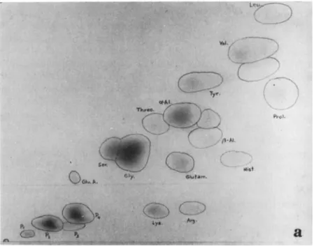
ever, show that sporulation occurs when the hemolymph is still quite rich in amino acids (Fig. l c ) . T h e composition of the diseased hemo
lymph is different from that of normal and starved grubs (see Fig. 1 ) . Similar changes in the blood of polyhedrosis-infected larvae of Bombyx mori (increase in histidine, and decrease in aspartic acid, cysteine, glu
tamic acid, glutamine, threonine, tyrosine, and valine) have been re
ported by Ishimori and Muto (1951). In late stages of the polyhedral disease of silkworms, tissues are broken down, and the content of the hemolymph in free ninhydrin-positive substances increases very much
(Drilhon et al, 1951).
A specific loss of tyrosine combined with a deficiency of proteins in the hemolymph of larvae of Melolontha infected with Rickettsiella melo- lonthae (Krieg) has been shown by Krieg (1958). T h e deficiency of tyrosine prevents melanization in grubs which have died of rickettsiosis.
Addition of tyrosine to diseased hemolymph causes melanization; how
ever, the reaction is slower than in normal hemolymph, a fact which indicates that tyrosine oxidase is present, but somewhat reduced (prob
ably in connection with a reduction of the hemocytes). Although the
concentration of protein is reduced in the hemolymph of diseased grubs, the blood becomes more viscous. This indicates that the blood loses water; its volume is reduced, and the turgor of larvae is lowered.
Extreme loss of water in the hemolymph and subsequently in the tissues, has been reported by Bucher (1957) from larvae of Malacosoma pluviale infected with a gram-negative bacterium (see below). An ex
treme increase of the turgor leading to edematous swelling of the body has been found in larvae of Bombyx affected with gattine (Paillot, 1926), and in grubs of Melolontha infected with Moratorvirus lamellicornium
(Krieg and Huger, 1960). A similar condition as a consequence of a genetic lethal factor has been found in Drosophila melanogaster (Ha
dorn, 1949). In the hemolymph of lethal-translucida larvae, the concen
tration of free amino acids is greatly increased (Hadorn and Mitchell, 1951; Stumm-Zollinger, 1954), while the concentration of globulins is reduced (Chen, 1956). As a consequence, much fluid is kept in the
FIG. 1. Free ninhydrin-positive substances found in the hemolymph of second- instar larvae of Melolontha melolontha Linnaeus, (a) Normal hemolymph. (b) Hemolymph of grub, starved for 3 weeks: increase in histidine, valine, and leucine;
decrease in polypeptides (PI, P2, P4), lysine, arginine, glutamic acid, serine, glutamine, ß-alanine. (c) Hemolymph of grub infected with Bacillus fribourgensis Wille: increase in histidine, valine, leucine, and ß-alanine; decrease in all polypeptides, serine, threonine, and α-alanine. (Benz, unpublished results).
PHYSIOPATHOLOGY AND HISTOCHEMISTRY
1 0 . 313
hemolymph, which has about three times more volume than in normal larvae. In the genetic disease, however, the increase of free amino acids may be due to a disturbance in protein synthesis, leading to a condition comparable to the increased amino acid concentration in fifth-instar silkworms whose silk glands have been extirpated (Akao, 1943). In the two microbial diseases, the same phenomena could result from the break
down of tissues, as discussed above. Besides, the possibility of a faulty excretory mechanism has to be taken into consideration for all three cases.
A complete elimination of tyrosine-oxidase is found in the blood of grubs of Popillia japonica, infected with B. popilliae (Beard, 1945). It is combined with a small reduction in the oxidation-reduction potential (Steinkraus, 1957). A lowering of the oxidation-reduction potential leading to melanization of the hemolymph, has been reported from pupae of Platysamia cecropia, infected by A. flavus (Sussman, 1952c).
A lowering of the blood pH is usually observed in septicemic insects;
it may be due to oxygen consumption in the blood and release of carbon dioxide by the bacteria. An increase of the hemolymph pH with subse
quent paralysis, is found in caterpillars with strongly alkaline gut con
tents after intoxication with crystals of Bacillus thuringiensis Berliner.
Since the toxins of the crystalliferous bacteria increase the permeability of the gut linings, the alkaline intestinal fluids leak into the hemocoel, and increase the pH of the weakly buffered hemolymph (coincidentally the pH of the gut is lowered). T h e high pH of the blood may act on the nervous system and lead to paralysis (Angus and Heimpel, 1956; Heim- pel and Angus, 1959). In some species, the leakage of the gut contents into the hemolymph can also cause paralysis without raising the blood pH above neutral. Experiments with larvae of Pieris brassicae Linnaeus revealed that intrahemocoelic injection of diluted gut contents caused paralysis within 1 to 2 minutes, although the pH of the blood did not rise above pH 7 (Benz, 1962b).
E. Physiopathology of the Hemocytes
Since immunity is treated in a separate chapter of this book, we may restrict ourselves to certain cytological, histochemical, and physiopatho- logical aspects of immune reactions.
In Rhodnius, the most abundant type of hemocytes are the phago
cytic "amoebocytes," which are characterized by their content of rounded, oval, or rodlike glassy inclusions. These inclusions stain supravitally with gentian violet, taking on a slate-blue tint. They stain intense green with Masson's trichrome, blue-black with chrome-hematoxylin of Go- mori, and deep red with fuchsin-paraldehyde. They are osmiophilic and
10. PHYSIOPATHOLOGY AND HISTOCHEMISTRY 315 clearly demonstrable with the osmium-gallate method, which is charac
teristic for unsaturated lipids and sulfhydryl groups (Wigglesworth, 1957). They share all these staining properties with the connective tissues and the neurosecretory material. But the most important prop
erty of the inclusions is their strong PAS reaction, which is found in connective tissue, but not in neurosecretory material. Thus the inclu
sions have all staining properties in common with the basement mem
branes and the connective tissues, and it is claimed by Wigglesworth (1956) that they are precursors of the connective tissue substance.
Lotmar (1945) suggested also a nutritive function of the hemocytes.
In addition, they influence the secretory activity of the thoracic gland, which produces the molting hormones. It has been demonstrated by Wigglesworth (1955) that the blocking of the phagocytic amoebocytes by trypan blue, India ink, or iron saccharate greatly delays molting in Rhodnius.
From these facts we may theoretically conclude that infections of the insect blood must have serious consequences, because the hemocytes are then not free to fulfill their functions in molting and metamorphosis.
It is well known that chronic infections delay development and very often prevent further molting, and especially metamorphosis. T h e situ
ation is even worse when hemocytes are destroyed. One of the best- known cases is probably the rickettsiosis of Melolontha melolontha (Krieg, 1958; Niklas, 1960). In the blood of grubs infected with Rickett- siella melolonthae, the number of phagocytes and other hemocytes is considerably reduced. Except for larvae infected in the first instar, which as a rule die in the second instar, all larvae die in the instar in which they have been infected. Hemocytes may also be destroyed by viroses such as Wassersucht in Melolontha spp. (Krieg and Huger, 1960) and nuclear polyhedroses and granuloses of Lepidoptera.
"Milky disease" of Melolontha melolontha caused by Bacillus sp.
(probably Bacillus fribourgensis Wille) leads to pycnosis in the hemo
cytes (Hurpin and Vago, 1958).
F. Physiopathology of the Adipose Tissue
In very young insects, the fat-body cells are scarcely distinguishable from hemocytes. T h e relations between these two tissues are probably very close. At the time of molting, the cells of the fat body multiply by mitosis. T h e primary function of these fat cells (trophocytes) ap
pears to be the accumulation, storage, resynthesis, and ultimate release of the products of digestion. Thus, they have a dominant position in insect metabolism, comparable to the function of the liver in verte
brates.
T h e reserves of the fat-body cells play a vital part in molting, meta
morphosis, maturation of gametes, starvation, and hibernation. Inani
tion, as well as hibernation of insects, leads to the depletion of the stored reserves. In diapausing and hibernating larvae, the fat body stores waste products. Grubs of Melolontha accumulate large amounts of uric acid in the cells of the fat body during winter (Wille et al., 1956).
Many microbes have a specific tropism to the adipose tissue, e.g., some Bergoldiavims spp. and Empusa spp. Since the fat body is an important center of metabolism, its destruction in larvae prevents molt
ing, and especially metamorphosis, and eventually leads to death. In adult insects, it may prevent the formation of gametes.
T h e rickettsioses of Coleoptera and Diptera cause the complete de
pletion of the reserves of the fat body; consequently the cytoplasm of the fat cells becomes extremely homogeneous in appearance, except for large numbers of strongly refractive crystals (Wille and Martignoni, 1952). Although these crystals are of a proteinaceous nature, they lack tyrosine (Krieg, 1958). Histopathological studies on the rickettsial dis
ease of Tipula paludosa Meigen indicate that the crystals are derived from the albuminoid granules of the cytoplasm (Huger, 1959). T h e albuminoid granules contain the same amino acids as the crystals, and in addition tyrosine (Krieg, 1959). T h e reduction or absence of tyro
sine in the hemolymph has already been mentioned. This specific loss of tyrosine cannot be explained at present.
T h e infection of the fat cells of Melolontha spp. with Moratorvirus lamellicornium leads to the specific destruction of the albuminoid gran
ules, which seem to play an essential part in molting. According to Krieg and Huger (1960), the albuminoid granules are transformed into a virogenic stroma, releasing virus particles. Since the albuminoid gran
ules are said to originate from chondrioconts (Paillot and Noel, 1926), they may contain RNS, a fact which would support the interpretation of Krieg and Huger.
Fat deposits in the adipose tissue of lepidopterous larvae as a rule are not used up, if the cells are infected with a Borrelinavirus. T h e same is usually true for granuloses. But since some granuloses induce mitotic activity in the cells of the fat body, the reserve material in the fat-body cells may become largely depleted.
G. Physiopathology of Excretion
Several diseases are known to interfere with the function of the Mal- pighian tubes and to affect the excretion of waste products in insects.
A typical example is the amoebic disease of Apis mellifera. T h e proto- zoon Valkampfia (Malpighamoeba) mellificae (Prell), parasitizes the
10. PHYSIOPATHOLOGY AND HISTOCHEMISTRY 317 Malpighian tubes which become packed with cysts in extreme cases.
Malamoeba locustae (King and Taylor) produces a similar effect in grasshoppers whose Malpighian tubes become swollen and are eventually destroyed. In both cases, mechanical blockage of the Malpighian tubes prevents proper excretion of waste products, thus probably leading to toxemia which in turn may produce the comatose condition found in heavily infected grasshoppers. Similar conditions may be involved in
^microsporidian diseases affecting the Malpighian tubes, such as Nosema muscularis Weiser in Porthetria dispar.
In larvae of A. mellifera, the infection with Bacillus larvae White may lead to an extreme increase in the volume of hemolymph combined with a marked reduction of the fat body (Jaeckel, 1930). This condi
tion is probably caused by the early degeneration of the Malpighian tubes.
As mentioned before, insufficient excretion might be involved in the edematous syndrome of gat tine in the silkworm, and of the Wassersucht in Melolontha.
Another type of excretory disease is found in Malacosoma pluviale, infected by a gram-negative bacterium which causes abnormal excretion of water by the gut (Bucher, 1957). T h e water lost by regurgitation l^and diarrhea is not replaced by feeding. Additional water is secreted
[ by the epithelial cells of the gut, which in turn draw on the reserves j φ£ the blood. Thus, dehydration becomes the immediate cause of death.
Since the beginning of faulty excretion coincides with the sporulation of the bacteria, it is probable that it is produced by a bacterial endo
toxin liberated at the time of spore formation.
H. Physiopathology of the Nervous System and of Behavior
It is well known that many diseases destroy the nervous system, as confirmed by reports on the aspergillosis of Platysamia cecropia by Suss
man (1952a) and on nuclear polyhedroses of Lepidoptera by Breindl (1938), Lotmar (1941), Martignoni (1954), and Benz (1961). T h e destruction of the ganglia affects locomotion, respiration, circulation, and digestion.
A less direct action on the nervous system is achieved by the toxic products of some mycoses. Thus, Burnside (1930) found an ether-solu
ble substance from the medium of A. flavus to be toxic to bees. Tou
manoff (1931) reports that the bees' first reaction to fungal toxins is hyperactivity, followed by fatigue and death. A steam-acetone extract of mycelia of Beauveria bassiana appears to have insecticidal action against certain mosquito larvae (Dresner, 1947), and the germinating hyphae have been reported to secrete a substance with a knockdown effect on
house flies (Dresner, 1949). According to Schaerffenberg (1957), the toxin of B. bassiana produces in larvae and adults of Coleoptera progres
sive paralysis; the first signs were found in the tibia-tarsi of the third pair of legs, followed by those of the second and first pair. T h e para
lyzed hind legs are dragged along. T h e movements progressively lose coordination and become slower. After 1 to 2 days, complete paralysis sets in, and the insects die in upright position without falling sideward.
T h e symptoms in grasshoppers infected with Cloaca cloacae var. acri- diorum (d'Herelle) resemble the symptoms produced by fungal toxins.
However, the dying grasshoppers show convulsive movements and also spasms of the posterior legs until they fall (Steinhaus, 1949).
Many diseases are known to alter the behavior of insects. In most cases, the behavior changes shortly before death; the dying insects often take on a position which facilitates the dispersal of the infective agent (Thaxter, 1888). A very instructive example has been cited by Bünzli and Büttiker (1959). T h e ant Paltothyreus tarsatus (Fabricius) usually does not climb on plants. Individuals infected with Cordyceps myrmecophila Cesati were, however, found to be fixed by their mandi
bles to the stems of grass, and slender stromata grew out of their bodies.
T h e underlying physiological mechanisms of such forms of behavior are not well studied. Therefore, it is often impossible to decide whether distinct patterns of behavior are the result of an adaptive evolutionary process, or just accidental. In some cases, however, we may be sure that the pathological behavior is purely accidental; the transmission of the pathogen may even be rendered more difficult (as with the Ricke ttsi- ella disease, discussed below). Generally, the pathological behavior may be considered the result of a strongly altered or reversed taxis, or kinesis, induced by disturbed physiological conditions in the heavily diseased organism. Some reactions, like extensive undirected migration (positive orthokinesis), may be induced in many larvae at any time by starvation, etc. However, abnormal behavior may also result from a taxis or kinesis which is normally absent in the species concerned or which functions in other instars.
T h e best-known case is probably the Wipfelkrankheit (nuclear polyhedrosis) of Lymantria monacha. T h e heavily diseased larvae usually migrate to the tops of the trees, where they die in a hanging posi
tion, attached to the branches by one or several prolegs. Since the infec
tion of some insects with certain Entomophthorales as well as with certain bacteria produce the same behavior, we may be sure that this type of reaction must be caused by a relatively common mechanism. It has been suggested by Komarek and Breindl (1924) that the migration of diseased caterpillars of the nun moth to the tops of the trees, may be induced by
10. PHYSIOPATHOLOGY AND HISTOCHEMISTRY 319 a reduced tension of oxygen in the tissues, caused by the affection of the tracheae by the virus. Unpublished experiments with larvae of Tenebrio molitor Linnaeus, and caterpillars of Barrathra (Mamestra) brassicae (Linnaeus) showed that in an atmosphere with a high content of carbon dioxide, these larvae exhibit positive orthokinesis (which, in the first species, may be stronger than positive thigmotaxis), and a loss of nega
tive phototaxis; neither of the two species, however, nor caterpillars of Pieris brassicae, showed a tendency to move upward under increased partial pressure of carbon dioxide. Unfortunately, no species migrating to elevated points when diseased, has so far been investigated in this manner. T h e experiments mentioned above show, however, that migra
tion to elevated points under high tension of carbon dioxide cannot be considered to be a general reaction of insect larvae. I f the interpretation of Komarek and Breindl should be correct, it must be restricted to larvae of certain species. W e would then expect that in these species, the phenomenon of Wipfelkrankheit could be caused exclusively by aerobic microorganisms which consume too much oxygen from the host, and by infecting agents which destroy the tracheal system or very greatly increase the need for oxygen. Since newly hatched larvae of Lymantria show a negative geotaxis, and an extreme positive photo- taxis which directs them to their food, it might also be possible that migration to the top presents a starvation effect, as described for larvae of Neodiprion swainei Middleton (Smirnoff, 1960). On the other hand, the hanging head-downward by one or several prolegs is probably caused by a high tension of carbon dioxide in the insect tissues. Experiments with caterpillars of Pieris brassicae showed that the larvae take on the same position on their food plants when strongly narcotized with carbon dioxide. They will not lose the grip of the prolegs to a silk thread, even when intoxicated by the gas. T h i s reaction may be specific, for narcosis with ether simply leads to hanging in some position, and the larvae usually drop to the ground shortly before they die.
Polyhedrosis-infected larvae of N. swainei show pronounced and undirected migration and lose their gregarious habit (Smirnoff, 1960).
Later on, the larvae gather on branches with maximum light exposure.
After 3 to 4 days, they migrate in specific directions, settling on trees partly stripped of leaves, and there they die or fall to the ground.
Since the polyhedroses of sawflies affect the epithelial cells of the midgut and make digestion impossible, the positive orthokinesis and loss of the gregarious habit may be explained as a starvation effect. Most insect larvae migrate when deprived of food, and gregarious larvae, such as the caterpillars of P. brassicae, lose their gregarious habit when starved.
An interesting pathological form of behavior of grubs of Melolontha
melolontha and M. hippocastani Linnaeus infected with Rickettsiella melolonthae has been studied by Niklas (1957). Between October and December, when healthy grubs of Melolontha move deeper into the soil, diseased grubs crawl upward and die on the surface of the forest ground.
This reaction might be interpreted as a reverse temperature reaction.
On the other hand, experiments carried out by Niklas, could not dem
onstrate a definite reaction of diseased grubs to falling temperatures.
Diseased grubs generally showed a tendency to move upward, whether the temperature fell, rose, or remained constant. Since in rickettsiosis- infected grubs, the fat body is depleted just as at the end of hiberna
tion when the larvae move upward, one might be inclined to consider the depletion of the fat body to be a stimulus for upward movements.
Experience gained in laboratory breeding shows yearly two maxima for disease incidence—one in summer, the other in late autumn. If the upward movements of the diseased grubs were caused by factors other than temperature, one would expect the diseased grubs to ap
pear on the surface of the ground also in summer. This has not been observed so far. Since in late autumn, a temperature gradient exists in the ground, experiments with temperature gradients might help to solve this problem.
I I I . SPECIAL PHYSIOPATHOLOGY AND HISTOCHEMISTRY
In this section an attempt is made to present the physiopathological and histochemical aspects of a few relatively well-known diseases. T h e examples are limited to fungal, bacterial, rickettsial, and viral diseases.
Some facts concerning other diseases have been reported in the previous section.
A. Mycoses
Most physiopathological data on mycoses have been discussed in the section on general physiopathology. Since little is known on the physio
pathology of obligate parasitic fungi, we restrict our discussion to the classical white-muscardine fungus, Beauveria bassiana, and to the facul
tative parasitic fungus Aspergillus flavus.
1. Infection by Beauveria bassiana
All details mentioned without reference are taken from Schaerffen- berg (1957).
T h e spores of B. bassiana germinate only when the humidity is high.
T h e hyphae release chitinase, lipase, and proteinase (Huber, 1958) and penetrate the cuticle within 12 to 24 hours. T h e PAS reaction of the endocuticle has not been studied, but might become positive, as in in-
10. PHYSIOPATHOLOGY AND HISTOCHEMISTRY 321 fections with Spicaria sp. (Benz, unpublished). T h e cell walls of the infecting hyphae contain no chitin (Huber, 1958), contrary to mycelia of B. bassiana grown on chitinless artificial media. In the epidermis, a small mycelium is produced which grows radially from the center of infection. T h e epidermal cells around the infecting hyphae lose their acid fuchsinophilia, and large vacuoles appear in the distal ends of the cells (Paillot, 1930). T h e cuticle stains brown to black above these cells, possibly because some oxygen can penetrate through the half-digested cuticle. In the course of 1 to 2 days, the mycelium reaches the open hemocoel, where it grows slowly but buds off hyphal bodies which cir
culate and divide in the hemolymph. T h e hemocytes are soon destroyed;
the blood becomes more viscous and pasty later on. At the same time, circulation slows down and eventually stops, possibly because of the mechanical resistance of the hemolymph (Vittadini, 1853; Schaerffen- berg, 1957). T h e pH of the blood rises slightly, and general paralysis sets in, followed by death (Steinhaus, 1949). Beauveria bassiana pro
duces a toxin which may kill mosquito larvae and adult Musca sp.
(Dresner, 1947) as well as larvae of Melolontha sp. and Leptinotarsa decemlineata (Say). T h e amount of toxin produced during the para
sitic phase of the fungus is probably too small to kill the host, especially if the latter is infected by a few spores only. Vital organs are not de
stroyed in the living insect, but are invaded (with the exception of the gut) in the saprophytic phase of the fungus. This result agrees with the findings of de Bary (1884) concerning infections with Cordyceps militaris. During the formation of conidia (24 to 48 hours after death), a white chalky crust of crystalline nature is deposited on the surface of the mummy. According to Verson (cited by Steinhaus, 1949), this ma
terial is a double oxalate of magnesium and ammonium. On grubs of Schizonycha profuga Pering infected by the fungus, the crust is formed only when the larvae die in the soil, but not when larvae are kept in the laboratory (Bünzli and Büttiker, 1959).
2. Infection by Aspergillus flavus
T h e following account is based mainly on the work of Lepesme (1938, 1939).
In Schistocerca gregaria, infection invariably takes place via the thoracic tracheae. T h e inspiratory function of the thoracic tracheae may account for this fact (the abdominal spiracles serve for expira
tion) . Aspergillus flavus excretes a proteinase, a chitinase, and a lipase (Huber, 1958). It can infect larvae of the honey bee and of Pyrausta nubilalis through the int,egument, and is capable of penetrating the in
tersegmental membranes of the infected Schistocerca from inside to out-
side when the conidia are formed. Therefore, it is difficult to under
stand why the fungus is unable to infect Schistocerca through the integ
ument. Since in the living locust, A. flavus invades only the muscles (especially the flight muscles), it has been suggested by Lepesme that the spores need for germination a stimulant which is produced by the flight muscles and present in the neighboring thoracic tracheae. How
ever, the facts mentioned above may be interpreted as follows: (1) T h e integument of Schistocerca exerts too much resistance to the newly ger
minated hyphae of A. flavus, whereas the larval cuticle and the tracheal linings are more easily penetrated. (2) T h e conidiophores can grow through the cuticle of the infected Schistocerca because the fully devel
oped mycelium produces more enzymes than the newly germinated hyphae.
Infection is possible at high temperatures only (30 to 45 ° C ) . After infection, some hyphae are encapsulated by micronucleocytes. However, the mycelium grows so rapidly at high temperatures, that it usually overcomes the hemocytes. Aspergillus flavus needs large amounts of carbohydrates. Therefore, the fungus may grow in the gut of the honey bee (Vincens, 1923), but not in the gut of Schistocerca. T h e high gly
cogen content of the flight muscles causes the specific tropism of the fungus to this tissue.
In infected pupae of Platysamia cecropia, the consumption of oxygen rises. Increased breathing causes a quick loss of water through the spira
cles (Sussman, 1952b). Melanization has been reported by Lepesme (1938) and Sussman (1952c). In adult honey bees, the gut may be obstructed and, therefore, may swell considerably (Vincens, 1923). No gross histological changes take place during the parasitic phase of fungal growth; merely a few muscular fibrillae are destroyed. In the honey bee, the gut cells and the hemocoel are invaded only after the insect has died.
Death is brought on by different mechanisms. In pupae of P. cecro
pia, death is caused by the destruction of the tracheae and ganglia (Sussman, 1952a). In Schistocerca and Apis, death occurs so suddenly that it can be explained only as the action of a toxin. In the honey bee, the toxic effect of A. flavus has been demonstrated (Toumanoff, 1928).
B. Bacterioses
Many references to the physiopathology of bacterial diseases have been given in the previous section of this chapter. But little is known about the histochemistry of bacterial diseases. Some pathogenic bacteria, like Bacillus popilliae, do not produce significant histopathological changes. T h e crystalliferous bacteria, which are the best studied from
10. P H Y S I O P A T H O L O G Y AND H I S T O C H E M I S T R Y 323 the physiopathological point of view, are treated in a separate chapter.
Therefore we shall restrict our discussion here to American foulbrood, a bacterial disease which has been well studied with regard to histo
chemistry. T h e example of American foulbrood may stand for many other bacterioses caused by proteolytic bacteria.
American foulbrood results from the infection of larvae of A. melli
fera with Bacillus larvae White. T h e histopathological and histochem- ical data given below without further reference have been derived from the work of Jaeckel (1930).
Larvae of A. mellifera are infected orally with B. larvae. For some time after infection, the vegetative cells may be found in the midgut exclusively. According to Sturtevant (1924), the development of the bacillus is inhibited by the presence of reducing sugars. This should explain why the infected larvae usually die as fully grown larvae or pupae only, i.e., after they have stopped feeding. T h e conclusion by Sturtevant has been contradicted by T a r r (1938). Jaeckel ascribes the late larval death to the small doses of spores by which the bees are in
fected under normal conditions, since a quick development of the dis
ease, followed by early larval death, may be achieved by artificial infec
tion with heavy doses.
Soon after infection, the larvae show starvation effects, i.e., reduced body size, and reduced or retarded growth of the fat body (3-day-old larvae possess a fat body resembling that of healthy 2-day-old larvae).
T h e blood becomes rich in glycogen, probably because no fat is de
posited. Paradoxically, the growth of the ovaries is very much stimu
lated. They reach, or even surpass, the size of the ovaries of queen larvae of the same age. In all cases where excretion stops early, the fat body is even less developed, and the volume of the blood is increased considerably. Bacillus larvae produces large quantities of proteinase (Hoist and Sturtevant, 1940) which, when injected into the hemocoel or applied orally, are highly toxic for bees (Patel and Gochnauer, 1959), whereas in other insects, the toxic (lytic) action is achieved by injection into the hemocoel only (Patel and Cutkomp, 1961). In A. mellifera, the toxic effect shows up first in the degeneration of several or all cells of the midgut and the Malpighian tubes. T h e degenerated plasma is gran
ulate, vacuolate, or clumpy. T h e plasma clumps are highly eosinophilic.
T h e nuclei swell and degenerate by pycnosis, karyorrhexis, or chroma- tolysis. Since similar effects may be found in histolysis, it is probable that they result from the proteolytic action of B. larvae. When the ba
cilli get into the hemolymph, they multiply rapidly. Phagocytosis does not take place, although tissue residues are phagocytized by the hemo
cytes of infected larvae. T h e eosinophilic blood now stains with hema-
toxylin. This may be due to the formation of basic compounds such as indole, amines, and ammonia, as reported by Sturtevant (1924). T h e lytic enzymes quickly affect the whole nervous system. T h e ganglion cells stain feebly, and their nuclei show pycnotic degeneration, or pyc- nosis. T h e muscles degenerate in the same way as in normal histolysis, the nuclei showing chromatolysis without destruction of the nuclear membrane. Contrary to earlier reports, the fat body is attacked rather late. Little fat is found in diseased larvae, but the fat present is not used by the bacilli. T h e peripheral plasma of the oenocytes becomes basophilic, and the nuclei show karyorrhexis. Except for the chitinous parts, all organs are destroyed by the proteolytic action of B. larvae. At sporulation, the bacillus produces a water-soluble antibiotic capable of inhibiting the growth of many bacteria. T h e antibiotic action is said to be inhibited by glucose (Hoist, 1945).
C. Rickettsioses
Rickettsiae are obligate cell parasites which, in insects, have a rather marked tropism to the fat body and the plasmatocytes of the hemolymph.
An exception is Rickettsiella stethorae Hall and Badgley [ = Enterella stethorae (Hall et Badgley) Krieg] which develops in the intestinal cells (Hall and Badgley, 1957). T h e cells of the fat body, and the plas
matocytes, are characterized by their content of albuminoid granules, which stain gray with Heidenhain's hematoxylin, pink with eosin, and pale bluish to blue with azan (Huger, 1959). T h e number of albu
minoid granules varies during the larval molts (Paillot, 1937), it is highest shortly before metamorphosis. In the infected cells typical re- fractile pseudocrystals are formed. T h e number of crystals is in corre
lation with the number of albuminoid granules present in the infected cells of the corresponding developmental stage. No crystals are formed in the intestinal cells infected with R. stethorae. According to Huger (1959) and Krieg (1960), most crystals are formed within the albumi
noid granules. This transformation seems to result from a disturbed tyrosine metabolism in the host (Krieg, 1958, 1959). Vago (1959) ob
served that hemocytes containing large numbers of albuminoid gran
ules are not affected by rickettsiosis. In the light of Huger's findings, it probably means that the albuminoid granules of the healthy cells have not yet been transformed into crystals. However, crystals may also de
velop outside the albuminoid granules (see below). T h e crystals may be dissolved with concentrated acetic acid or ammonia (Müller-Kögler, 1958). They stain black with Heidenhain's iron hematoxylin; light red with azan (Huger, 1959) ; and, in alkaline solution only, with crystal violet, Giemsa's solution, and Wright's solution (Vago, 1959). T h e
10. PHYSIOPATHOLOGY AND HISTOCHEMISTRY 325 rickettsial organisms stain violet with Giemsa's solution (R. stetho- rae = r e d ) , red with Machiavello's stain and with azan, and blue-black to black with iron hematoxylin.
Histological sections, stained with Mallory's triple stain, reveal the progressive lysis of the cytoplasm of infected fat-body cells and subse
quent lysis of the cell membranes. Dense plasmatic areas and vacuoles are noted. In Melolontha, some cells of the intestinal tract and the Mal
pighian tubes stain irregular and become vacuolated. T h e development of the rickettsiae begins in small vesicles ("initial bodies"), which soon become packed with the pathogen and develop into "vacuoles," which eventually contain some fluid (Vago, 1959; Krieg, 1960). T h e rickettsial organisms form long, coiled chains in the "vacuoles" and may best be demonstrated after prolonged treatment (24 hours) with Pappenheim's panoptic stain (Vago, 1959). Since some pseudocrystals may also be formed in the rickettsial vesicles, which never contain albuminoid granules (Vago, 1959; Krieg, 1960), one must conclude that the forma
tion of crystals is not directly dependent on the presence of albuminoid granules.
Other biochemical and physiopathological characteristics of rickett- sioses have been discussed in Sections I I , D-F, H.
D. Vir oses
T h e most intimate association between the pathogen and its host is found in viroses. While the infecting virus unit is essentially nonliving, it is able to "borrow" life from a living host cell. Through the associa
tion of the virus with the host, both virus and cell lose their individu
ality and form a new unit with distinct physiological characteristics.
T h e infected cell is therefore not directly comparable to the uninfected cell. While the normal cell synthesizes mainly its own building blocks, the infected cell is induced to produce virus nucleic acids and virus protein. T h i s strange developmental behavior of viruses has stimulated much research.
After virus infection, different things may happen in a cell: (1) the virus unit may be integrated into the cell's genetic material and mul
tiply simultaneously with the genetic material (integrated virus); (2) it may cause a latent infection and multiply at such a moderate rate as not to destroy the infected cell (moderate virus) or as to cause only a chronic disease without external symptoms; (3) it may multiply very fast and bring on the destruction of the host cell (cytocidal virus).
These viruses may produce acute viroses which lead to the destruction of the infected tissues and cause death to the host.
Since little is known on the physiology of integrated viruses and