Neuropeptides 86 (2021) 102126
Available online 23 January 2021
0143-4179/© 2021 Elsevier Ltd. All rights reserved.
Increased expression of dendrin in the dorsal horn of the spinal cord during stress is regulated by sex hormones
Marija Juri ´ c
a, Marta Balog
b, Vedrana Ivi ´ c
b, Maria Bo ˇ skovi ´ c
a, Benjamin Benzon
a, Anita Racetin
a, Katarina Vukojevi ´ c
a, Ivana Bo ˇ cina
c, Nives Kevi ´ c
c, Ivana Restovi ´ c
d, K ´ alm ´ an F. Sz ucs ˝
e, R ´ obert G ´ asp ´ ar
e, Marija Heffer
b, Sandor G. Vari
f, Natalija Filipovi ´ c
a,*aUniversity of Split School of Medicine, ˇSoltanska 2, Split 21000, Croatia
bFaculty of Medicine, Osijek Josip Juraj Strossmayer University of Osijek, Huttlerova 4, Osijek 31000, Croatia
cFaculty of Science, University of Split, Ruđera Boˇskovi´ca 33, 21000 Split, Croatia
dDepartment of Teacher Education, University of Split Faculty of Humanities and Social Sciences, Poljiˇcka cesta 35, 21000 Split, Croatia
eDepartment of Pharmacology and Pharmacotherapy, Interdisciplinary Excellence Centre, University of Szeged, D´om t´er. 12., H-6720 Szeged, Hungary
fInternational Research and Innovation in Medicine Program, Cedars–Sinai Medical Center, Los Angeles, CA, USA
A R T I C L E I N F O Keywords:
Chronic stress Ovariectomy Synapse Dendrin Spinal cord Pain Castration
A B S T R A C T
Chronic stress has various effects on organisms and is sex-specific. The aim of the study was to describe the expression of synapse strengthening protein, dendrin, in the spinal cord (SC) and the dependence of its expression on chronic stress and sex hormones. Thirteen-month-old female and male rats were castrated (ovariectomy [F-OVX] or orchidectomy [M-ORX]) or sham-operated (F-SH or M-SH), respectively. At age 15 months, three 10-day-sessions of sham stress (control, C) or chronic stress (S) were conducted. Dendrin expression was present in the thoracic SC segments and the dorsal root ganglia (DRG). In the SC, dendrin expression was prominent in superficial laminae of the dorsal horn and lamina X (central canal). The M-ORX-S group had the highest dendrin expression in the dorsal horn, being significantly higher than the M-ORX-C or M- SH-S groups (P <0.05). Dendrin expression was significantly higher in the F-SH-S group than the F-SH-C group (P <0.05), as well as in the F-SH-S than the M-SH-S (P <0.05). Co-localization with the α-d-galactosyl-specific isolectin B4 (IB4) in central projections of the DRG neurons in the dorsal horn of the SC was 7.43 ±3.36%, while with the calcitonin gene-related peptide (CGRP) was 8.47 ±4.45%. Localization of dendrin was observed in soma and nuclei of neurons in the dorsal horn. Dendrin expression in pain-processing areas of the SC, the DRG neurons and their peripheral projections suggest possible roles in pain perception and modulation. Stress-induced increase in dendrin expression and its dependence on sex hormones may partially explain sex-specific pain hypersensitivity induced by stress.
1. Introduction
Chronic stress is a situation in which an individual continually ex- periences a challenging emotional and physiological event, over a long period of time (Myslivecek and Kvetnansky, 2006). Responses to stress,
and to pain, play protective roles for the purpose of maintaining the internal environment of the body (Lunde and Sieberg, 2020). However, when those two responses become chronically challenged, due to pro- longed activation of afferent pathways, maladaptive neuroplasticity in the areas linked to the nociceptive processing occurs (Abdallah and Abbreviations: AA, aromatase activity; AMPARs, α-amino-3-hydroxy-5-methyl-4-isoxazolepropionic acid receptors; CGRP, calcitonin gene-related peptide; DRG, dorsal root ganglia; F-OVX-C, female ovariectomized control (sham stress) group; F-OVX-S, female ovariectomized chronic stress group; F-SH-C, female sham- operated control (sham stress) group; F-SH-S, female sham-operated chronic stress group; IB4, α-d-galactosyl-specific isolectin B4; KIBRA, kidney and brain expressed protein; LTP, long-term potentiation; M-ORX-C, male orchidectomized control (sham stress) group; M-ORX-S, male orchidectomized chronic stress group;
M-SH-C, male sham-operated control (sham stress) group; M-SH-S, male sham-operated chronic stress group; NF200, neurofilament 200; PGP, protein gene product 9.5, also known as ubiquitin carboxyl-terminal hydrolase-1 (UCH-L1); SC, spinal cord.
* Corresponding author at: Laboratory for Neurocardiology, Department of Anatomy, Histology and Embryology, University of Split School of Medicine, ˇSoltanska 2, 21000 Split, Croatia.
E-mail address: natalija.filipovic@mefst.hr (N. Filipovi´c).
Contents lists available at ScienceDirect
Neuropeptides
journal homepage: www.elsevier.com/locate/npep
https://doi.org/10.1016/j.npep.2021.102126
Received 30 May 2020; Received in revised form 15 January 2021; Accepted 17 January 2021
Geha, 2017; Li et al., 2020; Liu et al., 2019; Woda et al., 2016). The maladaptive chronic stress-induced neuroplasticity could explain chronic pain conditions such as irritable bowel syndrome, fibromyalgia, migraine (Greenwood-Van Meerveld and Johnson, 2017; Woda et al., 2016), and other pain involving syndromes often associated with anxi- ety or depressive disorders (Ji et al., 2018). A considerable body of ev- idence indicates the presence of gender differences in responses to stress and pain (Elzahaf et al., 2016; Mills et al., 2019). Basic and clinical studies of the past decade have given attention to the fluctuation of gonadal hormones as a major reason for gender differences in nocicep- tive sensitivity to acute, inflammatory and neuropathic pain (Aloisi, 2017; Melchior et al., 2016). Although many studies have reported a pro-nociceptive role of estradiol in somatic and visceral pain models (Ji et al., 2011; Spooner et al., 2007), other studies suggested the opposite (Coulombe et al., 2011; Craft, 2007). For example, in the colorectal distension model of visceral pain, the nociceptive role of estradiol de- pends on which receptor is activated (alpha or beta) (Cao et al., 2012; Ji et al., 2011).
In contrast, findings of the testosterone role in nociception are consistent – its action is protective (antinociceptive) (Aloisi et al., 2003;
Aloisi et al., 2004; Fanton et al., 2017; White and Robinson, 2015). The pro-nociceptive role of estradiol and antinociceptive role of testosterone are the results of different changes in spinal excitatory or inhibitory glutamatergic receptors under stress (Ji et al., 2018). All this considered, the prevalence of chronic pain conditions is significantly higher in women and also depends on the female reproductive cycle (Puri et al., 2005; Mills et al., 2019; Woda et al., 2016).
Dendrin is a protein kinase substrate with multiple sites available for phosphorylation assumed to be an important protein for synaptic plas- ticity (Neuner-Jehle et al., 1996). The role in synaptic plasticity is related to its dendritic localisation in neocortical forebrain neurons (Herb et al., 1997). Dendrin-coding mRNA is transported into the den- drites where the protein synthesis takes place, presumably to ensure rapid and economical delivery of dendrin to where it is needed. This process enables focused switches in synaptic properties (Steward, 1995).
To the best of our knowledge, the presence of dendrin protein in the nervous system has been described only in the rat forebrain (Rhyner et al., 1990). As yet, there are no publications of its distribution or po- tential function in the spinal cord (SC).
Chronic pain is a widespread condition that affects one-quarter of the adult population with serious impacts on quality of life (Abdallah and Geha, 2017; Mills et al., 2019). For most patients, there are still no adequate means for pain management (Abdallah and Geha, 2017).
Therefore, a better understanding of the way in which chronic stress leads to maladaptive pain responses that manifest as some form of a chronic pain condition is needed. We hypothesized that dendrin plays a role in maladaptive plasticity and pain perception during chronic stress.
Hence, we examined its expression in the SC and the dorsal root ganglia (DRG) and their peripheral projections. Furthermore, we proposed that sex hormones are involved in dendrin expression and sex-specific sensitivity to pain during exposure to chronic stress.
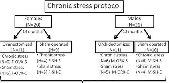
Fig. 1.Study design. F-OVX-S – female ovariectomized chronic stress group; F-SH-S – female sham-operated chronic stress group; F-OVX-C – female ovariectomized control (sham-stress) group; F-SH-C – female sham-operated control (sham stress) group; M-ORX-S – male orchidectomized chronic stress group; M-SH-S – male sham-operated chronic stress group; M-ORX-C – male orchidectomized control (sham-stress) group; M-SH-C – male sham-operated control (sham-stress) group.
2. Materials and methods 2.1. Animal experiment
Twenty female and twenty-one male Sprague Dawley-Charles River rats (Charles River, Wilmington, MA, USA) were used in the study (Fig. 1). Animals were housed in standard cages with two animals per cage at room temperature (21 ± 2 ◦C), with humidity of 40–60%.
Standard laboratory conditions, including rat chow and water ad libi- tum, and 12 h light/12 h dark cycle, were applied at all times except during some stressors of the chronic stress protocol (see below). Ex- periments were carried out at the Animal Facility of the Faculty of Pharmacy, University of Szeged, and approved by the National Scientific Ethical Committee on Animal Experimentation in Hungary (approval number: IV./3796/2015).
2.2. Ovariectomy and orchidectomy
At the age of 13 months, the rats were anesthetized with isoflurane (Florane®, Abbott Laboratories Ltd., Queenborough, UK). One group of female rats (N =11) was ovariectomized and one group of male rats (N
=11) was orchidectomized, while the remaining of the animals were sham-operated (females: N =9; males: N =10). Ovariectomy of female rats was made after midline abdominal incision, under general anes- thesia with isoflurane (Forane® isofluranum, Abbott Laboratories Ltd., Queenborough, UK) (Li et al., 2014; Rodriguez-Landa et al., 2019). Male rats were castrated according to Idris (Idris, 2012). After surgery, food and water were available ad libitum. The animals were closely moni- tored for 72 h. The stress experienced by the animals during surgery was considered irrelevant because the surgeries were performed at least 4 weeks before the chronic stress protocol, as previously described (Balog et al., 2015a).
2.3. Chronic stress protocol
Two months after the surgeries (age 15 months), the ovariectomized, orchidectomized and sham-operated groups of animals were divided into two groups as follows (Fig. 1): a chronic stress group with 22 rats (6 ovariectomized [F-OVX-S] and 4 sham-operated females [F-SH-S]; 6 orchidectomized [M-ORX-S] and 6 sham-operated males [M-SH-S]) and a sham stress (control) group with 19 rats (5 ovariectomized [F-OVX-C]
and 5 sham-operated females [F-SH-C]; 5 orchidectomized [M-ORX-C]
and 4 sham-operated males [M-SH-C]).
The chronic stress protocol consisted of three separate sessions, each lasting 10 days (Balog et al., 2015a; Balog et al., 2015b; Ivic et al., 2016).
The rats were subjected to a different stressor (see below and Table 1) or to sham stress each day. Rats in the sham stress groups were exposed to the same environment as the chronic stress groups, but without the stressors (except for the food deprivation and glucose tolerance test [GTT] stressors). After each session, there was a period of three weeks with no stressors.
The stressors included (Table 1): (i) Food deprivation for 12 h in the chronic stress groups, but no comparable stressor in the sham stress groups; (ii) GTT in the chronic stress groups, but no comparable stressor in the sham stress groups; (iii) Cold restraint, in which the chronic stress groups were restrained in metal tubes for 60 min in a cold room at +4 ◦C, whereas the sham stress groups was just exposed to tubes in the same room at ambient temperature; (iv) Light overnight, in which the lights were on from 9:00 pm – 9:00 am for the chronic stress groups, whereas the sham stress groups were also under the lights but with half of the cage covered; (v) Noise overnight, in which alarms sounded at irregular intervals from 9:00 pm–9:00 am for the chronic stress groups, whereas radio music was played at low volume in the background during same time period for the sham stress groups; (vi) Swim test, in which each rat in the chronic stress groups swam for 3 min in a bucket filled with cold water, whereas each rat in the sham stress groups was placed in a bucket without water for 3 min; and (vii) Cage rotation, in which each cage was rotated for 40 min after placing it on a laboratory shaker, while the cages for the sham groups were put on a shaker that was turned off for 40 min.
After completion of the chronic stress protocol, the rats were euthanized using isoflurane anesthesia at the age of 17 and one-half months. The cranial thoracic SC tissues (Th1-Th3 levels) were collected for analysis.
2.4. Western blot 2.4.1. Protein isolation
After a 2-month-old male, Sprague Dawley rat was euthanized with isoflurane anesthesia, whole brain tissue and the SC were removed and snap-frozen. The tissue was homogenized using a Minilys (Bertin Technologies SAS, Montigny-le-Bretonneux, France) in ice-cold RIPA buffer (50 mM Tris, 1 mM EDTA, 150 mM NaOH, 1% Triton, 0.5% DOC, 0.1% SDS, pH 7.4) with added protease and phosphatase inhibitors (1:1000 aprotinin, 1:1000 Na-vanadate, 1:200 phenylmethylsulfonyl fluoride [PMSF], 1:1000 leupeptin). Homogenized tissues were incu- bated for 2 h at +4 ◦C with constant agitation, followed by centrifuga- tion for 20 min at 12000 rpm at +4 ◦C. The supernatants containing protein lysate were collected into new tubes. Protein concentration was measured using Pierce™ BCA Protein Assay Kit (Cat. No. 23225, Thermo Scientific, Waltham, MA, USA), according to the manufacturer’s instructions.
2.4.2. Antibodies and Western blot analysis
The proteins were subjected to standard protocols for sodium dodecyl sulfate-polyacrylamide gel electrophoresis (SDS-PAGE) and immunoblotting (Henderson and Wolf, 1992). Then the proteins were transferred onto nitrocellulose membranes (GE Healthcare, Chicago, Illinois, USA) and immunostained with anti-dendrin antibody (1:500, AB15299-I, EMD Millipore, Temecula, USA) and β-actin antibody (1:5000, A5316, Merck KGaA, Darmstadt, Germany). The bands were detected using HRP-conjugated secondary antibodies (1:6000, P0448, DakoCytomation, Carpinteria, CA, USA) and ECL Plus substrate (Thermo Fisher Scientific Inc., Waltham, MA, USA).
2.5. Immunohistochemical staining
SC tissue samples were dissected and fixated by immersion in 4%
buffered paraformaldehyde. All the spinal cord specimens that we compared, including control and treated groups were treated the same way. After washing in phosphate buffer saline (PBS) and dehydration in rising concentrations of ethanol, dehydrated samples were embedded in Table 1
Daily stressors that were applied to rats in the chronic stress groups†during the three sessions of the chronic stress protocol.
Day Stressors during 1st Session Stressors during 2nd and 3rd Sessions
1. Food deprivation (12h) Cage rotation (40 min)
2. GTT‡ Swim test
3. Cold restraint (+4 ◦C, 60 min) +
food deprivation Cold restraint (+4 ◦C, 60 min) + food deprivation
4. GTT Noise overnight (9:00 pm - 9:00 am)
5. Light overnight (9:00 pm - 9:00 am) Light overnight (9:00 pm - 9:00 am) 6. Cage rotation (40 min) Cage rotation (40 min)
7. Swim test Swim test
8. Noise overnight (9:00 pm - 9:00 am) Noise overnight (9:00 pm - 9:00 am) 9. Cold restraint (+4 ◦C, 60 min) +
food deprivation Cold restraint (+4 ◦C, 60 min) + food deprivation
10. GTT GTT
†Chronic stress groups (N =22) included 6 ovariectomized female rats, 4 sham-operated female rats, 6 orchidectomized male rats and 6 sham-operated male rats.
‡Glucose tolerance test.
paraffin before serial sectioning (5 μm thick transverse sections) and mounted on glass slides. Sections were deparaffinized in xylene and rehydrated in descending concentrations of ethanol in water as previ- ously described (Agnic et al., 2018; Filipovic et al., 2017; Poljicanin et al., 2015).
Next, the sections were washed in PBS and the protein block (ab64226, Abcam, Cambridge, UK) was applied for 30 min. Then the sections were incubated overnight in a humid chamber with rabbit anti- dendrin polyclonal primary antibody (1:100, AB15299-I, EMD Milli- pore, Temecula, USA). After incubation, the sections were washed in PBS three times for 10 min and Alexa Fluor 488-conjugated donkey anti- rabbit IgG H&L secondary antibody (1:400, ab150073, Abcam, Cam- bridge, UK) was applied for 1 h. After washing in PBS, nuclei were stained with 4′6′-diamidino-2-phenylindole (DAPI) solution for 2 min, and cover-slipped. Control staining, with the exclusion of the primary antibody, resulted in no staining of tissue.
We also performed dual immunohistochemistry with rabbit anti- dendrin and FITC-conjugated α-d-galactosyl-specific isolectin B4 (IB4) (1:200, ALX-650-001F, Enzo, Farmingdale, NY, USA), as well as rabbit anti-dendrin and goat polyclonal to calcitonin gene-related peptide (CGRP) primary antibody (1:1000, ab36001, Abcam, Cambridge, UK) with the application of appropriate secondary antibodies (Rhodamine Red™-X (RRX) AffiniPure Donkey Anti-Goat IgG (H +L) 705–295-003;
Rhodamine (TRITC) AffiniPure Donkey Anti-Rabbit IgG (H + L) 711–025-152; both from Jackson ImmunoResearch Laboratories, Inc., West Grove, PA, USA).
Skin, heart and dorsal root ganglia (DRG) samples were obtained from the bank of animal tissues of the Laboratory for Neurocardiology (3-month-old male Sprague Dawley rats; tissue fixed in 4% para- formaldehyde in PBS and embedded in paraffin as described above).
Skin and heart were stained with a mixture of primary antibodies: rabbit anti-dendrin polyclonal antibody (1:100, AB15299-I, EMD Millipore, Temecula, USA) and mouse monoclonal antibody against protein gene product 9.5 (anti-PGP9.5, 1:500, 480,012, Invitrogen, Camarillo CA, USA), with an appropriate anti-rabbit secondary antibody, described above (Alexa Fluor 488-conjugated donkey anti-rabbit IgG H&L) and Rhodamine Red-X-AffiniPure Donkey Anti-Mouse IgG (H + L) anti- bodies (715–295-151; Jackson ImmunoResearch Laboratories, Inc., West Grove, PA, USA). Dual immunohistochemistry of DRG neurons was carried out with anti-dendrin and following primary antibodies: FITC- conjugated IB4, goat anti-CGRP and mouse anti-neurofilament 200kD (NF200) monoclonal antibody (1:100, MAB5266, EMD Millipore, Temecula, USA) with appropriate rhodamine X-conjugated donkey anti- mouse secondary antibody.
2.5.1. Data acquisition and quantification immunohistochemical analysis The sections were observed under an epifluorescent microscope (Olympus BX61, Tokyo, Japan). Images were captured using a cooled digital camera DP71 (Olympus, Tokyo, Japan) and Olympus CellA software (Olympus Life Sciences Microscopy, Tokyo, Japan). The pho- tomicrographs were taken at objective magnification factors of ×4 (to capture the entire SC segment); ×20 (to capture only the dorsal horn of the SC); and ×40 and ×100 (for capturing the details) (final magnifi- cation ×40, ×200, ×400 and ×1000; respectively). The photomicro- graphs were processed and subsequently analyzed by using ImageJ software (National Institutes of Health, Bethesda, MD, USA) and pro- cessing protocol was adjusted from already published (Lueti´c et al., 2020; Milardovi´c et al., 2020). First, the fluorescence leakage reduction was performed by subtraction of red counter-signal from green fluo- rescence and then the median filter was used with a radius of 5.0 pixels.
Subsequently, each picture was adjusted to the threshold method (tri- angle thresholding algorithm method) and analyzed by measuring the fluorescence percentage area.
Analysis of overlapping regions obtained by dual immunohisto- chemistry in the dorsal horn of the SC and neurons of the DRG, after image processing as described above, was carried out using Adobe
Photoshop (Adobe Inc., San Jose, California, USA) by merging them and calculating the fluorescence percentage area for every protein by itself and the overlapping part.
2.6. Statistical analyses
Mead’s resource equation was used for sample size estimation, where the degree of freedom is well above 20. Hence, the study power was above 80%.
PAST 3.22 Software (Øyvind Hammer, Natural History Museum, University of Oslo, Norway) was used for statistical analyses (Hammer, Harper, & Ryan, 2001). To determine significant differences among groups, ANOVA with Welch correction for unequal variances was used.
The difference between particular groups was tested by the t-test with Welch correction for unequal variances. Statistical significance was considered at P <0.05. We did not use multiple comparisons correction because all of the comparisons were preplanned. Normality and homo- geneity of data were tested by Anderson-Darling and Levene’s tests, respectively. Differences between groups were expressed as a fold change calculated as the relative difference to sham stressed animals.
2.7. Tissue preparation for electron microscopy
Tissues were prepared for electron microscopy by using an immu- nogold immunohistochemistry protocol (Filipovic et al., 2020; Kosovic et al., 2020; Vitlov Uljevic et al., 2019). Samples were fixed in 4%
paraformaldehyde in PBS, washed in PBS and then cut into 20 μm thick sections on a vibratome (Vibratome Series 1000, Pelco 101, Ted Pella, Inc., Redding, CA, USA). Following the wash in PBS, sections were incubated first in 50% ethanol for permeabilization and then in primary rabbit anti-dendrin polyclonal antibody (1:100, AB15299-I, EMD Mil- lipore, Temecula, USA) for 48 h at +4 ◦C. After incubation, sections were rinsed in PBS and then incubated overnight with gold-conjugated donkey anti-rabbit secondary antibody (1:1000, 711-205-152, Jackson Immuno Research Laboratories, Inc., West Grove, PA, USA). Following another wash in PBS, sections were post-fixed in 1% osmium tetroxide (1 h), then dehydrated in ethanol and embedded in Durcupan ACM resin (Sigma-Aldrich Inc. St. Louis, Missouri, USA). The size of gold particles used in this study was 12 nm. The sections were observed with a transmission electron microscope (JEM JEOL 1400, Jeol Ltd., Tokyo, Japan).
3. Results
3.1. Western immunoblot specificity analysis of dendrin in brain and SC We tested the specificity of the dendrin antibody using Western blots of the brain and SC homogenates from a 2-month-old male rat. There was a specific strong band at 72 kDa in the brain. In SC, the same band but with weaker intensity was found (Fig. 2a).
3.2. Specific localization pattern of dendrin in SC
Localization and quantification of dendrin expression were analyzed in thoracic segments (Th1-Th3 levels) of the SC from female and male rats in the experimental groups. As expected, dendrin expression was found in neurons and all parts of the gray matter. The expression was strong in large lower motor neurons of the ventral horn. In addition, a distinct expression of dendrin was seen in the dorsal horn. The most prominent staining with the highest intensity was found in laminae I and II, and in the area around the central canal (lamina X) (Fig. 2b).
Intensity profiles for dendrin expression in the SC were plotted. In most of the groups, there was a decreasing intensity of dendrin immu- nofluorescence from dorsal to the ventral area in sections of the SC. The highest intensity was found in the F-SH-S, F-OVX-C and F-OVX-S groups (Fig. 2c) and the M-ORX-S group (Fig. 2d). The lowest intensity was
observed in the F-SH-C group (Fig. 2c) and the M-SH-C, M-SH-S and M- ORX-C groups (Fig. 2d).
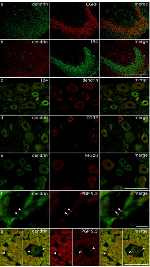
3.3. Overlapping of dendrin immunofluorescence signal with markers for primary afferent nociceptive fibers and DRG neurons
In the dorsal horn, we found dendrin immunoreactivity overlapping with IB4 and CGRP in central projections of DRG primary afferent nociceptive fibers (Fig. 3a and b). Overlapping of the threshold per- centage area for IB4 was 7.43 ±3.36% of the total area while over- lapping for CGRP was 8.47 ±4.45% of the total area, with no significant difference between groups. Because of these data, we explored the possible expression of dendrin in DRG neurons. Surprisingly, we found strong dendrin immunoreactivity in the soma of DRG neurons that were immunoreactive for IB4, CGRP and NF200 (Fig. 3c-e). We also found the co-localization of dendrin with PGP 9.5 (Protein gene product 9.5, UCH- L1, PARK5) in peripheral neuronal projections in the skin and heart (Fig. 3f and g). PGP 9.5 is a pan-neuronal marker, present in neurons, as well as in the nerve fibers in the central and peripheral nervous system (Lee et al., 2012).
3.4. Dendrin expression differences between groups based on chronic stress, castration, gender, and sex hormones
We investigated the effects of chronic stress, castration (orchid- ectomy or ovariectomy), gender and the combination of these factors on
dendrin expression in the SC. To assess the effect of chronic stress on dendrin expression we compared the two male sham-operated groups:
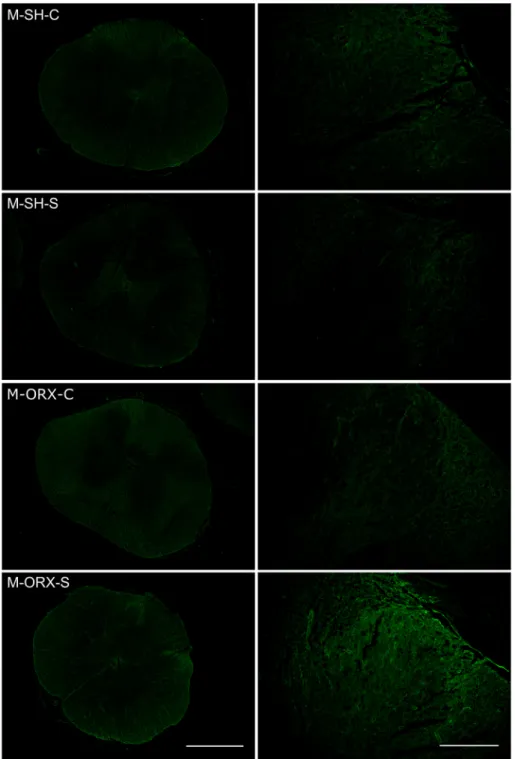
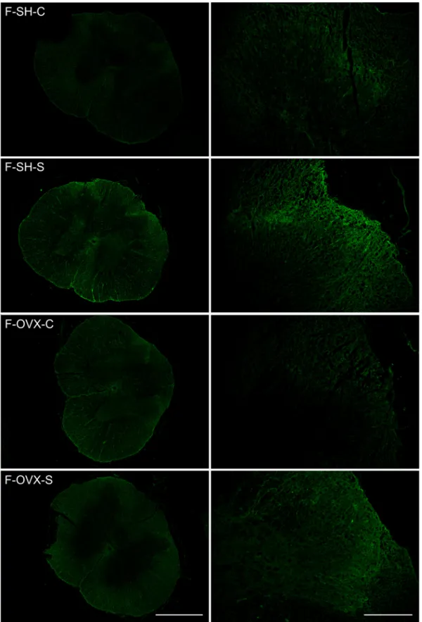
M-SH-S and M-SH-C. Chronic stress did not increase dendrin expression (relative difference =0.35-fold; 95% confidence interval [CI] -0.426 to 1.127; P =0.4534) (Figs. 4, 5). In contrast, when the female sham- operated groups were compared, dendrin expression nearly quadru- pled in F-SH-S rats compared to F-SH-C rats, and the difference was statistically significant (relative difference =3.669-fold; 95% CI 1.261 to 6.079; P =0.0445) (Figs. 4, 6).
To evaluate the effect of castration alone we compared the two male control groups (i.e., sham stress): M-SH-C vs. M-ORX-C (relative differ- ence =0.376-fold; 95% CI -0.731 to 1.483; P =0.4709), and the two female control groups: F-SH-C vs. F-OVX-C group (relative difference = 1.387-fold; 95% CI -0.083 to 2.857; P =0.0809). Neither comparison showed a significant difference (Fig. 4).
The effect of gender by itself did not influence dendrin expression (M-SH-C vs. F-SH-C; relative difference =0.577-fold; 95% CI -0.673 to 1.827; P =0.3170) (Fig. 4). However, when exposed to chronic stress female rats had a significant 10-fold increase in dendrin expression compared to male rats (F-SH-S vs. M-SH-S relative difference =10.358- fold; 95% CI 5.351 to 15.353; P =0.0322). Moreover, when sham- stressed animals were castrated there was also a significant 5-fold in- crease in dendrin expression for female rats compared to male rats (F- OVX-C vs. M-ORX-C relative difference =5.034-fold; 95% CI 1.407 to 8.662; P =0.0266).
To examine how stress might influence the expression of dendrin in Fig. 2.(a) Western blot analysis characterization of the dendrin antibody. There was a strong band at 72 kDa in the brain and a weaker band at the same position in the spinal cord (SC). The β-actin protein was used as a loading control at 42 kDa. (b) Intensity distribution heatmap for dendrin expression in the SC of rats in the F- SH-S group. Intensity range (pixel value 0–255): 0–6 (dark blue), 6–10 (bright blue), 10–12 (white), 12–16 (bright green), 16–20 (yellow), and 20–22 (bright red). (c, d) Intensity plot profiles for dendrin expression along the dorsoventral axis through the superficial laminae of the dorsal horn. Individual intensity plots for each of the female groups (c) and male groups (d) are shown. The distance from the dorsal root entering zone (DREZ) to the central canal is presented on the X-axis. The intensity of dendrin expression is expressed as the mean pixel value on the Y-axis.
castrated animals, we compared the two male orchidectomized groups:
M-ORX-S and M-ORX-C. In castrated male rats, chronic stress dramati- cally and significantly increased dendrin expression by almost 12-fold (relative difference =11.653-fold; 95% CI 7.807 to 15.5; P =0.0003) (Figs. 4, 5). In contrast, the combination of castration and chronic stress
did not significantly affect dendrin expression in female rats (F-OVX-S vs. F-OVX-C relative difference =1.015-fold; 95% CI -0.813 to 2.843; P
=0.2204) (Figs. 4, 6).
These findings led us to evaluate the influence of sex hormones in chronically stressed animals (M-SH-S vs. M-ORX-S; F-SH-S vs. F-OVX-S).
Fig. 3. Dendrin expression in the dorsal horn of spinal cord, dorsal root ganglia (DRG) neurons and peripheral nerves. (a, b) Expression of dendrin in the dorsal horn. Representative photomicrographs after thresholding the images of immunostained sections.
(a) dendrin (green), calcitonin gene-related peptide (CGRP, red) and co-localization of these proteins (“merge”). (b) dendrin (red), α-d-galactosyl-specific isolectin B4 (IB4, green) and co-localization (merge).
Magnification factor x200, scale bar: 200 μm. (c, d, e) Representative photomicrographs of DRG neurons after immunostaining. Dendrin expression in DRG neurons and co-localization with three neuronal markers: (c) IB4, (d) CGRP and (e) neurofilament 200 (NF200). Magnification factor x400, scale bar: 100 μm. (f, g) Expression of dendrin in peripheral nerves of rat skin and heart. Representative photomicro- graphs of (f) skin and (g) heart made by immuno- staining for dendrin (green) and co-localization with neuronal marker protein gene product 9.5 (PGP 9.5, red). In the heart, dendrin immunoreactive neuronal fibers were found between cardiomyocytes, sub- endo/epicardial areas (not shown) and around blood vessels. Magnification factor x1000 in (f), x400 in (g), scale bar: 100 μm (both). Arrowheads indicate dendrin immunoreactive nerve fibers (green) recog- nized by co-localization with PGP 9.5 (red). In rightmost panels, arrows indicate co-localization (orange). (a, b – M-SH-C; c-g - 3-month-old male Sprague Dawley rats).
In male rats, castration significantly augmented the effect of chronic stress on dendrin expression by 11-fold (relative difference =11.166- fold; 95% CI 7.945 to 14.388; P = 0.0005) (Figs. 4, 5). Whereas castration in females did not show a synergistic effect with chronic stress (relative difference =0.029-fold; 95% CI -1.093 to 1.153; P =0.9469) (Figs. 4, 6).
3.5. Ultrastructural characterization of dendrin in the dorsal horn of the SC
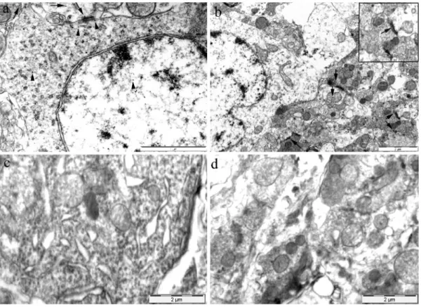
Transmission electron microscopy confirmed dendrin immunoreac- tivity in the dorsal horn neurons of the SC from M-SH-C rats, by using immunogold staining. As expected, gold particles were found in the postsynaptic area as well as in the neuronal soma and nucleus. It seems that the immunoreaction was distributed randomly throughout the neuronal soma and nucleus. Although less numerous, the gold particles were found in the presynaptic area and axon terminals confirming the immunoreactivity for dendrin in these areas as well. The immuno- reaction in these areas usually was distributed near the presynaptic membrane close to synaptic vesicles and mitochondria (Fig. 7).
4. Discussion
Gonadal hormones greatly influence the perception and modulation of how stressors impact an organism (Heck and Handa, 2019). In this study, our main focus was to explore the potential interaction between the effects of chronic stress and gonadal hormones in the SC. A promi- nent difference in the amount of dendrin expression was found.
To the best of our knowledge, there is no data available about den- drin expression in the SC before our study. Besides the expected presence of dendrin in neurons, we found dendrin expression concentrated in the SC areas associated with pain processing, namely the superficial laminae of the dorsal horn and lamina X (encircling the central canal). We also found overlapping areas of dendrin with IB4- and CGRP- immunoreactive central projections of the primary sensory neurons in superficial laminae of the dorsal horn. Furthermore, we found strong dendrin immunoreactivity in the soma of DRG neurons, as well as in their peripheral projections in the skin and viscera (heart). Taken together, these findings may suggest that the possible alterations in dendrin expression are related to the refinement of sensory information, in particular the processing of pain.
Previous studies have described dendrin as a protein bound to the
dendritic spines, which means it belongs to the group of proteins with dendritically localized neuronal mRNAs (Pinkstaff et al., 2001).
Although the expression is associated with synapses, there is also a characterization of dendrin in neuronal soma with accumulation on polyribosomes (Neuner-Jehle et al., 1996). By using transmission elec- tron microscopy in combination with immunogold labeling, we found similar results in soma but also with a presence in the neuronal nucleus.
A mechanism underlying the accumulation of dendrin in the nucleus might be possible through modulation of its interaction with the syn- aptic scaffolding protein MAGI/S-SCAM. Change in interaction with MAGI/S-SCAM may be responsible for the regulation of dendrin release from the synapse and its movement into the nucleus where it may then act as a retrograde messenger (Kremerskothen et al., 2006). The pre- synaptic presence of dendrin in the central projections of DRG neurons is not clear. On the presynaptic terminal, dendrin may also contribute to the LTP modulation, through increasing or decreasing the release of neurotransmitters through some other interaction yet to be discovered, important for the effective reorganization of neural circuits (Yang and Calakos, 2013).
Despite the above-mentioned findings, the exact role of dendrin in neurons is still unclear. It is believed that the role of dendrin in the brain is closely related to plastic events at the synaptic level, mainly because of its connection with the kidney and brain expressed protein (KIBRA), a synaptic scaffold protein responsible for regulating spatial learning and memory (Ji et al., 2019; Kremerskothen et al., 2003). It was found that the nature of the interaction between KIBRA and dendrin is important for learning and memory (Ji et al., 2019). After application of inhibitory peptide and causing disruption in KIBRA-dendrin interaction, synaptic AMPAR (α-amino-3-hydroxy-5-methyl-4-isoxazolepropionic acid re- ceptor) expression is reduced with consequences of attenuation of the excitatory synaptic transmission, long-term potentiation (LTP) (Ji et al., 2019), synaptic strength and plasticity (Diering and Huganir, 2018;
Herb et al., 1997; Kawata et al., 2006; Kremerskothen et al., 2006;
Makuch et al., 2011; Schochet et al., 2008). Previous studies have shown that chronic stress is linked to maladaptive pain responses because of its impact on pain processing areas via influence on glutamate receptor expression (Yuen et al., 2016). Taking all of the above into consider- ation, it can be assumed that exposure to chronic stress can cause changes in dendrin expression and through that, alteration of the SC synaptic plasticity. We found a significant increase in dendrin expression in the dorsal horn of rats exposed to chronic stress. However, this response was sex-dependent, observed only in female rats. Moreover, it seems that male sex hormones have a suppressive effect because an in- crease in dendrin expression induced by chronic stress appeared in male rats only after orchidectomy (i.e. after steep down-regulation of testosterone).
These findings may be a possible explanation for the different effects of gonadal hormones upon dendritic spine density and branching. It was previously found that testosterone in males induces dendritic branching but has no effect on spine density. In contrast, it was found that estradiol increases dendritic spine density (Todd et al., 2007). Both of these ef- fects were related to AMPAR trafficking. In addition, there is an indi- cation that females have more potential for forming fear memory by acute stressors than males, which is mediated through an increase in LTP induction and AMPARs (GluR1) in the lateral nucleus of the amygdala (Chen et al., 2014). On the other hand, the down-regulation of testos- terone after a gonadectomy resulted in the excitability and plasticity enhancement via LTP in the CA3 area as a counterbalance to the decreased amount of spine synapses (Hojo et al., 2009; Skucas et al., 2013). The state of excitability of the spinal neurons is therefore critical to proper discrimination of noxious stimuli and it could be related to changes in dendrin expression. On the other hand, sex-dependent dif- ferences of dendrin expression, especially after sex hormones depletion, may depend on aromatase activity (AA). In the study, Roselli et al.
described the dependence of AA and castration in the hypothalamus, while in females the estrous cycle and castration do not affect AA, in Fig. 4. Expression of dendrin in the dorsal horn of the rat spinal cord in the
experimental groups. Dendrin expression was quantified by measuring fluo- rescence percentage area (% area); * P <0.05;*** P <0.001 - statistically significant difference between indicated groups; # P <0.05 - statistically sig- nificant difference between genders. Abbreviations: SH-C, sham-operated con- trol group (sham stress); SH-S, sham-operated chronic stress group; Gx-C, orchidectomized/ovariectomized control group (sham stress); Gx-S, orchid- ectomized/ovariectomized chronic stress group.
male rats castration drastically decreases AA (Roselli and Resko, 1993).
Furthermore, Dickens et al. reported changes in AA due to acute stress exposure depending on the sex and brain region of Japanese quail (Dickens et al., 2011).
In conclusion, a better understanding of the neurobiological back- ground of the connection between chronic stress and pain can help us elucidate the numerous pathological changes that underlie the onset of stress-related conditions. The presence of dendrin in nociceptive areas of the dorsal horn of the SC, and its expression in the DRG and peripheral neurons, suggest its possible role in nociceptive pathway modulation as a possible linkage in which the influence of stress and pain overlap.
Further studies are needed to explain the role of the stress-induced
increase in dendrin expression that was found only in non- ovariectomized female and orchidectomized male rats, in sex-specific pain hypersensitivity induced by stress.
Author contributions
All authors have full access to all data in the study and take re- sponsibility for the integrity of the data and the accuracy of the data analysis. N.F., R.G., M.H. and S.G.V. - conceptualization; N.F., M.J., R.
G., M.H. and S.G.V. - methodology; M.J., N.F., M.Ba., V.I., M.Bo., A.R., I.
B., N.K., I.R., K.F.S., K.V., R.G., M.H. and S.G.V. - investigation; N.F., M.
J., B.B. - formal analysis; N.F., K.V., R.G., M.H. and S.G.V. - resources; M.
Fig. 5.Representative photomicrographs showing dendrin expression in the dorsal horn of spinal cords of different groups of male rats. Original magnification: x40, scale bar: 2 mm; and x200, scale bar: 100 μm. Abbreviations: M-SH-C, male sham-operated control (sham stress) group; M-SH-S, male sham-operated chronic stress group; M-ORX-C, male orchidectomized control (sham stress) group; M-ORX-S, male orchidectomized chronic stress group.
J. and N.F. writing-original draft; M.J., M.Ba., V.I., M.Bo., B.B., A.R., I.
B., N.K., I.R., R.G., K.F.S., M.H., K.V., S.G.V. and N.F. - writing-review and editing; M.J., I.B., N.F. - visualization; N.F. - supervision; N.F. - project administration; N.F., K.V., R.G., M.H. and S.G.V. - funding
acquisition.
Fig. 6. Representative photomicrographs showing dendrin expression in the dorsal horn of spinal cords of different groups of female rats. Original magnification:
x40, scale bar: 2 mm; and x200, scale bar: 100 μm. Abbreviations: F-SH-C, female sham-operated control (sham stress) group; F-SH-S, female sham-operated chronic stress group; F-OVX-C, female ovariectomized control (sham stress) group; F-OVX-S, female ovariectomized chronic stress group.
Ethical committee approval
Experiments were carried out at the Animal Facility of Faculty of Pharmacy, University of Szeged, approval number: IV./3796/2015.
Declaration of Competing Interest
The authors declare that they have no conflicts of interest.
Acknowledgements
This study was funded by the Regional Cooperation for Health, Sci- ence and Technology (RECOOP HST) Association formed by Cedars- Sinai Medical Center (CSMC, Los Angeles, CA, USA) RECOOP Grant # 001; the Ministry of Science Education and Sports, Republic of Croatia support (K. Vukojevi´c, N. Filipovi´c, I. Boˇcina and N. Kevi´c); the PhD programme “Biology of Neoplasms” School of Medicine, University of Split and the Croatian Scientific Foundation (HRZZ; grant no. HRZZ-IP- 2014-09-2324); Ministry of Human Capacities (Hungary grant 20391-3/
2018/FEKUSTRAT).
References
Abdallah, C.G., Geha, P., 2017. Chronic pain and chronic stress: two sides of the same coin? Chronic Stress (Thousand Oaks). 1.
Agnic, I., Filipovic, N., Vukojevic, K., Saraga-Babic, M., Grkovic, I., 2018. Isoflurane post- conditioning influences myocardial infarct healing in rats. Biotechnic Histochem. 93, 354–363.
Aloisi, A.M., 2017. Why we still need to speak about sex differences and sex hormones in pain. Pain Therapy 6, 111–114.
Aloisi, A.M., Ceccarelli, I., Fiorenzani, P., 2003. Gonadectomy affects hormonal and behavioral responses to repetitive nociceptive stimulation in male rats. Ann. N. Y.
Acad. Sci. 1007, 232–237.
Aloisi, A.M., Ceccarelli, I., Fiorenzani, P., De Padova, A.M., Massafra, C., 2004.
Testosterone affects formalin-induced responses differently in male and female rats.
Neurosci. Lett. 361, 262–264.
Balog, M., Miljanovic, M., Blazetic, S., Labak, I., Ivic, V., Viljetic, B., Borbely, A., Papp, Z., Blazekovic, R., Vari, S.G., Fagyas, M., Heffer, M., 2015a. Sex-specific chronic stress response at the level of adrenal gland modified sexual hormone and leptin receptors.
Croatian Med. J. 56, 104–113.
Balog, M., Mlinarevic, D., Seric, V., Miljanovic, M., Blazekovic, R., Degmecic, I.V., Blazetic, S., Orsolic, I., Vari, S.G., Heffer, M., 2015b. Plasma content of glucose, C- reactive protein, uric acid and cholesterol in male, female and ovariectomized rats upon acute and chronic stress–a path for development of cardiovascular diseases.
Coll. Antropol. 39, 385–392.
Cao, D.Y., Ji, Y., Tang, B., Traub, R.J., 2012. Estrogen receptor beta activation is antinociceptive in a model of visceral pain in the rat. J. Pain. 13, 685–694.
Chen, L.S., Tzeng, W.Y., Chuang, J.Y., Cherng, C.G., Gean, P.W., Yu, L., 2014. Roles of testosterone and amygdaloid LTP induction in determining sex differences in fear memory magnitude. Horm. Behav. 66, 498–508.
Coulombe, M.A., Spooner, M.F., Gaumond, I., Carrier, J.C., Marchand, S., 2011. Estrogen receptors beta and alpha have specific pro- and anti-nociceptive actions.
Neuroscience 184, 172–182.
Craft, R.M., 2007. Modulation of pain by estrogens. Pain 132 (Suppl. 1), S3–12.
Dickens, M.J., Cornil, C.A., Balthazart, J., 2011. Acute stress differentially affects aromatase activity in specific brain nuclei of adult male and female quail.
Endocrinology 152, 4242–4251.
Diering, G.H., Huganir, R.L., 2018. The AMPA receptor code of synaptic plasticity.
Neuron 100, 314–329.
Elzahaf, R.A., Johnson, M.I., Tashani, O.A., 2016. The epidemiology of chronic pain in Libya: a cross-sectional telephone survey. BMC Public Health 16, 776.
Fanton, L.E., Macedo, C.G., Torres-Chavez, K.E., Fischer, L., Tambeli, C.H., 2017.
Activational action of testosterone on androgen receptors protects males preventing temporomandibular joint pain. Pharmacol. Biochem. Behav. 152, 30–35.
Filipovic, N., Vukojevic, K., Bocina, I., Saraga, M., Durdov, M.G., Kablar, B., Saraga- Babic, M., 2017. Immunohistochemical and electronmicroscopic features of mesenchymal-to-epithelial transition in human developing, postnatal and nephrotic podocytes. Histochem. Cell Biol. 147, 481–495.
Filipovic, N., Bocina, I., Restovic, I., Grobe, M., Kretzschmar, G., Kevic, N., Masek, T., Vitlov Uljevic, M., Juric, M., Vukojevic, K., Saraga-Babic, M., Vuica, A., 2020.
Ultrastructural characterization of vitamin D receptors and metabolizing enzymes in the lipid droplets of the fatty liver in rat. Acta Histochem. 122, 151502.
Greenwood-Van Meerveld, B., Johnson, A.C., 2017. Stress-induced chronic visceral pain of gastrointestinal origin. Front. Syst. Neurosci. 11, 86.
Fig. 7. Representative transmission electron microscopy photomicrographs of dendrin immunoreactivity in dorsal horn neurons of the spinal cord. (a) Immunoglod staining for dendrin in axon terminals and presynaptic area (arrows) as well as in neuronal soma, postsynaptic area and nucleus (arrowheads). Scale bar: 1 μm. (b) Immunogold staining for dendrin in the axon terminals and presynaptic area (arrows). Scale bar: 2 μm. (c) Negative control in the area of neuronal soma. Scale bar: 2 μm. (d) Negative control in the area of axon terminals. Scale bar: 2 μm.
Hammer, Ø., Harper, D.A.T., Ryan, P.D., 2001. Paleontological statistics software package for education and data analysis. Palaeontologia Electronica 4, 9.
Heck, A.L., Handa, R.J., 2019. Sex differences in the hypothalamic-pituitary-adrenal axis’ response to stress: an important role for gonadal hormones.
Neuropsychopharmacology. 44, 45–58.
Henderson, C.J., Wolf, C.R., 1992. Immunodetection of proteins by western blotting. In:
Manson, M.M. (Ed.), Immunochemical Protocols. Humana Press, Totowa, NJ, pp. 221–233.
Herb, A., Wisden, W., Catania, M.V., Marechal, D., Dresse, A., Seeburg, P.H., 1997.
Prominent dendritic localization in forebrain neurons of a novel mRNA and its product, dendrin. Mol. Cell. Neurosci. 8, 367–374.
Hojo, Y., Higo, S., Ishii, H., Ooishi, Y., Mukai, H., Murakami, G., Kominami, T., Kimoto, T., Honma, S., Poirier, D., Kawato, S., 2009. Comparison between hippocampus-synthesized and circulation-derived sex steroids in the hippocampus.
Endocrinology 150, 5106–5112.
Idris, A.I., 2012. Ovariectomy/orchidectomy in rodents. Methods Mol. Biol. 816, 545–551.
Ivic, V., Blazetic, S., Labak, I., Balog, M., Vondrak, L., Blazekovic, R., Vari, S.G., Heffer, M., 2016. Ovariectomy and chronic stress lead toward leptin resistance in the satiety centers and insulin resistance in the hippocampus of Sprague-Dawley rats.
Croatian Med. J. 57, 194–206.
Ji, Y., Tang, B., Traub, R.J., 2011. Spinal estrogen receptor alpha mediates estradiol- induced pronociception in a visceral pain model in the rat. Pain 152, 1182–1191.
Ji, Y., Hu, B., Li, J., Traub, R.J., 2018. Opposing roles of Estradiol and testosterone on stress-induced visceral hypersensitivity in rats. J. Pain. 19, 764–776.
Ji, Z., Li, H., Yang, Z., Huang, X., Ke, X., Ma, S., Lin, Z., Lu, Y., Zhang, M., 2019. Kibra modulates learning and memory via binding to Dendrin. Cell Rep. 26 (2064–2077), e2067.
Kawata, A., Iida, J., Ikeda, M., Sato, Y., Mori, H., Kansaku, A., Sumita, K., Fujiwara, N., Rokukawa, C., Hamano, M., Hirabayashi, S., Hata, Y., 2006. CIN85 is localized at synapses and forms a complex with S-SCAM via dendrin. J. Biochem. 139, 931–939.
Kosovic, I., Filipovic, N., Benzon, B., Vukojevic, K., Saraga, M., Glavina Durdov, M., Bocina, I., Saraga-Babic, M., 2020. Spatio-temporal patterning of different connexins in developing and postnatal human kidneys and in nephrotic syndrome of the Finnish type (CNF). Sci. Rep. 10, 8756.
Kremerskothen, J., Plaas, C., Buther, K., Finger, I., Veltel, S., Matanis, T., Liedtke, T., Barnekow, A., 2003. Characterization of KIBRA, a novel WW domain-containing protein. Biochem. Biophys. Res. Commun. 300, 862–867.
Kremerskothen, J., Kindler, S., Finger, I., Veltel, S., Barnekow, A., 2006. Postsynaptic recruitment of Dendrin depends on both dendritic mRNA transport and synaptic anchoring. J. Neurochem. 96, 1659–1666.
Lee, J., Ladd, A., Hagert, E., 2012. Immunofluorescent triple-staining technique to identify sensory nerve endings in human thumb ligaments. Cells Tissues Organs 195, 456–464.
Li, L.H., Wang, Z.C., Yu, J., Zhang, Y.Q., 2014. Ovariectomy results in variable changes in nociception, mood and depression in adult female rats. PLoS One 9, e94312.
Li, Y.X., An, H., Wen, Z., Tao, Z.Y., Cao, D.Y., 2020. Can oxytocin inhibit stress-induced hyperalgesia? Neuropeptides 79, 101996.
Liu, X., Zhu, M., Ju, Y., Li, A., Sun, X., 2019. Autophagy dysfunction in neuropathic pain.
Neuropeptides 75, 41–48.
Lueti´c, M., Vitlov Uljevi´c, M., Maˇsek, T., Benzon, B., Vukojevi´c, K., Filipovi´c, N., 2020.
PUFAs supplementation affects the renal expression of pannexin 1 and connexins in diabetic kidney of rats. Histochem. Cell Biol. 153, 165–175.
Lunde, C.E., Sieberg, C.B., 2020. Walking the tightrope: a proposed model of chronic pain and stress. Front. Neurosci. 14, 270.
Makuch, L., Volk, L., Anggono, V., Johnson, R.C., Yu, Y., Duning, K., Kremerskothen, J., Xia, J., Takamiya, K., Huganir, R.L., 2011. Regulation of AMPA receptor function by the human memory-associated gene KIBRA. Neuron 71, 1022–1029.
Melchior, M., Poisbeau, P., Gaumond, I., Marchand, S., 2016. Insights into the mechanisms and the emergence of sex-differences in pain. Neuroscience 338, 63–80.
Milardovi´c, I., Vitlov Uljevi´c, M., Vukojevi´c, K., Kosti´c, S., Filipovi´c, N., 2020. Acta Histochem. 122, 151580.
Mills, S.E.E., Nicolson, K.P., Smith, B.H., 2019. Chronic pain: a review of its epidemiology and associated factors in population-based studies. Br. J. Anaesth. 123, e273–e283.
Myslivecek, J., Kvetnansky, R., 2006. The effects of stress on muscarinic receptors.
Heterologous receptor regulation: yes or no? Auton. Autacoid Pharmacol. 26, 235–251.
Neuner-Jehle, M., Denizot, J.P., Borbely, A.A., Mallet, J., 1996. Characterization and sleep deprivation-induced expression modulation of dendrin, a novel dendritic protein in rat brain neurons. J. Neurosci. Res. 46, 138–151.
Pinkstaff, J.K., Chappell, S.A., Mauro, V.P., Edelman, G.M., Krushel, L.A., 2001. Internal initiation of translation of five dendritically localized neuronal mRNAs. Proc. Natl.
Acad. Sci. U. S. A. 98, 2770–2775.
Poljicanin, A., Filipovic, N., Vukusic Pusic, T., Soljic, V., Caric, A., Saraga-Babic, M., Vukojevic, K., 2015. Expression pattern of RAGE and IGF-1 in the human fetal ovary and ovarian serous carcinoma. Acta Histochem. 117, 468–476.
Puri, V., Cui, L., Liverman, C.S., Roby, K.F., Klein, R.M., Welch, K.M., Berman, N.E., 2005. Ovarian steroids regulate neuropeptides in the trigeminal ganglion.
Neuropeptides 39, 409–417.
Rhyner, T.A., Borbely, A.A., Mallet, J., 1990. Molecular cloning of forebrain mRNAs which are modulated by sleep deprivation. Eur. J. Neurosci. 2, 1063–1073.
Rodriguez-Landa, J.F., Hernandez-Lopez, F., Cueto-Escobedo, J., Herrera-Huerta, E.V., Rivadeneyra-Dominguez, E., Bernal-Morales, B., Romero-Avendano, E., 2019.
Chrysin (5,7-dihydroxyflavone) exerts anxiolytic-like effects through GABAA receptors in a surgical menopause model in rats. Biomed. Pharmacotherapy. 109, 2387–2395.
Roselli, C.E., Resko, J.A., 1993. Aromatase activity in the rat brain: hormonal regulation and sex differences. J. Steroid Biochem. Mol. Biol. 44, 499–508.
Schochet, T.L., Bremer, Q.Z., Brownfield, M.S., Kelley, A.E., Landry, C.F., 2008. The dendritically targeted protein Dendrin is induced by acute nicotine in cortical regions of adolescent rat brain. Eur. J. Neurosci. 28, 1967–1979.
Skucas, V.A., Duffy, A.M., Harte-Hargrove, L.C., Magagna-Poveda, A., Radman, T., Chakraborty, G., Schroeder, C.E., MacLusky, N.J., Scharfman, H.E., 2013.
Testosterone depletion in adult male rats increases mossy fiber transmission, LTP, and sprouting in area CA3 of hippocampus. J. Neurosci. 33, 2338–2355.
Spooner, M.F., Robichaud, P., Carrier, J.C., Marchand, S., 2007. Endogenous pain modulation during the formalin test in estrogen receptor beta knockout mice.
Neuroscience 150, 675–680.
Steward, O., 1995. Targeting of mRNAs to subsynaptic microdomains in dendrites. Curr.
Opin. Neurobiol. 5, 55–61.
Todd, B.J., Schwarz, J.M., Mong, J.A., McCarthy, M.M., 2007. Glutamate AMPA/kainate receptors, not GABA(A) receptors, mediate estradiol-induced sex differences in the hypothalamus. Dev. Neurobiol. 67, 304–315.
Vitlov Uljevic, M., Starcevic, K., Masek, T., Bocina, I., Restovic, I., Kevic, N., Racetin, A., Kretzschmar, G., Grobe, M., Vukojevic, K., Saraga-Babic, M., Filipovic, N., 2019.
Dietary DHA/EPA supplementation ameliorates diabetic nephropathy by protecting from distal tubular cell damage. Cell Tissue Res. 378, 301–317.
White, H.D., Robinson, T.D., 2015. A novel use for testosterone to treat central sensitization of chronic pain in fibromyalgia patients. Int. Immunopharmacol. 27, 244–248.
Woda, A., Picard, P., Dutheil, F., 2016. Dysfunctional stress responses in chronic pain.
Psychoneuroendocrinology 71, 127–135.
Yang, Y., Calakos, N., 2013. Presynaptic long-term plasticity. Front. Synaptic Neurosci. 5, Yuen, E.Y., Wei, J., Yan, Z., 2016. Estrogen in prefrontal cortex blocks stress-induced 8.
cognitive impairments in female rats. J. Steroid Biochem. Mol. Biol. 160, 221–226.