Article
New 19-Residue Peptaibols from Trichoderma Clade Viride
Tamás Marik1, Chetna Tyagi1,2 ID, Gordana Raci´c3, Dávid Rakk1,2, András Szekeres1, Csaba Vágvölgyi1 ID and LászlóKredics1,*ID
1 Department of Microbiology, Faculty of Science and Informatics, University of Szeged, Közép fasor 52, H-6726 Szeged, Hungary; mariktamas88@gmail.com (T.M.); cheta231@gmail.com (C.T.);
rakkdavid@gmail.com (D.R.); andras.j.szekeres@gmail.com (A.S.); csaba@bio.u-szeged.hu (C.V.)
2 Doctoral School in Biology, Faculty of Science and Informatics, University of Szeged, H-6726 Szeged, Hungary
3 Faculty of Environmental Protection, Educons University, Vojvode Putnika 87, 21208 Sremska Kamenica, Serbia; gordana.racic84@gmail.com
* Correspondence: kredics@bio.u-szeged.hu; Tel.: +36-62-544-516
Received: 2 July 2018; Accepted: 10 August 2018; Published: 12 August 2018
Abstract:Trichoderma koningiopsisandT. gamsiibelong to cladeVirideofTrichoderma, the largest and most diverse group of this genus. They produce a wide range of bioactive secondary metabolites, including peptaibols with antibacterial, antifungal, and antiviral properties. The unusual amino acid residues of peptaibols, i.e.,α-aminoisobutyric acid (Aib), isovaline (Iva), and the C-terminal 1,2-amino alcohol make them unique among peptides. In this study, the peptaibiomes ofT. koningiopsisand T. gamsiiwere investigated by HPLC-ESI-MS. The examined strains appeared to produce 19-residue peptaibols, most of which are unknown from literature, but their amino acid sequences are similar to those of trikoningins, tricholongins, trichostrigocins, trichorzianins, and trichorzins. A new group of peptaibols detected inT. koningiopsisare described here under the name “Koningiopsin”. Trikoningin KA V, the closest peptaibol compound to the peptaibols produced by these two strains, was selected for structural investigation by short MD simulation, which revealed that many residues show high preference for left handed helix formation. The bioactivity of the peptaibol mixtures produced by T. koningiopsisandT. gamsiiwas tested on agar plates against bacteria, yeasts, and filamentous fungi.
The results revealed characteristic differences in bioactivities towards the different groups of target microorganisms, which can be explained with the differences in their cell wall structures.
Keywords:Trichoderma; Viride clade; peptaibol; liquid chromatography; mass spectrometry; bioactivity
1. Introduction
Within the filamentous fungal genusTrichoderma(Ascomycota, Hypocreales, Hypocreaceae) comprising more than 250 species [1], clade Viride forms one of the largest and most diverse groups.
The majority ofTrichodermaspecies were described after the year 2000, only a few species were initially included in the genus [2,3]. Bissett [4] proposed to includeH. rufa/T. virideand its relatives inTrichoderma section Trichoderma along withT. koningiiOudem. andT. atrovirideP. Karst. The monophyly of this group, earlier referred to asTrichodermasection Trichoderma [5] and recently as clade Viride, was confirmed after DNA sequence analysis of the internal transcribed spacers 1 and 2 (ITS 1 and 2), as well as fragments of the actin (act), calmodulin (cal) and translation elongation factor 1α(tef1) genes [6].
Since the work of Lieckfeldt et al. [7], many additional species and cultures referable to clade Viride were obtained and the taxonomy of this clade has also been revised [8]. Species in this clade can be isolated from very diverse sources with a wide geographic distribution [9] and they were also reported as beneficial organisms of industrial, agricultural, and medicinal fields [10,11].
Microorganisms2018,6, 85; doi:10.3390/microorganisms6030085 www.mdpi.com/journal/microorganisms
Trichodermaspecies are known to produce a broad range of bioactive secondary metabolites with antibacterial, antifungal, and antiviral properties [12]. Among them, peptaibols are non-ribosomal peptides containing unusual amino acid residues likeα-aminoisobutyric acid (Aib), as well as isovaline (Iva), and a C-terminal 1,2-amino alcohol (e.g., Leuol, Valol, Pheol, Tyrol, Ileol, Alaol, or Prool) [13,14].
The biosynthesis of these peptides significantly differs from the ribosomal pathway: they are assembled by large, modular enzymes known as non-ribosomal peptide synthetases (NRPS) [15,16]. The amino acid sequence of peptaibols usually appears as a short linear helical structure, therefore several molecules need to aggregate for the formation of ion channels, which are able to cause membrane damage in lipid bilayers [17].
The information available in the literature about the peptaibol profiles ofT. koningiopsisand T. gamsiiis limited. Peptaibols produced byT. koningiopsiswere previously identified as trikoningin KA V, together with other 11-residue compounds, trikoningin KB I and KB II [18]. To the best of our knowledge, other peptaibol compounds produced by T. koningiopsis were not previously reported in the literature.Trichoderma gamsiiis a widespread species of the genus, also known as an endophyte of the traditional Chinese medicinal plantPanax notoginseng. Although the investigation of peptaibol production was not yet carried out for this species,T. gamsiiwas shown to produce numerous secondary metabolites including cytochalasans [19–22], the spiro-cytochalasan trichodermone [21], trichoderamides A and B originating from the PKS-NRPS hybrid pathway [23], trichodenols A and B [23], trichoderpyrone [24], as well as volatile organic compounds like dimethyl disulfide, dibenzofuran, methanethiol, and ketones [25]. Among the detected cytochalasans (trichalasins A, B, C, D, E, F, G, H, aspochalasins D, I, J, K, M, P, and aspergillin PZ), aspochalasins D and I displayed weak inhibitory activity against the HeLa cancer cell line [19,20], trichalasin G proved to be modestly inhibitory to the human cancer cell line MDA-MB-231 [22], while trichoderpyrone displayed weak cytotoxic activities against A549, HepG2, and HeLa cancer cell lines [24].
The aim of the present study was to investigate the peptaibiomes of these two species and to characterise their antibiotic activity against a broad spectrum of microorganisms.
2. Materials and Methods
2.1. Strains and Culture Conditions
All strains used in this study are deposited in the Szeged Microbiology Collection (SZMC;
www.szmc.hu). TheTrichodermastrains selected for the investigation of their peptaibol production, T. gamsiiSZMC 1656 andT. koningiopsisSZMC 12500, were identified by Nucleotide BLAST analysis (https://blast.ncbi.nlm.nih.gov/Blast.cgi) of a part of the tef1 gene amplified according to Castagnoli et al. [26], and proved to be very closely related toT. gamsiiS582 (GenBank: KJ665495) andT. koningiopsisUNISS 17b-36a (GenBank: EF488124), respectively. BothTrichodermastrains were maintained on malt extract agar (MEA) supplemented with yeast extract (0.25 g L−1yeast extract, 0.5 g L−1malt extract, 1 g L−1glucose, 2 g L−1agar in distilled water; pH 6.5). To increase peptaibol production, strains were inoculated to large (40×40 cm) plates containing malt extract agar (MEA) medium (30 g L−1malt extract, 3 g L−1soy peptone, 15 g L−1agar in distilled water; pH 5.5) and incubated for 7 days at 25◦C. Bacteria (Escherichia coliSZMC 0582,Micrococcus luteusSZMC 0264 Pseudomonas aeruginosaSZMC 0568,Staphylococcus aureusSZMC 0579) and fungi (Candida boidiniiSZMC 0673,Kluyveromyces lactisSZMC 0683,Saccharomyces cerevisiaeSZMC 0425,Schizosaccharomyces pombe SZMC 0142,Alternaria alternataSZMC 16085,Fusarium solanispecies complex SZMC 11467,Rhizoctonia solani SZMC 6252J, Phoma cucurbitacearumSZMC 16088, T. aggressivum f. europaeum SZMC 1811, T. pleurotiSZMC 12454,T. koningiopsisSZMC 12500,T. gamsiiSZMC 1656) involved in the bioactivity tests were maintained on LB (Luria-Bertani) agar medium (10 g L−1tryptone, 5 g L−1yeast extract, 10 g L−1NaCl and 20 g L−1agar-agar in distilled water; pH 7) and MEA completed with yeast extract (see above), respectively.
2.2. Peptaibol Extraction
After 7 days of incubation on MEA medium, mycelium of the cultures was harvested from the plates and collected. Then, 300 mL chloroform/methanol 2/1 (v/v) solution was added and the mixture was shaken for 2 h. The lower phase was collected and evaporated to dryness (IKA RV 10;
IKA Works, Wilmington, NC, USA), The extraction steps were repeated three times in total. After the extraction, the dry residue was dissolved in MeOH, centrifuged in a Biofuge Primo centrifuge (Heraeus, Hanau, Germany) and stored at−20◦C. The samples were diluted 100×for HPLC-MS analysis and set to 100µg mL−1for inhibition tests.
2.3. Analytical Procedures
The crude peptaibol extracts were measured by using an HPLC-ESI-MS instrument with an Agilent 1100 system (Agilent Technologies, Palo Alto, CA, USA) controlled by a ChemStation software (A09.03; Agilent Technologies, Palo Alto, CA, USA). The system was equipped with a binary pump, a vacuum degasser, aµWell-plate autosampler, as well as a Jones Model 7990 Space column heater (Jones Chromatography Ltd., Lakewood, CO, USA). Peptaibol separation was carried out on Gemini NX-C18 HPLC column (150 mm×2.0 mm, 3µm; Phenomenex Inc., Torrance, CA, USA). Solvent A was H2O with 0.05% (v/v) trifluoroacetic acid (TFA), while Solvent B was acetonitrile/methanol 1/1 (v/v) with 0.05% (v/v) TFA. The flow rate was set to 0.2 mL min−1, the gradient program for Solvent B to 65%—0 min, 65%—5 min, 80%—45 min, 100%—70 min, 100%—75 min, 65%—76 min, 65%—81 min, the column temperature to 40◦C, and the injection volume to 5µL. The ESI-IT-MS instrument was Varian 500 MS (Agilent Technologies, Palo Alto, CA, USA) with ESI source in positive mode at normal scan speed and controlled by the 500-MS Mass Spec module driver of the Varian Workstation software (6.6/SP1; Varian Inc., Palo Alto, CA, USA). ESI parameters were set to the following values: spray chamber temperature: 50◦C, drying gas (N2) pressure: 30 psi, drying gas temperature: 350◦C, nebuliser gas (N2) pressure: 50 psi, needle voltage: 5704 V, spray shield voltage:
600 V. The general parameters were set as the maximum scan times at 2.78, 2µScans averaged, data rate at 0.36 Hz and multiplier offset at 0. The ionization control parameters were set as target TIC wet at 100% and max ion time at 250,000µsec, scan parameters as capillary voltage was set at 66 V, RF loading at 147%, while the MS scan parameters were set as low massm/zat 100, high massm/zat 2000.
The MS2measurements of selected y7fragments were carried out with the following excitation storage level (m/z)/excitation amplitude (V) conditions:m/z754.5 (204.5/2.95),m/z755.5 (204.8/2.96)m/z768.5 (208.0/3.00), andm/z769.5 (208.3/3.00).
Based on a calibration with alamethicin standard (Sigma-Aldrich Ltd., Budapest, Hungary), the peptaibol contents of the crude extracts were also calculated.
2.4. Nomenclature of the Identified Peptaibols
The newly identified peptaibol compounds obtained fromT. gamsiiSZMC 1656 were named according to their elution order (I, II, . . . n), attached to the prefix ‘Pept’. In the case of compounds eluting close to each other and differing in their characteristic ion fragments (b12and y7), Latin letters (a and b) are following the Roman numerals. The sequences obtained fromT. koningiopsisSZMC 12500 were named Koningiopsins and numbered with Roman numerals (I, II, . . . n) based on the elution order, and the different variants are distinguished by Latin letters (a and b) as mentioned before.
2.5. Sequence Selection and Force Field Library Generation for Non-Standard Residues
Trikoningin KA V (TKV) with the primary structure of AcAib1-Gly2-Ala3-Aib4-Ile5-Gln6-Aib7- Aib8-Aib9-Ser10-Leu11-Aib12-Pro13-Val14-Aib15-Ile16-Gln17-Gln18-Leuol19was selected for molecular dynamics studies. Aib and Leuol are non-standard (non-proteinogenic) amino acid residues in the selected sequence. The R.E.D server [27] was used for calculation of their partial charges and creating force field libraries. R.E.D stands for RESP ESP charge derive [28]. RESP (restrained electrostatic
potential) was used to calculate the charges with a HF/6-311G(d) basis set and Gaussian09 as quantum mechanical program interface. For the Aib residue, two conformations, i.e., α-helix (Φ= −63.8, Ψ=−38.3) andβ-sheet or C5(Φ=−157.2,Ψ= 161.9) were used. These were modified based on the strategy described by Cieplak et al. [29]. A slightly different strategy was used to calculate the charges for Leuol where two molecules, ethanol and Leu, were used to form the Leuol unit. The results include the charges calculated in the molecule files and a script to make force field libraries for these forces (Supplementary Data 1). The sequence was built by supplying residue units from the scratch using
“tleap” after sourcing the library files of non-standard amino acids.
2.6. Molecular Dynamics Simulations of Trikoningin KA V
The MD calculation was carried out with Amber14 [30] using ff14SB force field available on the NIIF server via University of Szeged. The first step was “energy minimization” to stabilize the system. The maximum number of cycles was set at 10,000 (maxcyc) with convergence criteria of 0.01. The Steepest descent algorithm was used for the first 100 cycles (ncyc) and then switched to conjugate-gradient algorithm. The energy minimization outputs were used for setting up the production run with 50,000,000 steps which correspond to 10,000 ps (frames) and therefore, 100 ns of total simulation time. The generalized born implicit solvent method was used to study this system.
The whole system was maintained at 300 K using Langevin thermostat (ntt = 3, gamma ln = 1.0).
The time step was set to 2 fs and no cutoff was applied for non-bonding interactions. The resultant trajectories were visualized in VMD (Visual Molecular Dynamics) [31]. Further secondary structure analysis was done bycpptraj[32] module of AmberTools18.
2.7. Testing the Inhibitory Effects of Peptaibol Extracts to Strains of Bacteria, Yeasts, and Filamentous Fungi For inhibition tests with the bacteria, LB agar medium was incubated at 37◦C based on the method of Marik et al. [33]. The same protocol was used for the inhibition assays with yeasts and filamentous fungi by using MEA supplemented with yeast extract. Agar plugs cut from the colonies of the fungal strains were placed in the centre of the plates and holes (5 mm in diameter) were bored around in 3 cm distance from the centre of the plate. Two-fold dilution series of the 100 mg mL−1 crude peptaibol extracts—which were also examined for their peptaibol composition—were tested, with methanol as control. The cultures were incubated at 25◦C. Photographs were taken with a Nikon Coolpix S2600 camera at two stages, when the edge of the culture reached the control hole and when it reached the edge of the Petri-dishes. Three parallel experiments were set up to measure the inhibition zones.
3. Results and Discussion
3.1. Identification, Sequencing, and Quantitation of Peptaibol Compounds Produced by T. gamsii and T. koningiopsis
Peptaibols produced by the examined species were identified based on the protocol described by Marik et al. [34]. The sequences were determined based on the observation of the characteristic ions of the compounds ([M + Na]+, [M + 2Na]2+, b12and y7ion) and the retention time. The bnfragments could be identified after the MS measurements, while the y7fragments could only be observed after MS2 investigations. The characteristic mass difference∆m = 213 Da could be observed in all MS spectra, resulting from the Gln6–Aib7 bond due to its stability under the fragmentation conditions of ESI-MS [35–37]. The b14fragments were not detected on the MS spectra after the Aib–Pro bond between positions 12 and 13, furthermore, the y7-AA(19-15) ions could also not be detected with MS2, therefore the amino acids in positions 14 and 15 (Vxx14-Aib15) were predicted based on the Comprehensive Peptaibotics Database showing the frequent presence of the Aib-Pro-Vxx-Aib motif in this region [14]. The sequences of the compounds identified from the two examined strains and listed in Tables1and2are derived from de novo MS-based sequencing. As no amino acid analysis
has been performed, a discrimination between isobaric amino acids was not possible. The diagnostic fragment ions of the peptaibols found in this study are shown in Tables S1–S4, presented according to Röhrich et al. [38]. The peptaibols produced byT. gamsiiSZMC 1656 strain (Table1, Figures S1, S3 and S5) proved to be completely different from the ones detected in the extract ofT. koningiopsisSZMC 12500 (Table2, Figures S2, S4 and S5). The main differences between the peptaibols of the two species could be identified in 4 positions of their sequences. In the 2nd position, peptaibols produced by T. gamsiiSZMC 1656 contain Gly or Ala in Pept-X, -XI, and -XII, while only Ala was observed in this position in the peptaibol sequences identified fromT. koningiopsisSZMC 12500. Another difference is at the 5th residue of the sequences, where mostly Lxx (Leu/Ile), in some cases Vxx (Val/Iva) was identified in the sequences ofT. gamsiiSZMC 1656, while the compounds ofT. koningiopsisSZMC 12500 exhibited mostly Aib at this position. The third main difference between the sequences of the two examined species was observed at the 9th position, where mostly Aib was identified in the peptaibols produced byT. gamsiiSZMC 1656, while those fromT. koningiopsisSZMC 12500 mostly showed Lxx and in some cases Aib. The 18th position contains Gln in the sequences ofT. gamsiiSZMC 1656, butT. koningiopsisSZMC 12500 produces compounds with Glu in this position causing 2 more variants of the y7ion. The sequences Pept-Vb, -VIb, and -VII were matching with trikoningin KA V, though the isomeric positions of Vxx and Lxx were not identified. All other sequences proved to be new and showed similarities to the peptaibol groups of trikoningins, tricholongins, trichostrigocins, and trichorzianins. Apart from the groups shown in Table1, the newly identified sequences also showed high similarity to trichorzin HAs, which, however, are only 18-residue peptaibols devoid of the Gln/Glu residue [39].
Trikoningin KA V, identified firstly fromT. koningii[40], is a peptaibol sequence positionally isomeric with sequences Pept-Vb, -VIb, and -VII. This compound was also found to be produced by T. koningiopsis, along with the 11-residue lipopeptaibols trikoningin KB I and KB II [18].
Trichostrigocins were previously identified fromT. strigosum[41–43] and later also from the extracts of T. paraviridescensandT. trixiaeas trichostrigocin-like compounds [26]. Tricholongins were detected in T. longibrachiatum[44] andT. strigosum[41], while trichorzianins are known fromT. atroviride[45–48].
Further 19-residue peptaibols closely related to those of the present study include hypophellins fromT. phellinicola(syn. Hypocrea phellinicola), hypopulvins fromT. pulvinatum(syn. H. pulvinata), gelatinosins fromT. gelatinosum(syn.H. gelatinosa), voglmayrins fromT. voglmayrii(syn.H. voglmayrii), minutisporins from T. minutisporum(syn. H. minutispora) and hypocitrins fromT. citrinum (syn.
H. citrina) [38,48,49]. This indicates that within the genus the ability to produce 19-residue peptaibols is not restricted to clade Viride of section Trichoderma, but also occurring in sections Hypocreanum (T. phellinicola, T. pulvinatum, T. citrinum) and Pachybasium (T. minutisporum), as well as in lone lineages (T. voglmayrii, T. gelatinosum).
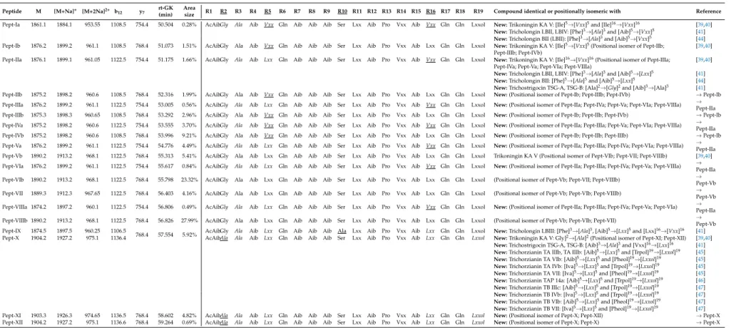
Table 1. Sequences of the newly identified peptaibol compounds produced byT. gamsiiSZMC 1656 and their similarities to known peptaibols available in the
“Peptaibiotics Database”.
Peptide M [M+Na]+ [M+2Na]2+ b12 y7 rt-GK (min)
Area
size R1 R2 R3 R4 R5 R6 R7 R8 R9 R10 R11 R12 R13 R14 R15 R16 R17 R18 R19 Compound identical or positionally isomeric with Reference Pept-Ia 1861.1 1884.1 953.55 1108.5 754.4 50.504 0.28% AcAibGly Ala Aib Vxx Gln Aib Aib Aib Ser Lxx Aib Pro Vxx Aib Vxx Gln Gln Lxxol New:Trikoningin KA V: [Ile]5→[Vxx]5and [Ile]16→[Vxx]16 [39,40]
New:Tricholongin LBII, LBIV: [Phe]3→[Ala]3and [Aib]5→[Vxx]5 [41]
New:Tricholongin BII (LBII): [Phe]3→[Ala]3and [Aib]5→[Vxx]5 [44]
Pept-Ib 1876.2 1899.2 961.1 1108.5 768.4 51.073 1.51% AcAibGly Ala Aib Vxx Gln Aib Aib Aib Ser Lxx Aib Pro Vxx Aib Lxx Gln Gln Lxxol New:Trikoningin KA V: [Ile]5→[Vxx]5(Positional isomer of Pept-IIb; [39,40]
Pept-IIIb; Pept-IVb)
Pept-IIa 1876.1 1899.1 961.05 1122.5 754.4 51.175 1.66% AcAibGly Ala Aib Lxx Gln Aib Aib Aib Ser Lxx Aib Pro Vxx Aib Vxx Gln Gln Lxxol New:Trikoningin KA V: [Ile]16→[Vxx]16(Positional isomer of Pept-IIIa; [39,40]
Pept-IVa; Pept-Va; Pept-VIa; Pept-VIIIa)
New:Tricholongin LBII, LBIV: [Phe]3→[Ala]3and [Aib]5→[Lxx]5 [41]
New:Tricholongin BII: [Phe]3→[Ala]3and [Aib]5→[Lxx]5 [44]
New:Trichostrigocin TSG-A, TSG-B: [Ala]2→[Gly]2and [Aib]3→[Ala]3 [41]
Pept-IIb 1875.2 1898.2 960.6 1108.5 768.4 52.316 1.99% AcAibGly Ala Aib Vxx Gln Aib Aib Aib Ser Lxx Aib Pro Vxx Aib Lxx Gln Gln Lxxol New:(Positional isomer of Pept-Ib; Pept-IIIb; Pept-IVb) →Pept-Ib Pept-IIIa 1876.2 1899.2 961.1 1122.5 754.4 53.005 0.56% AcAibGly Ala Aib Lxx Gln Aib Aib Aib Ser Lxx Aib Pro Vxx Aib Vxx Gln Gln Lxxol New:(Positional isomer of Pept-IIa; Pept-IVa; Pept-Va; Pept-VIa; Pept-VIIIa) →
Pept-IIa Pept-IIIb 1875.3 1898.3 960.65 1108.5 768.4 53.292 2.96% AcAibGly Ala Aib Vxx Gln Aib Aib Aib Ser Lxx Aib Pro Vxx Aib Lxx Gln Gln Lxxol New:(Positional isomer of Pept-Ib; Pept-IIb; Pept-IVb) →Pept-Ib Pept-IVa 1875.2 1898.2 960.6 1122.5 754.4 53.555 3.70% AcAibGly Ala Aib Vxx Gln Aib Aib Aib Ser Lxx Aib Pro Vxx Aib Vxx Gln Gln Lxxol New:(Positional isomer of Pept-IIa; Pept-IIIa; Pept-Va; Pept-VIa; Pept-VIIIa) →
Pept-IIa Pept-IVb 1875.2 1898.2 960.6 1108.5 768.4 53.996 9.21% AcAibGly Ala Aib Vxx Gln Aib Aib Aib Ser Lxx Aib Pro Vxx Aib Lxx Gln Gln Lxxol New:(Positional isomer of Pept-Ib; Pept-IIb; Pept-IIIb) →Pept-Ib Pept-Va 1876.2 1899.2 961.1 1122.5 754.4 54.776 4.49% AcAibGly Ala Aib Lxx Gln Aib Aib Aib Ser Lxx Aib Pro Vxx Aib Vxx Gln Gln Lxxol New:(Positional isomer of Pept-IIa; Pept-IIIa; Pept-IVa; Pept-VIa; Pept-VIIIa) →
Pept-IIa Pept-Vb 1890.2 1913.2 968.1 1122.5 768.4 55.313 5.41% AcAibGly Ala Aib Lxx Gln Aib Aib Aib Ser Lxx Aib Pro Vxx Aib Lxx Gln Gln Lxxol Trikoningin KA V (Positional isomer of Pept-VIb; Pept-VII; Pept-VIIIb) [39,40]
Pept-VIa 1876.2 1899.2 961.1 1122.5 754.4 55.617 0.84% AcAibGly Ala Aib Lxx Gln Aib Aib Aib Ser Lxx Aib Pro Vxx Aib Vxx Gln Gln Lxxol New:(Positional isomer of Pept-IIa; Pept-IIIa; Pept-IVa; Pept-Va; Pept-VIIIa) → Pept-IIa Pept-VIb 1890.2 1913.2 968.1 1122.5 768.4 55.798 23.32% AcAibGly Ala Aib Lxx Gln Aib Aib Aib Ser Lxx Aib Pro Vxx Aib Lxx Gln Gln Lxxol (Positional isomer of Pept-Vb; Pept-VII; Pept-VIIIb) →
Pept-Vb Pept-VII 1889.3 1912.3 967.65 1122.5 768.4 56.403 4.16% AcAibGly Ala Aib Lxx Gln Aib Aib Aib Ser Lxx Aib Pro Vxx Aib Lxx Gln Gln Lxxol (Positional isomer of Pept-Vb; Pept-VIb; Pept-VIIIb) →
Pept-Vb Pept-VIIIa 1874.2 1897.2 960.1 1122.5 754.4 56.806 0.49% AcAibGly Ala Aib Lxx Gln Aib Aib Aib Ser Lxx Aib Pro Vxx Aib Vxx Gln Gln Lxxol New:(Positional isomer of Pept-IIa; Pept-IIIa; Pept-IVa; Pept-Va; Pept-VIa) →
Pept-IIa Pept-VIIIb 1890.2 1913.2 968.1 1122.5 768.4 56.826 27.99% AcAibGly Ala Aib Lxx Gln Aib Aib Aib Ser Lxx Aib Pro Vxx Aib Lxx Gln Gln Lxxol (Positional isomer of Pept-Vb; Pept-VIb; Pept-VII) →
Pept-Vb Pept-IX 1874.5 1897.5 960.25 1106.5
768.4 57.554 5.92% AcAibGly Ala Aib Lxx Gln Aib Aib Aib Ala Lxx Aib Pro Vxx Aib Lxx Gln Gln Lxxol New:Tricholongin LBIII: [Phe]3→[Ala]3, [Aib]5→[Lxx]5and [Lxx]16→[Vxx]16 [41]
Pept-X 1904.2 1927.2 975.1 1136.4 AcAibAla Ala Aib Lxx Gln Aib Aib Aib Ser Lxx Aib Pro Vxx Aib Lxx Gln Gln Lxxol New:Trikoningin KA V: Gly]2→[Ala]2(Positional isomer of Pept-XI; Pept-XII) [39,40]
New:Trichostrigocin TSG-A, TSG-B: [Aib]3→[Ala]3and [Vxx]16→[Lxx]16 [41]
New:Trichorzianin TA IIIb, TA IIIb: [Aib]5→[Lxx]5and [Trpol]19→[Lxxol]19 [45]
New:Trichorzianin TA VIb: [Aib]5→[Lxx]5and [Pheol]19→[Lxxol]19 [45]
New:Trichorzianin TA IVb: [Iva]5→[Lxx]5and [Trpol]19→[Lxxol]19 [45]
New:Trichorzianin TA VII: [Iva]5→[Lxx]5and [Pheol]19→[Lxxol]19 [45]
New:Trichorzianin TAP 14a: [Aib]5→[Lxx]5and [Trpol]19→[Lxxol]19 [46]
New:Trichorzianin TB IIIc: [Aib]5→[Lxx]5and [Trpol]19→[Lxxol]19 [47]
New:Trichorzianin TB IVb: [Iva]5→[Lxx]5and [Trpol]19→[Lxxol]19 [47]
New:Trichorzianin TB VIb: [Aib]5→[Lxx]5and [Pheol]19→[Lxxol]19 [47]
New:Trichorzianin TB VII: [Iva]5→[Lxx]5and [Pheol]19→[Lxxol]19 [47]
Pept-XI 1903.3 1926.3 974.65 1136.5 768.4 58.602 4.82% AcAibAla Ala Aib Lxx Gln Aib Aib Aib Ser Lxx Aib Pro Vxx Aib Lxx Gln Gln Lxxol New:(Positional isomer of Pept-X; Pept-XII) →Pept-X Pept-XII 1904.2 1927.2 975.1 1136.6 768.4 59.264 0.69% AcAibAla Ala Aib Lxx Gln Aib Aib Aib Ser Lxx Aib Pro Vxx Aib Lxx Gln Gln Lxxol New:(Positional isomer of Pept-X; Pept-X) →Pept-X
Variable residues are underlined in the table header, minor sequence variants are underlined in the sequences. Amino acid exchanges in new sequences are italicised.
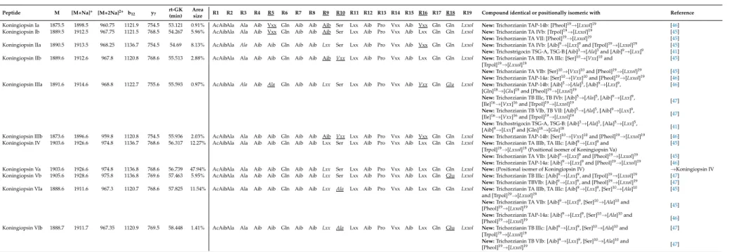
Table 2.Sequences of the newly identified peptaibol compounds produced byT. koningiopsisSZMC 12500 and their similarities to known peptaibols available in the
“Peptaibiotics Database”.
Peptide M [M+Na]+ [M+2Na]2+ b12 y7 rt-GK (min)
Area
size R1 R2 R3 R4 R5 R6 R7 R8 R9 R10 R11 R12 R13 R14 R15 R16 R17 R18 R19 Compound identical or positionally isomeric with Reference Koningiopsin Ia 1875.5 1898.5 960.75 1121.9 754.5 53.121 0.91% AcAibAla Ala Aib Vxx Gln Aib Aib Aib Ser Lxx Aib Pro Vxx Aib Vxx Gln Gln Lxxol New:Trichorzianin TAP-14b: [Pheol]19→[Lxxol]19 [46]
Koningiopsin Ib 1889.5 1912.5 967.75 1121.5 768.5 54.267 5.96% AcAibAla Ala Aib Vxx Gln Aib Aib Aib Ser Lxx Aib Pro Vxx Aib Lxx Gln Gln Lxxol New:Trichorzianin TA IVb: [Trpol]19→[Lxxol]19 [45]
New:Trichorzianin TA VII: [Pheol]19→[Lxxol]19 [45]
Koningiopsin IIa 1890.5 1913.5 968.25 1136.7 754.5 54.69 8.13% AcAibAla Ala Aib Aib Gln Aib Aib Lxx Ser Lxx Aib Pro Vxx Aib Vxx Gln Gln Lxxol New:Trichorzianin TA IVb: [Aib]9→[Lxx]9and [Trpol]19→[Lxxol]19 [45]
New:Trichostrigocin TSG-A, TSG-B:[Aib]3→[Ala]3and [Aib]9→[Lxx]9 [41]
Koningiopsin IIb 1889.6 1912.6 967.8 1120.8 768.6 55.513 2.88% AcAibAla Ala Aib Aib Gln Aib Aib Aib Vxx Lxx Aib Pro Vxx Aib Lxx Gln Gln Lxxol New:Trichorzianin TA IIIb, TA IIIc: [Ser]10→[Vxx]10and [45]
[Trpol]19→[Lxxol]19
New:Trichorzianin TA VIb: [Ser]10→[Vxx]10and [Pheol]19→[Lxxol]19 [45]
New:Trichorzianin TAP-14a: [Ser]10→[Vxx]10and [Pheol]19→[Lxxol]19 [46]
Koningiopsin IIIa 1891.6 1914.6 968.8 1122.7 755.6 55.593 0.97% AcAibAla Ala Aib Ala Gln Aib Aib Lxx Ser Lxx Aib Pro Vxx Aib Vxx Gln Glu Lxxol New:Trichorzianin TAP-14b: [Aib]5→[Ala]5, [Aib]9→[Lxx]9, [46]
[Gln]18→[Glu]18and [Pheol]19→[Lxxol]19
New:Trichorzianin TB IIIc, TB IVb: [Aib]5→[Ala]5, [Aib]9→[Lxx]9,
[Ile]16→[Vxx]16and [Trpol]19→[Lxxol]19 [47]
New:Trichorzianin TB VIb, TB VII: [Aib]5→[Ala]5, [Aib]9→[Lxx]9,
[Ile]16→[Vxx]16and [Trpol]19→[Lxxol]19 [47]
New:Trichostrigocin TSG-A, TSG-B: [Aib]3→[Ala]3, [Ala]5→[Lxx]5,
[Aib]9→[Lxx]9and [Gln]18→[Glu]18 [41]
Koningiopsin IIIb 1873.6 1896.6 959.8 1120.8 754.5 55.936 2.03% AcAibAla Ala Aib Aib Gln Aib Aib Aib Vxx Lxx Aib Pro Vxx Aib Vxx Gln Gln Lxxol New:Trichorzianin TAP-14b: [Ser]10→[Vxx]10and [Pheol]19→[Lxxol]19 [46]
Koningiopsin IV 1903.6 1926.6 974.8 1136.7 768.6 56.317 12.27% AcAibAla Ala Aib Aib Gln Aib Aib Lxx Ser Lxx Aib Pro Vxx Aib Lxx Gln Gln Lxxol New:Trichorzianin TA IIIb, TA IIIc: [Aib]9→[Lxx]9and [45]
[Trpol]19→[Lxxol]19(Positional isomer of Koningiopsin Va)
New:Trichorzianin TA VIb: [Aib]9→[Lxx]9and [Pheol]19→[Lxxol]19 [45]
New:Trichorzianin TAP-14a: [Aib]9→[Lxx]9and [Pheol]19→[Lxxol]19 [46]
Koningiopsin Va 1903.6 1926.6 974.8 1136.8 768.6 56.739 47.94% AcAibAla Ala Aib Aib Gln Aib Aib Lxx Ser Lxx Aib Pro Vxx Aib Lxx Gln Gln Lxxol New:(Positional isomer of Koningiopsin IV) →Koningiopsin IV Koningiopsin Vb 1905.6 1928.6 975.8 1136.8 769.6 57.463 5.95% AcAibAla Ala Aib Aib Gln Aib Aib Lxx Ser Lxx Aib Pro Vxx Aib Lxx Gln Glu Lxxol New:Trichorzianin TB IIIc: [Aib]9→[Lxx]9, and [Trpol]19→[Lxxol]19 [47]
New:Trichorzianin TBVIb: [Aib]9→[Lxx]9, and [Pheol]19→[Lxxol]19 [47]
Koningiopsin VIa 1888.6 1911.6 967.3 1120.7 768.6 57.825 11.54% AcAibAla Ala Aib Aib Gln Aib Aib Lxx Ala Lxx Aib Pro Vxx Aib Lxx Gln Gln Lxxol New:Trichorzianin TA IIIb, TA IIIc: [Aib]9→[Lxx]9, [Ser]10→[Ala]10 [45]
and [Trpol]19→[Lxxol]19
New:Trichorzianin TA VIb: [Aib]9→[Lxx]9, [Ser]10→[Ala]10and
[Pheol]19→[Lxxol]19 [45]
New:Trichorzianin TAP-14a: [Aib]9→[Lxx]9, [Ser]10→[Ala]10and
[Pheol]19→[Lxxol]19 [46]
Koningiopsin VIb 1888.7 1911.7 967.35 1120.9 769.5 58.448 1.41% AcAibAla Ala Aib Aib Gln Aib Aib Lxx Ala Lxx Aib Pro Vxx Aib Lxx Gln Glu Lxxol New:Trichorzianin TB IIIc: [Aib]9→[Lxx]9, [Ser]10→[Ala]10and [47]
[Trpol]19→[Lxxol]19
New:Trichorzianin TB VIb: [Aib]9→[Lxx]9, [Ser]10→[Ala]10and
[Pheol]19→[Lxxol]19 [47]
Variable residues are underlined in the table header, minor sequence variants are underlined in the sequences. Amino acid exchanges in new sequences are italicised. Positions R14 and R15 were predicted based on the Comprehensive Peptaibotics Database [14].
In certain cases, minor differences were observed between the presently detected and the previously reported peptaibol sequences showing amino acid exchanges only at selected positions of the peptide chain (Tables1and2). Trichorzianins differ in the position 19 of the peptide chain.
This position plays a critical role in the lifetime of the opened ion channel as the substitution of Pheol to Leuol/Ileol has led to increased lifetime of the open channel [50]. This was also observed previously in the case of peptaibol-formed channels, where the Pheol was substituted to Trpol in both trichorzianin B-IIIc (Trpol) and B-VII (Pheol) [51]. On the other hand, the investigation of synthetic alamethicin analogues—where all Aib residues were changed to Leu—revealed that the substitution of the C-terminal residues was not affecting the lifetime of the open channel [52]. A secondary structural study was also carried out for the purified compound trichorzianin TA VII in association with sodium dodecyl sulfate (SDS) micelles, which revealed formation of two right-handed helical segments (1–8 and 11–19) linked by aβ-turn [53]. The novelty of the peptaibols produced by T. koningiopsis SZMC 12500 is in the variation of the C-terminus, which is critical in the lifetime of the ion channels. The name “Koningiopsin” was introduced for these novel compounds.
The calculated contents of the whole peptaibol molecules were 214.28 µg mL−1 and 101.26µg mL−1 in the crude extracts of T. gamsii SZMC 1656 and T. koningiopsis SZMC 12500, respectively. In the case ofT. gamsiiSZMC 1656, Pept VIIIb and Pept VIb were the most abundant sequences of peptaibols. The sum of the amount of these two molecules was approx. 50%, while the concentration of other peptaibols remained below 10%. In the extract ofT. koningiopsisSZMC 12500, Pept XVIIa accounted for almost half of the peptaibols produced. A new method for quantification of peptaibols based on the length of different peptides was described by Van Bohemen et al. [54]. The different length of peptaibols results in different structures, furthermore, shorter peptaibols (11–14 residues) contain more Pro leading to a structural deformation [55]. Based on the alamethicin standard, only the longer (17–20-residue) peptaibols can be quantified with high accuracy.
3.2. Structural Elucidation of Trikoningin KA V Based on Short Molecular Dynamics
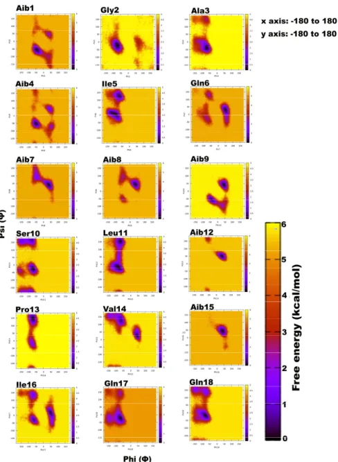
Trikoningin KA V (TKV, positionally isomeric with sequences Pept-Vb, -VIb, and -VII ofT. gamsii) is a 19-residue peptaibol with seven Aib residues constituting its sequence. Aib is an achiral residue, which has been shown to promote helix formation and can exist in both right- and left-handed helix regions on the Ramachandran plot [56–59]. To determine the propensities of each residue for a given secondary structural region on the Ramachandran plot, their relative free energies were calculated, which clearly describe an energetically favourable conformation (Figure1). The darkest scatter populations indicate energetically preferable conformations.
Unexpectedly, a strong preference was found for the left-handed helix region ofΦ-ψplots during this simulation, specifically for residues in the central region flanked by Gln6, Aib7, Aib8, Aib9, Aib12, Val14, Aib15, and Ile16. Except for Aib1 and Aib4, all other Aib residues show free energy minimum in the left-handed helix region. Most standard (proteinogenic) amino acid residues, Gly2, Ala3, Ser10, Leu11, Gln17, and Gln18, display an energy minimum in the right-handedα-helix region. Ile5, Ser10, Leu11, Pro13, and Ile16 also show preference for a poly-proline II region. This behaviour of Leu and Ile to occupyβ-space on the Ramachandran plot is expected due toβ-branching of their side-chains.
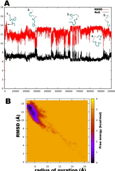
It is known that due to heavy side chains, they show lesser propensity to exist in a helix and, therefore, prefer formation ofβ-strands. The presence of three consecutive Aib residues in positions 7, 8, and 9 of the peptide chain seems to drive its conformation towards a left-handed helix, while the rest shows clear preference for right-handedness. This resulted in an overall unwinding of the helix, which never seems to form a continuous spiral shape. Further experiments with higher sampling power are required to confirm these results. The calculation of root-mean-square-deviation (RMSD) values based on the coordinates of peptide backbone atoms C, CA, and N for each frame with respect to the average structure has been provided. A similar result was obtained for radius of gyration (RoG) values, which is the root-mean-square-distance of peptide components from their center of mass calculated for each frame. The preliminary investigation revealed that the overall conformation (obtained from the
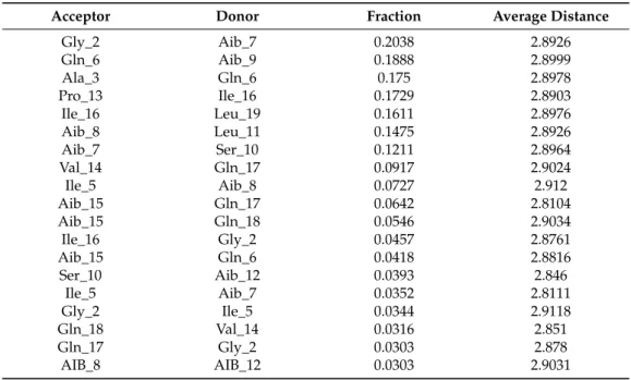
trajectory with RMSD value between 12 to 14Å, denoted by structures 3 and 4) resembles a hairpin with turn structures that never assumes a spiral shape (Figure2A). Structures 2 and 5 with an almost linear structure show lower RMSD values than 12 Å, which is not energetically favoured. The free-energy landscape as a function of RMSD and radius of gyration is shown in Figure2B, which clearly indicates that structures with RoG value of less than 8 Å and RMSD values between 12–14 Å are energetically favoured. When compared with the hydrogen bonding pattern within the backbone, mostly i+3→i H-bonds were found that denote 310helix probably in left-handed conformation as indicated byΦ-ψ plots (Table3). Ile5→Gly2, Aib8→Ile5, Aib9→Gln6, Ser10→Aib7, Ile16→Pro13, and Gln17→Val14 are examples of left-handed 310helix bonds while Gln6→Ala3, Leuol19→Ile16, Leu11→Aib8 are examples of right-handed 310helix. Fewγ-turn populations are also seen by Aib7→Ile5, Aib12→Ser10, and Gln17→Aib15 as energetically stable. This means that the highly bent structure resembling aβ-hairpin with the N- and C-terminals in close proximity to each other is energetically favoured in comparison to a linear backbone.
Microorganisms 2018, 6, x FOR PEER REVIEW 11 of 19
Figure 1. Free energy-based Ramachandran plots for each Trikoningin KA V residue during 100 ns long implicit water simulation. The x and y axes range from −180 to +180. The darkest red regions indicate toward minimum energy secondary structural regions favoured by each residue during the simulation.
Unexpectedly, a strong preference was found for the left-handed helix region of Ф-ψ plots during this simulation, specifically for residues in the central region flanked by Gln6, Aib7, Aib8, Aib9, Aib12, Val14, Aib15, and Ile16. Except for Aib1 and Aib4, all other Aib residues show free Figure 1.Free energy-based Ramachandran plots for each Trikoningin KA V residue during 100 ns long implicit water simulation. The x and y axes range from−180 to +180. The darkest red regions indicate toward minimum energy secondary structural regions favoured by each residue during the simulation.
Figure 2. (A). The root-mean-square-deviation (RMSD in red color) and radius of gyration (RoG in black) with corresponding three-dimensional structures of trikoningin KA V, (B). Free energy landscape as a function of RMSD and RoG shows energetically favoured conformations with RMSD between 12–14 Å and RoG value less than 8 Å.
Table 3. Backbone H-bonds of Trikoningin KA V along with their frequency of occurrence given by fraction, average distance, and angle.
Acceptor Donor Fraction Average Distance
Gly_2 Aib_7 0.2038 2.8926
Gln_6 Aib_9 0.1888 2.8999
Ala_3 Gln_6 0.175 2.8978
Pro_13 Ile_16 0.1729 2.8903
Ile_16 Leu_19 0.1611 2.8976
Aib_8 Leu_11 0.1475 2.8926
Aib_7 Ser_10 0.1211 2.8964
Val_14 Gln_17 0.0917 2.9024
Ile_5 Aib_8 0.0727 2.912
Figure 2.(A). The root-mean-square-deviation (RMSD in red color) and radius of gyration (RoG in black) with corresponding three-dimensional structures of trikoningin KA V, (B). Free energy landscape as a function of RMSD and RoG shows energetically favoured conformations with RMSD between 12–14 Å and RoG value less than 8 Å.
Table 3.Backbone H-bonds of Trikoningin KA V along with their frequency of occurrence given by fraction, average distance, and angle.
Acceptor Donor Fraction Average Distance
Gly_2 Aib_7 0.2038 2.8926
Gln_6 Aib_9 0.1888 2.8999
Ala_3 Gln_6 0.175 2.8978
Pro_13 Ile_16 0.1729 2.8903
Ile_16 Leu_19 0.1611 2.8976
Aib_8 Leu_11 0.1475 2.8926
Aib_7 Ser_10 0.1211 2.8964
Val_14 Gln_17 0.0917 2.9024
Ile_5 Aib_8 0.0727 2.912
Aib_15 Gln_17 0.0642 2.8104
Aib_15 Gln_18 0.0546 2.9034
Ile_16 Gly_2 0.0457 2.8761
Aib_15 Gln_6 0.0418 2.8816
Ser_10 Aib_12 0.0393 2.846
Ile_5 Aib_7 0.0352 2.8111
Gly_2 Ile_5 0.0344 2.9118
Gln_18 Val_14 0.0316 2.851
Gln_17 Gly_2 0.0303 2.878
AIB_8 AIB_12 0.0303 2.9031
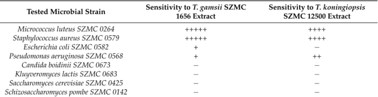
3.3. Inhibitory Effects of Peptaibol Extracts Towards Bacteria, Yeasts, and Filamentous Fungi
Micrococcus luteusandS. aureusproved to be sensitive to both peptaibol extracts (Table4),E. coli was more resistant, whileP. aeruginosashowed higher sensitivity to the extracts fromT. koningiopsis than to those fromT. gamsii. M. luteusandS. aureusare Gram-positive bacteria, whileE. coliandP.
aeruginosaare Gram-negative ones, thus their sensitivity showed correlation with the type of their cell wall. Studies on the bioactivity of paracelsins [60] or alamethicin [61] showed similar results, i.e., that Gram-positive bacteria proved to be more sensitive to peptaibols. Testing the peptaibols trichorzianine A1 and B1 on Gram-positive and Gram-negative bacteria also revealed similar results, furthermore, synergistic effect could also be detected between peptaibols and different cell membrane-affecting (MACs) and cell wall-degrading enzymes (CWDEs) [62,63]. In a study of Lorito et al. [64], the inhibition ofβ-D-glucan synthase was reported as a specific effect of peptaibol antibiotics. On the other hand, in the study of Cutler et al. [65], the purified peptaibol identified as trikoningin KA V (also known as koningin A) seemed to be inactive against both Gram-positive and Gram-negative bacteria. No inhibition zones could be observed in the case of yeasts (Table4), though in another study, minimum inhibition could be observed after the treatment of trichokonins produced byT. koningiionS. cerevisiae CGMCC2.395 andC. albicansCGMCC2.538 [66]. Complete inhibition could not be observed among the fast-growing fungi (Table5), though. Interestingly, the producerT. koningiopsisandT. gamsiistrains seemed to be more sensitive to their own peptaibol extracts thanT. aggressivumf. europaeumand T. pleuroti, known as the causal agents of green mould disease occurring in mushroom cultivation (Table5). The latter two species are also known to produce peptaibols [67], but their peptaibiomes are entirely different from those ofT. koningiopsisSZMC 12500 andT. gamsiiSZMC 1656, they produce 18-residue hypomurocin-like peptaibols and tripleurins, respectively.
Table 4.Bioactivity of concentrated peptaibol extracts (100 mg mL−1) fromTrichoderma gamsiiSZMC 1656 andT. koningiopsisSZMC 12500 towards bacteria and yeasts.
Tested Microbial Strain Sensitivity toT. gamsiiSZMC 1656 Extract
Sensitivity toT. koningiopsis SZMC 12500 Extract
Micrococcus luteus SZMC 0264 +++++ ++++
Staphylococcus aureus SZMC 0579 +++++ ++++
Escherichia coli SZMC 0582 + −
Pseudomonas aeruginosa SZMC 0568 + ++
Candida boidinii SZMC 0673 − −
Kluyveromyces lactis SZMC 0683 − −
Saccharomyces cerevisiae SZMC 0425 − −
Schizosaccharomyces pombe SZMC 0142 − −
−, absence of inhibition; diameter of inhibition zone: +, 5–7 mm; ++, 7–9 mm, +++, 9–11 mm, ++++, 11–13 mm diameter, +++++, 13–15 mm.
Table 5. Bioactivity of concentrated peptaibol extracts (100 mg mL−1) and their two-fold serial dilutions fromTrichoderma gamsiiSZMC 1656 andT. koningiopsisSZMC 12500 towards cultures of filamentous fungi.
Tested Filamentous Fungal Strain Sensitivity toT. gamsiiSZMC 1656 Extract
Sensitivity toT. koningiopsis SZMC 12500 Extract
Alternaria alternataSZMC 16085 +++++ * +++ *
Fusarium solanispecies complex SZMC 11467 ++ +
Rhizoctonia solaniSZMC 6252J ++ * +++ *
Phoma cucurbitacearumSZMC 16088 ++ * ++++ *
T. aggressivumf.europaeumSZMC 1811 ++ +
T. pleurotiSZMC 12454 ++ +
T. gamsiiSZMC 1656 +++ +++
T. koningiopsisSZMC 12500 +++ +++
inhibition of mycelial growth at dilution steps: +, 1st–2nd; ++, 3rd–4th; +++, 5th; ++++, 6th; +++++, 7 th. *, mycelial growth was completely stopped.
TheF. solanispecies complex member appeared to be more sensitive to the peptaibol extract of T. gamsiithan to the one ofT. koningiopsis. Inhibition could also be detected in the case ofA. alternata, R. solaniandP. cucurbitacearum, all growing very slowly (8 days, 12 days, and 11 days, respectively, till they reach the MeOH hole on the plates), and interestingly, the mycelial growth of these filamentous fungi stopped where the peptaibols were added into the holes and could not reach the edge of the plates (Table5).
4. Conclusions
In this study, the peptaibiome composition ofT. koningiopsisandT. gamsiiwas identified by HPLC-ESI-MS measurements, which revealed a total of 30 peptaibol sequences. A structurally close compound, trikoningin KA V, was selected from the literature for structural elucidation, which revealed fluctuating right- and left-handed helical conformations. The examination of their antibiotic activity against a broad spectrum of different microorganisms showed that Gram-positive bacteria were strongly inhibited, while Gram-negative bacteria seemed to be less sensitive to the peptaibol extracts tested. Inhibitory effects of the studied peptaibol extracts could not be observed on yeasts, while filamentous fungi showed considerable sensitivity.
Supplementary Materials:The following are available online athttp://www.mdpi.com/2076-2607/6/3/85/s1, Supplementary Data 1: Detailed description for residue parameterization, Figure S1: Extracted ion chromatograms (EIC) resulting from full scan measurements of crude extracts fromT. gamsiiSZMC 1656, Figure S2: Extracted ion chromatograms (EIC) resulting from full scan measurements of crude extracts prepared fromT. koningiopsisSZMC 12500, Figure S3: Typical b ion series of described peptaibols ranged from b12atm/z1106, Figure S4: Typical b ion series of described peptaibols ranging from b12atm/z1120, Figure S5: Typical MS2spectra of y-ions 754.5 (A), 755.5 (B), 768.5 (C) and 769.5 (D) resulting from the full scan measurements of crude peptaibol extracts, Table S1:
Diagnostic fragment ions of peptaibols detected with the full scan MS measurement of peptaibol extracts from
plate cultures ofT. gamsiiSZMC 1656, Table S2: Diagnostic fragment ions of peptaibols detected with the full scan MS measurement of peptaibol extracts from plate cultures ofT. koningiopsisSZMC 12500, Table S3: Diagnostic fragment ions of acylium ion (y7) detected with MS2measurements of peptaibol extracts from plate cultures of T. gamsiiSZMC 1656, Table S4: Diagnostic fragment ions of acylium ion (y7) detected with MS2measurements of peptaibol extracts from plate cultures ofT. koningiopsisSZMC 12500.
Author Contributions: Conceptualization, A.S., C.V., L.K.; Data analysis, T.M., C.T., G.R., D.R.; Funding acquisition, A.S., C.V., L.K.; Investigation, T.M., G.R., D.R., A.S.; Methodology, T.M., G.R., D.R., A.S.; Project administration, A.S., C.V., L.K.; Resources, AS., C.V., L.K.; Software, C.T.; Supervision, A.S., C.V., L.K.; Validation, D.R., A.S.; Visualization, T.M., C.T.; Writing—original draft, T.M.; Writing—review & editing, A.S., C.V., L.K.
Funding:This research was funded by grant GINOP-2.3.2-15-2016-00012 (Széchenyi 2020 Programme). LK was supported by the János Bolyai Research Scholarship (Hungarian Academy of Sciences). AS was supported by the New National Excellence Program of the Ministry of Human Capacities (ÚNKP-16-4).
Conflicts of Interest:The authors declare no conflict of interest.
References
1. Bissett, J.; Gams, W.; Jaklitsch, W.; Samuels, G.J. AcceptedTrichodermanames in the year 2015.IMA Fungus 2015,6, 263–295. [CrossRef] [PubMed]
2. Bisby, G.R.Trichoderma viridePers. ex Fries, and notes onHypocrea.Mycol. Res.1939,23, 149–168. [CrossRef]
3. Rifai, M.A. A revision of the genusTrichoderma.Mycol. Pap.1969,116, 1–56.
4. Bissett, J. A revision of the genusTrichoderma. II. Infrageneric classification.Can. J. Bot.1991,69, 2357–2372.
[CrossRef]
5. Kullnig-Gradinger, C.M.; Szakacs, G.; Kubicek, C.P. Phylogeny and evolution of the genusTrichoderma: A multigene approach.Mycol. Res.2002,106, 757–767. [CrossRef]
6. Samuels, G.J.; Dodd, S.L.; Lu, B.S.; Petrini, O.; Schroers, H.J.; Druzhinina, I.S. TheTrichoderma koningii aggregate species.Stud. Mycol.2006,56, 67–133. [CrossRef] [PubMed]
7. Lieckfeldt, E.; Samuels, G.J.; Nirenberg, H.I.; Petrini, O. A morphological and molecular perspective of Trichoderma viride: Is it one or two species?Appl. Environ. Microbiol.1999,65, 2418–2428. [PubMed]
8. Jaklitsch, W.M.; Samuels, G.J.; Dodd, S.L.; Lu, B.S.; Druzhinina, I.S.Hypocrea rufa/Trichoderma viride: A reassessment, and description of five closely related species with and without warted conidia.Stud. Mycol.
2006,56, 135–177. [CrossRef] [PubMed]
9. Kredics, L.; Hatvani, L.; Naeimi, S.; Körmöczi, P.; Manczinger, L.; Vágvölgyi, C.; Druzhinina, I. Biodiversity of the genusHypocrea/Trichodermain different habitats. InBiotechnology and biology of Trichoderma; Gupta, V.K., Schmoll, M., Herrera-Estrella, A., Upadhyay, R.S., Druzhinina, I., Tuohy, M.G., Eds.; Elsevier: New York, NY, USA, 2014; pp. 3–24. ISBN 978-0-444-59576-8.
10. Verma, M.; Brar, S.K.; Tyagi, R.D.; Surampalli, R.Y.; Valéro, J.R. Antagonistic fungi,Trichodermaspp.: Panoply of biological control.Biochem. Eng. J.2007,37, 1–20. [CrossRef]
11. Schuster, A.; Schmoll, M. Biology and biotechnology ofTrichoderma.Appl. Microbiol. Biotechnol. 2010,87, 787–799. [CrossRef] [PubMed]
12. Reino, J.L.; Guerrero, R.F.; Hernández-Galán, R.; Collado, I.G. Secondary metabolites from species of the biocontrol agentTrichoderma.Phytochem. Rev.2008,7, 89–123. [CrossRef]
13. Degenkolb, T.; Brückner, H. Peptaibiomics: Towards a myriad of bioactive peptides containing C(alpha)-dialkylamino acids?Chem. Biodivers.2008,5, 1817–1843. [CrossRef] [PubMed]
14. Stoppacher, N.; Neumann, N.K.N.; Burgstaller, L.; Zeilinger, S.; Degenkolb, T.; Brückner, H.; Schuhmacher, R.
The comprehensive peptaibiotics database.Chem. Biodivers.2013,10, 734–743. [CrossRef] [PubMed]
15. Marahiel, M.A. Protein templates for the biosynthesis of peptide antibiotics.Chem. Biol.1997,4, 561–567.
[CrossRef]
16. Bushley, K.E.; Turgeon, B.G. Phylogenomics reveals subfamilies of fungal nonribosomal peptide synthetases and their evolutionary relationships.BMC Evol. Biol.2010,10, 26. [CrossRef] [PubMed]
17. Duclohier, H. Helical kink and channel behaviour: A comparative study with the peptaibols alamethicin, trichotoxin and antiamoebin.Eur. Biophys. J.2004,33, 169–174. [CrossRef] [PubMed]
18. McMullin, D.R.; Renaud, J.B.; Barasubiye, T.; Sumarah, M.W.; Miller, J.D. Metabolites ofTrichodermaspecies isolated from damp building materials.Can. J. Microbiol.2017,63, 621–632. [CrossRef] [PubMed]