C A S E R E P O R T Open Access
Sarcoidosis lymphoma syndrome - the value of PET-CT in the diagnosis
Adrian Kis1, Noemi Eszes1*, Lilla Tamasi1, Gyorgy Losonczy1, Attila Csekeo2, Judit Csomor3and Veronika Muller1
Abstract
We report a 52-year-old patient who developed B-cell non-Hodgkin’s lymphoma subsequent to sarcoidosis.
Sarcoidosis was diagnosed 16 years ago and remained asymptomatic for 14 years after steroid treatment. She presented with new symptoms of arthralgia, photosensitivity, butterfly erythema, autoimmune antibodies (ANA, chromatin positivity) associated with progression of the known left upper lobe lesion on the chest X-ray suggesting primary autoimmune disease (systemic lupus erythematosus). As steroid treatment was not effective, we started bolus cyclophosphamide therapy after which progression was seen on the chest X-ray. Computed tomography (CT)-guided needle biopsy confirmed malignancy of indefinable origin. Despite of the well-known
fluorodeoxyglucose (FDG) avidity in active sarcoidosis, a FDG-positron emission tomography (PET) scan was performed to stage the primary tumour. Intensive FDG uptake was detected in the affected lung segment, with moderate uptake in mediastinal lymph nodes. The patient underwent left upper lobectomy. The histology showed pulmonary mucosa-associated lymphoma (bronchus-associated lymphoid tissue (BALT) lymphoma) in the lung tissue, while only sarcoidosis was present in the mediastinal lymph nodes. Bone marrow biopsy was negative.
The association between sarcoidosis and lymphoma is known as sarcoidosis lymphoma syndrome, which is a rare disease. PET-CT was helpful in the differentiation of sarcoidosis and malignancy in this patient. It is important to be aware of the risk of lymphoma in sarcoidosis and FDG-PET, used for adequate purpose, can help the diagnosis.
Keywords:Sarcoidosis, Malignancy, Sarcoidosis lymphoma syndrome, PET-CT
Background
Sarcoidosis is a chronic granulomatous inflammatory multisystem disorder of unknown origin. The associ- ation between sarcoidosis and malignancy - particularly lymphoproliferative disease, such as non-Hodgkin’s lymphoma - has been previously described [1-3]. In sar- coidosis lymphoma syndrome, sarcoidosis is followed by the development of the lymphoproliferative disorder [4]. Higher fluorodeoxyglucose (FDG)-uptake may be seen in both diseases; therefore, positron emission tomography-computed tomography (PET-CT) cannot exclude or prove the presence of malignancy in sarcoid- osis patients. In our case report of sarcoidosis lymph- oma syndrome, we demonstrate how PET-CT could help within the decision making process.
Case presentation
The 52-year-old female patient first presented in 1992 with erythema nodosum, alopecia and a dry cough. Chest X-ray and computed tomography (CT) showed bilateral hilar lymphadenopathy and lung parenchyma involve- ment. Histology confirmed sarcoidosis, which was treated with systemic steroids for six months, after which per- sisting diabetes mellitus developed. After 14 asymptomatic years without immunosuppressive therapy, she was re- ferred to our department with symptoms of dry cough, snoring and mild anaemia. On the chest X-ray, progres- sion of the known left upper lobe lesion was noted (Figure 1). As primarily progression of sarcoidosis was suspected, steroid treatment was restarted. During the im- munosuppressive therapy arthralgia, butterfly erythema, photosensitivity appeared, associated with anti-nuclear and anti-chromatin antibody positivity. A lung function test revealed decreased diffusion capacity of carbon monoxide (CO). According to the American College of Rheumatology’s criteria, an additional diagnosis of systemic
* Correspondence:noemi.eszes@gmail.com
1Department of Pulmonology, Semmelweis University, 1125, Diós árok 1/C, Budapest, Hungary
Full list of author information is available at the end of the article
© 2013 Kis et al.; licensee BioMed Central Ltd. This is an Open Access article distributed under the terms of the Creative Commons Attribution License (http://creativecommons.org/licenses/by/2.0), which permits unrestricted use, distribution, and reproduction in any medium, provided the original work is properly cited.
lupus erythematosus (SLE) was established. Due to inef- fective response to steroid treatment, bolus cyclophos- phamide (500 mg/m2 monthly) therapy was instituted.
Repeated chest X-ray showed rapid and significant worsening of the left upper lobe lesion following the first dose (Figure 2). Beside the left upper lobe lesion, en- larged mediastinal lymph nodes and a small nodular lesion in the left lower lobe (segment 10) were noted on the chest CT. Histology of the transbronchial biopsy from the left upper lobe showed nonspecific lymphocytic inflammation.
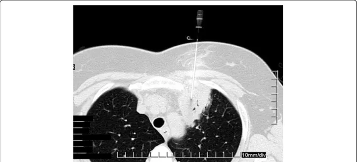
CT-guided needle aspiration of the left upper lobe lesion was performed to get proper diagnosis (Figure 3). Cytology confirmed malignancy of indefinable, but possible renal ori- gin. Abdominal ultrasound and CT scan were negative.
To localize the primary tumour and its stage, FDG- PET-CT scan was made, knowing that active sarcoidosis is associated with increased FDG uptake. Intensive FDG
uptake was detected in the affected lung segment, with only moderate uptake in mediastinal lymph nodes (Figure 4). As the nodule in the left lower lobe showed moderate FDG uptake similar to mediastinal lymph nodes, it was suggested to represent sarcoidosis. Taking into con- sideration the very intensive FDG uptake and cytology confirmed malignancy of the left upper lobe, our surgical team came to the decision of performing a left upper lob- ectomy and extensive lymph node sampling.
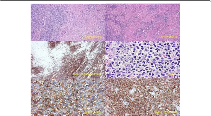
Postoperative histology showed pulmonary mucosa- associated lymphoma (bronchus associated lymphoid tissue (BALT) lymphoma) in the left upper lobe lung tis- sue, while only sarcoidosis was present in the mediastinal lymph nodes. Immunophenotyping demonstrated a B cell lineage phenotype: CD20, BCL2 and lambda light chain positivity on the cell surface, with CD10 and CD5 negativ- ity (Figure 5). The tumour was confined only to the lung parenchyma of the left upper lobe; haematological exami- nations, including bone marrow biopsy, were negative, confirming the diagnosis of stage I BALT lymphoma. After surgery the patient received only respiratory rehabilitation.
Three-year follow-up with regular chest-abdominal CT scans showed no progression or recurrence of the diseases (Figures 6 and 7).
Sarcoidosis is a multisystem granulomatous disorder of uncertain origin. The diagnosis of sarcoidosis is based on clinical, radiological, biochemical and histological findings.
These findings are erythema nodosum, lymphopenia, hypergammaglobulinemia, anergy, hypercalcemia and ele- vated angiotensin-converting enzyme. The non-caseating granulomas are B-cell negative [1]. The results should be carefully interpreted, because sarcoid-like granulomas can be found in many other disorders, such as chronic inflam- matory, infectious and neoplastic diseases [2]. Incidence is influenced by age, race and geographical location. The subacute form diagnosed in most patients under the age of 30 affects mainly intrathoracic organs and the duration of the disease is generally less than two years. The chronic form begins more often over the age of 40 and might in- volve extrathoracic organs [1].
Brincker and Wilbek have matched all patients who had been diagnosed with respiratory sarcoidosis during the period 1962 to 1971 against the data of the Cancer Regis- try. About 1.5 times higher incidence of cancer was seen in sarcoidosis patients. Lung cancer occurred 3 times and malignant lymphoma 11.5 times more frequently than in the control population [3]. In a later study, Brincker found three features of malignancies following sarcoidosis based on the analysis of 29 case reports in the literature [1]: the median age of the patients was 41 years at the diagnosis of sarcoidosis, which indicates a late onset, and is more often associated with a chronic type [2]. Lymphoproliferative disease (LD) has occurred with a median interval of 24 months after the diagnosis of sarcoidosis [3]. Hodgkin’s
Figure 1Chest X-ray of the patient.After 14 asymptomatic years, new symptoms occurred. The chest X-ray showed progression of the known left upper lobe lesion.
Figure 2Repeat chest X-ray.Repeat chest X-ray showed rapid and significant worsening of the left upper lobe lesion.
disease was diagnosed more frequently than other types of lymphomas. Further analysis of 17 cases revealed that LD developed 5.5 times more frequently than expected, which may suggest that the chronic active type of sarcoidosis is a predisposing factor for LD. Brincker introduced the term sarcoidosis lymphoma syndrome for this specific condi- tion [4]. In a retrospective cohort study Askling and his colleagues assessed the risk for malignancy in sarcoidosis.
They linked a sarcoidosis incidence study (Uppsala Cohort) and patients identified with sarcoidosis in the Swedish Inpatient Register to population-based registers (Cancer Register, Register of Causes of Death, Register of
Total Population). Significantly increased risk for malig- nant diseases including lung, stomach, small intestine and liver, for melanoma and non-melanoma skin cancer, non- Hodgkin’s lymphoma (NHL) and leukaemia was noted.
For non-Hodgkin’s lymphoma the risk was nearly doubled five to nine years after the diagnosis of sarcoidosis [5].
There are many theories explaining the possible rela- tionship of this association. The changes in the number and the functions of immune cells in sarcoidosis are well described, suggesting dysfunction in the immunoregula- tory pathways leading to granuloma formation. A clone may escape in an environment with defective T suppressor
Figure 3Computed tomography (CT) guided needle biopsy of the left upper lobe lesion.
Figure 4Fluorodeoxyglucose (FDG)-positron emission tomography (PET) scan.
cells regulation resulting in the development of lymphoid malignancy. In this context, chronic sarcoidosis represents a more prolonged and more severe form of the disease.
Steroid treatment, by further compromising the immune system, might be an additional predisposing factor [2].
There are some cases in which LD developed subsequent to sarcoidosis [6,7], but only a few case reports present malignancy preceding sarcoidosis. Suen et al. described six patients who developed malignancy first, followed by
the diagnosis of sarcoidosis. The median interval between the diagnoses of the two diseases was only nine months [8]. There are also some patients with NHL, who devel- oped sarcoidosis subsequent to the diagnosis of LD.
Kornackeret al.reported two cases, where NHL was iden- tified and treated for 3 and 10 months before the diagnosis of sarcoidosis. They proposed that sarcoidosis had been triggered by immunological disturbance induced by chemotherapy or associated with the lymphoma. They
Figure 5Immunophenotyping of pulmonary mucosa-associated lymphoma.B cell lineage phenotype was demonstrated: CD20, BCL2 and lambda light chain positivity on the cell surface with CD10 and CD5 negativity.
Figure 6Repeat chest computed tomography (CT) scan one year after surgery.One-year follow-up showed no progression of the diseases.
Figure 7Repeat chest X-ray one year after surgery.One-year follow-up showed no progression of the diseases.
supposed an infectious agent, which could have spread due to the immunosuppression (caused by chemotherapy) resulting in granuloma formation. Alternatively antineo- plastic therapy might have reduced suppressor T cells leading to lymphocyte activation observed in sarcoidosis [9]. Sybert et al. reported a patient with osteosarcoma who developed granulomatous lymphadenopathy and multiple pulmonary nodules confirmed as sarcoidosis following cessation of treatment. They suggested that immunosuppression inhibited the development of sarcoid- osis, which became symptomatic when chemotherapy was terminated [10].
FDG-PET is a sensitive method for the staging of sev- eral malignancies, the underlying biological principle is based on the Warburg effect [11]. FDG is transported into cells by glucose transporter GLUT-1 and is metabo- lized to FDG-6-phosphate and trapped [12]. The degree of the FDG uptake depends on the number of trans- porters and on the metabolic rate. Physiological activity is detected in the brain, myocardium and genitourinary tract. Due to increased metabolism, malignant tissues typically demonstrate higher FDG uptake than benign le- sions and normal tissues. In patients with sarcoidosis, FDG-PET could be used for monitoring the response to treatment because FDG uptake correlates with disease activity but is not useful for initial diagnosis as it could be misinterpreted as a malignancy [13].
In a study with 21 patients, Baeet al.found that BALT lymphomas show heterogeneous but identifiable FDG up- take on PET scans [14]. They analyzed the role of FDG- PET for staging and follow-up of patients with extranodal marginal zone mucosa (mucosa associated lymphoid tis- sue, MALT) lymphomas. A total of 42 patient scans and clinical information were reviewed. MALT lymphomas have high FDG avidity and PET scan is usable for detec- tion of areas of transformation and for staging. In 34 of the 42 patients, there was FDG avidity, which demon- strated that FDG-PET scan is a possible diagnostic tool for the detection of MALT lymphoma in the majority of patients. Eleven patients who had BALT lymphoma in their lung all showed focal FDG uptake on PET scans. The authors suggested that with the advance and spread of the technology, its role in cancer - especially in MALT lymph- omas - will expand and will lead to more accurate staging and better management of the disease [15].
According to the current literature, our patient had chronic sarcoidosis and had been treated with systemic steroids. The development of autoimmunity (SLE) and its treatment with cyclophosphamide induced changes in the slow growing tumour. This is in line with the theory that abnormal immune function contributes to the de- velopment of LD, and that immunosuppressive therapy may enhance it [16,17]. The development of lymphoma presented over a decade.
Conclusions
According to previous observations, sarcoidosis predis- poses for lymphoid malignancies. Sarcoidosis and lymph- oma both can cause increased FDG uptake in mediastinal lymph nodes, thus FDG-PET-CT cannot prevent the need for histological verification [18]. In our patient suffering from sarcoidosis, malignancy was only confirmed by cy- tology so FDG-PET-CT was an important diagnostic tool in the further decision-making process. The progression of the lung lesion, later confirmed as BALT lymphoma, occured following steroid and cyclophosphamide treat- ment (both agents used in the treatment of lymphomas), which was retrospectively interesting and unexpected. Fu- ture studies are needed to assess the full potential of FDG-PET-CT in the diagnosis of sarcoidosis lymphoma syndrome.
Consent
Written informed consent was obtained from the patient for publication of this case report and any accompanying images. A copy of the written consent is available for re- view by the Editor-in-Chief of this journal.
Abbreviations
ANA:Antinuclear antibodies; BALT: Bronchus associated lymphoid tissue;
CO: Carbon-monoxide; CT: Computed tomography;
FDG: Fluorodeoxyglucose; LD: Lymphoproliferative disease; MALT: Mucosa associated lymphoid tissue; NHL: Non-Hodgkin’s lymphoma; PET: Positron emission tomography; SLE: Systemic lupus erythematosus.
Competing interests
The authors have no competing interests to declare.
Authors’contributions
AK wrote the paper. NE and LT have been involved in drafting the manuscript. GYL and VM drafted the manuscript and revised it critically for important intellectual content. ACS performed the lobectomy. JCS carried out the histological examination and immunophenotyping. All authors read and approved the final manuscript.
Acknowledgement
This work is in memory of Pal Magyar, who helped the authors with writing this article.
Author details
1Department of Pulmonology, Semmelweis University, 1125, Diós árok 1/C, Budapest, Hungary.2Koranyi National Institute for Tuberculosis and Pulmonology, 1121, PihenőStreet 1, Budapest, Hungary.31st Department of Pathology and Experimental Cancer Research, Semmelweis University, 1085, Üllői Street 26, Budapest, Hungary.
Received: 7 November 2012 Accepted: 5 September 2013 Published: 18 September 2013
References
1. Brincker H:Interpretation of granulomatous lesions in malignancy.
Acta Oncol1992,31:85–89.
2. Karakantza M, Matutes E, MacLennan K, O'Connor NT, Srivastava PC, Catovsky D:Association between sarcoidosis and lymphoma revisited.
J Clin Pathol1996,49:208–212.
3. Brincker H, Wilbek E:The incidence of malignant tumours in patients with respiratory sarcoidosis.Br J Cancer1974,29:247–252.
4. Brincker H:The sarcoidosis-lymphoma syndrome.Br J Cancer1986, 54:467–473.
5. Askling J, Grunewald J, Eklund A, Hillerdal G, Ekbom A:Increased risk for cancer following sarcoidosis.Am J Respir Crit Care Med1999,160:1668–1672.
6. Oliwiecki S, Kotecha B, Kingston T, Rothera MP:Sarcoidosis-lymphoma syndrome.J R Soc Med1992,85:176–177.
7. Suvajdzic N, Milenkovic B, Perunicic M, Stojsic J, Jankovic S:Two cases of sarcoidosis-lymphoma syndrome.Med Oncol2007,24:469–471.
8. Suen JS, Forse MS, Hyland RH, Chan CK:The malignancy-sarcoidosis syndrome.Chest1990,98:1300–1302.
9. Kornacker M, Kraemer A, Leo E, Ho AD:Occurrence of sarcoidosis subsequent to chemotherapy for non-Hodgkin’s lymphoma: report of two cases.Ann Hematol2002,81:103–105.
10. Sybert A, Butler TP:Sarcoidosis following adjuvant high-dose methotrexate therapy for osteosarcoma.Arch Intern Med1978,138:488–489.
11. Warburg O, Posener K, Negelein E:Über den stoffwechsel der carcinomzelle.Biochem Z1924,152:309–344.
12. Gallagher BM, Fowler JS, Gutterson NI, MacGregor RR, Wan CN, Wolf AP:
Metabolic trapping as a principle of radiopharmaceutical design: some factors responsible for the biodistribution of [18F] 2-deoxy-2-fluoro-D- glucose.J Nucl Med1978,19:1154–1161.
13. Love C, Thomas MB, Tronco GG, Palestro CJ:FDG PET of infection and inflammation.Radiographics2005,25:1357–1368.
14. Bae YA, Lee KS, Han J, Ko YH, Kim BT, Chung MJ, Kim TS:Marginal zone B-cell lymphoma of bronchus-associated lymphoid tissue.Chest2008, 133:433–440.
15. Beal KP, Yeung HW, Yahalom J:FDG-PET scanning for detection and staging of extranodal marginal zone lymphomas of the MALT type: a report of 42 cases.Ann Oncol2005,16:473–480.
16. Kristinsson SY, Goldin LR, Björkholm M, Koshiol J, Turesson I, Landgren O:
Genetic and immune-related factors in the pathogenesis of lymphoproliferative and plasma cell malignancies.Haematologica2009, 94:1581–1589.
17. Bernatsky S, Ramsey-Goldman R, Joseph L, Boivin JF, Costenbader KH, Urowitz MB, Gladman DD, Fortin PR, Nived O, Petri MA, Jacobsen S, Manzi S, Ginzler EM, Isenberg D, Rahman A, Gordon C, Ruiz-Irastorza G, Yelin E, Bae SC, Wallace DJ, Peschken CA, Dooley MA, Edworthy SM, Aranow C, Kamen DL, Romero-Diaz J, Askanase A, Witte T, Barr SG, Criswell LA,et al:
Lymphoma risk in systemic lupus: effects of disease activity versus treatment.Ann Rheum Dis2013: [Epub ahead of print].
18. Spagnolo P, Luppi F, Roversi P, Cerri S, Fabbri LM, Richeldi L:Sarcoidosis:
challenging diagnostic aspects of an old disease.Am J Med2012, 125:118–125.
doi:10.1186/1477-7819-11-235
Cite this article as:Kiset al.:Sarcoidosis lymphoma syndrome - the value of PET-CT in the diagnosis.World Journal of Surgical Oncology 201311:235.
Submit your next manuscript to BioMed Central and take full advantage of:
• Convenient online submission
• Thorough peer review
• No space constraints or color figure charges
• Immediate publication on acceptance
• Inclusion in PubMed, CAS, Scopus and Google Scholar
• Research which is freely available for redistribution
Submit your manuscript at www.biomedcentral.com/submit