Article
New Species of the Genus Curvularia:
C. tamilnaduensis and C. coimbatorensis from Fungal Keratitis Cases in South India
Noémi Kiss1, Mónika Homa1,2, Palanisamy Manikandan3,4 , Arumugam Mythili5,
Krisztina Krizsán6, Rajaraman Revathi7, Mónika Varga1, Tamás Papp1,2 , Csaba Vágvölgyi1 , LászlóKredics1,* and Sándor Kocsubé1,*
1 Department of Microbiology, Faculty of Science and Informatics, University of Szeged, 6726 Szeged, Hungary; kissnoemi621@gmail.com (N.K.); homamoni@gmail.com (M.H.);
varga.j.monika@gmail.com (M.V.); pappt@bio.u-szeged.hu (T.P.); csaba@bio.u-szeged.hu (C.V.)
2 MTA-SZTE “Lendület” Fungal Pathogenicity Mechanisms Research Group, 6726 Szeged, Hungary
3 Department of Medical Laboratory Sciences, College of Applied Medical Sciences, Majmaah University, Al Majmaah 11952, Saudi Arabia; manikandanpalanisamy@gmail.com
4 Greenlink Analytical and Research Laboratory India Private Ltd., Coimbatore, Tamil Nadu 641014, India
5 Department of Microbiology, Dr. G.R. Damodaran College of Science, Coimbatore, Tamil Nadu 641014, India; mythilia1689@gmail.com
6 Synthetic and Systems Biology Unit, Institute of Biochemistry, Biological Research Centre, Hungarian Academy of Sciences, 6726 Szeged, Hungary; krizsank@gmail.com
7 Aravind Eye Hospital and Postgraduate Institute of Ophthalmology, Coimbatore, Tamil Nadu 641014, India;
revathi@aravind.org
* Correspondence: kredics@bio.u-szeged.hu; (L.K.); shigsanyi@gmail.com; (S.K.)
Received: 6 December 2019; Accepted: 18 December 2019; Published: 20 December 2019
Abstract:Members of the genusCurvulariaare melanin-producing dematiaceous fungi of increasing clinical importance as causal agents of both local and invasive infections. This study contributes to the taxonomical and clinical knowledge of this genus by describing two newCurvulariaspecies based on isolates from corneal scrapings of South Indian fungal keratitis patients. The phylogeny of the genus was updated based on three phylogenetic markers: the internal transcribed spacer (ITS) region of the ribosomal RNA gene cluster as well as fragments of the glyceraldehyde-3-phosphate dehydrogenase (gpdh) and translation elongation factor 1-α(tef1α) genes. The maximum likelihood phylogenetic tree constructed from the alignment of the three concatenated loci revealed that the examined isolates are representing two new, yet undescribed,Curvulariaspecies. Examination of colony and microscopic morphology revealed differences between the two species as well as between the new species and their close relatives. The new species were formally described asCurvularia tamilnaduensisN. Kiss
& S. Kocsubésp. nov. andCurvularia coimbatorensisN. Kiss & S. Kocsubésp. nov. Antifungal susceptibility testing by the broth microdilution method of CLSI (Clinical & Laboratory Standards Institute) revealed that the type strain ofC. coimbatorensisis less susceptible to a series of antifungals than theC. tamilnaduensisstrains.
Keywords: Curvularia; keratitis; taxonomy; antifungal susceptibility; Curvularia coimbatorensis;
Curvularia tamilnaduensis
1. Introduction
The fungal genus Curvularia (Ascomycota, Pleosporales, Pleosporaceae) comprises of dematiaceous, melanin-producing molds with various lifestyles including saprophytism, plant endophytism [1], plant parasitism [2], and human pathogenicity [3].
Pathogens2020,9, 9; doi:10.3390/pathogens9010009 www.mdpi.com/journal/pathogens
The genus-level identification ofCurvulariawas performed traditionally by the examination of pigmentation, as well as the morphology of the septate conidia and hyphae [3]. The first sequence-based species-level identification attempts targeted the internal transcribed spacer (ITS) region of the ribosomal RNA gene cluster, which alone, however, proved inappropriate, either for the purposes of exact diagnosis [4] or for the phylogenetic resolution of the genus and the clarification of its relationship to the closely related generaBipolaris,Cochliobolus, andDrechslera[3]. Multilocus sequence typing (MLST) involving fragments of the nuclear ribosomal large subunit RNA (LSU) as well as the glyceraldehyde-3-phosphate dehydrogenase (gpdh) and translation elongation factor 1-α(tef1a) genes in addition to ITS had resulted in the recently accepted phylogenetic concept of the genus Curvularia[5], which was applied in more recent works [6–8]. Recently, the genus involves more than 100 described species, which can be divided into six clades (americana, eragrostidis, hominis, lunata, spicifera, and trifolii) according to Madrid et al. [7] based on MLST of four loci (ITS, LSU,gpdh, and the RNA polymerase II subunitrpb2).
Krizsán et al. [3] reviewed the clinical importance of the genus Curvularia, and identified Curvularia australiensis,Curvularia geniculata,Curvularia hawaiiensis,Curvularia lunata,Curvularia pallescens, andCurvularia spiciferaas the species most frequently isolated from clinical samples. Further members of the genus with confirmed clinical relevance includeCurvularia americana,Curvularia chlamydospora, Curvularia hominis,Curvularia muehlenbeckiae,Curvularia pseudolunata[7],Curvularia brachyspora[9], Curvularia senegalensis [10,11], Curvularia clavata [12], Curvularia tuberculata [13], and Curvularia inaequalis [14–16]. A Curvularia infection in humans is designated as curvulariosis, a subtype of phaeohyphomycoses (i.e., fungal infections caused by dematiaceous fungi) [3]. The resulting diseases include deep and disseminated infections [3,17–19], infections complicating peritoneal dialysis [14,20,21], respiratory infections including sinusitis and bronchopulmonary mycosis [3,10,22], urinary tract infections [23], as well as localized infections affecting the skin, nail [4,24,25], and the eye. Among eye infections, the involvement of Curvularia spp. is most frequent in keratitis—a suppurative, ulcerative disease of the cornea, but endophthalmitis and chronic dacryocystitis cases have also been reported [3,26].
In this study, we describe two new species of the genusCurvularia, the type strains of which were isolated from corneal scraping samples derived from South Indian patients diagnosed with fungal keratitis.
2. Results
2.1. Strain Selection and Case Details
About two thirds of the dematiaceous fungi isolated from corneal ulcers in the Aravind Eye Hospital, Coimbatore, Tamil Nadu, India belong to the genusCurvularia(unpublished data). The four strains involved in this study were selected retrospectively based on the inability of reliable species-level identification of someCurvulariaisolates by ITS sequence analysis. Details available of the cases are presented in Table1. All four patients were diagnosed with fungal corneal ulcer. The corneal scrapings from the ulcers were in all cases positive for fungal filaments in direct microscopy (both 10% KOH and Gram staining). None of the cases had a history of contact lens wear. History of falling dust (2) and mud (1) into the eye was recorded as predisposing factors. Based on the typical clinical picture and the KOH report, topical antifungal therapy was started with natamycin (5% suspension) and econazole drops (2%) every half an hour, along with homatropine (1%) administered three times a day. Unfortunately, the patients were lost to follow up after one or two visits.
Table 1.Case details of the fungal keratitis infections.
Strain Age Sex Clinical Diagnosis
Corneal
Scraping Therapy Outcome
SZMC 22225 80 Male Fungal
corneal ulcer 11 July 2012 NAT, ECZ, HTR
Lost to follow up after two visits
SZMC 22226 66 Male Fungal
corneal ulcer 2 March 2013 NAT, ECZ, HTR
Lost to follow up after one visit
SZMC 26758 40 Male Fungal
corneal ulcer 21 March 2011 NAT, ECZ, HTR
Lost to follow up after one visit
SZMC 26759 NA NA Fungal
corneal ulcer NA NA Lost to follow up
NAT: natamycin (5%); ECZ: econazole (2%), HTR: homatropine (1%); NA: data not available.
2.2. Updated Phylogeny of the Genus Curvularia
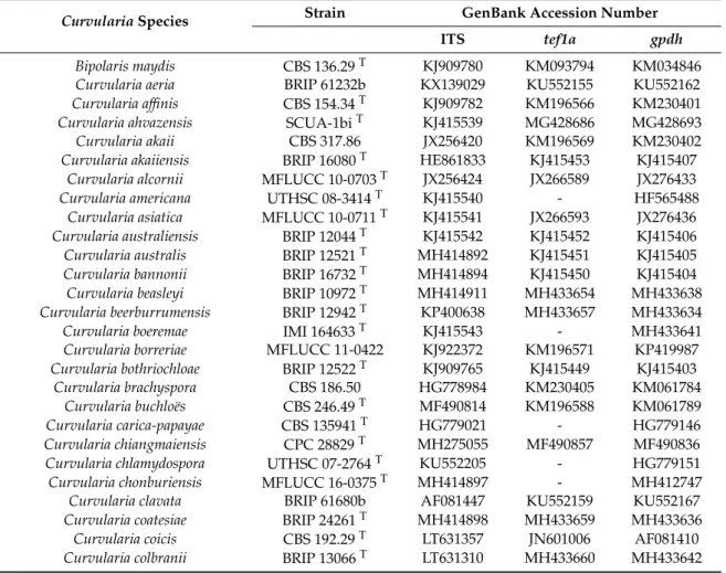
Table2shows the strains and sequences involved in the phylogenetic analysis of the genus Curvularia, including four isolates derived from cases of fungal keratitis diagnosed and treated in the Aravind Eye Hospital, Coimbatore, Tamil Nadu, India. Thetef1αdataset consisted of 902 characters of nucleotide alignment without binary characters. Thegpdhdataset contained 684 characters with 601 characters of nucleotide alignment and 63 binary characters derived from indel coding. The length of the ITS alignment was 1193 characters long, containing 896 bp of nucleotide data and 297 binary characters.
Table 2.Sequences used for the phylogenetic analysis.
CurvulariaSpecies Strain GenBank Accession Number
ITS tef1a gpdh
Bipolaris maydis CBS 136.29T KJ909780 KM093794 KM034846
Curvularia aeria BRIP 61232b KX139029 KU552155 KU552162
Curvularia affinis CBS 154.34T KJ909782 KM196566 KM230401
Curvularia ahvazensis SCUA-1biT KJ415539 MG428686 MG428693
Curvularia akaii CBS 317.86 JX256420 KM196569 KM230402
Curvularia akaiiensis BRIP 16080T HE861833 KJ415453 KJ415407
Curvularia alcornii MFLUCC 10-0703T JX256424 JX266589 JX276433
Curvularia americana UTHSC 08-3414T KJ415540 - HF565488
Curvularia asiatica MFLUCC 10-0711T KJ415541 JX266593 JX276436
Curvularia australiensis BRIP 12044T KJ415542 KJ415452 KJ415406
Curvularia australis BRIP 12521T MH414892 KJ415451 KJ415405
Curvularia bannonii BRIP 16732T MH414894 KJ415450 KJ415404
Curvularia beasleyi BRIP 10972T MH414911 MH433654 MH433638
Curvularia beerburrumensis BRIP 12942T KP400638 MH433657 MH433634
Curvularia boeremae IMI 164633T KJ415543 - MH433641
Curvularia borreriae MFLUCC 11-0422 KJ922372 KM196571 KP419987
Curvularia bothriochloae BRIP 12522T KJ909765 KJ415449 KJ415403
Curvularia brachyspora CBS 186.50 HG778984 KM230405 KM061784
Curvularia buchloës CBS 246.49T MF490814 KM196588 KM061789
Curvularia carica-papayae CBS 135941T HG779021 - HG779146
Curvularia chiangmaiensis CPC 28829T MH275055 MF490857 MF490836
Curvularia chlamydospora UTHSC 07-2764T KU552205 - HG779151
Curvularia chonburiensis MFLUCC 16-0375T MH414897 - MH412747
Curvularia clavata BRIP 61680b AF081447 KU552159 KU552167
Curvularia coatesiae BRIP 24261T MH414898 MH433659 MH433636
Curvularia coicis CBS 192.29T LT631357 JN601006 AF081410
Curvularia colbranii BRIP 13066T LT631310 MH433660 MH433642
Table 2.Cont.
CurvulariaSpecies Strain GenBank Accession Number
ITS tef1a gpdh
Curvularia comoriensis CBS 110673 KJ415544 - LT715841
Curvularia crassiseptum CBS 503.90T HG778985 - LT715882
Curvularia crustacea BRIP 13524T MF490815 KJ415448 KJ415402
Curvularia cymbopogonis CBS 419.78 KJ415545 - HG779129
Curvularia dactyloctenicola CPC 28810T LT631356 MF490858 MF490837
Curvularia dactyloctenii BRIP 12846T JN192375 KJ415447 KJ415401
Curvularia deightonii CBS 537.70 MH414899 - LT715839
Curvularia ellisii CBS 193.62T HG778986 JN601007 JN600963
Curvularia eragrosticola BRIP 12538T KJ909781 MH433661 MH433643
Curvularia eragrostidis CBS 189.48 HG778987 - HG779154
Curvularia geniculata CBS 187.50 JN192376 KM230410 KM083609
Curvularia gladioli CBS 210.79 KJ415546 - HG779123
Curvularia graminicola BRIP 23186aT KJ415547 JN601008 JN600964
Curvularia harveyi BRIP 57412T KJ415548 KJ415446 KJ415400
Curvularia hawaiiensis BRIP 11987T KJ415549 KJ415445 KJ415399
Curvularia heteropogonicola BRIP 14579T HG779011 KJ415444 KJ415398
Curvularia heteropogonis CBS 284.91T JN192380 JN601013 JN600969
Curvularia hominis CBS 136985T KJ922375 - HG779106
Curvularia homomorpha CBS 156.60T HG778991 JN601014 JN600970
Curvularia inaequalis CBS 102.42T MH861533 KM196574 KM061787
Curvularia intermedia CBS 334.64 MH414900 - HG779155
Curvularia ischaemi CBS 630.82T MH855025 - LT715790
Curvularia kenpeggii BRIP 14530T MH414901 MH433662 MH433644
Curvularia kusanoi CBS 137.29 JX256429 JN601016 LT715862
Curvularia lamingtonensis BRIP 12259T JF812154 MH433663 MH433645
Curvularia lunata CBS 730.96T MH414902 JX266596 JX276441
Curvularia malina CBS 131274T HE792934 KR493095 KP153179
Curvularia mebaldsii BRIP 12900T MF139088 MH433664 MH433647
Curvularia micropus CBS 127235 KJ909770 - LT715859
Curvularia microspora GUCC6272T MG846737 MF139115 MF139106
Curvularia miyakei CBS 197.29T KP400647 KM196568 KM083611
Curvularia mosaddeghii IRAN 3131CT KJ415550 MH392152 MH392155
Curvularia muehlenbeckiae CBS 144.63T MH414910 KM196578 KP419996
Curvularia neergaardii BRIP 12919T KJ415551 KJ415443 KJ415397
Curvularia neoindica IMI 129790T MF490816 MH433667 MH433649
Curvularia nicotiae BRIP 11983T JN601033 KJ415442 KJ415396
Curvularia nodosa CPC 28800T KP400650 MF490859 MF490838
Curvularia nodulosa CBS 160.58 JN192384 JN601019 JN600975
Curvularia oryzae CBS 169.53T KJ922380 KM196590 KP645344
Curvularia ovariicola CBS 470.90T MH275056 JN601020 JN600976
Curvularia pallescens CBS 156.35T KJ415552 KM196570 KM083606
Curvularia pandanicola MFLUCC 15-0746T HG778995 MH412763 MH412748
Curvularia papendorfii CBS 308.67T MH414905 KJ415441 KJ415395
Curvularia perotidis CBS 350.90T KY905678 KM230407 HG779138
Curvularia petersonii BRIP 14642T MH414906 MH433668 MH433650
Curvularia pisi CBS 190.48T KJ415553 KY905697 KY905690
Curvularia platzii BRIP 27703bT KJ922373 MH433669 MH433651
Curvularia portulacae BRIP 14541T KJ922376 KJ415440 KJ415393
Curvularia prasadii CBS 143.64T MF490819 KM230408 KM061785
Curvularia protuberata CBS 376.65T HE861842 KM196576 KM083605
Curvularia pseudobrachyspora CPC 28808T HE861838 MF490862 MF490841
Table 2.Cont.
CurvulariaSpecies Strain GenBank Accession Number
ITS tef1a gpdh
Curvularia pseudolunata UTHSC 09-2092T JN192386 - HF565459
Curvularia pseudorobusta UTHSC 08-3458 MH414907 - HF565476
Curvularia ravenelii BRIP 13165T KJ415555 JN601024 JN600978
Curvularia reesii BRIP 4358T KJ909783 MH433670 MH433637
Curvularia richardiae BRIP 4371T KX139030 KJ415438 KJ415391
Curvularia robusta CBS 624.68T KJ415556 KM196577 KM083613
Curvularia rouhanii SCUA-2bi-2T HG779001 MG428687 MG428694
Curvularia ryleyi BRIP 12554T KY905679 KJ415437 KJ415390
Curvularia senegalensis CBS 149.71 KJ415558 - HG779128
Curvularia soli CBS 222.96T MH414904 KY905698 KY905691
Curvularia sorghina BRIP 15900T KP400655 KJ415435 KJ415388
Curvulariasp. BRIP 17068b KP400654 MH433666 MH433648
Curvulariasp. AR5117 HE861826 KP735698 KP645349
Curvulariasp. MFLUCC 120177 JN192387 KP735697 KP645348
Curvulariasp. UTHSC 8809 MH414908 - HF565477
Curvularia spicifera CBS 274.52 KJ909777 JN601023 JN600979
Curvularia sporobolicola BRIP 23040bT MH275057 MH433671 MH433652
Curvularia subpapendorfii CBS 656.74T HG779023 KM196585 KM061791
Curvularia thailandicum MFLUCC 15-0747T JN192388 MH412764 MH412749
Curvularia trifolii CBS 173.55 KJ415559 - HG779124
Curvularia tripogonis BRIP 12375T KC424596 JN601025 JN600980
Curvularia tropicalis BRIP 14834T JX256433 KJ415434 KJ415387
Curvularia tsudae ATCC 44764T HG779024 KC503940 KC747745
Curvularia tuberculate CBS 146.63T MF490822 JX266599 JX276445
Curvularia uncinate CBS 221.52T HG779026 - HG779134
Curvularia variabilis CPC 28815T KP400652 MF490865 MF490844
Curvularia verruciformis CBS 537.75 MH414909 - HG779133
Curvularia verruculosa CBS 150.63 MH275058 KP735695 KP645346
Curvularia warraberensis BRIP 14817T AF071338 MH433672 MH433653
Curvularia xishuangbannaensis KUMCC 17-0185T KJ909780 MH412765 MH412750
Curvularia gudauskasii DAOM 165085 KX139029 KM093794 AF081393
Curvularia tamilnaduensissp. nov. SZMC 22226T* MN628311 MN628303 MN628307 SZMC 26758 * MN628308 MN628300 MN628304 SZMC 26759 * MN628309 MN628301 MN628305 Curvularia coimbatorensissp. nov. SZMC 22225T* MN628310 MN628302 MN628306
Ttype strain; * Strains examined during the present study. Sequences derived from the present study are set in bold.
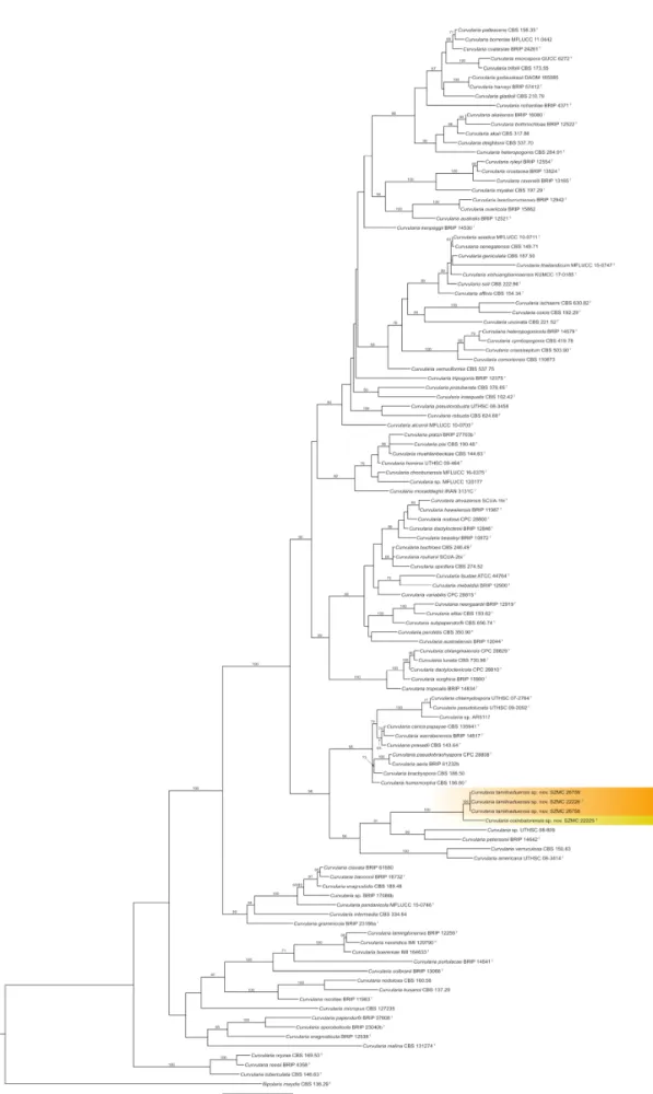
On the phylograms obtained from each of the three loci, the four keratitis isolates of this study were resolved as two new species with over 80% of confidence values (data not shown), one of them represented by the single isolate SZMC 22225, while the other one by isolates SZMC 22226, SZMC 26758, and SZMC 26758. As the individual inferences were largely congruent, the three loci were concatenated and partitioned. The phylogenetic tree obtained from the concatenated dataset is shown in Figure1.
Pathogens 2020, 9, x FOR PEER REVIEW 6 of 13
Figure 1. Maximum likelihood phylogeny of the genus Curvularia inferred from the concatenated internal transcribed spacer (ITS), translation elongation factor 1-α (tef1a), and glyceraldehyde-3-phosphate dehydrogenase (gpdh) sequences. The isolates examined in this study are
shown as the new speciesCurvularia tamilnaduensisandCurvularia coimbatorensis(highlighted in color).
Sequences of the referenceCurvulariastrains were collected from the GenBank Nucleotide database (Table1). Bootstrap support values greater than 60% are shown above the branches.Bipolaris maydis CBS 136.29 was used to root the tree. Abbreviations of culture collections: BRIP: Plant Pathology Herbarium, Queensland, Australia; CBS: Westerdijk Fungal Biodiversity Institute culture collection, The Netherlands; CPC: Cultures of Pedro Crous, housed at Westerdijk Fungal Biodiversity Institute;
DAOM: Canadian National Mycological Herbarium, Ottawa, Canada; GUCC: Guizhou University Culture Collection, Guizhou, China; IMI: CABI Bioscience, Eggham, UK; IRAN: Iranian Fungal Culture Collection, Iranian Research Institute of Plant Protection, Tehran, Iran; KUMCC: Culture Collection of Kunming Institute of Botany, Kunming, China; MFLUCC: Mae Fah Luang Culture Collection, Chiang Rai, Thailand; SCUA: Collection of Fungal Cultures, Department of Plant Protection, Shahid Chamran University of Ahvaz, Iran; SZMC: Szeged Microbiology Collection, Szeged, Hungary; UTHSC:
University of Tennessee Health Science Center, Memphis, USA.T: type strain.
2.3. Taxonomy and Related Information
Curvularia coimbatorensisN. Kiss & S. Kocsubésp. nov. (Figure2). MycoBank accession number:
MB 833656. The etymology is referring to the city in Tamil Nadu, South India where the type strain was isolated.
Pathogens 2020, 9, x FOR PEER REVIEW 7 of 13
Figure 1. Maximum likelihood phylogeny of the genus Curvularia inferred from the concatenated internal transcribed spacer (ITS), translation elongation factor 1-α (tef1a), and glyceraldehyde-3- phosphate dehydrogenase (gpdh) sequences. The isolates examined in this study are shown as the new species Curvularia tamilnaduensis and Curvularia coimbatorensis (highlighted in color). Sequences of the reference Curvularia strains were collected from the GenBank Nucleotide database (Table 1).
Bootstrap support values greater than 60% are shown above the branches. Bipolaris maydis CBS 136.29 was used to root the tree. Abbreviations of culture collections: BRIP: Plant Pathology Herbarium, Queensland, Australia; CBS: Westerdijk Fungal Biodiversity Institute culture collection, The Netherlands; CPC: Cultures of Pedro Crous, housed at Westerdijk Fungal Biodiversity Institute;
DAOM: Canadian National Mycological Herbarium, Ottawa, Canada; GUCC: Guizhou University Culture Collection, Guizhou, China; IMI: CABI Bioscience, Eggham, UK; IRAN: Iranian Fungal Culture Collection, Iranian Research Institute of Plant Protection, Tehran, Iran; KUMCC: Culture Collection of Kunming Institute of Botany, Kunming, China; MFLUCC: Mae Fah Luang Culture Collection, Chiang Rai, Thailand; SCUA: Collection of Fungal Cultures, Department of Plant Protection, Shahid Chamran University of Ahvaz, Iran; SZMC: Szeged Microbiology Collection, Szeged, Hungary; UTHSC: University of Tennessee Health Science Center, Memphis, USA. T: type strain.
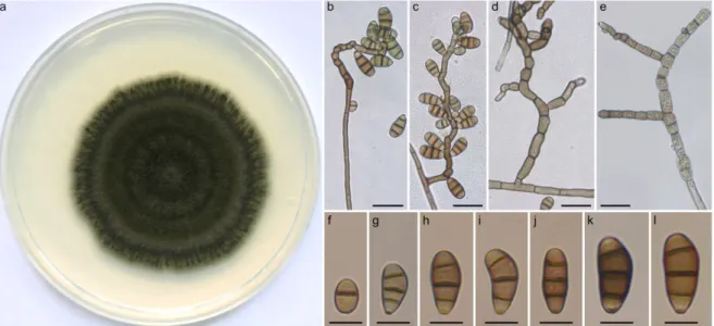
Figure 2. Morphological features of Curvularia coimbatorensis SZMC 2225. (a) Colony morphology on PDA (potato dextrose agar) medium after 7 d at 25 °C; (b,c) conidiophores with septate conidia; (d) branching conidiophores; (e) swollen cells; (f–l) septate conidia. Scale bars: (b–e) 20 µm; (f–l) 10 µm.
Curvularia tamilnaduensis N. Kiss & S. Kocsubé sp. nov. (Figure 3). MycoBank accession number:
MB 833657. The etymology is referring to the state of South India where the type strain and the other two examined strains were isolated.
Vegetative hyphae septate, subhyaline to brown, branched, smooth walled, but often heavily asperulate, 2–3 µm in width. Colonies on PDA reaching approximately 6–7 cm in diameter after 7 d at 25 °C, surface lanose, aerial mycelium abundant, margin fimbriate, olivaceous green.
Conidiophores erect, usually unbranched, in most cases uniformly brown, sometimes with paler tip, seminematous, septate, slightly flexuous, rarely geniculate towards the apex, up to 125 µm long, 2.5–
4 µm wide. Conidiogenous cells integrated, terminal or intercalary, smooth, pale brown to brown, mono- or polytretic, proliferating sympodially. Chlamydospores present, subglobose, terminal and intercalary, 8–22 µm in diameter. Conidia ellipsoidal to clavate to obovoid, asymmetrical with paler basal and apical cells, usually curved at the third cell from the base which is darker than the other cells, (15-)20–23(-28) × (7-)8–10(-11) µm, (2-)3-distoseptate with non-protuberant, thickened, and darkened hila.
Specimens examined: India, Coimbatore, human corneal scraping from corneal ulcer, 2013, (holotype: freeze dried culture specimen in the Szeged Microbiological Collection (SZMC) at the
Figure 2.Morphological features ofCurvularia coimbatorensisSZMC 2225. (a) Colony morphology on PDA (potato dextrose agar) medium after 7 days at 25◦C; (b,c) conidiophores with septate conidia;
(d) branching conidiophores; (e) swollen cells; (f–l) septate conidia. Scale bars: (b–e) 20µm; (f–l) 10µm.
Vegetative hyphae septate, subhyaline to brown, branched, smooth, 3–4µm in width. Colonies on PDA reaching approximately 4–6 cm in diameter after 7 days at 25◦C, surface funiculose, margin fimbriate, olivaceous black to olivaceous grey, velutinous with sparse aerial mycelium. Conidiophores erect, often branched, in most cases uniformly brown, sometimes pale brown at apex, seminematous, septate, flexuous, in most cases geniculate towards the apex, up to 210µm long, 3–4µm wide, basal cells sometimes swollen. Conidiogenous cells integrated, terminal, or intercalary with sympodial proliferation, smooth, brown, mono- or polytretic. Chlamydospores not observed. Conidia ellipsoidal to clavate to obovoid, asymmetrical with paler end cells, usually curved at the third cell from the base, (13-)16–18(-23)×(7-)8–9(-10)µm, 3-distoseptate, hila slightly protuberant, thickened and darkened.
Specimens examined: India, Coimbatore, human corneal scraping from corneal ulcer, 2012, (holotype: freeze dried culture specimen in the Szeged Microbiological Collection (SZMC) at the
Department of Microbiology, Faculty of Science and Informatics, University of Szeged, Hungary, SZMC 22225, includes ex-type culture).
Curvularia tamilnaduensisN. Kiss & S. Kocsubésp. nov. (Figure3). MycoBank accession number:
MB 833657. The etymology is referring to the state of South India where the type strain and the other two examined strains were isolated.
Pathogens 2020, 9, x FOR PEER REVIEW 8 of 13
Department of Microbiology, Faculty of Science and Informatics, University of Szeged, Hungary, SZMC 22226, includes ex-type culture); India, Coimbatore, human corneal scraping from corneal ulcer, 2011, (SZMC 26758); India, Coimbatore, human corneal scraping from corneal ulcer, 2011–2013, (SZMC 26759).
Figure 3. Morphological features of Curvularia tamilnaduensis SZMC 2226. (a) Colony morphology on PDA medium after 7 d at 25 °C; (b) conidiophores with septate conidia; (c) subglobose intercalary chlamydospore; (d–f) septate conidia. Scale bars: (b,c) 20 µm; (d–i) 10 µm.
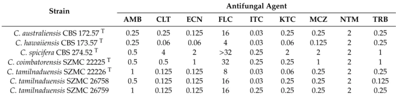
2.4. Antifungal Susceptibilities of Curvularia Strains Isolated from Fungal Keratitis
The minimum inhibitory concentrations (MIC) of nine antifungal agents towards C.
coimbatorensis SZMC 22225, C. tamilnaduensis SZMC 22226, SZMC 26758, and SZMC 26759, as well as the type strains of C. australiensis (CBS 172.57), C. hawaiiensis (CBS 173.57), and C. spicifera (CBS 274.52) are shown in Table 3. The MIC of natamycin was 2 µg mL−1 for both new species and all other strains tested, while substantial differences between them could be observed in the case of clotrimazole, econazole, miconazole, and terbinafine, with the type strain of C. coimbatorensis having 4, 8, 4, and 4–
8 times higher values, respectively. Among the tested isolates, the type strain of C. spicifera proved to be the less susceptible to clotrimazole, econazole, fluconazole, ketoconazole, and miconazole. Notable strain-to-strain variations between the C. tamilnaduensis strains could be observed only in the case of itraconazole and ketoconazole with detected MIC ranges of 0.03–0.25 and 0.06–0.25, respectively.
3. Discussion
The phylogenetic tree obtained from the concatenated dataset of three loci presents an update about the phylogeny of Curvularia, which is mostly in agreement with the recently published phylogenies of this genus (Figure 1). C. ischaemi formed a clade with C. coicis, which is in contradiction with the results of Tan et al. [8] and Tibpromma et al. [27], where C. ischaemi formed a sister clade to C. gladioli, but in agreement with the phylogram obtained by Madrid et al. [7] and Manamgoda et al.
[28]. Our analysis placed C. perotidis as a sister clade to C. australiensis, however, other studies [7,8,27,29] suggested that this species is closer to C. spicifera. The placement of C. variabilis was also different from previously published articles [8,29]. According to the analyses of Tan et al. [8] and Marin-Felix et al. [29], C. variabilis forms a clade with C. hawaiiensis, C. nodosa, C. dactyloctenicola, and C. beasleyi, however, in this study we found C. variabilis as a sister clade of C. tsudae and C. mebaldsii.
The same authors found C. tripogonis, C. pseudorobusta, C. robusta, C. alcornii, C. protuberata, and C.
inaequalis as members of two distinct monophyletic clades, while our results indicate that these species are closely related and paraphyletic, however, none of the topologies have strong statistical supports. The observed slight differences between the previous inferences and our analyses did not
Figure 3.Morphological features ofCurvularia tamilnaduensisSZMC 2226. (a) Colony morphology on PDA medium after 7 days at 25◦C; (b) conidiophores with septate conidia; (c) subglobose intercalary chlamydospore; (d–f) septate conidia. Scale bars: (b,c) 20µm; (d–i) 10µm.
Vegetative hyphae septate, subhyaline to brown, branched, smooth walled, but often heavily asperulate, 2–3µm in width. Colonies on PDA reaching approximately 6–7 cm in diameter after 7 days at 25◦C, surface lanose, aerial mycelium abundant, margin fimbriate, olivaceous green. Conidiophores erect, usually unbranched, in most cases uniformly brown, sometimes with paler tip, seminematous, septate, slightly flexuous, rarely geniculate towards the apex, up to 125µm long, 2.5–4µm wide.
Conidiogenous cells integrated, terminal or intercalary, smooth, pale brown to brown, mono- or polytretic, proliferating sympodially. Chlamydospores present, subglobose, terminal and intercalary, 8–22µm in diameter.Conidiaellipsoidal to clavate to obovoid, asymmetrical with paler basal and apical cells, usually curved at the third cell from the base which is darker than the other cells, (15-)20–23(-28)
×(7-)8–10(-11)µm, (2-)3-distoseptate with non-protuberant, thickened, and darkened hila.
Specimens examined: India, Coimbatore, human corneal scraping from corneal ulcer, 2013, (holotype: freeze dried culture specimen in the Szeged Microbiological Collection (SZMC) at the Department of Microbiology, Faculty of Science and Informatics, University of Szeged, Hungary, SZMC 22226, includes ex-type culture); India, Coimbatore, human corneal scraping from corneal ulcer, 2011, (SZMC 26758); India, Coimbatore, human corneal scraping from corneal ulcer, 2011–2013, (SZMC 26759).
2.4. Antifungal Susceptibilities of Curvularia Strains Isolated from Fungal Keratitis
The minimum inhibitory concentrations (MIC) of nine antifungal agents towardsC. coimbatorensis SZMC 22225,C. tamilnaduensisSZMC 22226, SZMC 26758, and SZMC 26759, as well as the type strains ofC. australiensis(CBS 172.57),C. hawaiiensis(CBS 173.57), andC. spicifera(CBS 274.52) are shown in Table3. The MIC of natamycin was 2µg mL−1for both new species and all other strains tested, while substantial differences between them could be observed in the case of clotrimazole, econazole, miconazole, and terbinafine, with the type strain ofC. coimbatorensishaving 4, 8, 4, and 4–8 times higher values, respectively. Among the tested isolates, the type strain ofC. spiciferaproved to be the less susceptible to clotrimazole, econazole, fluconazole, ketoconazole, and miconazole. Notable
strain-to-strain variations between theC. tamilnaduensisstrains could be observed only in the case of itraconazole and ketoconazole with detected MIC ranges of 0.03–0.25 and 0.06–0.25, respectively.
Table 3. Antifungal susceptibilities of theCurvularia coimbatorensisand Curvularia tamilnaduensis strains in comparison with the type strains ofCurvularia australiensis, Curvularia hawaiiensis, and Curvularia spicifera determined by the CLSI (Clinical & Laboratory Standards Institute) broth microdilution method (minimum inhibitory concentrations (MIC) values inµg mL−1).
Strain Antifungal Agent
AMB CLT ECN FLC ITC KTC MCZ NTM TRB
C. australiensisCBS 172.57T 0.25 0.25 0.125 16 0.03 0.25 0.25 2 0.25
C. hawaiiensisCBS 173.57T 0.25 0.06 0.06 4 0.03 0.06 0.125 2 0.25
C. spiciferaCBS 274.52T 0.5 4 2 >32 0.25 2 2 2 1
C. coimbatorensisSZMC 22225T 0.5 0.5 1 32 0.25 0.25 1 2 1
C. tamilnaduensisSZMC 22226T 1 0.125 0.125 8 0.03 0.06 0.25 2 0.25
C. tamilnaduensisSZMC 26758 0.5 0.125 0.125 16 0.03 0.25 0.25 2 0.125
C. tamilnaduensisSZMC 26759 1 0.125 0.125 16 0.25 0.25 0.25 2 0.25
T: type strain; AMB: amphotericin B; CLT: clotrimazole; ECN: econazole; FLC: fluconazole; ITC: itraconazole; KTC:
ketoconazole; MCZ: miconazole; NTM: natamycin; TRB: terbinafine.
3. Discussion
The phylogenetic tree obtained from the concatenated dataset of three loci presents an update about the phylogeny ofCurvularia, which is mostly in agreement with the recently published phylogenies of this genus (Figure1).C. ischaemiformed a clade withC. coicis, which is in contradiction with the results of Tan et al. [8] and Tibpromma et al. [27], whereC. ischaemiformed a sister clade toC. gladioli, but in agreement with the phylogram obtained by Madrid et al. [7] and Manamgoda et al. [28]. Our analysis placedC. perotidisas a sister clade toC. australiensis,however, other studies [7,8,27,29] suggested that this species is closer toC. spicifera. The placement ofC. variabiliswas also different from previously published articles [8,29]. According to the analyses of Tan et al. [8] and Marin-Felix et al. [29],C. variabilis forms a clade withC. hawaiiensis,C. nodosa,C. dactyloctenicola, andC. beasleyi, however, in this study we foundC. variabilisas a sister clade ofC. tsudaeandC. mebaldsii. The same authors foundC. tripogonis, C. pseudorobusta,C. robusta,C. alcornii,C. protuberata, andC. inaequalisas members of two distinct monophyletic clades, while our results indicate that these species are closely related and paraphyletic, however, none of the topologies have strong statistical supports. The observed slight differences between the previous inferences and our analyses did not affect the validity of any of the previously described species, and some of them might be the result of the slightly broader taxon sampling.
One of the newly described species,C. coimbatorensisis only known from the type specimen isolated from corneal ulcer. Phylogenetic analysis based on three loci placedC. coimbatorensisas a sister clade to the other newly described speciesC. tamilnaduensis. The two species are closely related, but can be distinguished bytef1a,gpdh, and ITS sequences, with percentage identities of 99%, 98%, and 99%, respectively.C. petersonii[8] is also closely related and can be distinguished by all three loci (98% in tef1a, 93% ingpdhand 96% in ITS).C. coimbatorensisdiffers fromC. tamilnaduensisin colony morphology, the lack of chlamydospores, and the size of conidia.C. petersoniiis very similar in colony morphology, however, has significantly shorter (up to 110µm) and only slightly geniculate conidiophores bearing narrower (5-)5.5–6(-7) conidia [8].C. coimbatorensishas longer conidiophores.
The phylogenetic analysis based on three loci placed the other newly described species, C. tamilnaduensisas a sister clade to the recently described species C. petersonii. C. tamilnaduensis can be reliably distinguished from the ex-type ofC. petersoniibytef1a,gpdhand ITS sequences with percentage identities of 99%, 95%, and 96%, respectively. The two species also differ by morphology, as C. petersoniihas not been reported to produce chlamydospores and has different conidial dimensions (17–19×5.5–6) [8]. C. americana[7] andC. verruculosa[30] are also related species with considerable amount of genetic distances and none of these species have been reported before to have chlamydopores.
C. americana has 4(-5)-distoseptate and wider (7–15 µm) conidia, while C. verruculosahas mostly 3-distoseptate conidia, but also wider (12–17µm) than those ofC. tamilnaduensis.
The antifungal susceptibilities of the examined strains ofC. coimbatorensisandC. tamilnaduensis to amphotericin B, clotrimazole, econazole, fluconazole, itraconazole, ketoconazole, miconazole, natamycin, and terbinafine were within the MIC ranges reported for other clinically relevantCurvularia species in the study of Guarro et al. [11] and the review of Krizsán et al. [3]. The type strain of C. coimbatorensisproved to be less susceptible than the strains ofC. tamilnaduensisto all antifungals except for natamycin. For itraconazole and ketoconazole our results are in agreement with the study of Guarro et al. [11], who reported that amphotericin B, itraconazole, miconazole and ketoconazole are highly effective against a series ofCurvulariaspecies known from fungal keratitis (C. brachyspora, C. clavata,C. geniculata,C. lunata,C. pallescens,C. senegalensis, andC. verruculosa).
4. Materials and Methods
4.1. Curvularia Strains, Culture Conditions, and Morphological Examination
The Curvularia strains involved in this study derived from corneal scrapings from fungal corneal ulcers of keratitis patients attending the Aravind Eye Hospital and Postgraduate Institute of Ophthalmology, Coimbatore, India. All cases were initially screened by experienced ophthalmologists, and the corneal scrapings were collected following the clinical diagnosis of fungal keratitis. The samples were initially processed microbiologically for the isolation of the causative agents as described earlier [31]. The corneal scrapings of all patients were subjected to Gram stain, Giemsa stain, and 10%
KOH wet mount. Culture methods involved direct inoculation of specimens onto 5% sheep blood agar, chocolate agar, non-nutrient agar, potato dextrose agar, thioglycolate broth, and brain–heart infusion broth. The microbial cultures were considered positive only if the growth of the same organism was demonstrated on two or more solid media, or there was confluent growth at the site of inoculation on one solid medium with consistent direct microscopic findings. The isolates were deposited in the Szeged Microbiology Collection (SZMC, Szeged, Hungary) under the accession numbers SZMC 22225, SZMC 22226, SZMC 26758, and SZMC 26759. Colony morphology of the isolates was examined on PDA (BioLab, Budapest, Hungary) medium after 7 days of incubation at 25◦C under normal day/night light conditions. Micromorphological characters were examined with a Leica DMI 4000B (Leica, Wetzlar, Germany) microscope equipped with a Leica DFC 295 camera. Microscopic features were examined in lactic acid (100%v/v) on glass slides. Conidiophores were studied in the same mounting fluid with the transparent tape method. Conidiophores and conidia were measured using the software ImageJ v2.52a (National Institute of Mental Health, Bethesda, MD, USA). Size ranges of the conidia were derived from 50 measurements. Lengths and widths are given as (minimum value) mean size minus SD-mean size plus SD (maximum value).
4.2. DNA Extraction, Amplification, Sequencing, and Phylogenetic Analysis
Genomic DNA was isolated from the examinedCurvulariastrains SZMC 22225, SZMC 22226, SZMC 26758, and SZMC 26759 with the Masterpure™Yeast DNA Purification Kit (Epicentre Biotechnologies, Madison, WI, USA) according to the manufacturer’s instructions. Fragments oftef1aandgpdhwere amplified as described previously [5,32,33]. The ITS region of the ribosomal RNA gene cluster was amplified according to White et al. [34]. Sequencing of the amplicons was carried out on a 3500 Genetic Analyzer (Thermo Fisher Scientific, Waltham, MA, USA) by the sequencing service of the Biological Research Centre, Szeged, Hungary. Resulting sequences were deposited in the GenBank Nucleotide database (www.ncbi.nlm.nih.gov) under the accession numbers shown in Table2.
Sequences of the four clinical isolates were aligned with publicly available sequences of 108 previously describedCurvulariaspecies, as well asBipolaris maydis as the outgroup (Table2).
Phylogenetic analyses were conducted using three loci (tef1α,gpdhand ITS). Sequences of all three loci were aligned with the phylogeny-aware sequence alignment tool Canopy v0.1.4 using RAxML as tree
estimator and PRANK [35] with the -F option as the aligner with 10 iterations and seed decomposition strategy. Alignments of the three loci were concatenated and partitioned by region. Thetef1αsequences formed one partition while in the case ofgpdhsequences the dataset was partitioned to exons and introns. The ITS dataset was divided to rDNA and ITS1-ITS2 regions. Alignments ofgpdhand ITS datasets contained high number of indels with important phylogenetic signal, therefore gaps were coded as absence/presence characters by SequenceMatrix v1.8 [36] using the simple indel coding algorithm [37]. The two indel matrices were concatenated and added as a single partition to the dataset.
Maximum likelihood analysis was performed using RAxML-NG v0.9.0 [38] under the GTR model with gamma-distributed rate heterogeneity using empirical base frequencies. As indel-based datasets do not contain constant sites, the ascertainment bias correction described by Lewis [39] was used for this partition. Statistical support of the best ML tree was obtained with 1000 thorough bootstrap replicates.
4.3. Antifungal Susceptibility Testing
In vitro antifungal susceptibility tests were carried out according to the CLSI M38-A2 broth microdilution method [40]. Nine antifungal agents: amphotericin B, clotrimazole, econazole, fluconazole, itraconazole, ketoconazole, miconazole, natamycin and terbinafine (Sigma-Aldrich, Budapest, Hungary) were examined. Microtiter plates were incubated at 35◦C for 72 h. Plates were evaluated both spectrophotometrically with a Spectrostar Nano microplate reader (BMG Labtech, Ortenberg, Germany) and by visual examination.
5. Conclusions
The present study demonstrates, that although the phylogeny of the genusCurvulariais resolved and well established, further expansion can be expected both in the list of describedCurvulariaspecies and in the known spectrum of clinically relevant members of the genus. The collection of further keratitis isolates from the genusCurvulariaand gaining data about their antifungal susceptibilities are therefore tasks of increasing importance. Furthermore, comparing the infectivity of variousCurvularia species causing keratitis—including the recently described ones—in animal keratitis models would be an intriguing topic for future research.
Author Contributions: Conceptualization, S.K., L.K., T.P. and C.V.; methodology, N.K., A.M., M.H. and S.K.;
software, S.K.; validation, R.R., P.M., M.H., C.V., M.V. and S.K.; formal analysis, K.K., T.P. and M.V.; investigation, N.K., A.M., P.M., K.K., M.H., M.V. and S.K.; resources, R.R., P.M., A.M., T.P. and C.V.; data curation, N.K., S.K., K.K., L.K. and M.H.; writing—original draft preparation, N.K., S.K., L.K.; writing—review and editing, N.K., S.K., L.K., P.M., T.P. and C.V.; visualization, N.K., M.H. and S.K.; supervision, S.K.; project administration, S.K., T.P., L.K., and P.M.; funding acquisition, S.K., T.P., L.K. and P.M. All authors have read and agreed to the published version of the manuscript.
Funding:This research was funded by grants NKFI PD-116609 (National Research, Development and Innovation Office, Hungary), GINOP-2.3.2-15-2016-00035 (Széchenyi 2020 Programme) and also supported by the COST action HUPLANTcontrol (Control of Human Pathogenic Micro-organisms in Plant Production Systems, CA16110).
LK is grantee of the János Bolyai Research Scholarship (Hungarian Academy of Sciences) and the Bolyai Plus Scholarship (New National Excellence Programme). TP and MH are supported by the grants LP2016-8/2016 and by the FIKP program (TUDFO/4738-1/2019 ITM) of the Ministry of Human Capacities.
Acknowledgments:The authors wish to thank Venkatapathy Narendran (Aravind Eye Hospital and Postgraduate Institute of Ophthalmology, Coimbatore, Tamil Nadu, India), Coimbatore Subramanian Shobana (Department of Microbiology, PSG College of Arts and Science, Coimbatore, Tamil Nadu, India) and Kanesan Panneer Selvam (Department of Microbiology, M.R Government Arts College, Mannargudi, Tamil Nadu, India) for constantly supporting the research efforts on fungal keratitis within the frames of the Indo-Hungarian Fungal Keratitis Research Group.
Conflicts of Interest:The authors declare no conflict of interest.
References
1. Bengyella, L.; Iftikhar, S.; Nawaz, K.; Fonmboh, D.J.; Yekwa, E.L.; Jones, R.C.; Njanu, Y.M.T.; Roy, P.
Biotechnological application of endophytic filamentousBipolarisandCurvularia: A review on bioeconomy impact.World J. Microbiol. Biotechnol.2019,35, 69. [CrossRef] [PubMed]
2. Kusai, N.A.; Azmi, M.M.Z.; Zulkifly, S.; Yusof, M.T.; Zainudin, N.A.I.M. Morphological and molecular characterization ofCurvulariaand related species associated with leaf spot disease of rice in Peninsular Malaysia.Rend. Lincei Sci. Fis. Nat.2016,27, 205–214. [CrossRef]
3. Krizsán, K.; Papp, T.; Manikandan, P.; Shobana, C.S.; Chandrasekaran, M.; Vágvölgyi, C.; Kredics, L.
Clinical Importance of the GenusCurvularia. InMedical Mycology: Current Trends and Future Prospects;
Razzaghi-Abyaneh, M., Shams-Ghahfarokhi, M., Rai, M., Eds.; CRC Press: Boca Raton, FL, USA, 2016;
pp. 147–204. [CrossRef]
4. Yanagihara, M.; Kawasaki, M.; Ishizaki, H.; Anzaw, K.; Udagawa, S.; Mochizuki, T.; Sato, Y.; Tachikawa, N.;
Hanakawa, H. Tiny keratotic brown lesions on the interdigital web between the toes of a healthy man caused byCurvulariaspecies infection and a review of cutaneousCurvulariainfections.Mycoscience2010,51, 224–233.
[CrossRef]
5. Manamgoda, D.S.; Cai, L.; McKenzie, E.H.C.; Crous, P.W.; Madrid, H.; Chukeatirote, E.; Shivas, R.G.; Tan, Y.P.;
Hyde, K.D. A phylogenetic and taxonomic re-evaluation of theBipolaris-Cochliobolus-Curvulariacomplex.
Fungal Divers.2012,56, 131–144. [CrossRef]
6. Paredes, K.; Capilla, J.; Sutton, D.A.; Mayayo, E.; Fothergill, A.W.; Guarro, J. Virulence ofCurvulariain a murine model.Mycoses2013,56, 512–515. [CrossRef] [PubMed]
7. Madrid, H.; da Cunha, K.C.; Gené, J.; Dijksterhuis, J.; Cano, J.; Sutton, D.A.; Guarro, J.; Crous, P.W. Novel Curvulariaspecies from clinical specimens.Persoonia2014,33, 48–60. [CrossRef] [PubMed]
8. Tan, Y.P.; Crous, P.W.; Shivas, R.G. Cryptic species ofCurvulariain the culture collection of the Queensland Plant Pathology Herbarium.MycoKeys2018,35, 1–25. [CrossRef] [PubMed]
9. Marcus, L.; Vismer, H.F.; van der Hoven, H.J.; Gove, E.; Meewes, P. Mycotic keratitis caused byCurvularia brachyspora(Boedjin). A report of the first case.Mycopathologia1992,119, 29–33. [CrossRef] [PubMed]
10. Travis, W.D.; Kwon-Chung, K.J.; Kleiner, D.E.; Geber, A.; Lawson, W.; Pass, H.I.; Henderson, D. Unusual aspects of allergic bronchopulmonary fungal disease: Report of two cases due toCurvulariaorganisms associated with allergic fungal sinusitis.Hum. Pathol.1991,22, 1240–1248. [CrossRef]
11. Guarro, J.; Akiti, T.; Horta, R.A.; Morizot Leite-Filho, L.A.; Gené, J.; Ferreira-Gomes, S.; Aguilar, C.;
Ortoneda, M. Mycotic keratitis due toCurvularia senegalensisand in vitroantifungal susceptibilities of Curvulariaspp.J. Clin. Microbiol.1999,37, 4170–4173. [PubMed]
12. Fan, Y.M.; Huang, W.M.; Li, S.F.; Wu, G.F.; Li, W.; Chen, R.Y. Cutaneous phaeohyphomycosis of foot caused byCurvularia clavata.Mycoses2009,52, 544–546. [CrossRef] [PubMed]
13. Vasikasin, V.; Nasomsong, W.; Srisuttiyakorn, C.; Mitthamsiri, W.; Oer-Areemitr, N.; Changpradub, D.
Disseminated phaeohyphomycosis caused by Curvularia tuberculata in a previously healthy man.
Mycopathologia2019,184, 321–325. [CrossRef]
14. Pimentel, J.D.; Mahadevan, K.; Woodgyer, A.; Sigler, L.; Gibas, C.; Harris, O.C.; Lupino, M.; Athan, E.
Peritonitis due toCurvularia inaequalisin an elderly patient undergoing peritoneal dialysis and a review of six cases of peritonitis associated with otherCurvulariaspp.J. Clin. Microbiol.2005,43, 4288–4292. [CrossRef]
[PubMed]
15. Posteraro, B.; Scarano, E.; La Sorda, M.; Torelli, R.; De Corso, E.; Mulé, A.; Paludetti, G.; Fadda, G.;
Sanguinetti, M. Eosinophilic fungal rhinosinusitis due to the unusual pathogenCurvularia inaequalis.Mycoses 2010,53, 84–88. [CrossRef]
16. Cruz, R.; Barthel, E.; Espinoza, J. Allergic rhinosinusitis byCurvularia inaequalis(Shear) Boedijn.Rev. Chil.
Infectol.2013,30, 319–322. (In Spanish) [CrossRef] [PubMed]
17. Flanagan, K.L.; Bryceson, A.D. Disseminated infection due to Bipolaris australiensis in a young immunocompetent man: Case report and review. Clin. Infect. Dis. 1997, 25, 311–313. [CrossRef]
[PubMed]
18. Filizzola, M.J.; Martinez, F.; Rauf, S.J. Phaeohyphomycosis of the central nervous system in immunocompetent hosts: Report of a case and review of the literature.Int. J. Infect. Dis.2003,7, 282–286. [CrossRef]
19. Gadgil, N.; Kupferman, M.; Smitherman, S.; Fuller, G.N.; Rao, G.Curvulariabrain abscess.J. Clin. Neurosci.
2013,20, 173–175. [CrossRef]
20. Vachharajani, T.J.; Zaman, F.; Latif, S.; Penn, R.; Abreo, K.D.Curvularia geniculatafungal peritonitis: A case report with review of literature.Int. Urol. Nephrol.2005,37, 781–784. [CrossRef]
21. Diskin, C.J.; Stokes, T.J.; Dansby, L.M.; Radcliff, L.; Carter, T.B. Case report and review: Is the tendency for Curvulariatubular obstruction significant in pathogenesis?Perit. Dial. Int.2008,28, 678–679.
22. Saenz, R.E.; Brown, W.D.; Sanders, C.V. Allergic bronchopulmonary disease caused byBipolaris hawaiiensis presenting as a necrotizing pneumonia: Case report and review of literature.Am. J. Med. Sci. 2001,321, 209–212. [CrossRef] [PubMed]
23. Robson, A.M.; Craver, R.D.Curvulariaurinary tract infection: A case report.Pediatr. Nephrol.1994,8, 83–84.
[CrossRef] [PubMed]
24. Safdar, A.Curvularia—Favorable response to oral itraconazole therapy in two patients with locally invasive phaeohyphomycosis.Clin. Microbiol. Infect.2003,9, 1219–1223. [CrossRef] [PubMed]
25. Fernandez, M.; Noyola, D.E.; Rossmann, S.N.; Edwards, M.S. Cutaneous phaeohyphomycosis caused by Curvularia lunataand a review ofCurvulariainfections in pediatrics.Pediatr. Infect. Dis. J.1999,18, 727–731.
[CrossRef] [PubMed]
26. Dave, V.P.; Joseph, J.; Pathengay, A.; Pappuru, R.R.; Das, T. Clinical presentations, diagnosis, and management outcomes ofCurvulariaendophthalmitis and a review of literature.Retina2018. [CrossRef] [PubMed]
27. Tibpromma, S.; Hyde, K.D.; Bhat, J.D.; Mortimer, P.E.; Xu, J.; Promputtha, I.; Doilom, M.; Yang, J.B.;
Tang, A.M.C.; Karunarathna, S.C. Identification of endophytic fungi from leaves of Pandanaceae based on their morphotypes and DNA sequence data from southern Thailand.MycoKeys2018,33, 25–67. [CrossRef]
[PubMed]
28. Manamgoda, D.S.; Rossman, A.Y.; Castlebury, L.A.; Crous, P.W.; Madrid, H.; Chukeatirote, E.; Hyde, K.D.
The genus Bipolaris.Stud. Mycol.2014,79, 221–288. [CrossRef]
29. Marin-Felix, Y.; Senwanna, C.; Cheewangkoon, R.; Crous, P.W. New species and records ofBipolarisand Curvulariafrom Thailand.Mycosphere2017,8, 1556–1574. [CrossRef]
30. Sivanesan, A. Graminicolous species ofBipolaris,Curvularia,Drechslera,Exserohilumand their teleomorphs.
Mycol. Pap.1987,158, 1–261.
31. Mythili, A.; Babu Singh, Y.R.; Priya, R.; Shafeeq Hassan, A.; Manikandan, P.; Panneerselvam, K.; Narendran, V.;
Shobana, C.S.In vitroand comparative study on the extracellular enzyme activity of molds isolated from keratomycosis and soil.Int. J. Ophthalmol.2014,7, 778–784. [CrossRef]
32. Schoch, C.L.; Crous, P.W.; Groenewald, J.Z.; Boehm, E.W.A.; Burgess, T.I.; de Gruyter, J.; de Hoog, G.S.;
Dixon, L.J.; Grube, M.; Gueidan, C.; et al. A class-wide phylogenetic assessment of Dothideomycetes.
Stud. Mycol.2009,64, 1–15. [CrossRef] [PubMed]
33. Berbee, M.; Pirseyedi, M.; Hubbard, S.Cochliobolusphylogenetics and the origin of known, highly virulent pathogens, inferred from ITS and glyceraldehyde-3-phosphate dehydrogenase gene sequences.Mycologia 1999,91, 964–977. [CrossRef]
34. White, T.J.; Bruns, T.D.; Lee, S.; Taylor, J.W. Amplification and direct sequencing of fungal ribosomal genes for phylogenetics. InPCR Protocols: A Guide to Methods and Applications; Innis, M.A., Gelfand, D.H., Sninsky, J.J., White, J.W., Eds.; Academic Press: San Diego, CA, USA, 1990; pp. 315–322.
35. Löytynoja, A. Phylogeny-aware alignment with PRANK.Meth. Mol. Biol.2014,1079, 155–170. [CrossRef]
36. Vaidya, G.; Lohman, D.J.; Meier, R. SequenceMatrix: Concatenation software for the fast assembly of multigene datasets with character set and codon information.Cladistics2011,27, 171–180. [CrossRef]
37. Simmons, M.P.; Ochoterena, H. Gaps as characters in sequence-based phylogenetic analysis.Syst. Biol.2000, 49, 369–381. [CrossRef]
38. Kozlov, A.M.; Darriba, D.; Flouri, T.; Morel, B.; Stamatakis, A. RAxML-NG: A fast, scalable, and user-friendly tool for maximum likelihood phylogenetic inference.Bioinformatics2019,35, 4453–4455. [CrossRef]
39. Lewis, P.O. A likelihood approach to estimating phylogeny from discrete morphological character data.
Syst. Biol.2001,50, 913–925. [CrossRef]
40. Clinical and Laboratory Standards Institute. Reference Method for Broth Dilution Antifungal Susceptibility Testing of Yeasts, 3rd ed.; Approved Standard, CLSI Document M27-A3; Clinical and Laboratory Standards Institute: Wayne, PA, USA, 2008.
©2019 by the authors. Licensee MDPI, Basel, Switzerland. This article is an open access article distributed under the terms and conditions of the Creative Commons Attribution (CC BY) license (http://creativecommons.org/licenses/by/4.0/).