Endoscopic Versus Microscopic Stapes Surgery Outcomes: A Meta- analysis and Systematic Review
Alexandros Koukkoullis, MD, MRCS(ENT) ; István Tóth, MD; Noémi Gede, PhD; Zsolt Szakács, MD;
Péter Hegyi, MD, PhD, DSc, MAE; Gábor Varga, PhD, DSc; István Pap, MD; Kinga Harmat, MD;
Adrienn Németh, MD, PhD; István Szanyi, MD, PhD; László Lujber, MD, PhD;
Imre Gerlinger, MD, PhD, DSc; Péter Révész, MD, PhD
Objective:Compare intraoperative and postoperative outcomes of endoscopic and microscopic stapes surgery to provide objective evidence on whether the former is a better alternative than the latter.
Methods: We performed a systematic review and meta-analysis for studies that compared endoscopic stapes surgery with microscopic stapes surgery. Only studies that met predetermined criteria were selected and assessed for bias and quality.
Primary outcomes were postoperative air–bone gap (ABG) and chorda tympani nerve injury. Secondary outcomes were aver- age operating time, tympanic membrane (TM) perforation, and postoperative taste disturbance, pain, and dizziness. We calcu- lated pooled odds ratios (ORs) with 95% confidence intervals (CIs) for dichotomous outcomes and weighted mean difference (WMD) with 95% CI for continuous outcomes. A confidence interval starting above 1.0 was considered as statistically signifi- cant.I2andχ2tests were used to quantify statistical heterogeneity. We used funnel plots to look for publication bias and per- formed a sensitivity analysis.
Results:Six nonrandomized cohort studies were eligible. The primary outcomes were ABG < 10 dB: OR = 1.80 (95% CI:
0.96 to 3.38), ABG = 11 dB to 20 dB: OR = 1.49 (95% CI: 0.76 to 2.93), ABG > 20 dB: OR = 2.51 (95% CI: 0.77 to 8.22), and chorda tympani injury: OR = 3.51 (95% CI: 1.55 to 7.93). Secondary outcomes were taste: OR = 2.36 (95% CI: 1.01 to 5.51), average operation time: WMD = 0.14 (95% CI: −11.69 to 11.98), TM perforation: OR = 1.70 (95% CI: 0.44 to 6.58); pain:
OR = 0.84 (95% CI: 0.36 to 1.96), and dizziness: OR = 2.15 (95% CI: 0.94 to 4.89).
Conclusions:Endoscopic stapes surgery is a valid alternative to the microscope.
Key Words:Endoscopic, microscopic, stapedotomy, stapedectomy, otosclerosis.
Level of Evidence:2a
Laryngoscope, 130:2019–2027, 2020
INTRODUCTION
The microscope is the conventional method used by ear surgeons to view the middle ear structures, allowing for binocular vision and the freedom of both hands. Its main disadvantage is the need for an unobstructed, direct view of the operating area.1 The endaural incision, dril- ling of the bony auditory canal, and regular repositioning of the patient and the surgeon are a few examples of how the operation has to adapt to the microscope. Neverthe- less, the microscope has proven itself as the tool of choice in stapes surgery with reliable outcomes.2
In contrast, the endoscope has only recently been introduced as an alternative viewing apparatus for middle ear surgery. Since its beginnings more than 50 years ago, it has become the modern way to visualize middle ear structures, gradually making its way into the surgical realm.3,4Initially, its use was limited as an adjuvant tool to improve detection in cholesteatoma surgery and in endoscopically assisted ear surgery.5–9Soon, surgeons real- ized that it can be used to replace the microscope entirely for certain operations such as stapes surgery, with out- comes comparable to the microscopes.10The endoscope can
This is an open access article under the terms of the Creative Commons Attribution-NonCommercial License, which permits use, distri- bution and reproduction in any medium, provided the original work is properly cited and is not used for commercial purposes.
From the Department of Otorhinolaryngology–Head and Neck Surgery (A.K.,I.T.,I.P., K.H., A.N.,I.S.,L.L., I.G., P.R.), University of Pécs Medical School, Pécs, Hungary; Institute for Translational Medicine, Szentágothai Research Centre (N.G., Z.S., P.H.), University of Pécs Medical School, Pécs, Hungary; First Department of Medicine (P.H.), University of Szeged, Szeged, Hungary; Momentum Gastroenterology Multidisciplinary Research Group (P.H.), Hungarian Academy of Sciences– University of Szeged, Szeged, Hungary; and the Department of Oral Biology (G.V.), Semmelweis University, Budapest, Hungary.
Additional supporting information may be found in the online version of this article.
Editor’s Note: This Manuscript was accepted for publication on September 19, 2019.
A.K. andI.T. sharefirst authorship.
The study was supported by the Economic Development and Inno- vation Operative Program Grant (GINOP 2.3.2-15-2016-00048) and by the Human Resources Development Operational Program Grant (EFOP- 3.6.2-16-2017-00006) from the National Research, Development, and Inno- vation Office.
The authors have no other funding,financial relationships, or con- flicts of interest to disclose.
Send correspondence to Alexandros Koukkoullis, MD, Department of Otorhinolaryngology–Head and Neck Surgery, University of Pécs Medical School, Munkácsy M. utca 2, Pécs 7621, Hungary. E-mail:
alexandroskoukkoullis@gmail.com DOI: 10.1002/lary.28353 The Laryngoscope
© 2019 The Authors.The Laryngoscope published by Wiley Periodicals, Inc. on behalf of The American Laryngological, Rhinological and Otological Society, Inc.
offer a close-up view of the stapes footplate, with minimal drilling of the external auditory canal and reduced manip- ulation of the chorda tympani nerve.11–13 The magnified, wide-field view and the improved illumination provide a safer manipulation of the stapes superstructure, the foot- plate, and the chorda tympani. In their study, Bennett et al. concluded that all of the regions of the middle ear have better visualization by the endoscope (0, 30, and 45) compared to the microscope.14 However, the endo- scope is not without its drawbacks, which include the loss of three-dimensional vision and the use of one hand, resulting in surgeons being reluctant to change from the microscope because of a possible long learning curve.1,15It has also been reported anecdotally that heat emanating from the light of the endoscope can also injure the chorda tympani nerve, and the loss depth perception can impact the choice of prosthesis length and thus hearing outcomes.
Irrigation is therefore paramount with both techniques.
Recently, there has been a number of publications comparing the two modalities and only one review, but no meta-analysis that attempted to collect all evidence on the field.16 Therefore, we decided to systematically review the literature for studies that compared endoscopic with micro- scopic stapes surgery to perform a meta-analysis to test our hypothesis: endoscopy performs better than microscopy.
MATERIALS AND METHODS
This meta-analysis adheres to Preferred Reporting Items for Systematic Reviews and Meta-analysis guidelines and was registered onto the PROSPERO under the registration number CRD42018095617.17
Search Query
A search was performed in the PubMed, Embase, Cochrane, Web of Science, ClinicalTrials, World Health Organization, and Scopus databases from inception up to June 6, 2018. Our search query was the combination of free text terms and Medical Subject Headings as follows: (endoscop*OR microscop*OR conventional OR traditional) AND (stapedotomy OR stapedectomy OR stapes OR otosclerosis). Cited and citing articles or relevant papers were hand searched to ensure the detection of all available records.
Selection and Eligibility
Yield of search was compiled in reference manager software (EndNote 7.4; Clarivate Analytics, Philadelphia, PA, USA) to remove overlaps between databases and duplicate records. Remaining records were selected by title, abstract, and full text by two authors in duplicate (A.K. and I.T.). If an agreement could not be reached, the dispute was resolved with the help of a third investigator (P.R.).
We chose studies whose populations were patients suffering from conductive hearing loss due to stapesfixation and undergoing stapes surgery, which was carried out either with the endoscope or the microscope, and recorded intraoperative and/or postoperative outcomes.18Only controlled studies were included. Primary out- comes were the average postoperative air–bone gap (ABG) and injury to the chorda tympani nerve. Secondary outcomes were the average operating times, intraoperative tympanic membrane (TM) perforation, and postoperative complications of taste distur- bance, pain, and dizziness. We searched for observational and exper- imental (randomized and nonrandomized) controlled studies. Case
reports, case series, letters, editorials, comments, and review arti- cles were excluded, as well as conference abstracts.
Data Extraction
Our review team registered the collected data onto a predefined Excel (Microsoft, Redmond, WA) table. Two indepen- dent authors collected the data (A.K. andI.T.), and the table was reviewed by a third author (P.R.). We collected data for the pri- mary outcomes and several secondary outcomes but only ana- lyzed those reported by at least three studies.
Study Quality Assessment
The quality of the studies was also independently assessed by two authors (A.K. andI.T.) according to the Newcastle-Ottawa Scale for cohorts, a validated quality assessment instrument for nonrandomized trials, which consists of three domains of quality:
selection, comparability, and exposure assessment.19 Because bias cannot be described with numerical value, we graded each section as having an either high-, low-, or uncertain-level risk of bias. This was because the authors were uncertain of the risk of some of the elements found in each of the study methods. Any discrepancies were resolved through discussion. If an agreement could not be reached, the dispute was settled with the help of a third investigator (P.R.) and the senior statistician.
Statistical Analysis
Statistical analysis was performed by using Stata 11 SE (StataCorp, College Station, TX). We calculated pooled odds ratios (ORs) with 95% confidence intervals (CIs), in some cases by means of the Peto method because of rare events, for dichotomous out- comes and weighted mean difference (WMD) with 95% CI for con- tinuous outcomes.20 A CI starting above 1.0 was considered as statistically significant. The zero events were handled with continu- ity correction. We only considered results credible if they included three or more studies. We applied the random-effects model with DerSimonian-Laird estimation.I2andχ2tests were used to quan- tify statistical heterogeneity and gain probability values, respec- tively, and were interpreted as per the identifying and measuring heterogeneity section in the Cochrane handbook.20To check for publication bias, a visual inspection of funnel plots was performed.
Sensitivity analysis was performed by omitting studies (one by one) from the analyses and recalculating to investigate the impact of the individual studies on the summary estimate.19
RESULTS
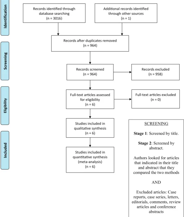
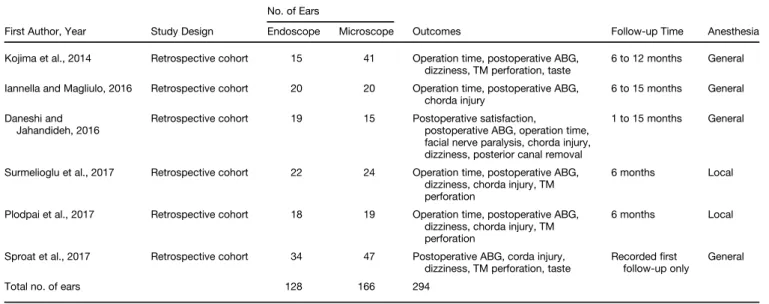
The search yielded a total of 3,017 articles. After excluding duplicates, we screened the remaining records for eligibility and found six articles for qualitative and quantita- tive synthesis (Fig. 1).15,21–25Characteristics of the included studies are presented in Table I. All the six articles were nonrandomized cohort studies, with the exposure group being all patients who underwent fully endoscopic stapes surgery, and the control group being all patients who under- went microscopic stapes surgery during the study period.
A summary of our analysis results can be seen in Table II.
Primary Outcomes
Average postoperative ABG.All studies evaluated average postoperative ABG, but onlyfive studies15,21–23,25
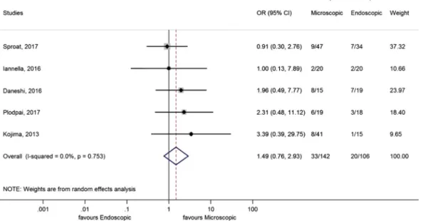
performed the evaluation according to the Committee on Hearing and Equilibrium of the American Academy of Otolaryngology–Head and Neck Surgery.26 We analyzed the ABG in three groups. Group 1 was ABG of 10 dB or less (Fig. 2). There was no significant difference (OR = 1.80 (95% CI: 0.96 to 3.38)), and theI2and χ2sta- tistical analysis suggested homogeneity (I2 = 0.0%, P= .529). When the Sproat et al. study was removed dur- ing sensitivity analysis, there was a statistically signifi- cant difference in favor of the endoscope.23 Group 2 was ABG between 11 dB and 20 dB (Fig. 3). There was no sig- nificant difference (OR = 1.49 [95% CI: 0.76 to 2.93]), and
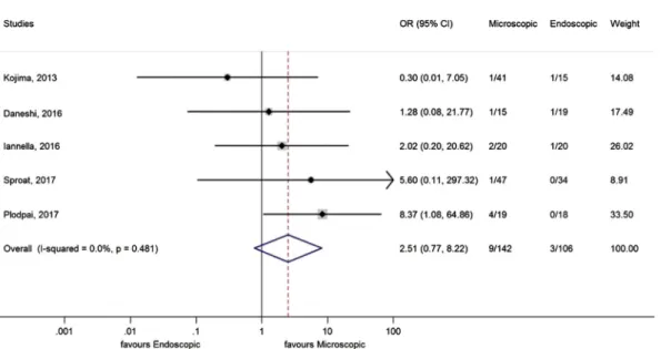
the I2 and χ2statistical analysis suggested homogeneity (I2= 0.0%,P= .753). Group 3 was ABG > 20 dB (Fig. 4).
Patients were more than two times likely to end up in this group if they were operated on with a microscope (OR = 2.51 [95% CI: 0.77 to 8.22]), but this was not statis- tically significant, and the I2 and χ2 statistical analysis suggested homogeneity (I2= 0.0%,P= .673).
Chorda tympani injury. Injury to the nerve was more than three times likely to happen with microscopic stapes surgery (OR = 3.51 [95% CI: 1.55 to 7.93]) and was statistically significant (Fig. 5). TheI2and χ2statistical analysis suggested homogeneity (I2 = 0.0%, P = .924).
Records idenfied through database searching
(n = 3016)
ScreeningIncludedEligibilitynoitacifitnedI
Addional records idenfied through other sources
(n = 1)
Records aer duplicates removed (n = 964)
Records screened (n = 964)
Records excluded (n = 958)
Full-text arcles assessed for eligibility
(n = 6)
Full-text arcles excluded (n = 0)
Studies included in qualitave synthesis
(n = 6)
Studies included in quantave synthesis
(meta-analysis) (n = 6)
SCREENING Stage 1: Screened by title.
Stage 2: Screened by abstract.
Authors looked for articles that indicated in their title
and abstract that they compared the two methods
AND
Excluded articles: Case reports, case series, letters, editorials, comments, review
articles and conference abstracts
Fig. 1. Flow diagram of the systematic review and meta-analysis. [Colorfigure can be viewed in the online issue, which is available at www.
laryngoscope.com.]
During sensitivity analysis, if the Surmelioglu et al. study was excluded, then there was no statistically significant difference.24
Secondary Outcomes
Postoperative taste disturbance. Four stud- ies15,21,23,24
evaluated postoperative taste disturbance, which was more than two times as likely to happen with microscopic stapes surgery (OR = 2.36 [95% CI: 1.01 to 5.51]) and was statistically significant. TheI2andχ2sta- tistical analysis suggested mild heterogeneity (I2= 21.2%,P= .283).
Average operating time. Five studies compared the average operating times.15,21,22,24,25
The average oper- ating time for the mean operating times reported with
the endoscope was 64.6 minutes and for the microscope was 69.6 minutes. However, only three studies were included in the meta-analysis due to insufficient publi- shed data such as the standard deviation.15,24,25 Our results showed no significant difference between the two operations (WMD = 0.14 [95% CI:−11.69 to 11.98]). The results of the I2and χ2statistics suggested considerable heterogeneity (I2= 82.8%,P= .003).
Tympanic Membrane (TM) perforation. Four studies21,23–25 evaluated TM perforation. TM perforation was not significantly different between the endoscopic and microscopic approach (OR = 1.70 [95% CI: 0.44 to 6.58]). TheI2andχ2statistical analysis suggested homo- geneity (I2= 0.0%,P= .983).
Postoperative pain.Four studies15,22,24,25
evaluated postoperative pain, which was the same for microscopic TABLE I.
Characteristics of Included Studies.
First Author, Year Study Design
No. of Ears
Outcomes Follow-up Time Anesthesia
Endoscope Microscope
Kojima et al., 2014 Retrospective cohort 15 41 Operation time, postoperative ABG, dizziness, TM perforation, taste
6 to 12 months General Iannella and Magliulo, 2016 Retrospective cohort 20 20 Operation time, postoperative ABG,
chorda injury
6 to 15 months General Daneshi and
Jahandideh, 2016
Retrospective cohort 19 15 Postoperative satisfaction,
postoperative ABG, operation time, facial nerve paralysis, chorda injury, dizziness, posterior canal removal
1 to 15 months General
Surmelioglu et al., 2017 Retrospective cohort 22 24 Operation time, postoperative ABG, dizziness, chorda injury, TM perforation
6 months Local
Plodpai et al., 2017 Retrospective cohort 18 19 Operation time, postoperative ABG, dizziness, chorda injury, TM perforation
6 months Local
Sproat et al., 2017 Retrospective cohort 34 47 Postoperative ABG, corda injury, dizziness, TM perforation, taste
Recordedfirst follow-up only
General
Total no. of ears 128 166 294
ABG = air–bone gap; TM = tympanic membrane.
TABLE II.
Summary of OR Statistical Analysis.
Outcome
No. of Studies Included
No. of Events/Total No. of Cases Result Analyzed
95% Confidence Interval
I2 χ2 Statistically Significant
Endoscope Microscope Lower Upper
Primary outcomes
Group 1: ABG = 0–10 dB 5 83/106 100/142 OR = 1.80 0.96 3.38 0% 0.529 No
Group 2: ABG = 11–20 dB 5 20/106 33/142 OR = 1.49 0.76 2.93 0% 0.753 No
Group 3: ABG = 21–30 dB 5 3/106 9/142 OR = 2.51 0.77 8.22 0% 0.481 No
Chorda tympani injury 6 3/128 20/166 OR = 3.51 1.55 7.93 0% 0.924 Yes
Secondary outcomes
Average operation time 3 Not applicable Not applicable WMD = 0.14 −11.69 11.98 82.8% 0.003 No
Postoperative dizziness 6 35/128 70/166 OR = 2.15 0.94 4.89 0.0% 0.49 No
Postoperative pain 4 63/91 95/ 132 OR = 0.84 0.36 1.96 64.2% 0.039 No
Postoperative taste disturbance 4 7/91 19/132 OR = 2.36 1.01 5.51 21.2% 0.283 Yes
Tympanic membrane perforation 4 2/89 5/131 OR = 1.70 0.44 6.58 0.0% 0.983 No
ABG = air–bone gap; OR = odds ratio; WMD = weighted mean difference.
and endoscopic stapes surgery (OR = 0.84 [95% CI: 0.36 to 1.96]). TheI2andχ2statistical analysis suggested signifi- cant heterogeneity (I2= 64.2%,P= .039).
Postoperative dizziness. All studies evaluated postoperative dizziness, and there was no statistical dif- ference (OR = 2.15 [95% CI: 0.94 to 4.89]). TheI2and χ2 statistical analysis suggested mild heterogeneity (I2 = 0.0%, P = .49). Postoperative dizziness is signifi- cantly higher with microscopic stapes surgery when the Iannella and Magliulo study is removed for sensitivity analysis.15
Risk of Bias Analysis
The risk of bias within individual studies is graphi- cally represented in Table III. The Plodpai et al.25 and Surmelioglu et al.24 studies were judged to have a low risk of bias, Iannella and Magliulo15 and Daneshi and Jahandideh22 moderate risk, and Kojima et al.21 and Sproat et al.23high risk.
The main reason for studies being assessed as high risk was not explicitly stating their sources and methods (e.g., the randomization process, patient selection, patient records, theater logbooks, audiology database).
Fig. 2. Forest plot of odds ratio (OR) with a 95% confidence interval (CI) of patients with average postoperative air–bone gap <10 dB. No sta- tistical difference. [Colorfigure can be viewed in the online issue, which is available at www.laryngoscope.com.]
Fig. 3. Forest plot of odds ratio (OR) with a 95% confidence interval (CI) of patients with average postoperative air–bone gap 10 dB to 20 dB.
No statistical difference. [Colorfigure can be viewed in the online issue, which is available at www.laryngoscope.com.]
The selection process was of particular interest as none of the studies were blinded, and surgeons might have chosen patients with ideal ears for the endoscopic proce- dures, a major confounder, or overestimated their results. Some of the authors tried to mitigate this selec- tion bias by stating there was randomization but did not describe the process.
An uncertain risk of bias in some studies was that patients for each group were selected from different time periods. This was due to the surgeons operating initially with the microscope before they changed to the endo- scope, and using the data collected during those two periods for comparison. The difference of the surgeon’s skill level between the two techniques could be considered Fig. 4. Forest plot of odds ratio (OR) with a 95% confidence interval (CI) of patients with average postoperative air–bone gap >20 dB. No sta- tistical difference. [Colorfigure can be viewed in the online issue, which is available at www.laryngoscope.com.]
Fig. 5. Forest plot of odds ratio (OR) with a 95% confidence interval (CI) of patients who suffered chorda tympani injury. The zero events were handled with continuity correction. Injury is most likely with (favors) the microscope. [Colorfigure can be viewed in the online issue, which is available at www.laryngoscope.com.]
as a confounder that would have influenced the selection process as mentioned earlier. In addition, we judged that to include patients who had been operated on in the past (revision surgeries), patients who were not followed up for a minimum of 6 months, or to exclude patients lost to follow-up due to inadequate data were causes of high risk of bias.
Most importantly, there was no standardized way of reporting outcomes and statistical results, which we judged to carry the highest risk of bias. This was espe- cially true in the case of the chorda tympani injury and postoperative taste disturbance. All studies were able to comment on whether the nerve was injured, but most of them did no grade the injury level, which could range from mild manipulation to severing the nerve. Similarly, there was no clarification on the definition of taste distur- bance or how it was measured and for how long. Only two studies mentioned that the disturbance was tran- sient.15,24We recommend that future studies state if the nerve was manipulated or severed and avoid using the term injury. In addition, taste needs to be measured with a validated method, and any changes after surgery need to be reviewed in follow-up, stating if it was transient or permanent. The correlation of nerve injury with taste dis- turbance also needs to be statistically analyzed, as some of the studies indicated no correlation between the two.
With regard to ABG measurements, previous endo- scopic and microscopic stapes surgery case series have demonstrated improvement in hearing by closure of the average ABG rather than just the average postoperative ABG.11,12,18 Only Surmelioglu et al. published their results in this manner, which we believe to be a more accurate way to demonstrate surgical effectiveness.24 Hence, we recommend that in the future results are pub- lished both ways to assist analysis.
Other than Iannella and Magliulo, no other study clar- ified how operation time was measured, which made assess- ment of this parameter extremely difficult.15Surgeries done under local anesthesia took longer, which is a confounding factor, and we assume this was due to patient movement during the procedure.24,25 All studies compared dizziness, but only two attempted to quantify it with respect to the duration (number of days after surgery).15,21Similarly, only
two out of four studies attempted to quantify pain with respect to severity and the need for medication.15,22
DISCUSSION
The main advantage of the endoscope over the micro- scope is better visualization of the middle ear structures, requiring less bone removal and chorda tympani nerve manipulation, which is a cause of postoperative taste dis- turbance.15 Our study concluded that the hearing out- comes of the endoscopic approach are similar to those of the microscope. However, there would have been a statis- tical difference for ABG <10 dB favoring the endoscope if we had not included the results of Sproat et al. in our analysis.23 In that study they compared the outcomes from separate time periods. We stated earlier that the surgeon’s skill level with the endoscope might not be the same as with the microscope, which in turn might have affected their outcomes. At the same time, Sproat et al.
were the only ones to use an otology audit tool to collect data and provided the largest number of cases for analy- sis. It could be inferred that the other studies over- estimated their results in favor of the endoscope, especially in the absence of blinding.
The chorda tympani can clearly be visualized during the procedure with either intervention, making easy for surgeons to include in their notes if it was injured, and in turn to include it in their study outcomes. However, the level of manipulation is not stated, and thus the term is vague, just as the terms used for postoperative taste dis- turbance in the absence of a validated tool and follow-up.
Although our study has shown statistically significant dif- ference in favor of the endoscope, this result must be taken in context with the significant level of bias calcu- lated in our analysis. In the Surmelioglu et al. study, the microscopic intervention, which was performed by either of two surgeons, had a very high level of chorda tympani injury compared to the endoscope, which was performed only by the more experienced of the two surgeons.24The surgeon’s experience, a confounder, could have influenced the outcomes in this study. Wefind no statistical differ- ence in the chorda tympani injury when we remove Surmelioglu et al. from our analysis. Furthermore, a TABLE III.
Risk of Bias Analysis.
Daneshi and Jahandideh, 2016
Kojima et al., 2014
Sproat et al., 2017
Plodpai et al., 2017
Iannella and Magliulo, 2016
Surmelioglu et al., 2017
Representativeness of the study ✓ X X ✓ ✓ ✓
Selection of the nonexposed cohort ✓ X X ✓ ? ✓
Ascertainment of exposure ? ? ✓ ✓ ? ✓
Demonstration that outcome of interest was not present at start of the study
? X X ✓ ? ✓
Comparability of cohorts on the basis
of the design or analysis controlled for confounders
? ? ✓ ✓ ? X
Assessment of outcome X X ? ? X X
Was follow-up long enough for outcomes to occur X ✓ X ✓ ✓ ✓
Adequacy of follow-up of cohorts ✓ X X ✓ ✓ ✓
✓= low risk; X = high risk; ? = uncertain risk.
secondary meta-analysis (see Supporting Table I in the online version of this article) of the risk difference (RD) was not statistically significant, which would further sup- port the hypothesis that there is no advantage with the endoscope.
Theoretically, endoscopically operated patients would suffer less with postoperative dizziness because of reduced manipulation of the stapes footplate due to better visualization. Equally, they would have complained less about postoperative pain due to less drilling of the exter- nal auricular canal. However, our study has shown that there was no statistical difference with either outcome. In experienced hands, TM perforation numbers will be mini- mal no matter which approach is used.
The operating time was the most poorly measured and published outcome, and this reflects the retrospective nature of the studies. Although there is no significant dif- ference in our analysis, a significant heterogeneity is seen in our results.
It was mentioned several times in the literature that the endoscope allows views with minimal or even without the need of drilling the bone of the posterior canal. This outcome was only sufficiently recorded by two stud- ies.15,22 Future studies should record if the drilling was done and try to quantify it (e.g., minimal, moderate, and extensive). Also, there is anecdotal evidence that depending on the surgeon’s dominant hand they would drill one side and not the other due to space constraints.
Learning curve was a particularly recurring theme.
The Iannella and Magliulo study was able to plot a learn- ing curve for endoscopic stapes surgery by analyzing the average operating times of a surgeon by groups of cases and by time intervals.15 In the first 10 cases done with the endoscope, the average operation time was signifi- cantly slower than the last 10 cases. However, they found no statistical difference between the average surgical times for endoscopic and microscopic approaches in the last 4-month period of their 1-year study and in the last 10 out of 20 cases. This would suggest that the learning curve for the endoscope is 10 cases. This is much smaller than the 60 to 80 cases for conventional microscopic sur- gery as published by Yung et al.27Nonetheless, the learn- ing curve has often been framed as the main cause for surgeons’ reluctance to change to the endoscope due to probable longer operation time.1,21Although the evidence is weak and not an outcome of this study, we believe sur- geons should not be afraid to operate with the endoscope, as the learning curve is not long and outcomes are similar or better than the microscopes, even at the early stages.
Study Limitations
Our meta-analysis used nonrandomized studies, which were done retrospectively and contained significant bias that reduced its level of evidence. By including only articles that directly compared the two surgeries we have limited our data pool. This has resulted in a theoretical increased risk of publication bias, which could not be assessed using the sym- metry of the funnel plot due to the low number of eligible studies (see Supporting Information, Appendix A and B in the online version of this article). The authors contemplated
the use of indirect comparison especially, as these can offer outcomes from a larger number of cases without great incon- sistency from a direct comparison.28However, this would not have decreased the risk of bias as the main risk factors of randomization, selection and publication would still exist, and new confounding factors would creep in.
CONCLUSION
Overall, our study indicates endoscopic and micro- scopic stapes surgery have similar audiological success, with some data suggesting a lower risk of chorda tympani injury and postoperative taste disturbance with the endo- scope. However, we acknowledge the limitations of our study and would like to encourage prospective randomize controlled trials to validate our results.
BIBLIOGRAPHY
1. Tarabichi M. Endoscopic middle ear surgery.Ann Otol Rhinol Laryngol 1999;108:39–46.
2. Vincent R, Sperling NM, Oates J, Jindal M. Surgicalfindings and long-term hearing results in 3,050 stapedotomies for primary otosclerosis: a prospec- tive study with the otology-neurotology database.Otol Neurotol2006;27(8 suppl 2):S25–S47.
3. Mer SB, Derbyshire AJ, Brushenko A, et al. Fiberoptic endoscopes for exam- ining the middle ear.Arch Otolaryngol1967;85:387–393.
4. Marchioni D, Soloperto D, Villari D, et al. Stapes malformations: the con- tribute of the endoscopy for diagnosis and surgery. Eur Arch Otorhinolaryngol2016;273:1723–1729.
5. Thomassin JM, Korchia D, Duchon-Doris JM. Residual cholesteatoma: its prevention by surgery with endoscopic guidance. Rev Laryngol Otol Rhinol (Bord)1991;112:405–408.
6. McKennan KX. Endoscopic‘second look’mastoidoscopy to rule out residual epitympanic/mastoid cholesteatoma.Laryngoscope1993;103:810–814.
7. Tarabichi M. Endoscopic management of acquired cholesteatoma.Am J Otol 1997;18:544–549.
8. Kojima H, Tanaka Y, Yaguchi Y, Miyazaki H, Murakami S, Moriyama H.
Endoscope-assisted surgery via the middle cranial fossa approach for a petrous cholesteatoma.Auris Nasus Larynx2008;35:469–474.
9. Carter MS, Lookabaugh S, Lee DJ. Endoscopic-assisted repair of superior canal dehiscence syndrome.Laryngoscope2014;124:1464–1468.
10. Poe DS. Laser-assisted endoscopic stapedectomy: a prospective study.
Laryngoscope2000;110(5 pt 2 suppl 95):1–37.
11. Nogueira Junior JF, Martins MJ, Aguiar CV, et al. Fully endoscopic stapes surgery (stapedotomy): technique and preliminary results. Braz J Otorhinolaryngol2011;77:721–727.
12. Sarkar S, Banerjee S, Chakravarty S, et al. Endoscopic stapes surgery: our experience in thirty-two patients.Clin Otolaryngol2013;38:157–160.
13. Migirov L, Wolf M. Endoscopic transcanal stapedotomy: how I do it.Eur Arch Otorhinolaryngol2013;270:1547–1549.
14. Bennett ML, Zhang D, Labadie RF, Noble JH. Comparison of middle ear visualization with endoscopy and microscopy. Otol Neurotol 2016;37:
362–366.
15. Iannella G, Magliulo G. Endoscopic versus microscopic approach in stapes surgery: are operative times and learning curve important for making the choice?Otol Neurotol2016;37:1350–1357.
16. Hunter JB, Zuniga MG, Leite J, et al. Surgical and audiological outcomes in endoscopic stapes surgeries across four institutions.Otolaryngol Head Neck Surg2016;154:1093–1098.
17. Moher D, Liberati A, Tetzlaff J, Altman DG; the PRISMA Group. Preferred reporting items for systematic reviews and meta-analyses: the PRISMA statement.PLoS Med2009;6:e1000097.
18. Schardt C, Adams MB, Owens T, Keitz S, Fontelo P. Utilization of the PICO framework to improve searching PubMed for clinical questions.BMC Med Inform Decis Mak2007;7:16.
19. Wells GA, Shea B, O’Connell D, et al. The Newcastle-Ottawa Scale (NOS) for assessing the quality if nonrandomized studies in meta-analyses. Available at: http://www.ohri.ca/programs/clinical_epidemiology/oxford.asp. Accessed June, 2018.
20. Higgins JPT, Green S, eds. Cochrane Handbook for Systematic Reviews of Interventions, Version 5.1.0. London, United Kingdom: The Cochrane Col- laboration; 2011.
21. Kojima H, Komori M, Chikazawa S, et al. Comparison between endoscopic and microscopic stapes surgery.Laryngoscope2014;124:266–271.
22. Daneshi A, Jahandideh H. Totally endoscopic stapes surgery without pack- ing: novel technique bringing most comfort to the patients.Eur Arch Otorhinolaryngol2016;273:631–634.
23. Sproat R, Yiannakis C, Iyer A. Endoscopic stapes surgery: a comparison with microscopic surgery.Otol Neurotol2017;38:662–666.
24. Surmelioglu O, Ozdemir S, Tarkan O, Tuncer U, Dagkiran M, Cetik F. Endoscopic versus microscopic stapes surgery.Auris Nasus Larynx2017;44:253–257.
25. Plodpai Y, Atchariyasathian V, Khaimook W. Endoscope-assisted stapedotomy with microdrill: comparison with a conventional technique.
J Med Assoc Thai2017;100:190–196.
26. Committee on Hearing and Equilibrium guidelines for the evaluation of results of treatment of conductive hearing loss. American Academy of
Otolaryngology-Head and Neck Surgery Foundation, Inc. Otolaryngol Head Neck Surg1995;113:186–187.
27. Yung MW, Oates J, Vowler SL. The learning curve in stapes surgery and its implication to training.Laryngoscope2006;116:67–71.
28. Song F, Xiong T, Parekh-Bhurke S, et al. Inconsistency between direct and indirect comparisons of competing interventions: meta-epidemiological study.BMJ2011;343:d4909.