SUMOylation of PHYTOCHROME INTERACTING FACTOR 3 promotes photomorphogenesis in Arabidopsis thaliana
P´eter Bernula1 , Alad´ar Pettko-Szandtner´ 2 , Anita Hajdu1, L´aszlo Kozma-Bogn´ ar´ 1,3 , Eve-Marie Josse4 , Eva´ Ad´´ am1,5 , Ferenc Nagy1 and Andr´as Viczian´ 1
1Institute of Plant Biology, Biological Research Centre, Szeged H-6726, Hungary;2Laboratory of Proteomics Research, Biological Research Centre, Szeged H-6726, Hungary;3Department of Genetics, Faculty of Sciences and Informatics, University of Szeged, Szeged H-6726, Hungary;4School of Biological Sciences, Institute of Molecular Plant Sciences, University of Edinburgh, Edinburgh, EH9 3JH, UK;5Department of Medical Genetics, Faculty of Medicine, University of Szeged, Szeged H-6720, Hungary
Author for correspondence:
Andr´as Viczi´an Email: aviczian@brc.hu Received:17 March 2020 Accepted:7 October 2020
New Phytologist(2021)229:2050–2061 doi: 10.1111/nph.17013
Key words: Arabidopsis thaliana, light sig- nalling, photomorphogenesis, phytochrome, PIF3, SUMO, SUMOylation.
Summary
In Arabidopsis thaliana, phytochrome B (phyB) is the dominant receptor of photomor- phogenic development under red light. Phytochrome B interacts with a set of downstream regulatory proteins, including PHYTOCHROME INTERACTING FACTOR 3 (PIF3). The inter- action between PIF3 and photoactivated phyB leads to the rapid phosphorylation and degra- dation of PIF3 and also to the degradation of phyB, events which are required for proper photomorphogenesis.
Here we report that PIF3 is SUMOylated at the Lys13 (K13) residue and that we could detect this posttranslational modification in a heterologous experimental system and alsoin planta.
We also found that the SUMO acceptor site mutant PIF3(K13R) binds more strongly to the target promoters than its SUMOylated, wild-type counterpart. Seedlings expressing PIF3 (K13R) show an elongated hypocotyl response, elevated photoprotection and higher tran- scriptional induction of red-light responsive genes compared with plantlets expressing wild- type PIF3.
These observations are supported by the lower level of phyB in plants which possess only PIF3(K13R), indicating that SUMOylation of PIF3 also alters photomorphogenesis via the reg- ulation of phyB levels. In conclusion, whereas SUMOylation is generally connected to differ- ent stress responses, it also fine-tunes light signalling by reducing the biological activity of PIF3, thus promoting photomorphogenesis.
Introduction
Light sensing plant photoreceptor molecules are responsible for light perception and initiation of the signalling responses neces- sary for survival and achieving optimal fitness in the ever-chang- ing light environment. There are five phytochromes (phyA–E) in Arabidopsis which are responsible for red and far-red light sens- ing (Nagy & Schafer, 2002; Legris et al., 2019). Among them, phyB is dominant and is responsible for photomorphogenic development in red light. Phytochromes are synthesized in the red-light absorbing inactive conformer Pr, which is converted to the far-red light absorbing biologically active Pfr form upon red light illumination. Photoactivated phyB governs diverse sig- nalling pathways and interacts with a set of basic helix-loop-helix (bHLH) transcription factors known as PHYTOCHROME INTERACTING FACTORs (PIFs) (Leivar & Quail, 2011).
One of them, PIF3, is involved in the repression of photomor- phogenic development in the dark but promotes some light-in- duced responsesafter the onset of light (Al-Sady et al., 2008;
Leivar et al., 2008; Shinet al., 2009; Stephenson et al., 2009;
Zhanget al., 2013). Light-activated phyB Pfr is accumulated in the nucleus and interacts with PIF3, and this leads to a fast decrease in PIF3 protein levels (Ni et al., 1999; Bauer et al., 2004; Parket al., 2004). PIF3 is phosphorylated by several differ- ent kinases on multiple residues (Ni et al., 2013, 2017; Shin et al., 2016; Linget al., 2017) and this posttranslational modifi- cation (PTM) is necessary for ubiquitination and subsequent degradation of the protein (Park et al., 2004; Al-Sady et al., 2006). It has been shown that, alongside fast PIF3 degradation, there are other mechanisms by which the amount of active PIF3 is reduced after the onset of light: thePIF3transcript level is also decreased in light (Shi et al., 2016), phyB sequesters PIF3 and releases it from a DNA target (Parket al., 2012, 2018), and addi- tionally a recent study demonstrated that phyB inhibits PIF3 translation by intron retention (Donget al., 2020).
Interestingly, despite the fact that the rapid decrease in the amount of available PIF3 is one of the first molecular events of photomorphogenesis, PIF3 has role in hypocotyl and cotyledon growth under light/dark photocycles (Soyet al., 2012); fine-tun- ing circadian responses (Soy et al., 2016); developing freezing
tolerance (Jianget al., 2017); regulating stomatal opening (Wang et al., 2010) and protecting young seedlings from reactive oxygen species (Chenet al., 2013). Additional reports indicate that PIF3 mediates hormonal responses, and together with other PIFs it is involved in the integration of light and hormonal signalling (Lau
& Deng, 2010; Yang et al., 2012; Zhong et al., 2012, 2014;
Zhanget al., 2014; Liet al., 2016). It was also shown that PIF3 does not act alone but redundantly controls gene expression with other PIFs, forming a signalling hub with diverse functions (Zhanget al., 2013; Leivar & Monte, 2014; Pfeifferet al., 2014).
Arabidopsis expresses four isoforms of the Small Ubiquitin- like MOdifier (SUMO) proteins, SUMO1–3 and SUMO5 (Kurepa et al., 2003; van den Burg et al., 2010; Hammoudi et al., 2016). The attachment of SUMO is a PTM among eukary- otes which involves activation, conjugation and ligation of SUMO to lysine amino acids located in a conserved sequence motif of the target protein (Novatchkovaet al., 2004; Parket al., 2011; Vierstra, 2012; Castano-Miquel et al., 2013; Tomanov et al., 2014; Augustine & Vierstra, 2018). SUMO and ubiquitin proteins show high structural similarities; however, whereas ubiq- uitination typically directs the target protein to proteasomal degradation (Vierstra, 2009), SUMOylation has more diverse outcomes, for example changes in stability, enzyme activity, nuclear localization, protein interaction, etc. of the target protein (reviewed by (Augustine & Vierstra, 2018)). Tightly controlled enzymatic de-SUMOylation of the targets opens additional regu- latory pathways (Yateset al., 2016). It has been observed that the overall SUMOylation of the plant proteome is increased during various stress responses (e.g. heat, drought, salt, pests, etc.) and also under developmental changes (flowering, growth, etc.) (Kurepaet al., 2003; Murtaset al., 2003; Miuraet al., 2005; Lee et al., 2007; Contiet al., 2008; Milleret al., 2010, 2013; Bailey et al., 2016; Caiet al., 2017; Casta˜no-Miquelet al., 2017; Rytz et al., 2018) (recently reviewed by Castroet al., 2012; Elrouby, 2015, 2017; Augustine & Vierstra, 2018; Vermaet al., 2018).
Light signalling pathways have a drastic impact on plant devel- opment, and their protein components are also targets of SUMOylation. For example, SUMOylation of the available phyB pool is increased by red light and reaches high levels in the middle of the day in plants grown under diurnal conditions. It has also been shown that phyB SUMOylation inhibits phyB–
PIF5 binding and attenuates light signalling (Sadanandomet al., 2015). In addition to the phyB photoreceptor, CONSTITUTIVE PHOTOMORPHOGENIC 1 (COP1), another important component of light signalling, is also SUMOylated. This ubiquitin ligase has key functions in main- taining etiolated growth and preventing photomorphogenesis in darkness. SUMOylation increases the activity of COP1, and the fact that COP1 regulates the abundance of the SIZ1 SUMO ligase links light signalling tightly to various stress responses that are mediated by SUMOylation (Kim et al., 2016; Lin et al., 2016; Hammoudiet al., 2018; Mazuret al., 2019).
A recent study showed that FAR-RED ELONGATED HYPOCOTYL 1 (FHY1), a nuclear transporter of the photoacti- vated phytochrome A (phyA), is SUMOylated, and that this modification accelerates FHY1 degradation (Qu et al., 2020).
These results indicate that light signalling is modified by SUMOylation, targeting not only the photoreceptor but also downstream components of the signal transduction pathways.
Our data indicate that PIF3, another key light signalling com- ponent and phyB direct interactor, is also SUMOylated. We noticed that SUMOylation of PIF3 leads to decreased PIF3 activ- ity, thus promoting cotyledon expansion and inhibiting hypocotyl elongation in red light, and attenuating PIF3-mediated gene induction and photoprotection. These phenotypic responses could be a result of the weaker DNA binding affinity of SUMOylated PIF3 and of the higher stability of phyB in those plants which contain SUMOylated PIF3. Our work suggests that, in contrast to the previously identified PTMs, SUMOyla- tion of PIF3 modulates the activity of this transcription factor, rather than the amount available.
Materials and Methods
Plant materials
Throughout this study we used thepif3-3(Monte et al., 2004) mutant of Arabidopsis thaliana(Columbia ecotype). Plant trans- formation was performed using the floral dip method, and trans- genic lines containing a single transgene locus were selected.pifq/
35S:PIF3-YFPhas already been published (Pfeifferet al., 2012).
ThephyB-9mutant (Reedet al., 1993) was used as a control, as shown in Fig. 2(a) (see later).
Molecular cloning
PIF3(K13R)was generated using the QuikChange Site-Directed Mutagenesis Kit (Agilent, Santa Clara, CA, USA) according to the manufacturer’s instructions. The coding sequences of PIF3 and PIF3(K13R) were fused to the coding region of YELLOW FLUORESCENT PROTEIN(YFP) asBamHI-SmaI fragments in the pPCV binary vector (Bauer et al., 2004). PIF3 and PIF3 (K13R) coding regions were cloned as BamHI-NotI fragments into the pET28 vector (Novagen, Madison, WI, USA) for bacte- rial protein expression. All clones were checked by sequencing.
Supporting Information Table S1 shows the sequence of the oligonucleotides used for cloning.
Seedling phenotyping
Measurement of hypocotyl elongation and data evaluation were performed as described previously (Adam et al., 2013; Dobos et al., 2019). In the survival test, seedlings were grown in the dark on Murashige & Skoog (MS) medium plates for 4 d and were then placed under white light irradiation (100μmol m−2s−1) for 2 d. The ratio of total to survived seedlings was calculated.
All experiments were repeated at least three times.
SUMO binding site predictions
The in silicoSUMO binding site predictions for different PIF3 homologues were performed using the current version (as of 3
May 2020) of the GPS-SUMOonline tool at medium sensitivity (http://sumosp.biocuckoo.org; Zhaoet al., 2014). The alignment was performed using CLUSTAL O (v.1.2.4; www.ebi.ac.uk). The GenBank accessions of the used sequences are as follows: A.
thaliana (NP_001318964); Eutrema salsugineum (XP_006417565); Citrus clementina(XP_006423962); Capsella rubella (XP_006303172); Eucalyptus grandis (XP_010070103);
Aquilegia coerulea (PIA60627); Gossypium barbadense (PPS10307); Cucumis sativus (XP_011648884); Vitis vinifera (RVW77362); Cephalotus follicularis (GAV72297); Populus trichocarpa (XP_006382253); Physcomitrella patens (XP_024361305);Arachis duranensis(XP_015939567).
Total plant protein extraction and immunoblotting
Total plant protein extract preparation and Western blot analysis were performed as previously described (Vanhaelewyn et al., 2019). We used the following primary antibodies: anti-PIF3 (Baueret al., 2004), anti-SUMO1 (Agrisera), anti-T7 (Novagen), anti-ACTIN (Sigma-Aldrich), anti-PHYB (generous gift of Peter H. Quail, UC, Berkeley), and the following secondary antibod- ies: Polyclonal Swine Anti-Rabbit Immunoglobulins/HRP (Dako, Glostrup, Denmark), and Goat Anti-Mouse IgG Peroxi- dase Conjugated antibody (Invitrogen). The signals were visual- ized using Immobilon Western HRP Substrate (Millipore) according to the manufacturer’s recommendations using a cooled digital camera (Hamamatsu Orca-II, Hamamatsu, Japan).
Immunoprecipitation of PIF3-YFP
The immunoprecipitation procedure has been described in detail elsewhere (Orosa & Viczian, 2019). Briefly, 1 g of frozen plant material was ground in liquid nitrogen and mixed with 1.8 ml extraction buffer (30 mM Tris-HCl, pH 8.8, 1% sodium dodecyl sulphate (SDS), 1% (w/v) Triton X100, 50 mM sodium-bisulfite, 20 mM N-ethylmaleimide (Sigma), 1 mM phenylmethylsulfonyl fluoride (Roche), 1 piece per 10 ml buffer cOmplete ULTRA Mini Protease Inhibitor tablet (Roche)). Thawed samples were col- lected into 2 ml reaction tubes and cleared using centrifugation (20 000g, 4°C, 15 min). The supernatant was added to 30µl anti-GFP agarose beads (Chromotec, Planegg-Martinsried, Ger- many) equilibrated in extraction buffer. Samples were rotated in a roller drum for least 30 min (20 rpm, 4°C), the supernatant was discarded and the beads were washed four times with extraction buffer. After washing, 30µl 2×R loading buffer (125 mM Tris- HCl, pH 6.8, 4% SDS, 20% glycerol, 10% 2-mercaptoethanol, 0.02% bromophenol-blue) was added to each sample. After 5 min of incubation at 95°C, samples were centrifuged (20 000g, 1 min) to settle the agarose beads before loading the supernatant onto denaturing acrylamide gel.
Transcript level determination
Seedlings were surface sterilized, placed on MS agar plates and kept at 4°C for 3 d. After 6 h of white light irradiation (100 μmol m−2s−1, Lumilux XT T8 L 36 W/865 fluorescent
tubes; Osram, Munich, Germany) they were kept in darkness for 5 d at 22°C. Seedlings were irradiated with 10µmol m−2s−1 red light (Snap-Lite LED light source; Quantum Devices Inc., Barneveld, WI, USA) for 60 min and snap frozen in liquid nitro- gen. Total plant RNA was isolated using the Nucleospin Plant II Maxi kit (Macherey-Nagel, Duren, Germany). The reverse tran-¨ scription reaction was performed using the RevertAid First Strand cDNA Synthesis Kit (Thermo Scientific, Waltham, MA, USA), whereas the quantitative polymerase chain reaction was performed using the qPCRBIO Sygreen Mix (PCR Biosystems, London, UK) according to the manufacturer’s instructions. Table S1 shows the sequence of the oligonucleotides used for the qRT- PCR assays. The mRNA levels are presented as relative to the constitutively expressed TUBULIN2/3 mRNA transcript (Endo et al., 2007).
In vitroSUMOylation assay
PIF3 pET28 and PIF3(K13R) pET28 constructions were trans- formed into Escherichia coli strain BL21 containing plasmids expressing the SUMO-activating (E1) and conjugating (E2) enzymes and one of the four SUMO isoforms (Okada et al., 2009).
Ten milliliters Luria Bertani (LB) cultures were incubated at 37°C until an optical density at 660nm (OD660) of 0.6 was reached, after which β-D-1-thiogalactopyranoside (IPTG, Biosynth AG, Staad, Switzerland) was added to reach the final concentration of 0.5 mM for each culture. The bacterial cultures were further incubated overnight at 16°C. Cells were harvested by centrifugation (3000g, 4°C, 20 min), and the pellet was re- suspended in 1 ml sterile water. 10 µl cell suspension was mixed in 20µl 2×R loading buffer and denaturated (95°C, 10 min).
We collected the debris by centrifugation (20 000g, 1 min) and immediately loaded 28μl of the supernatant onto denaturing polyacrylamide gel. PIF3 and PIF3-SUMO were detected using anti-T7 antibody.
Expression and purification ofin vitroSUMOylated PIF3 for electrophoretic mobility shift assay (EMSA), co-im- munoprecipitation (Co-IP) assay and tandem mass spec- trometry (MS/MS)
PIF3-pET28 and PIF3(K13R)-pET28 constructions were trans- formed into E. coli strain BL21 containing plasmids expressing the SUMO-activating (E1) and conjugating (E2) enzymes and one of the four SUMO isoforms (Okadaet al., 2009). One liter bacterial culture in LB broth was incubated at 37°C until an OD660value of 0.6 was reached, and IPTG was added to reach a final concentration of 0.5 mM for each culture, followed by incubation for 4.5 h at 37°C. Cells were collected and re-sus- pended in 12 ml lysis buffer (50 mM NaH2PO4, 300 mM NaCl, 10 mM Imidazole (PanReac AppliChem, Darmstadt, Germany), pH 8.0) and then 10 mg lysosyme (Fluka, Buchs, Switzerland), one cOmplete ULTRA Mini Protease Inhibitor tablet, 20 mM N-ethylmaleimide (NEM, Sigma) and 1 mM phenylmethylsulfonyl fluoride (PMSF, Roche) were added to
each sample. Samples were incubated on ice for 30 min followed by 12 rounds of sonication for 10 s. The supernatant was cleared by centrifugation (20 000g, 4°C, 25 min) and loaded onto 3 ml Ni-NTA agarose beads (Qiagen), equilibrated in lysis buffer.
Samples were rotated on a roller drum for 1 h at 4°C (20 rpm).
Beads were collected (1000g, 4°C, 2 min) and washed three times using ice cold washing buffer (50 mM NaH2PO4, 300 mM NaCl, 20 mM Imidazole). The proteins were eluted from the beads using 2 ml of elution buffer (5 mM NaH2PO4, 300 mM NaCl, 250 mM Imidazole) three times, and the samples were concentrated using Amicon Ultracel 30 K filter tubes (Merck Millipore, Darmstadt, Germany) according to the manu- facturer’s instructions.
Electrophoretic mobility shift assay
To produce double-stranded probes, equal amounts of comple- mentary oligonucleotides were mixed at a final concentration of 40µM in 10 mM Tris-HCl (pH 7.5), 1 mM EDTA, and 50 mM NaCl, heated to 95°C for 5 min, and left to cool down in a block heater to room temperature overnight. The 50 end of the forward oligonucleotide was labelled with biotin (Thermo Scien- tific). PIF3 and PIF3(K13R) proteins with an N-terminal 6×His tag were expressed inE. coliBL21 cells and purified using an Ni- NTA agarose matrix according to the manufacturer’s recommen- dations (QIAexpressionist, Qiagen). In the binding reactions 10 mM Tris-HCl (pH 7.5), 85 mM KCl, 5% (v/v) glycerol, 0.1μg- µl−1poly(dI-dC), 40 fmol probe and variable amounts ofE. coli expressed and purified PIF3 proteins were mixed to a final vol- ume of 20µl. The reactions were incubated at room temperature for 20 min and loaded on native 4% polyacrylamide gels. Gels were run in 0.5×(Tris-borate-EDTA) TBE and electro-blotted to a nylon membrane (Hybond-N+, Amersham) in 0.5×TBE.
Detection of the biotin-specific signal was performed using the Chemiluminescent Nucleic Acid Detection Module (Thermo Scientific) according to the manufacturer’s instruction. Signals were detected as described for the Western blots.
Accession numbers
The accession numbers for the Arabidopsis genes used in this study are as follows: PIF3, AT1G09530; ELIP2, AT4G14690;
CHS, AT5G13930; PHYB, AT2G18790; PIL1, AT2G46970;
TUB2/3, AT5G62700, AT5G62690.
Details of mass spectrometry and yeast two-hybrid assays are available in Methods S1.
Results
Identification of the SUMOylated residue in PIF3
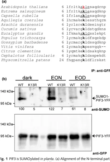
We performed anin silicosearch to identify potential SUMO tar- get residues in the Arabidopsis PIF3 protein. The GSP-SUMOsoft- ware (Zhaoet al., 2014) identified lysine 13 (K13) as a potential SUMO attachment target with high probability as part of a con- ventional consensus motif ψ–K–X–E/D (ψ, hydrophobic
residue, X, any amino acid, D, aspartic acid, E, glutamic acid) (Hendriks & Vertegaal, 2016; Augustine & Vierstra, 2018). Fur- thermore, we also noticed that a lysine residue located in a similar position is a potential SUMOylation target at the N-terminus of PIF3 among different plant species (Fig. 1a). To demonstrate the PIF3 SUMOylationin vitro, we expressed this protein in a bacte- rial system which reconstitutes Arabidopsis SUMOylation in
(a)
(b)
Fig. 1PIF3 is SUMOylatedin planta. (a) Alignment of the N-terminal part of PIF3 protein sequences from different species. Numbers indicate the corresponding amino acid position of each protein sequence. The Lys13 amino acid in the Arabidopsis PIF3 (red) was identified as a SUMO attachment site by the GPS-SUMOonline tool. Lysine amino acids at the N- terminal part of the PIF3 homologues from different plant species were also identified as SUMOylation sites (bold) by GPS-SUMO. Underlined sequences were found as consensus (ψ–K–X–E/D) SUMOylation motifs, whereas the others were identified as non-consensus sites. (b) Arabidopsispif3seedlings expressing the35S:PIF3-YFPor35S:PIF3(K13R)-YFPtransgenes were grown on Murashige & Skoog medium under an 8 h : 16 h, light : dark photoperiod (white light, 100µmol m−2s−1) or in darkness for 7 d (dark).
Samples were harvested at the end of the light phase (EOD) and at the end of the night (EON). PIF3-YFP and PIF3(K13R)-YFP were
immunoprecipitated by using GFP-Trap agarose beads. Western blotting was used to analyse samples containing identical amounts of fusion proteins (loading control, lower panel). PIF3-YFP and PIF3(K13R-YFP) were detected using anti-GFP, whereas Arabidopsis SUMO1 conjugated PIF3-YFP was visualized using anti-SUMO1 antibody. The numbers below the
immunoblot image show the relative amount of SUMO-PIF3-YFP, with the signal normalized to that of the dark-grown sample (100%).
E. coliby expressing Arabidopsis SUMO proteins along with the necessary SUMO-conjugating enzymes (Okadaet al., 2009). We found the following: first, we can detect SUMO-PIF3 conjugates;
second, all plant-expressed active SUMOs (SUMO1, SUMO2, SUMO3, SUMO5) were conjugated to PIF3; third, only the conjugatable SUMOs were attached to PIF3; and fourth, we could not detect SUMO conjugated to the PIF3(K13R) mutant in which the lysine 13 was replaced by arginine (Fig. S1). Fur- thermore, we were also able to confirm the SUMOylation of lysine 13 using liquid chromatography–tandem mass spectrome- try (LC-MS/MS) (Fig. S2).
To test PIF3 SUMOylationin planta, we fused PIF3 and PIF3 (K13R) to YELLOW FLUORESCENT PROTEIN (YFP) and expressed PIF3-YFP and PIF3(K13R)-YFP at high levels under the control of the constitutive viral p35S promoter in the pif3 mutant background. To enrich the PIF3 content of our sample, and to get rid of other SUMOylated proteins, we immunoprecip- itated PIF3-YFP and PIF3(K13R)-YFP using GFP-Trap agarose beads. Next, we tested these samples with Western blot analysis, applying the anti-SUMO1 antibody. We found that, similar to thein vitrobacterial system, PIF3 is also SUMOylatedin planta, whereas we could not detect a SUMO signal for the PIF3(K13R) mutant protein (Fig. 1b). In addition, we noted that PIF3- SUMO conjugates also occur in etiolated seedlings and in plantlets grown under light/dark cycles, and accumulate to higher levels during the dark phase (Figs 1b, S3a).
SUMOylation reduces the biological activity of PIF3 in light To examine the biological role of PIF3 SUMOylation, we expressedp35S:PIF3-YFPandp35S:PIF3(K13R)-YFPtransgenes in the pif3 mutant background and chose those lines which express the chimeric proteins at the same level (Fig. S3b–d).
Afterwards, we measured the light-induced inhibition of hypocotyl elongation in the lines grown under constant red light,
an assay which is widely used to monitor the performance of phyB-mediated light signalling. We noticed that those plants that express the wild-type (WT) PIF3 have longer hypocotyls in red light (hyposensitive photomorphogenic response), compared with the background pif3 mutant plants (Fig. 2a). This result replicates observations from earlier studies (Kim et al., 2003;
Baueret al., 2004; Monteet al., 2004). More interestingly, those seedlings which express the SUMO acceptor site mutant PIF3 (K13R) show an even more hyposensitive hypocotyl elongation
(a)
(b) (c)
(d) Fig. 2SUMOylation of PIF3 results in reduced biological activity in light.
(a) Hypocotyl length of Arabidopsis seedlings grown at different fluence rates of red light was determined after 4 d of growth and was normalized to the corresponding dark values. Error bars indicate SE. The following lines were examined: Col, Columbia wild type;pif3, mutant lacking functional PIF3;phyB-9, mutant lacking functional phyB. The following transgenic lines in thepif3background were also examined: PIF3,p35S:
PIF3-YFP; PIF3(K13R),p35S:PIF3(K13R)-YFP; hPIF3,p35S:PIF3-YFP expressed at high level; hPIF3(K13R),p35S:PIF3(K13R)-YFPexpressed at high level. Asterisks indicate significant differences between the PIF3 and PIF3(K13R) lines, according to Student’st-test:***,P<0.005. (b) Hypocotyl length ofpif3seedlings expressing the35S:PIF3-YFPor35S:
PIF3(K13R)-YFPtransgenes were grown on Murashige & Skoog medium under an 8 h : 16 h, light : dark photoperiod (white light,
100µmol m−2s−1) for 7 d. Error bars indicate SE. Asterisks indicate
significant differences between the marked lines, according to Student’st- test:***,P<0.005. (c) Cotyledon area of the seedlings propagated as described in (b). (d) Survival rate of 4-d-old etiolated Arabidopsis seedlings was calculated after 2 d of white light irradiation (100μmol m−2s−1).
Error bars depict SE. Asterisks indicate significant differences between the marked lines, according to Student’st-test:**,P<0.05,***,P<0.005.
The lines used are the same as those detailed for (a).
phenotype, despite the fact that the PIF3-YFP and PIF3(K13R)- YFP proteins are expressed at the same levels. The inhibition of the hypocotyl elongation response is even weaker in two other plant lines expressing PIF3-YFP and PIF3(K13R)-YFP proteins at higher but equal levels (Fig. S3b) and they respond to light only under higher fluences. We suspect that the response is satu- rated and the high level of PIF3 expression masks the subtle dif- ferences between the lines (Fig 2a). PIF3 also plays key roles in the promotion of hypocotyl growth and inhibition of cotyledon expansion in seedlings grown under light/dark photoperiods (Soy et al., 2012, 2016). Under these growth conditions, PIF3-YFP and PIF3(K13R)-YFP proteins are expressed in the transgenic plants at the same levels (Fig. S3e–f). The longer hypocotyls and the smaller cotyledons of the plants expressing PIF3(K13R) indi- cate that PIF3 SUMOylation promotes seedling photomor- phogenic growth under light/dark photoperiods similarly to under constant irradiation (Fig. 2b,c).
It is well established that PIF3 is required for proper Chl accumu- lation and seedling greening. Lack of PIF3 results in overaccumula- tion of protochlorophyllide, a precursor of Chl, causing photobleaching and cell death when etiolated seedlings are trans- ferred to light conditions (Shinet al., 2009; Stephensonet al., 2009;
Chen et al., 2013). To test this response, we transferred etiolated seedlings to white light conditions and 2 d later calculated the ratio of viable to dead seedlings. The survival rate was 78.2% for the WT plants, but only 13.8% of the pif3 mutant seedlings survived the light treatment. Expressing WT PIF3-YFP in thepif3 background increases the number of surviving plants dramatically and comple- ments the mutant phenotype, whereas PIF3(K13R)-YFP, having a mutated SUMO acceptor site, induces an even higher survival rate (Fig. 2d). Those lines which express PIF3-YFP or PIF3(K13R)-YFP at higher levels induce equally high responses. The response is most likely saturated, and subtle differences between the lines are masked in these high-level expressors (Fig. 2d).
In conclusion, these results indicate that the SUMO acceptor site mutant PIF3(K13R) molecules trigger stronger PIF3-medi- ated responses than the WT counterparts, in other words, SUMOylation reduces the biological activity of PIF3 in young seedlings in light.
Binding to target promoters is affected by the SUMOylation of PIF3
Light-induced degradation in etiolated seedlings is a characteris- tic property of PIF3 (Bauer et al., 2004; Parket al., 2004). We found that the amounts of both PIF3-YFP and PIF3(K13R)-YFP decreased quickly after the onset of red light irradiation, and no difference in the degradation of the two proteins can be detected, noting the obvious limitations of the Western blot hybridisation analysis, which may hide subtle temporal differences between the sample collection time points (Fig. S4). Not only the light-in- duced degradation but also the intracellular localization and nuclear complex formation of PIF3(K13R)-YFP resembles its WT counterpart (Fig. S5). Collectively, these results indicate that SUMOylation does not alter the light-induced degradation and intracellular localization of PIF3.
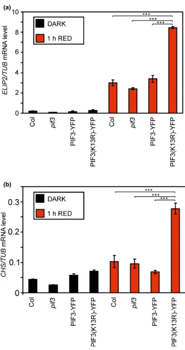
Transcription factor PIF3 is involved in the mediation of light-induced gene expression. This response is among the first molecular mechanisms which interrupts etiolated growth and contributes to proper photomorphogenic development after the onset of light (Al-Sadyet al., 2008). We examined the induction of the genes encoding early light-induced protein 2 (ELIP2) and chalcone synthase (CHS) and found that a short red light treat- ment increases their expression level. This increase is higher in those plants which express PIF3(K13R)-YFP than in PIF3-YFP
(a)
(b)
Fig. 3PIF3-dependent induction of early response genes. Five-day-old dark-grown Arabidopsis seedlings (black columns) were irradiated with 10µmol m−2s−1red light for 1 h (red columns) and the levels ofELIP2(a) orCHS(b) transcripts were measured by reverse transcription quantitative polymerase chain reaction (qRT-PCR). The mRNA levels were normalized toTUBULIN (TUB)levels. Error bars show SE calculated from three independent experiments. Asterisks indicate significant difference according to Student’st-test:***,P<0.005.
expressors at a very early stage of the photomorphogenic develop- ment after 1 h of red light irradiation (Fig. 3).
Although the composition of protein complexes containing PIF3 and regulating transcription is obscure, the direct binding of PIF3 to certain target promoters and the strength of this bind- ing can be examined using EMSA. We expressed and isolated PIF3 and PIF3(K13R) from anE. colistrain which is not able to SUMOylate proteins, and we found no differences between the DNA binding of the WT and the K13R mutant (Fig. S6a). This indicates that a single amino acid exchange does not alter PIF3 binding to DNA. Next, we expressed and purified PIF3 and PIF3(K13R) fromE. colicells which contained all of the neces- sary elements of the Arabidopsis SUMOylation pathway and used these to perform an EMSA with a G-box motif (CACGTG)-containing promoter section of the PIF3-like 1 (PIL1), which has been shown to be a binding target of PIF3 (Zhang et al., 2013). Applying the same amount of WT and mutant protein, we found that PIF3(K13R), showing no detectable SUMOylation, binds with higher affinity to the target DNA sequence (Figs 4, S7). We received the same results when
using the G-box containing promoter elements of ELIP2 and CHS (Fig. S8). Collectively, these results indicate that SUMOylation can modulate the binding affinity of PIF3 to dif- ferent target promoters.
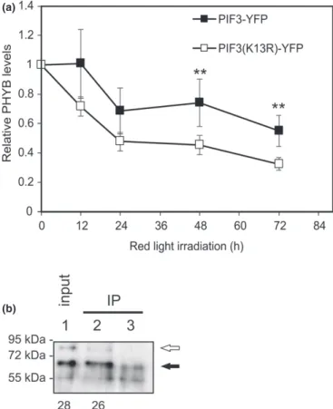
The SUMOylation state of PIF3 affects the stability of PHYB
A few years ago, it was revealed that not only is the degradation of PIF3 is induced by light, but PIF3 and PHYB interact in a protein complex and co-degrade (Niet al., 2014). This process decreases
Fig. 4Electromobility shift assay shows that DNA binding is stronger for PIF3(K13R) than PIF3. Equal amounts of biotin-labelled double-stranded probes were incubated with Arabidopsis PIF3 or PIF3(K13R) expressed and purified fromEscherichia coliwhich also expressed Arabidopsis SUMO activation and conjugation enzymes together with SUMO3. The amounts of PIF3 and PIF3(K13R) proteins used in the binding reaction are indicated at the top of the image. Binding reactions were resolved on 6% native polyacrylamide gels. The 26-nuclotide-long probe represented a fragment of thePIL1promoter carrying a single G-box element at the centre (PIL1a probe from Zhanget al., (2013)). PIF3-DNA and PIF3(K13R)-DNA complex are indicated by an arrow, whereas free (non-bound) probes are indicated by an asterisk. The independent repetition experiment is presented in Supporting Information Fig. S7(c).
(a)
(b)
Fig. 5SUMOylation of PIF3 stabilizes phyB but did not affect PIF3-PHYB binding. (a) Eight-day-old etiolatedpif3mutant Arabidopsis seedlings expressing PIF3-YFP or PIF3(K13R)-YFP were irradiated for 72 h with red light (30µmol m−2s−1) and the level of PHYB was monitored by Western blot hybridization using anti-PHYB antibody. The experiment was repeated four times. Error bars indicate SE, and asterisks denote the level of significance according to Student’st-test,**,P<0.05. (b) His-tagged Arabidopsis PIF3 was expressed inEscherichia colicells containing plasmids for expression of Arabidopsis SUMO-activating (E1) and conjugating (E2) enzymes and SUMO3. We affinity purified PIF3 using Ni- NTA agarose beads and mixed with total plant protein extract, made from 35S:PHYB-YFP/pifqtransgenic (lane 1 and lane 2) or Columbia seedlings (negative control, lane 3). After protein complex purification using anti- GFP-bound agarose beads (IP, lane 2 and lane 3), proteins were subjected to Western blot analysis using anti-His antibody. Lane 1 shows the input without immunoprecipitation (IP). The empty arrow indicates protein bands corresponding to SUMOylated PIF3, and the filled arrow indicates non-SUMOylated PIF3. The numbers below the immunoblot image represent the detected SUMO-PIF3 signal as a percentage of that of the corresponding band for the non-SUMOylated PIF3 in the same sample.
the amount of available active photoreceptors thus fine-tunes pho- tomorphogenesis. We wanted to examine how SUMOylation of PIF3 influences PHYB levels in our transgenic lines equally expressing PIF3-YFP or PIF3(K13R)-YFP. During continuous red light irradiation, using Western blotting, we could detect less phyB in the line expressing PIF3(K13R)-YFP (Figs 5a, S9a), indicating that PIF3 SUMOylation stabilizes PHYB.
This observation can be explained by the difference in stability of the complexes formed by SUMOylated PIF3 and phyB, and the SUMO acceptor site mutant PIF3(K13R) and phyB. We found that K13R mutation per sedoes not alter PIF3 binding to phytochromes (Fig. S6b), and to test the effect of PIF3 SUMOylation on phyB binding, we expressed PIF3 in anE. coli strain which is able to SUMOylate proteins. Fig. S7(a,b) shows that the entire quantity of available PIF3 is not SUMOylated in the bacterial cells, and a considerable amount of nonSUMOylated protein is also present. We incubated this mixture of PIF3 and SUMO-PIF3 together with native protein extract made from PHYB-YFP expressor plants. We then purified PHYB-YFP-con- taining complexes using anti-GFP antibody coupled agarose beads and examined whether immunoprecipitation (IP) altered the ratio of PHYB-bound SUMO-PIF3 to PIF3. Our Western blot analysis indicates that the ratio of SUMO-PIF3 to PIF3 is similar in the
‘before IP’ (input) and ‘after IP’ lanes, indicating that phyB binds to SUMOylated PIF3 and to non-SUMOylated PIF3 with equal affinity (Figs 5b, S9b). We also note that the possibility of slight differences in SUMO-PIF3–phyB binding under different experi- mental conditions cannot be excluded.
Discussion
Among the posttranslational modifications of PIF3, phosphoryla- tion and ubiquitination have been detected and studied extensively so far. It has been found that phosphorylation of PIF3 occurs at numerous amino acid residues, and the more phosphorylated state of the molecule leads to subsequent ubiquitination and degrada- tion of the protein (Niet al., 2013), similar to other PIF3 homo- logues (PIF4 and PIF5) (Shen et al., 2007 Lorrain et al., 2008).
This is one of the earliest key steps in the switching of plant skoto- morphogenesis to photomorphogenic development.
Here we report that PIF3 is SUMOylated at the N-terminal part of the molecule, and the PIF3 molecule possessing the K13R mutation shows no detectable SUMOylation. These results do not exclude the possibility that SUMO could be attached to other amino acids, but they strongly indicate that the major SUMOyla- tion site of PIF3 is the lysine 13. We also note that the SUMOylated pool of PIF3 represents only a minor portion of the total PIF3 amount, as we could only detect PIF3-SUMO using a SUMO-specific antibody and not with anti-PIF3.
Despite the low amount of PIF3-SUMO in plants, our pheno- typic analyses indicated that even this subtle quantity has an effect on plant photomorphogenesis. We found that seedlings expressing PIF3(K13R)-YFP show hyposensitive light responses, and have longer hypocotyls and smaller cotyledons in light than those expressing PIF3-YFP (Fig. 2a–c). PIF3 is an overall nega- tive regulator of photomorphogenesis (Leivar et al., 2008), and
our data indicate that the biological activity of PIF3 is decreased by SUMOylation in light. This conclusion is further confirmed by experiments in which PIF3 acts as a positive regulator of gene s 2d, 3). Thus, taken together, these datasets suggest that the PIF3(K13R) SUMO acceptor mutant functions as a ‘hyperactive’
PIF3, exhibiting increased activity in different responses.
To find a mechanistic explanation for this phenomenon, we hypothesized that the following properties/functions of PIF3 might be affected by SUMOylation: (a) DNA binding, (b) degra- dation in light, (c) intracellular localization, (d) complex forma- tion, and (e) co-degradation with phyB. The following paragraphs discuss these possibilities.
PIF3 acts as a transcription factor, binding to G-box promoter elements (Zhang et al., 2013). We observed that PIF3(K13R) binds to these elements with higher affinity than its WT counter- part in anin vitroEMSA assay (Figs 4, S7–S8). This result is fur- ther supported by the light-induced gene expression in planta, which shows that the expression of PIF3(K13R) leads to higher expression levels of early light-response genes (Fig. 3). These data support the idea that SUMOylation decreases the activity of PIF3 in this manner by interfering with its DNA binding.
We monitored PIF3 degradation in irradiated etiolated seedlings and found that PIF3(K13R)-YFP degrades approxi- mately as quickly as its wild-type counterpart, PIF3-YFP. We also noticed that both chimeric proteins showed a characteristic upshifted band (Fig. S4) which is the result of protein phospho- rylation and has been reported previously (Niet al., 2013). We also found no difference in the intracellular localization and light-induced nuclear speckle formation of PIF3-YFP and PIF3 (K13R)-YFP (Fig. S5). Based on these results, obtained using the available experimental methods, we can conclude that PIF3 SUMOylation does not alter the light-induced degradation or intracellular localization of PIF3.
Light does not solely initiate the degradation of PIF3 but also destabilizes the PIF3–phyB complex, resulting also in the degra- dation of the photoreceptor (Niet al., 2014). We found that the expression of PIF3(K13R) results in a lower amount of detectable phyB compared with PIF3-expressor seedlings (Fig. 5a). These data indicate that SUMOylation of PIF3 increases the stability of phyB. Because phyB-driven responses depend on the degree of photoreceptor availability, we postulate that SUMO-regulated PIF3-phyB co-degradation can fine-tune photomorphogenic responses, explaining why a lack of PIF3 SUMOylation leads to hyposensitive responses. The SUMO acceptor site K13 is located close to the APB active phyB binding (APB) motif, which is responsible for phyB binding (Khannaet al., 2004). It is tempt- ing to speculate that attachment of the bulky SUMO peptide in the proximity of this motif interferes with PIF3-phyB binding.
Interestingly, we found that PIF3 SUMOylation does not alter the binding affinity of PIF3 to phyB (Figs 5b, S9b), indicating that PIF3 binding is not the key mechanism by which PIF3 SUMOylation modifies phyB levels. These observations resemble the results published by Ni et al. (2013) who found that phos- phorylation does not alter the binding affinity of PIF3 to phyB;
thus, it seems that the mode of action of these PTMs is not the modification of the stability of the PIF3–phyB complex.
Our data demonstrate that SUMOylation decreases the activ- ity of PIF3 in light signalling. It was shown previously that phyB is also a SUMO target and that SUMOylated phyB mediates impaired light signalling (Sadanandomet al., 2015). It is interest- ing to note that SUMOylation exerts opposite overall effects on signalling by decreasing the activity of different components with opposite impacts. On the one hand, in light, the phyB photore- ceptor promotes photomorphogenesis; thus, SUMOylation of this positive factor slightly decreases its activity. On the other hand, PIF3 is a negative component of light-dependent develop- ment, and its SUMOylation therefore results in loss of activity and thus more pronounced photomorphogenesis. We also notice that the degree of SUMOylation of the phyB pool is higher dur- ing the light part of the day, while that of PIF3 is higher during the dark hours. It is tempting to speculate that SUMOylated PIF3 could modulate phyB stability during the night and in cooperation with phyB phosphorylation – which modulates the thermal relaxation of the receptor (Medzihradszkyet al., 2013)– and that they could set the levels of available phyB Pfr by differ- ent mechanisms.
Although the finer details of PIF3 SUMOylation and its conse- quences are not known, we note its interesting signalling/regula- tory aspects. First, every earlier report showed that PIF3 signalling is regulated by changing the levels of available PIF3 protein.
Sophisticated and very effective molecular mechanisms can reduce PIF3 levels quickly and drastically following the onset of light. Dif- ferent kinases phosphorylate PIF3 under different conditions, but the consequences are the same: ubiquitination and degradation of the protein (Ling et al., 2017; Ni et al., 2017). SUMOylation, however, changes PIF3 activity but does not alter PIF3 stability drastically in light. In this respect, SUMOylation of COP1 is simi- lar as SUMOylation modifies (increases) COP1 activity (Linet al., 2016). Second, the formation of complexes of phyB and PIFs is a key step in the initiation of photomorphogenic signalling.
SUMOylation of PIF3 does not alter its binding affinity to phyB, but SUMOylation of phyB could affect the stability of the PIF3–- phyB complex, as has been shown for PIF5–phyB (Sadanandom et al., 2015). Balancing the SUMOylation of both proteins offers a possibility of further regulation of the complex dynamics. Third, PIF3 does not only play a role in light signalling but also in various hormonal signalling pathways, and it is tempting to speculate that SUMOylation can fine-tune them. Fourth, we propose that not only PIF3 but also its bHLH transcription factor homologues are potential targets of SUMOylation, which implies that there are further and even more complicated regulatory aspects of hor- monal, thermal and light signalling that are yet to be described.
These possibilities could be exciting new avenues of interest for future investigations.
Acknowledgements
This work was supported by grants from the Economic Develop- ment and Innovation Operative Program (GINOP-2.3.2-15- 2016-00001, GINOP-2.3.2-15-2016-00015 and GINOP-2.3.2- 15-2016-00032) and by the Hungarian Scientific Research Fund (OTKA, K-132633, ANN-128740).
Author contributions
PB, AH, AP-S, E-MJ, E´A and AV performed the experiments;´ FN, LK-B and AV supervised the experiments; FN, LK-B and AV designed the experiments and analysed the data; AV wrote the article, with contributions from all of the authors.
ORCID
Eva´ Ad´´ am https://orcid.org/0000-0003-3946-5158 P´eter Bernula https://orcid.org/0000-0002-5295-5568 Eve-Marie Josse https://orcid.org/0000-0002-3286-8264 L´aszlo Kozma-Bogn´´ ar https://orcid.org/0000-0002-8289- 193X
Ferenc Nagy https://orcid.org/0000-0002-6157-9269 Alad´ar Pettk´o-Szandtner https://orcid.org/0000-0002-9145- 4686
Andr´as Viczi´an https://orcid.org/0000-0003-2055-3430
References
Adam E, Kircher S, Liu P, Merai Z, Gonzalez-Schain N, Horner M, Viczian A, Monte E, Sharrock RA, Schafer Eet al. 2013.Comparative functional analysis of full-length and N-terminal fragments of phytochrome C, D and E in red light-induced signaling.New Phytologist200: 86–96.
Al-Sady B, Kikis EA, Monte E, Quail PH. 2008.Mechanistic duality of transcription factor function in phytochrome signaling.Proceedings of the National Academy of Sciences, USA105: 2232–2237.
Al-Sady B, Ni W, Kircher S, Schafer E, Quail PH. 2006.Photoactivated phytochrome induces rapid PIF3 phosphorylation prior to proteasome- mediated degradation.Molecular Cell23: 439–446.
Augustine RC, Vierstra RD. 2018.SUMOylation: re-wiring the plant nucleus during stress and development.Current Opinion in Plant Biology45: 143–154.
Bailey M, Srivastava A, Conti L, Nelis S, Zhang C, Florance H, Love A, Milner J, Napier R, Grant Met al. 2016.Stability of small ubiquitin-like modifier (SUMO) proteases OVERLY TOLERANT TO SALT1 and -2 modulates salicylic acid signalling and SUMO1/2 conjugation inArabidopsis thaliana.
Journal of Experimental Botany67: 353–363.
Bauer D, Viczian A, Kircher S, Nobis T, Nitschke R, Kunkel T, Panigrahi KCS, Adam E, Fejes E, Schafer Eet al. 2004.Constitutive photomorphogenesis 1 and multiple photoreceptors control degradation of phytochrome interacting factor 3, a transcription factor required for light signaling in Arabidopsis.Plant Cell16: 1433–1445.
van den Burg HA, Kini RK, Schuurink RC, Takken FLW. 2010.Arabidopsis small ubiquitin-like modifier paralogs have distinct functions in development and defense.Plant Cell22: 1998–2016.
Cai B, Kong X, Zhong C, Sun S, Zhou XF, Jin YH, Wang Y, Li X, Zhu Z, Jin JB. 2017.SUMO E3 Ligases GmSIZ1a and GmSIZ1b regulate vegetative growth in soybean.Journal of Integrative Plant Biology59: 2–14.
Castano-Miquel L, Mas A, Teixeira I, Seguı´ J, Perearnau A, Thampi BN,˜ Schapire AL, Rodrigo N, La Verde G, Manrique Set al. 2017.SUMOylation inhibition mediated by disruption of SUMO E1–E2 interactions confers plant susceptibility to necrotrophic fungal pathogens.Molecular Plant10: 709–720.
Castano-Miquel L, Segui J, Manrique S, Teixeira I, Carretero-Paulet L, Atencio F, Lois LM. 2013.Diversification of SUMO-activating enzyme in Arabidopsis:
implications in SUMO conjugation.Molecular Plant6: 1646–1660.
Castro PH, Tavares RM, Bejarano ER, Azevedo H. 2012.SUMO, a
heavyweight player in plant abiotic stress responses.Cellular and Molecular Life Sciences69: 3269–3283.
Chen D, Xu G, Tang W, Jing Y, Ji Q, Fei Z, Lin R. 2013.Antagonistic basic helix-loop-helix/bZIP transcription factors form transcriptional modules that integrate light and reactive oxygen species signaling inArabidopsis.Plant Cell 25: 1657–1673.
Conti L, Price G, O’Donnell E, Schwessinger B, Dominy P, Sadanandom A.
2008.Small ubiquitin-like modifier proteases OVERLY TOLERANT TO SALT1 and -2 regulate salt stress responses inArabidopsis.Plant Cell20:
2894–2908.
Dobos O, Horvath P, Nagy F, Danka T, Viczian A. 2019.A deep learning-based approach for high-throughput hypocotyl phenotyping.Plant Physiology181:
1415–1424.
Dong J, Chen H, Deng XW, Irish VF, Wei N. 2020.Phytochrome B induces intron retention and translational inhibition ofPHYTOCHROME- INTERACTING FACTOR3.Plant Physiology182: 159.
Elrouby N. 2015.Analysis of Small Ubiquitin-Like Modifier (SUMO) targets reflects the essential nature of protein SUMOylation and provides insight to elucidate the role of SUMO in plant development.Plant Physiology169:
1006–1017.
Elrouby N. 2017.Regulation of plant cellular and organismal development by SUMO.Advances in Experimental Medicine and Biology963: 227–247.
Endo M, Mochizuki N, Suzuki T, Nagatani A. 2007.CRYPTOCHROME2 in vascular bundles regulates flowering inArabidopsis.Plant Cell19: 84–93.
Hammoudi V, Fokkens L, Beerens B, Vlachakis G, Chatterjee S, Arroyo-Mateos M, Wackers PFK, Jonker MJ, van den Burg HA. 2018.The Arabidopsis SUMO E3 ligase SIZ1 mediates the temperature dependent trade-off between plant immunity and growth.PLoS Genetics14: e1007157.
Hammoudi V, Vlachakis G, Schranz ME, van den Burg HA. 2016.Whole- genome duplications followed by tandem duplications drive diversification of the protein modifier SUMO in Angiosperms.New Phytologist211: 172–185.
Hendriks IA, Vertegaal ACO. 2016.A comprehensive compilation of SUMO proteomics.Nature Reviews Molecular Cell Biology17: 581–595.
Jiang B, Shi Y, Zhang X, Xin X, Qi L, Guo H, Li J, Yang S. 2017.PIF3 is a negative regulator of theCBFpathway and freezing tolerance inArabidopsis.
Proceedings of the National Academy of Sciences, USA114: E6695–E6702.
Khanna R, Huq E, Kikis EA, Al-Sady B, Lanzatella C, Quail PH. 2004.A novel molecular recognition motif necessary for targeting photoactivated
phytochrome signaling to specific basic helix-loop-helix transcription factors.
Plant Cell16: 3033–3044.
Kim JY, Jang I-C, Seo HS. 2016.COP1 controls abiotic stress responses by modulating AtSIZ1 function through its E3 ubiquitin ligase activity.Frontiers in Plant Science7: 1182.
Kim J, Yi H, Choi G, Shin B, Song P-S, Choi G. 2003.Functional
characterization of phytochrome interacting factor 3 in phytochrome-mediated light signal transduction.Plant Cell15: 2399–2407.
Kurepa J, Walker JM, Smalle J, Gosink MM, Davis SJ, Durham TL, Sung D-Y, Vierstra RD. 2003.The small ubiquitin-like modifier (SUMO) protein modification system in Arabidopsis. Accumulation of SUMO1 and -2 conjugates is increased by stress.Journal of Biological Chemistry278:
6862–6872.
Lau OS, Deng XW. 2010.Plant hormone signaling lightens up: integrators of light and hormones.Current Opinion in Plant Biology13: 571–577.
Lee J, Nam J, Park HC, Na G, Miura K, Jin JB, Yoo CY, Baek D, Kim DH, Jeong JCet al. 2007.Salicylic acid-mediated innate immunity in Arabidopsis is regulated by SIZ1 SUMO E3 ligase.The Plant Journal49: 79–90.
Legris M, Ince YC, Fankhauser C. 2019.Molecular mechanisms underlying phytochrome-controlled morphogenesis in plants.Nature Communications10:
5219.
Leivar P, Monte E. 2014.PIFs: systems integrators in plant development.Plant Cell26: 56–78.
Leivar P, Monte E, Oka Y, Liu T, Carle C, Castillon A, Huq E, Quail PH.
2008.Multiple phytochrome-interacting bHLH transcription factors repress premature seedling photomorphogenesis in darkness.Current Biology18:
1815–1823.
Leivar P, Quail PH. 2011.PIFs: pivotal components in a cellular signaling hub.
Trends in Plant Science16: 19–28.
Li K, Yu R, Fan L-M, Wei N, Chen H, Deng XW. 2016.DELLA-mediated PIF degradation contributes to coordination of light and gibberellin signalling in Arabidopsis.Nature Communications7: 11868.
Lin X-L, Niu D, Hu Z-L, Kim DH, Jin YH, Cai B, Liu P, Miura K, Yun D-J, Kim W-Yet al. 2016.An Arabidopsis SUMO E3 Ligase, SIZ1, negatively
regulates photomorphogenesis by promoting COP1 activity.PLoS Genetics12:
e1006016.
Ling J-J, Li J, Zhu D, Deng XW. 2017.Noncanonical role ofArabidopsisCOP1/
SPA complex in repressing BIN2-mediated PIF3 phosphorylation and degradation in darkness.Proceedings of the National Academy of Sciences, USA 114: 3539–3544.
Lorrain S, Allen T, Duek PD, Whitelam GC, Fankhauser C. 2008.
Phytochrome-mediated inhibition of shade avoidance involves degradation of growth-promoting bHLH transcription factors.The Plant Journal53:
312–323.
Mazur MJ, Kwaaitaal M, Mateos MA, Maio F, Kini RK, Prins M, van den Burg HA. 2019.The SUMO Conjugation Complex Self-Assembles into Nuclear Bodies Independent of SIZ1 and COP1.Plant Physiology179: 168–183.
Medzihradszky M, Bindics J, Adam E, Viczian A, Klement E, Lorrain S, Gyula P, Merai Z, Fankhauser C, Medzihradszky KFet al. 2013.Phosphorylation of phytochrome B inhibits light-induced signaling via accelerated dark reversion inArabidopsis.The Plant Cell25: 535–544.
Miller MJ, Barrett-Wilt GA, Hua Z, Vierstra RD. 2010.Proteomic analyses identify a diverse array of nuclear processes affected by small ubiquitin-like modifier conjugation inArabidopsis.Proceedings of the National Academy of Sciences, USA107: 16512–16517.
Miller MJ, Scalf M, Rytz TC, Hubler SL, Smith LM, Vierstra RD. 2013.
Quantitative proteomics reveals factors regulating RNA biology as dynamic targets of stress-induced SUMOylation inArabidopsis.Molecular Cell Proteomics12: 449–463.
Miura K, Rus A, Sharkhuu A, Yokoi S, Karthikeyan AS, Raghothama KG, Baek D, Koo YD, Jin JB, Bressan RAet al. 2005.TheArabidopsisSUMO E3 ligase SIZ1 controls phosphate deficiency responses.Proceedings of the National Academy of Sciences, USA102: 7760–7765.
Monte E, Tepperman JM, Al-Sady B, Kaczorowski KA, Alonso JM, Ecker JR, Li X, Zhang Y, Quail PH. 2004.The phytochrome-interacting transcription factor, PIF3, acts early, selectively, and positively in light-induced chloroplast development.Proceedings of the National Academy of Sciences, USA101:
16091–16098.
Murtas G, Reeves PH, Fu Y-F, Bancroft I, Dean C, Coupland G. 2003.A nuclear protease required for flowering-time regulation in Arabidopsis reduces the abundance of SMALL UBIQUITIN-RELATED MODIFIER conjugates.
Plant Cell15: 2308–2319.
Nagy F, Schafer E. 2002.Phytochromes control photomorphogenesis by differentially regulated, interacting signaling pathways in higher plants.Annual Review of Plant Biology53: 329–355.
Ni M, Tepperman JM, Quail PH. 1999.Binding of phytochrome B to its nuclear signalling partner PIF3 is reversibly induced by light.Nature400:
781–784.
Ni W, Xu S-L, Chalkley RJ, Pham TND, Guan S, Maltby DA, Burlingame AL, Wang Z-Y, Quail PH. 2013.Multisite light-induced phosphorylation of the transcription factor PIF3 is necessary for both its rapid degradation and concomitant negative feedback modulation of photoreceptor phyB levels in Arabidopsis.Plant Cell25: 2679–2698.
Ni W, Xu S-L, Gonzalez-Grandio E, Chalkley RJ, Huhmer AFR, Burlingame AL, Wang Z-Y, Quail PH. 2017.PPKs mediate direct signal transfer from phytochrome photoreceptors to transcription factor PIF3.Nature Communications8: 15236.
Ni W, Xu S-L, Tepperman JM, Stanley DJ, Maltby DA, Gross JD, Burlingame AL, Wang Z-Y, Quail PH. 2014.A mutually assured destruction mechanism attenuates light signaling in Arabidopsis.Science344: 1160–1164.
Novatchkova M, Budhiraja R, Coupland G, Eisenhaber F, Bachmair A. 2004.
SUMO conjugation in plants.Planta220: 1–8.
Okada S, Nagabuchi M, Takamura Y, Nakagawa T, Shinmyozu K, Nakayama J, Tanaka K. 2009.Reconstitution ofArabidopsis thalianaSUMO pathways in E. coli: functional evaluation of SUMO machinery proteins and mapping of SUMOylation sites by mass spectrometry.Plant and Cell Physiology50:
1049–1061.
Orosa B, Viczian A. 2019.Detection of SUMOylated phytochromes in plants.
In: Hiltbrunner A, ed.Phytochromes. Methods in molecular biology,vol.2026.
New York, NY, USA: Humana Press, 69–83.
Park E, Kim J, Lee Y, Shin J, Oh E, Chung W-I, Liu JR, Choi G. 2004.
Degradation of phytochrome interacting factor 3 in phytochrome-mediated light signaling.Plant and Cell Physiology45: 968–975.
Park E, Kim Y, Choi G. 2018.Phytochrome B requires PIF degradation and sequestration to induce light responses across a wide range of light conditions.
Plant Cell30: 1277–1292.
Park E, Park J, Kim J, Nagatani A, Lagarias JC, Choi G. 2012.Phytochrome B inhibits binding of phytochrome-interacting factors to their target promoters.
The Plant Journal72: 537–546.
Park HC, Choi W, Park HJ, Cheong MS, Koo YD, Shin G, Chung WS, Kim W-Y, Kim MG, Bressan RAet al. 2011.Identification and molecular properties of SUMO-binding proteins inArabidopsis.Molecules and Cells32:
143–151.
Pfeiffer A, Nagel M-K, Popp C, Wust F, Bindics J, Viczian A, Hiltbrunner A, Nagy F, Kunkel T, Schafer E. 2012.Interaction with plant transcription factors can mediate nuclear import of phytochrome B.Proceedings of the National Academy of Sciences, USA109: 5892–5897.
Pfeiffer A, Shi H, Tepperman JM, Zhang Y, Quail PH. 2014.Combinatorial complexity in a transcriptionally centered signaling hub inArabidopsis.
Molecular Plant7: 1598–1618.
Qu G-P, Li H, Lin X-L, Kong X, Hu Z-L, Jin YH, Liu Y, Song H-L, Kim DH, Lin Ret al. 2020.Reversible SUMOylation of FHY1 regulates Phytochrome A signaling inArabidopsis.Molecular Plant13: 879–893.
Reed JW, Nagpal P, Poole DS, Furuya M, Chory J. 1993.Mutations in the gene for the red/far-red light receptor phytochrome B alter cell elongation and physiological responses throughout Arabidopsis development.Plant Cell5:
147–157.
Rytz TC, Miller MJ, McLoughlin F, Augustine RC, Marshall RS, Juan Y-T, Charng Y-Y, Scalf M, Smith LM, Vierstra RD. 2018.SUMOylome profiling reveals a diverse array of nuclear targets modified by the SUMO ligase SIZ1 during heat stress.Plant Cell30: 1077–1099.
Sadanandom A, Adam E, Orosa B, Viczian A, Klose C, Zhang C, Josse E-M, Kozma-Bognar L, Nagy F. 2015.SUMOylation of phytochrome-B negatively regulates light-induced signaling inArabidopsis thaliana.Proceedings of the National Academy of Sciences, USA112: 11108–11113.
Shen Y, Khanna R, Carle CM, Quail PH. 2007.Phytochrome induces rapid PIF5 phosphorylation and degradation in response to red-light activation.Plant Physiology145: 1043–1051.
Shi H, Shen X, Liu R, Xue C, Wei N, Deng XW, Zhong S. 2016.The red light receptor phytochrome B directly enhances substrate-E3 ligase interactions to attenuate ethylene responses.Developmental Cell39: 597–610.
Shin A-Y, Han Y-J, Baek A, Ahn T, Kim SY, Nguyen TS, Son M, Lee KW, Shen Y, Song P-Set al. 2016.Evidence that phytochrome functions as a protein kinase in plant light signalling.Nature Communications7: 11545.
Shin J, Kim K, Kang H, Zulfugarov IS, Bae G, Lee C-H, Lee D, Choi G. 2009.
Phytochromes promote seedling light responses by inhibiting four negatively- acting phytochrome-interacting factors.Proceedings of the National Academy of Sciences, USA106: 7660–7665.
Soy J, Leivar P, Gonzalez-Schain N, Martin G, Diaz C, Sentandreu M, Al-Sady B, Quail PH, Monte E. 2016.Molecular convergence of clock and
photosensory pathways through PIF3-TOC1 interaction and co-occupancy of target promoters.Proceedings of the National Academy of Sciences, USA113:
4870–4875.
Soy J, Leivar P, Gonzalez-Schain N, Sentandreu M, Prat S, Quail PH, Monte E.
2012.Phytochrome-imposed oscillations in PIF3 protein abundance regulate hypocotyl growth under diurnal light/dark conditions in Arabidopsis.The Plant Journal71: 390–401.
Stephenson PG, Fankhauser C, Terry MJ. 2009.PIF3 is a repressor of chloroplast development.Proceedings of the National Academy of Sciences, USA 106: 7654–7659.
Tomanov K, Zeschmann A, Hermkes R, Eifler K, Ziba I, Grieco M, Novatchkova M, Hofmann K, Hesse H, Bachmair A. 2014.Arabidopsis PIAL1 and 2 promote SUMO chain formation as E4-type SUMO ligases and are involved in stress responses and sulfur metabolism.Plant Cell26:
4547–4560.
Vanhaelewyn L, Bernula P, Van Der Straeten D, Vandenbussche F, Viczian A.
2019.UVR8-dependent reporters reveal spatial characteristics of signal
spreading in plant tissues.Photochemical & Photobiological Sciences18:
1030–1045.
Verma V, Croley F, Sadanandom A. 2018.Fifty shades of SUMO: its role in immunity and at the fulcrum of the growth-defence balance.Molecular Plant Pathology19: 1537–1544.
Vierstra RD. 2009.The ubiquitin-26S proteasome system at the nexus of plant biology.Nature Reviews Molecular Cell Biology10: 385–397.
Vierstra RD. 2012.The expanding universe of ubiquitin and ubiquitin-like modifiers.Plant Physiology160: 2–14.
Wang F-F, Lian H-L, Kang C-Y, Yang H-Q. 2010.Phytochrome B is involved in mediating red light-induced stomatal opening inArabidopsis thaliana.
Molecular Plant3: 246–259.
Yang D-L, Yao J, Mei C-S, Tong X-H, Zeng L-J, Li Q, Xiao L-T, Sun T, Li J, Deng X-Wet al. 2012.Plant hormone jasmonate prioritizes defense over growth by interfering with gibberellin signaling cascade.Proceedings of the National Academy of Sciences, USA109: E1192–1200.
Yates G, Srivastava AK, Sadanandom A. 2016.SUMO proteases: uncovering the roles of deSUMOylation in plants.Journal of Experimental Botany67:
2541–2548.
Zhang D, Jing Y, Jiang Z, Lin R. 2014.The chromatin-remodeling factor PICKLE integrates brassinosteroid and gibberellin signaling during skotomorphogenic growth inArabidopsis.Plant Cell26: 2472–2485.
Zhang Y, Mayba O, Pfeiffer A, Shi H, Tepperman JM, Speed TP, Quail PH.
2013.A quartet of PIF bHLH factors provides a transcriptionally centered signaling hub that regulates seedling morphogenesis through differential expression-patterning of shared target genes inArabidopsis.PLoS Genetics9:
e1003244.
Zhao Q, Xie Y, Zheng Y, Jiang S, Liu W, Mu W, Liu Z, Zhao Y, Xue Y, Ren J.
2014.GPS-SUMO: a tool for the prediction of sumoylation sites and SUMO-interaction motifs.Nucleic Acids Research42: W325–330.
Zhong S, Shi H, Xue C, Wang L, Xi Y, Li J, Quail PH, Deng XW, Guo H.
2012.A molecular framework of light-controlled phytohormone action in Arabidopsis.Current Biology22: 1530–1535.
Zhong S, Shi H, Xue C, Wei N, Guo H, Deng XW. 2014.Ethylene-orchestrated circuitry coordinates a seedling’s response to soil cover and etiolated
growth.Proceedings of the National Academy of Sciences, USA111:
3913–3920.
Supporting Information
Additional Supporting Information may be found online in the Supporting Information section at the end of the article.
Fig. S1PIF3 is SUMOylatedin vitro.
Fig. S2Detection of SUMOylated K13 in PIF3.
Fig. S3 PIF3 SUMOylation, PIF3-YFP and PIF3(K13R)-YFP transgene levels in thepif3mutant.
Fig. S4Degradation of PIF3-YFP and PIF3(K13R)-YFP in red light.
Fig. S5Intracellular localisation of PIF3(K13R) resembles that of the wild-type.
Fig. S6The K13R mutation does not alter the binding of PIF3 to phyB or the promoter sequence.
Fig. S7 Determination of protein amounts via electrophoretic mobility shift assays (EMSAs) and independent repetition for EMSA using thePIL1probe.