The correlation between the cardiovascular instability and the size of the developed ischaemic lesions in patients who
underwent carotid stenting
Sa´ndor Csizmadia1 , Zsofia Kasza´s1, Robert Klucsai2, E´va Bartha2 and Erika V€or€os2
Abstract
Aims: In this study we investigated the relationship between cardiovascular instability and the size of the developed ischaemic lesions during carotid stent implantation by diffusion-weighted (DWI) magnetic resonance (MR) examination.
Material and methods:We retrospectively analyzed anaesthesia reports and follow-up MR examinations after stent implan- tation of 192 patients in a 3-year period. Nineteen aspects of cardiovascular status were analyzed. We registered the duration of the intervention, low and high blood pressure (BP) values during anaesthesia and heart rates. The fluctuations of BP and heart rate and the time of their compensations after the stent expansion were also recorded. Values were compared with the number and the size of ischaemic lesions on DWI scans. We used Spearman and Kendall rank correlations and Welch’s tests for statistical analysis. Values ofp0.05 were considered as statistically significant.
Results:Decreased heart rate significantly correlated with the number (p¼0.0123) and size (p¼0.00323) of ischaemic lesions during stent expansion. Other cardiovascular parameters did not show any significant correlations.
Conclusions:Our results indicate that only heart rate attenuation affects the size of ischaemic lesions; thus the prevention of bradycardia is highly important.
Keywords
Carotid stent implantation, microembolization, hypoperfusion, diffusion-weighted magnetic resonance imaging, bradycardia
Introduction
Atherosclerosis leads to increased mortality and morbidity in the Western world. Morphologically, the atheromatous intimal plaque is pathological. It is a well-circumscribed lesion that bulges into the lumen, the cut surface of which is yellowish-white.
Symptoms are the result of complications caused by the plaque. The structure of the plaques is important in the evolution of symptoms. Instable, vulnerable pla- ques have a greater lipid core than the covering fibrous cap. In these cases, haemodynamic changes can lead to the rupture of these plaques easily. The stable plaque grows slowly for several years, gradually bulging into the lumen and progressively decreasing the amount of circulating blood in the stenotic lumen.1
Atherosclerosis can damage cervical blood vessels too. The symptoms of carotid stenosis are broad, and it can also be asymptomatic. It is considered as symp- tomatic when transitory ischaemic attack (TIA), amau- rosis fugax or ischaemic stroke has occurred on the ipsilateral side of the stenosis in the last 6 months
and neurological symptoms originate from carotid stenosis.2
There are several ways to treat carotid stenosis besides medications. Carotid angioplasty, the minimal- ly invasive, interventional procedure, competes with carotid endarterectomy as the oldest method. Each method has its advantages and disadvantages. During percutaneous transluminal angioplasty, a fluoroscopy- guided catheter is inserted into the carotid artery. On reaching the stenosis, a balloon is inflated and a stent is deployed to achieve the correct size of the lumen.3–5
Microembolization and hypoperfusion during the procedure are well-known adverse events accompanying
1Affidea Hungary Ltd. Budapest, Hungary
2Department of Radiology, University of Szeged, Hungary Corresponding author:
Sa´ndor Csizmadia, Sziva´rva´ny utca 23/A, Hodmez}ova´sa´rhely H-6800, Hungary.
Email: csizmadia.sandor.89@gmail.com
The Neuroradiology Journal 0(0) 1–9
!The Author(s) 2021 Article reuse guidelines:
sagepub.com/journals-permissions DOI: 10.1177/1971400920988667 journals.sagepub.com/home/neu
loon catheter dilatation and stent implantation. They may result in brain hypoperfusion through the baror- eceptors of the carotid system and its reflexes. At the bifurcation of the common carotid artery, carotid sinus and glomus caroticum can be found. Both areas are rich in baroreceptors. These receptors are sensitive to mechanical triggers such as the tension of the blood vessel’s wall. Mechanoreceptors affect the heart chro- notropically and dromotropically via the fibres of the vagus nerve and the thalamus. In the event of hyper- tension baroreceptor reflex decreases blood pressure (BP) by mitigating heart rate and total peripheral resis- tance.8–10
To sum up, invasive treatment of carotid stenosis can provoke cerebrovascular complications. However, therapy is essential since atheromatous plaques and stenosis are potentially harmful. The risks and benefits of the intervention should always be considered.
The aim of our retrospective study was to find cor- relation between the developed cardiovascular instabil- ity and the size and number of ischaemic lesions, both during the whole interventional procedure and imme- diately after carotid stent implantation using diffusion- weighted (DWI) MR examination.11,12
Methods and materials
Patients and intervention protocol
The medical records of 192 patients were examined retrospectively in the Department of Radiology at the University of Szeged. The patients received carotid stents between 30 June 2014 and 17 July 2017. We ana- lyzed plaques with the help of the previously obtained carotid computed tomography (CT) angiogram. We did not perform an intervention if the density of the plaque was less than 60 Hounsfield units (HU) or we found ulcerated plaque or concentric calcified plaque.
The degree of the stenosis was between 75% and 90%.
Interventions were performed under local anaesthesia with anaesthesiologist supervision. During the proce- dure the common femoral artery was punctured, the catheter was guided to the carotid stenosis, the balloon dilatation was performed and the stent was implanted thereafter. Wallstent (Boston Scientific, Marlborough, MA, USA) was implanted in every case. A embolic protection device was not used. All of the interventions
using DWI MR scans.
Examined cardiovascular parameters
For every intervention two different statuses were investigated according to the reports. Firstly, cardio- vascular parameters and their changes during the entire procedure were registered. Additionally, changes of cardiovascular parameters at the moment of the stent dilatation were recorded for all patients. The start and the end point were recorded to measure the whole duration of the procedures. We noted every case where hypotension occurred; furthermore, we recorded the minimum value and the duration of the periods of hypotension. Hypotension was defined as systolic BP lower than 100 mmHg or diastolic BP lower than 60 mmHg. We noted every case where hypertension occurred and recorded its duration and the highest BP value. In the case of hypertension, we divided the values into two subgroups depending on whether the systolic BP was over 140 mmHg or 160 mmHg.
The duration and the lowest heart rate of bradycardia were also recorded. The durations of tachycardia with the highest heart rate were noted too.
Bradycardia was determined as less than 60 beats per minute (bpm), tachycardia as more than 100 bpm.
If the BP or heart rate changed, we summarized the periods with the abnormal values and this was used for calculations.
We also examined numerous cardiovascular param- eters at the moment of the stent dilatation. The time interval between the starting point of the intervention and the point of the balloon dilatation was registered in every case. We registered the systolic and diastolic values of the BP immediately before and after stent implantation. We recorded the heart rate right before and after stent implantation also. We used the same border values given above. The starting point of car- diovascular instability at dilatation and the point at which the values returned to normal again, i.e. com- pensation, were recorded. Compensation time was determined as the period between them. If the values were not normalized during the intervention until the end of the procedure, we used the end point value as the end of the compensation. Where we noticed hypo- tension or bradycardia after the dilatation, we recorded the time that was needed for the registered parameters
to reset to normal values. Figure 2 shows the cardio- vascular parameters in an anaesthesiologist’s record.
Determining ischaemic lesions
All of the interventions were scheduled. The patients had no signs of acute stroke at the time of the proce- dure; therefore, we did not perform preoperative MR examinations. Twenty-four hours after the procedure we performed DWI and fluid-attenuated inversion recovery (FLAIR) sequences MR examinations. We used a 1.5 T Signa Excite MRI machine (General Electric Medical Systems, Fairfield, CT, USA). Areas with diffusion restriction were identified. The number, size, the side and the location (supra- or infratentorial) of these areas were noted. Images were analyzed by two independent radiologists. Figure 3 demonstrates ischaemic lesions on a DWI scan.
We compared the two-measurement series with each other to detect any differences regarding the number of diffusion-restricted areas. If a difference occurred, the images were revised. Apparent diffusion coefficient (ADC) values were measured in all of the questionable cases. If the ADC value of the lesion was less than 0.007, it was considered as a real diffusion restriction.
Figure 4 shows the ADC value measurement proce- dure. Extension was determined by the usage of antero-posterior (AP) and latero-lateral (LL) diame- ters. In this case the lesions were also measured by the two independent radiologists. The two
measurements were averaged and then multiplied to get the areas used for calculations. If a lesion occurred in more than one slice, then the areas of the same lesions were added to get its approximate three- dimensional size. These lesions were considered as one lesion regardless of the number of slices used to identify them.
Patients and exclusion criteria
Several patients were excluded because they were not appropriate for the aim of our study. In total, 37 patients were excluded because of inaccessible or incomplete anaesthesiologist’s records; 29 patients were excluded because of motion or metal artefact during MR examinations and we could not analyse the images satisfactorily. Additionally, another seven patients were excluded because we identified cerebral media artery occlusion or carotid occlusion, or we per- formed further interventions like subclavian dilatation.
One patient was excluded with statistical outlier values.
Extended ischaemic injuries were found in three cases, and they were considered as outliers also. In total, 77 patients were excluded because of the above- mentioned reasons.
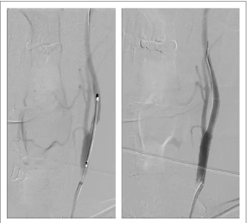
Medical records including cardiovascular parame- ters for the whole procedure and the number size of ischaemic lesions were complete for 115 patients (55 female, 60 male). The average age was 67.3 years (range 47 to 87 years). In 45 cases the procedure Figure 1. Digital subtraction angiography image of a 52-year-old male patient who received stent implantation on the left side.
was performed on the right side and in 70 cases on the left side.
Additional patients were excluded because of miss- ing values at the moment of dilatation. In 29 cases the exact time of dilatation was not recorded. In 86 cases the medical records of cardiovascular parameters at the moment of stent implantation and the number and size of ischaemic lesions was complete (44 female, 42 of male). The average age was 66.5 (range 47 to 87 years). Dilatation was performed on the right side in 38 cases and on the left side in 38 cases.
Statistical analysis
Data were analyzed by Microsoft Office ProPlus 365 Excel Analysis ToolPak and IBM SPSS Statistics Version 25 software. In total, 26 parameters were col- lected from anaesthesiologists’ records and six param- eters were collected from MR scans.
Non-parametric statistical tests were used since we hypothesized that the number and size of ischaemic lesions are not normally distributed. The following sta- tistical tests, which do not require normal distribution of the statistical groups, were used: Spearman rank Figure 3. Diffusion-weighted magnetic resonance image of a 72-year-old male patient after stent implantation on the left side.
Supratentorial ischaemic lesions are easily identifiable (blue arrows).
Figure 2. Cardiovascular parameters in an anaesthesiologist’s record. Systolic and diastolic BP values (black arrows), heart rate (orange arrow) and the moment of balloon dilatation (red arrow) were recorded. Parameters are shown in timeline (white arrow) by the record.
correlation, Kendall rank correlation, Welch test, Pearson rank correlation. p 0.05 was considered as statistically significant. Data were presented as mean standard deviation.
Results
Results of the entire procedure period
Values of the cardiovascular parameters. In total, 115 patients’ cardiovascular parameters were measured and averaged from the medical records of anaesthesi- ologists. The average time for the whole procedure was 5519 min (the shortest was 35 min, the longest 140 min). Hypotension was recorded in 26 cases, with the average time being 1719 min (the shortest lasted for 35 min, the longest 40 min); the lowest value was 75/
35 mmHg. Systolic values were over 140 mmHg in 100 cases, the average time for which was 4321 min (the shortest was 3 min, the longest 140 min); the highest value was 215/115 mmHg. Systolic values were over 160 mmHg in 59 cases, the average time for which was 3122 min (the shortest was 5 min, the longest 95 min). Bradycardia occurred in 55 cases, the average time for which was 1419 min (the shortest was 1 min, the longest 95 min); the lowest heart rate was 10 bpm.
Tachycardia occurred in six cases, the average time for which was 3435 min (the shortest was 5 min, the lon- gest 100 min); the highest value was 140 bpm.
Parameters of ischaemic lesions. Eighty-four out of 115 patients developed ischaemic lesions. In total, 469 lesions were analyzed. The average size was 3.433.32 mm (APLL). In 26 cases a lesion appeared in more than one slice because of its exten- sion. Out of 469 lesions, 262 were on the left side and 207 were on the right side. In total, 462 diffusion restricted lesions were supratentorial, and seven of them were infratentorial.
Statistical results for the whole procedure period. All recorded cardiovascular parameters were correlated
with the number and size of ischaemic lesion. Only one of the registered cardiovascular parameters did not correlate significantly. Table 1 summarises the sta- tistical analysis andp-values. The presence of tachycar- dia, its period and its maximum values cannot be interpreted due to the low number of cases.
Results at the moment of stent dilatation
Values of the cardiovascular parameters. Right before dila- tation the average systolic BP was 15219 mmHg, the diastolic was 7912 mmHg and heart rate was 7515 bpm. Right after dilatation the average systolic BP was 13424 mmHg, the diastolic was 7215 mmHg and heart rate was 6122 bpm. Out of 86 cases 15 patients developed hypotension; the shortest compensation time was 5 min, the longest compensation time was 25 min and the average compensation time was 116 min. Bradycardia occurred in 37 cases; its shortest compensation time was 2 min, the longest compensa- tion time was 60 min and the average compensation time was 810 min. Cardiovascular instability occurred in 44 cases, and in eight cases both bradycar- dia and hypotension occurred.
Parameters of ischaemic lesions. Sixty-one out of 86 patients had ischaemic lesions on MR scans after the procedure. In total, 333 diffusion-restricted lesions in 61 patients were registered. Twenty-six lesions extend- ed into more than one slice because of their size. In total, 330 lesions were supratentorial and three lesions were located infratentorially; 170 lesions were on the right side and 163 were on the left side. The average dimension was 3.583.49 mm (APLL).
Statistical results at the moment of stent dilatation
There was no significant correlation between the dura- tion of the procedure (starting time of the procedure and time of the dilatation) and the examined parame- ters of the ischaemic lesions. We used Kendall rank correlation for statistical analysis for number of lesions (p¼0.3402) and their size (p¼0.4792).Figure 4. Ischaemic lesion on the left side after stent implantation in a 65-year-old female patient. Diffusion restriction was proved by apparent diffusion coefficient measurement.
Statistically, we got more reliable results when the parameters of the ischaemic lesions were correlated with systolic and diastolic BP changes during dilatation compared to the usage of correlation with systolic and diastolic BP values before and after the dilatation. The average systolic BP decrease was 18.623.95 mmHg and the diastolic BP decrease was 7.7313.83 mmHg during the dilatation. There was no significant correla- tion (Kendall rank correlation) between the attenua- tion of systolic BP and the number of lesions (p¼0.287) and their size (p¼0.4661). We also could not prove a significant difference between the attenua- tion of diastolic BP and the number (p¼0.2832) and size (p¼0.3956) of the lesions. We used the heart rate decline during the dilatation for the statistical analysis, the average value for which was 1420.58 bpm. This alteration correlated significantly with the number of lesions (p¼0.0123) and their size (p¼0.00323) by Kendall rank correlation.
The compensation time of bradycardia did not cor- relate significantly with the lesion number (p¼0.06472) and size (p¼0.2744) by Kendall rank correlation. The p-value of the number of ischaemic lesions is approaching the point of p0.05 and can indicate a connection between the two variables. The correlation between the compensation time of hypoten- sion and ischaemic lesions could not be interpreted due to the low case number and the outlier values.
Generally, BP decreased after dilatation, but in most cases it did not decrease below 100 mmHg systolic or 60 mmHg diastolic value.
Discussion
Correlation between embolization and cardiovascular instability
Carotid stent implantation is accompanied by micro- embolization and hypoperfusion, which can lead to ischaemic brain injury. In serious cases stroke may
develop. Microembolization means that small particles of the plaques are detached and can reach the brain via circulation during and after the procedure.13,14
Several studies demonstrated that ischaemic brain injury after stent implantation cannot be caused by microembolization exclusively. At the moment of dila- tation hypotonia and brain hypoperfusion caused by cardiovascular instability can increase the chances of MR detectable ischaemic brain lesion develop- ment.15–17
It is already proven that carotid stent implantation increases cardiovascular instability, and the barorecep- tor reflex plays a major role in the evolving hypotonia.
Whether hypo- or hypertension occurs, turbulent blood flow increases the mechanical effects of cardiac instability to the injured blood vessel wall. The two processes potentiate the effect of each other. Several studies have investigated haemodynamic instability emphasizing the importance of the postoperative period. On the contrary just a few studies have examined the effect of intraprocedural cardiac instabil- ity.18–20
Our study demonstrates that balloon catheter dila- tation with stent implantation and the cardiac instabil- ity during the whole procedure do not significantly increase the number of MR detectable ischaemic lesions and extension 24 hours after the procedure.
Only patients with over 160 mmHg systolic BP approached the level of significance. This suggest that extremely high BP during the procedure can have an influence on the development of ischaemic lesions.
Regarding this, further investigations are required.
On analysis of our data, we did not find any significant correlation among the size of periprocedural ischaemic brain lesions, the amount of time till dilatation, the duration of the whole intervention and the systolic and diastolic BP changes during the procedure.
Therefore, the attenuation of systolic and diastolic BP does not mean there is an additional risk factor for ischaemic brain lesions, although in extreme cases
Presence of bradycardia Welch test p¼0.8981 p¼0.1147
Period of bradycardia Kendall rank correlation p¼0.9872 p¼0.5281
Minimum value of bradycardia Kendall rank correlation p¼0.2985 p¼0.3042
Presence, period and maximum value of tachycardia Not applicable BP: blood pressure.
it is necessary to correct them to maintain the integrity of the circulation. Heart rate attenuation during the dilatation procedure led to significant correlation with the ischaemic lesions’ number and size. There was no significant correlation between the compensa- tion time of bradycardia and the number of ischaemic lesions, but thep-value of 0.065 highly suggests a con- nection between them.
Cardiovascular instability during dilatation can be explained with the baroreceptor reflex. Activation of it has negative chronotropic and dromotropic effect via the parasympathetic nervous system. It is a simple idea to prevent or treat bradycardia and hypotonia with atropine.21,22
Atropine is a parasympatholytic drug, and it can be classified as an anticholinergic agent in other aspects.
At the parasympathetic end plates, it inhibits impulse conduction competitively by decreasing the parasym- pathetic tone. Atropine 0.5–1 mg is used to prevent or treat bradycardia and hypotonia during carotid stenting.23
Several studies have investigated whether atropine significantly decreases the incidence of hypotonia and bradycardia during the procedure. Cayne et al. demon- strated that receiving atropine as prevention before dilatation significantly decreases the incidence of hypo- tonia and bradycardia after dilatation.24On the other hand, Qureshi et al. demonstrated that receiving atro- pine as prevention paradoxically significantly increased the chance of postoperative bradycardia.25 This study recommends caution when using atropine. The Stenting versus Endarterectomy for Treatment of Carotid Artery Stenosis study7 suggests a protocol of 1 mg of atropine intravenously prophylactically.
According to our study, bradycardia must be com- pensated and larger heart rate attenuation must be pre- vented or corrected as soon as possible to avoid the evolution of ischaemic lesions. We cannot conclude the same regarding BP parameters, but it is important to maintain cardiovascular integrity with proper anaes- thesia during the procedure.
Limitations and additional influencing factors of embolus formation
For the objectivity we must consider the limitations of our study. We disregarded patients’ previous clinical history. Previous symptoms (stroke or TIA), comor- bidity and risk factors (smoking, body weight, lifestyle) in particular might be important. We documented the grade of stenosis and we also performed plaque analy- sis before the procedure with the help of the previously obtained CT angiography. Although these aspects are important and influence the extension of developed ischaemic lesions, previous studies have already exam- ined them. Therefore, we did not include these param- eters in the statistical tests.
Biasi GM et al. investigated whether preprocedural plaque analysis can provide more precise risk analysis
for periprocedural stroke.26 This study showed that hypoechogenic, soft plaques are associated with higher risk for stroke, and therefore they predispose to stroke. Additional studies indicate correlations among atherosclerotic plaque structure, plaque rupture and consecutive ischaemic brain lesions. Plaque morphology is one of the most important factors in post-procedural stroke, but this has already been proven.10,27
The structure of the plaque is not just significant in embolization, but it also has an important role in the development of cardiovascular instability. Gupta et al.
demonstrated that calcified plaques predispose individ- uals to cardiovascular instability.20 Owing to their stable structures, the force of dilatation mediates more effectively to the baroreceptors, thus activating the baroreceptor reflex. Several studies proved that the incidence of cardiovascular instability is strongly cor- related with the location of the stenosis. Stent implan- tation close to the carotid sinus significantly increased cardiovascular instability compared to more distant sites.20
Currently, there is no generally accepted guideline for proper stent selection. We implanted open Wallstents; therefore, the type of stent has no effect on our data. Csobay-Nova´k et al. proposed recommen- dations for the usage of different stent types.28 They have also demonstrated that cardiovascular instability occurs more frequently with implantation of shorter stents. It can be explained that shorter stents have stronger radial force to the narrowed blood vessel wall. Stronger radial force means greater transmural force stimulating the carotid system’s own receptors, thus increasing cardiovascular instability. Similarly, a significant correlation has been proved between circu- latory fluctuation and the grade of dilatation (differ- ence in the lumen diameters before and after the procedure).21,29–31
As outlined above, it is obvious that many studies have already examined carotid stenting, but several aspects of this procedure are still unclear. In our study we aimed to focus on some of those unanswered questions, and we should consider broadening our treatise by measuring other parameters.
An additional limitation of our study was that we did not investigate patients’ medication. According to other studies, this is an important parameter regarding the prevention and therapy of bradycardia.
Medications during anaesthesia are included in the anaesthesiologists’ record. We have been analysing them, and it will be the subject of our next study.
Tools which can prevent distal embolization are still an undiscovered area. Several physicians use these tools, but they cannot give full protection, espe- cially against prolonged embolization, and their inser- tion can lead to embolism.32,33We did not use embolic protection devises in any of the procedures because of the additional risk. Thus, we could not investigate their effect.
should be prevented or ceased quickly to save brain tissue. Most of the lesions are asymptomatic but can still cause the death of millions of neurons, which cannot be disregarded.
Despite our results, stent implantation can still be considered safe since intraoperative bradycardia is pre- ventable or treatable by proper anaesthesia. According to our results cardiovascular instability and post- procedural ischaemic lesions can accompany stent implantation. These effects are a technicality of the intervention and should be considered at surgical plan- ning, but they do not mean additional risk.
Conflict of interest
The authors declared no potential conflicts of interest with respect to the research, authorship, and/or publication of this article.
Funding
The author(s) received no financial support for the research, authorship, and/or publication of this article.
ORCID iD
Sa´ndor Csizmadia https://orcid.org/0000-0002-2153-4293
References
1. Robbins SL. Basic pathology. 9th ed. Amsterdam:
Elsevier, 2013, pp. 335–344.
2. Petty GW, Brown RD Jr, Whisnant JP, et al. Ischemic stroke subtypes: a population-based study of incidence and risk factors.Stroke1999; 30: 2513–2516.
3. Massara M, Notarstefano S, Gerardi P, et al. Unstable atherosclerotic plaque in the common carotid artery:
Diagnosis and treatment strategy. Semin Vasc Surg 2018; 31: 88–90.
4. Roffi M, Yadav JS. Carotid stenting. Circulation 2006;
114: 1–5.
5. Moran CJ, Cross DT, Derdeyn CP. Techniques of carot- id angioplasty and stenting. Neuroimaging Clin N Am 2007; 17: 337–353.
6. Gerard P, Stansby SM.Practical carotid artery stenting.
Berlin: Springer, 2009, pp. 143–158.
7. Caputo R, et al. Stenting versus endarterectomy for treatment of carotid-artery stenosis. N Engl J Med 2010; 363: 1663–1674.
8. Andani R, Khan YS. Anatomy, head and neck, carotid sinus. StatPearls Publishing, https://www.ncbi.nlm.nih.
Stroke2014; 45: 146–151.
12. Doig D. et al. Carotid anatomy does not predict the risk of new ischaemic brain lesions on diffusion-weighted imaging after carotid artery stenting in the ICSS-MRI substudy.Eur J Vasc Endovasc Surg2016; 51: 14–20.
13. Gossetti B, Gattuso R, Irace L, et al. Embolism to the brain during carotid stenting and surgery.Acta Chir Belg 2007; 107: 151–154.
14. Orlandi G, Fanucchi S, Fioretti C, et al. Characteristics of cerebral microembolism during carotid stenting and angioplasty alone.Arch Neurol2001; 58: 1410–1413.
15. Leisch F, Kerschner K, Hofmann R, et al. Carotid sinus reactions during carotid artery stenting: Predictors, inci- dence, and influence on clinical outcome. Catheter Cardiovasc Interv2003; 58: 516–523.
16. Pappada` G, Beghi E, Marina R, et al. Hemodynamic instability after extracranial carotid stenting. Acta Neurochirurgica2006; 148: 639–645.
17. Caplan LR, Ka SW, Gao S, et al. Is hypoperfusion an important cause of strokes? If so, how?Cerebrovasc Dis 2006; 21: 145–153.
18. Kojuri J, Ostovan MA, Zamiri N, et al. Hemodynamic instability following carotid artery stenting. Neurosurg Focus2011; 30: 6–12.
19. Altinbas A, Algra A, Brown MM, et al. Effects of carotid endarterectomy or stenting on hemodynamic complica- tions in the International Carotid Stenting Study: A ran- domized comparison.Int J Stroke2014; 9: 284–290.
20. Gupta R, Abou-Chebl A, Bajzer CT, et al. Rate, predic- tors, and consequences of hemodynamic depression after carotid artery stenting. J Am Coll Cardiol 2006; 47:
1538–1543.
21. Lavoie P, Rutledge J, Dawoud MA, et al. Predictors and timing of hypotension and bradycardia after carotid artery stenting. AJNR Am J Neuroradiol 2008; 29:
1942–1947.
22. Mlekusch W, Schillinger M, Sabeti S, et al. Hypotension and bradycardia after elective carotid stenting: frequency and risk factors.J Endovasc Ther2003; 10: 851–859.
23. Katzung BG, Masters SB, Trevor AJ. Basic & clinical pharmacology. 12th ed. New York: McGraw-Hill Education, 2012, pp. 115–124.
24. Cayne NS, Faries PL, Trocciola SM, et al. Carotid angioplasty and stent-induced bradycardia and hypoten- sion: Impact of prophylactic atropine administration and prior carotid endarterectomy. J Vasc Surg 2005; 41:
956–961.
25. Qureshi IA, Luft AR, Sharma M, et al. Frequency and determinants of postprocedural hemodynamic instability after carotid angioplasty and stenting.Stroke1999; 30:
2086–2093.
26. Biasi GM, Froio A, Diethrich EB, et al. Carotid plaque echolucency increases the risk of stroke in carotid stent- ing: The imaging in carotid angioplasty and risk of stroke (ICAROS) study.Circulation2004; 110: 756–762.
27. Redgrave JNE, Lovett JK, Gallagher PJ, et al.
Histological assessment of 526 symptomatic carotid pla- ques in relation to the nature and timing of ischemic symptoms: The Oxford plaque study. Circulation 2006;
113: 2320–2328.
28. Csobay-Nova´k C, Ba´ra´ny T, Zima E, et al. Role of stent selection in the incidence of persisting hemodynamic depression after carotid artery stenting. J Endovasc Ther2015; 22: 122–129.
29. Ahn SH, Prince EA, Dubel GJ. Carotid artery stenting:
Review of technique and update of recent literature.
Semin Intervent Radiol2013; 30: 288–296.
30. Mu¨ller-Hu¨lsbeck S, Sch€afer PJ, Charalambous N, et al.
Comparison of carotid stents: An in-vitro experiment focusing on stent design. J Endovasc Ther 2009; 16:
168–177.
31. Ackerstaff RGA, et al. Prediction of early cerebral out- come by transcranial Doppler monitoring in carotid bifurcation angioplasty and stenting. J Vasc Surg2005;
41: 618–624.
32. Chen CI, Iguchi Y, Garami Z, et al. Analysis of emboli during carotid stenting with distal protection device.
Cerebrovasc Dis2006; 21: 223–228.
33. Stojanov D, Ilic M, Bosnjakovic P, et al. New ischemic brain lesions on diffusion-weighted MRI after carotid artery stenting with filter protection: Frequency and relationship with plaque morphology. AJNR Am J Neuroradiol2012; 33: 708–714.