III./1.1. Classification of cerebrovascular diseases
(Stroke 1990;21:637-676)
a.) Asymptomatic cerebrovascular disorder (CVD)
b.) Cerebral dysfunction with global or focal neurological signs (= stroke) c.) Vascular dementia
d.) Hypertensive encephalopathy
Hypertensive encephalopathy is an acute condition associated with extremely high (e.g.
260/140 mmHg) blood pressure values. At such arterial pressure values, autoregulation is no longer functioning, and cerebral perfusion increases with increasing blood pressure. Cerebral edema is responsible for the clinical signs.
Vascular dementia is the impairment of cognitive performance due to disturbed cerebral circulation rather than a degenerative process. Vascular dementia progresses stepwise, in contrast to degenerative dementias – such as Alzheimer’s disease – where progression is slow and continuous. The pathological background of cognitive decline may be multiple lacunar infarcts or single large infarcts at so called “strategic locations”.
Definition of stroke:
Appears as a consequence of an acute disturbance of blood flow of the brain Associated with neurological signs (focal signs, disturbance of consciousness) Signs last longer than 24 hours or the patient dies within 24 hours
Transient ischemic attack (TIA) is a clinically distinct condition where clinical signs resolve within 24 hours
The definition of TIA is currently changing: clinical signs resolve within 1 hour and no permanent tissue damage is detectable with neuroimaging. If clinical signs resolve but tissue damage is present, the term transient signs with infarction (TSI) is used
Stroke types:
Ischemic stroke (85%)
territorial (in the distribution territory of large arteries, Fig. 1) thrombotic (local atherosclerotic plaque)
embolic (embolus = plug)
border zone (in the watershed areas of large vessels, Fig. 1) hemodynamic causes
Fig. 1: Territorial and border zone infarcts
small vessel disease lacunar infarct (Fig. 2)
Fig 2: Lacunar infarcts
Hemorrhagic stroke (15%)
intracerebral hemorrhage (bleeding within the brain tissue, Fig. 3) subarachnoid hemorrhage (bleeding into the subarachnoid space surrounding the brain, Fig. 4)
Fig. 3: Intracerebral hemorrhage
Fig. 4: Subarachnoid hemorrhage
In addition to the arterial disturbances of cerebral blood, venous disturbance of cerebral blood may also occur. This is called cerebral venous sinus thrombosis. However, the term “stroke” is used usually to denote cerebral blood flow disturbance on the arterial side.
Classifications of ischemic stroke
Three classifications of ischemic stroke are mentioned here.
A./ In Central-Europe, a classification based on arterial supply territories is traditionally used with diagnoses in Latin such as “Insufficientia circulationis in territorio arteriae cerebri mediae sinistrae” in Hungarian hospital discharge reports.
The following two classifications are used most commonly in other regions:
B./ The British Oxfordshire Community Stroke Project (OCSP) determined lesion size and location based on the clinical signs observed during physical examination, and set up four categories with prognostic significance.
C./ The North American TOAST classification was initially used in a clinical trial (the TOAST-trial). This classification differentiates five groups based on etiology.
A. Classification based on blood supply territories Large vessels (carotid, vertebrobasilar) Arteries of the Willis-circle
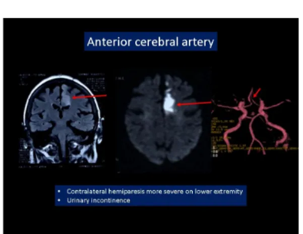
Anterior cerebral artery (ACA, Fig. 5)
weakness is more severe in the lower extremity urinary incontinence
Fig 5: Cerebral infarct in the territory of the anterior cerebral artery
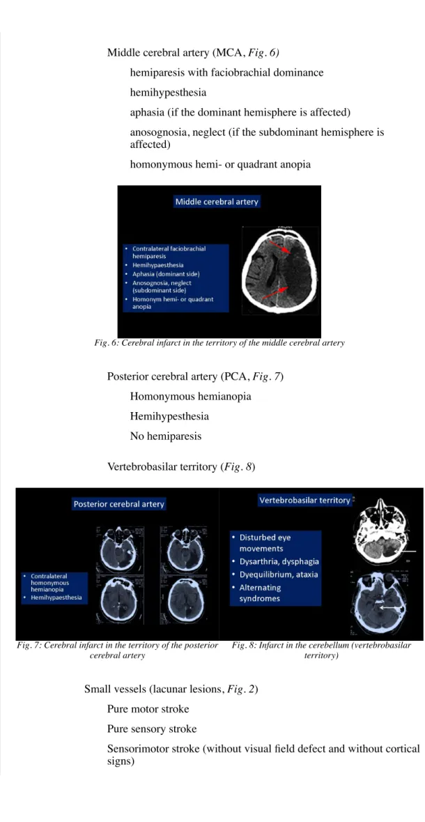
Middle cerebral artery (MCA, Fig. 6)
hemiparesis with faciobrachial dominance hemihypesthesia
aphasia (if the dominant hemisphere is affected) anosognosia, neglect (if the subdominant hemisphere is affected)
homonymous hemi- or quadrant anopia
Fig. 6: Cerebral infarct in the territory of the middle cerebral artery
Posterior cerebral artery (PCA, Fig. 7) Homonymous hemianopia Hemihypesthesia
No hemiparesis
Vertebrobasilar territory (Fig. 8)
Fig. 7: Cerebral infarct in the territory of the posterior cerebral artery
Fig. 8: Infarct in the cerebellum (vertebrobasilar territory)
Small vessels (lacunar lesions, Fig. 2) Pure motor stroke
Pure sensory stroke
Sensorimotor stroke (without visual field defect and without cortical signs)
Ataxic hemiparesis
Dysarthria-clumsy hand syndrome Brainstem alternating syndromes
Important alternating syndromes:
1. Hemiparesis alternans oculomotoria (Weber syndrome, mesencephalon)
2. Hemiparesis alternans facialis (Millard-Gubler syndrome, pons)
3. Hemiparesis alternans hypoglossa (Déjerine syndrome, medulla)
4. Alternating sensory syndrome (Wallenberg syndrome, dorsolateral medullary syndrome, no paresis)
B. Classification based on lesion location – clinical signs – prognosis:
Oxfordshire Community Stroke Project (Bamford-classification) Bamford et al (Fig. 9)
TACI (total anterior circulation infarct): hemiparesis, hemianopia AND cortical signs (aphasia, apraxia)
PACI (partial anterior circulation infarct): fewer signs than in TACI POCI (posterior circulation infarct): brainstem and cerebellar signs, hemianopia
LACI (lacunar infarct): pure motor stroke, pure sensory stroke, sensorimotor stroke, ataxic hemiparesis, dysarthria-clumsy hand syndrome. No visual field defect, no cortical signs.
Fig. 9: OCSP classification with CT examples
C. Classification based on etiology: the TOAST-criteria (Salerno, S. M. et. al.
Ann Intern Med 1996;124:21-26) Large vessel (atherothrombotic) Small vessel disease (lacunar) Cardioembolic
Other known etiology Unknown etiology


