TDDFT-ECD and DFT-NMR studies of thaigranatins A – E and granatumin L isolated from Xylocarpus granatum †
Attila M´andi, aJun Wu*band Tibor Kurt´an *a
TDDFT-ECD calculations were utilized to explain the mirror image or different ECD spectra of previously reported homochiral natural products thaigranatins A–E and granatumin L, the simple comparison of which would result in a wrong stereochemical conclusion. The configurational assignment was confirmed independently and geometrical parameters of the chromophores governing the ECD spectra were identified in the structurally related natural products by analyzing the ECD spectra and geometries of the low-energy computed conformers obtained by different methods. Different conformations of the furan-2-yl-d-lactone subunit were found responsible for the mirror image ECD spectra of the homochiral thaigranatins C–E. Two DFT13C NMR chemical shift calculation methods and DP4+ analysis were performed on the C-6 epimers of thaigranatin D, which together with the ECD calculation, could determine the absolute configuration of C-6 as (R).
Introduction
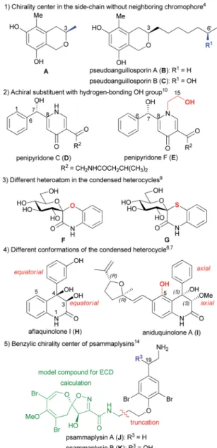
Due to the low sample amount requirement and efficient reproduction of the experimental electronic circular dichroism (ECD) spectra by time-dependent density functional theory ECD (TDDFT-ECD) calculations, ECD is considered the most widely applied microscale chiroptical method for the congurational assignment of natural products (NPs) containing chromo- phores with absorbance in the UV-vis spectral range.1–3 For structurally related NPs, the simple comparison of experimental ECD spectra is oen utilized to determine the absolute cong- uration (AC) if it was determined for one of the derivatives. For instance, the C-3 AC of pseudoanguillosporin B (C), an iso- chroman derivative with a 6-hydroxyhept-1-yl side-chain at C-3, was determined by comparing the ECD spectrum with that of the co-isolated pseudoanguillosporin A (B) containing only a C- 3 heptyl substituent (Fig. 1).4The remote C-60chirality center of Cwas far from the isochroman chromophore and it had prac- tically no contribution to the ECD prole, which justied the comparison of the ECD spectra of B and C. The absolute conguration ofBwas conrmed by the solution TDDFT-ECD calculation approach applied for the truncated model compound A, containing only a C-3 methyl group.4 Although chiroptical data of homochiral related derivatives are oen
similar, a subtle change in the planar structure or substitution pattern may result in rather different5–8or nearly opposite ECD spectra or optical rotation.9–13 TDDFT-ECD calculations of structurally related NPs having markedly different ECD spectra were found useful to interpret the differences.
For instance, the pyridine alkaloids penipyridones C (D) and F (E) had the same chromophoric system and absolute cong- uration butEhad an additionalN-2-hydroxyethyl group, which resulted in near mirror image ECD spectra and oppositely signed specic rotation (Fig. 1).10TDDFT-ECD calculations and analysis of the low-energy conformers revealed that in the presence of the achiralN-2-hydroxyethyl substituent, the addi- tional 15-OH group formed an intramolecular hydrogen bond with the 7-OH, which changed the relative orientation of the phenyl and g-pyridone chromophores.10 The replacement of oxygen heteroatom with sulfur in the benzene-condensed spiro derivatives F and G containing benzo[1,4]oxazin-3-one and benzo[1,4]thiazin-3-one chromophores resulted in signicantly different ECD spectra with mirror image transitions in the high- wavelength region, although the computed conformers had very similar geometries (Fig. 1).9
Aaquinolone I (H) and aniduquinolone A (I) shared a very similar substituted 4-aryl-3,4-dihydroquinolin-2(1H)-one chro- mophore but in the presence of the 5-OH group in the peri position ofI, the C-4 phenyl group adopted an axial orientation, while inH, having a hydrogen atom in theperiposition, the C-4 aryl group preferred the equatorial orientation in all the low- energy computed conformers (Fig. 1).6,7 Due to the different conformations of the condensed heterocycles, the ECD spectra ofHandIwere fairly different and comparison of ECDs could not be used to determine the AC ofH.7Psammaplysins A (J) and
aDepartment of Organic Chemistry, University of Debrecen, P. O. Box 400, 4002 Debrecen, Hungary. E-mail: kurtan.tibor@science.unideb.hu; Fax: +36-52-512-744
bSchool of Pharmaceutical Sciences, Southern Medical University, 1838 Guangzhou Avenue North, Guangzhou 510515, P. R. China. E-mail: wwujun2003@yahoo.com
†Electronic supplementary information (ESI) available. See DOI:
10.1039/d0ra03725g
Cite this:RSC Adv., 2020,10, 32216
Received 25th April 2020 Accepted 20th August 2020 DOI: 10.1039/d0ra03725g rsc.li/rsc-advances
PAPER
Open Access Article. Published on 01 September 2020. Downloaded on 9/1/2020 1:59:47 PM. This article is licensed under a Creative Commons Attribution-NonCommercial 3.0 Unported Licence.
View Article Online
View Journal | View Issue
B (K),rst representatives of the psammaplysin alkaloid family, differed in the additional C-19 benzylic hydroxyl group of K, which implied a C-19 chirality center in the side-chain of K (Fig. 1).14Regardless this benzylic chirality center, the experi- mental ECD spectra ofJandKwere found near identical, which conrmed that the side-chain with an aryl ether chromophore had negligible contribution to the ECD spectra. Thus a trun- cated model compound could be utilized for the ECD calcula- tions of J, which reduced the number of conformers and enabled the congurational assignment of J.14 The above examples may justify to explore the structural and chiroptical background for the different ECD proles of structurally related derivatives by ECD calculations, which would allow us avoiding wrong congurational assignments for analogous derivatives.
Thaigranatins A–E (1–5), limonoid derivatives containing a pentacyclic skeleton with a furan-3-yl substituent at thed-
synthetic interest.
The AC of thaigranatin A (1) was elucidated by single crystal X-ray analysis, while those of2and6were determined on the basis of their similar ECD spectra to that of thaigranatin A (1).16 The experimental solution ECD spectra of1and6were quite similar, while that of2 had a broad positive ECD band with a maximum at 217 nm, which were missing from those of1and 6. The ECD spectra of3–5were signicantly different from the others, which did not enable to utilize the simple comparison of the ECD spectra for the congurational assignment. Moreover, the ECD spectrum of5was found near mirror image of those of 3and4, which is quite puzzling considering their analogous structure with minor structural differences (Fig. 3). The abso- lute congurations of3–5 were proposed on the basis of the common biosynthetic origin with thaigranatin A (1).16In this
Fig. 1 Examples on the comparison of ECD spectra of structurally related derivatives.
Fig. 2 Structures of thaigranatins A–E (1–5) and granatumin L (6) isolated fromXylocarpus granatum.16
Open Access Article. Published on 01 September 2020. Downloaded on 9/1/2020 1:59:47 PM. This article is licensed under a Creative Commons Attribution-NonCommercial 3.0 Unported Licence.
work, we utilized TDDFT-ECD calculations of1–6with different combinations of methods to explain the ECD spectral differ- ences of 1–6 and the mirror image ECD spectra of3–5. This approach was tested whether it is suitable to prove the proposed homochirality of1–6and thus it can provide an independent congurational assignment for3–5. Furthermore, the AC of C-6 in the side-chain of4remained undetermined.16In this work we applied DFT-NMR 13C chemical shi calculations of the C-6 epimers to elucidate the AC of this chirality center.27–29
Semi-synthetic derivatives obtained by the derivatization of granatumin L (6) showed potent human immunodeciency virus 1 (HIV-1) and inuenza A virus (IAV) inhibitory activities, which may also justify to explore relationship between stereo- chemistry and characteristic ECD features and to determine solution geometries and conrm the absolute conguration.16
Results and discussion
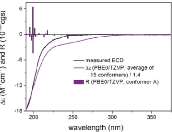
Thaigranatin A (1) had a saturatedd-lactone chromophore with a C-17 furan-3-yl substituent, ana,b-unsaturated ester moiety (C-32 to C-35), an isolated ester (C-7) and a double bond (D8,30) chromophore. In the ECD spectrum of1, an intense negative ECD band could be observed below 200 nm with aat negative plateau up to 270 nm. Since the absolute conguration was determined by single crystal X-ray diffraction analysis,16 the ECD calculation of1was utilized to validate the applied DFT/
TDDFT methods. The initial 29 Merck Molecular Force Field (MMFF) conformers of (1R,2S,3R,4S,5S,6R,9S,10R,13R,14S,17R)- 1were re-optimized at the B3LYP/6-31+G(d,p), the CAM-B3LYP/
TZVP30,31PCM/MeCN and the SOGGA11-X/TZVP32,33SMD/MeCN
levels, separately. ECD spectra computed at various levels for the low-energy conformers over 1% Boltzmann population (15, 15 and 17 conformers, respectively) reproduced well the exper- imental ECD spectrum (Fig. 4). Since the CAM-B3LYP/TZVP PCM/MeCN and the SOGGA11-X/TZVP SMD/MeCN levels gave almost the same ECD results, only the rst two levels were applied for the other derivatives in the DFT re-optimization step.
In all the CAM-B3LYP/TZVP PCM/MeCN conformers of1, the C-17 furan-3-yl group adopted anequatorialarrangement with syn coplanarorientation of the 17-H and C-21 atoms (u17-H,C-17,C-
20,C-21 ¼ 10.2) and the 14-H had axial orientation. The d- lactone ring had a distorted half-chair conformation with the C- 13 being out of plane, while the otherve atoms located in nearly one plane. The geometry of the low-energy conformers differed in the relative arrangements of the C-5 and C-3 substituents by rotation along the C-6 to C-7 and C-32 to C-33 sigma bonds (Fig. 5). In the lowest-energy conformer (conf A), the C-32 carbonyl oxygen wassyn coplanarwith the C-33 methyl Fig. 3 Comparison of the experimental ECD spectra of3–5in MeCN. Fig. 4 Experimental ECD spectrum of1in MeCN compared with the Boltzmann-weighted PBE0/TZVP PCM/MeCN ECD spectrum of (1R,2S,3R,4S,5S,6R,9S,10R,13R,14S,17R)-1. Level of optimization: CAM- B3LYP/TZVP PCM/MeCN. Bars represent the rotatory strength values of the lowest-energy conformer. Experimental ECD spectrum of1was reproduced with permission from ref. 16.
Fig. 5 Overlapped geometries and selected torsional angles of the six lowest-energy conformers of (1R,2S,3R,4S,5S,6R,9S,10R,13R,14S,17R)- 1. Level of optimization: CAM-B3LYP/TZVP PCM/MeCN.
Open Access Article. Published on 01 September 2020. Downloaded on 9/1/2020 1:59:47 PM. This article is licensed under a Creative Commons Attribution-NonCommercial 3.0 Unported Licence.
and D, conformers C and F hadsyn coplanararrangement of the C-32 carbonyl oxygen (s-transenone) and the C-33 methyl group but the C-5 hadgaucheorientation with the C-7 methoxy oxygen (uC-5,C-6,C-7,]O¼ 137.4). The positive CEs of conformers A, C, D, F were cancelled by the strong negative CEs of conformers B and E, in which the C-32 carbonyl oxygen wasanti periplanar with the C-33 methyl group (uO],C-32,C-33,C-36 ¼ 178.4, s-cis enone). Thus the rotation along the C-32 to C-33 bond of the a,b-unsaturated ester moiety and around the C-6 to C-7 bond was mostly responsible for the differences in the ECD spectra of the low-energy conformers of1.
Compared to compound1,2did not contain the conjugating D33,34double bond and the C-6 chirality center in the other side chain and the experimental ECD spectrum had a strong nega- tive CE below 210 nm and a weaker positive CE at 217 nm with a positive plateau up to 260 nm. Compound 2had 43 initial MMFF conformers, the gas-phase and solvent model re- optimizations of which yielded 6 and 10 low-energy conformers over 1% Boltzmann-population. Therstve low- energy CAM-B3LYP/TZVP PCM/MeCN conformers had differ- ences in the orientation of the C-3 ester group and the furan ring while the lactone ring showed only marginal differences (Fig. 7). The individual computed ECDs of these conformers were similar but that of conformer E showed a signicant red shidue to the opposite orientation of the furan ring (Fig. 8 and S2†). This red shi of the minor conformers obviously contributes to the high-wavelength positive shoulder of the experimental ECD spectrum. Similarly to the conformers of1,
thed-lactone ring had a distorted half-chair conformation in the low-energy conformers. The Boltzmann-weighted ECD spectra computed at all the applied combinations reproduced well the experimental spectrum including the positive CE and shoulder
(Fig. 9), which conrmed the
(1R,2S,3R,4S,5S,9S,10R,13R,14S,17R) absolute conguration of 2and hence the homochirality with1.16The difference in the experimental ECD spectra of1and2can be attributed to the absence of theD33,34double bond and the C-6 chirality center.
Compound3contained the same C-3a,b-unsaturated ester moiety as 1but it missed the C-6 chirality center due to the exchange of the 6-OH group with a hydrogen atom. Moreover, theD8,30double bond of1was shied toD8,14position in3and C-30 became a chirality center due to the presence of thesec- hydroxyl group. Besides the intense negative CE below 211 nm, the experimental ECD spectrum of 3 had a positive CE at
Fig. 6 Experimental ECD spectrum of1in MeCN compared with the PBE0/TZVP PCM/MeCN ECD spectra of the individual six lowest- energy CAM-B3LYP/TZVP PCM/MeCN conformers of (1R,2S,3R,4S,5S,6R,9S,10R,13R,14S,17R)-1.
Fig. 7 Overlapped geometries of thefive lowest-energy CAM-B3LYP/
TZVP PCM/MeCN conformers of (1R,2S,3R,4S,5S,9S,10R,13R,14S,17R)- 2.
Fig. 8 Experimental ECD spectrum of2in MeCN compared with the B3LYP/TZVP PCM/MeCN ECD spectra of the individualfive lowest- energy CAM-B3LYP/TZVP PCM/MeCN conformers of (1R,2S,3R,4S,5S,9S,10R,13R,14S,17R)-2. Experimental ECD spectrum of 2was reproduced with permission from ref. 16.
Open Access Article. Published on 01 September 2020. Downloaded on 9/1/2020 1:59:47 PM. This article is licensed under a Creative Commons Attribution-NonCommercial 3.0 Unported Licence.
220 nm accompanied with a at positive shoulder having maximum at 286 nm.
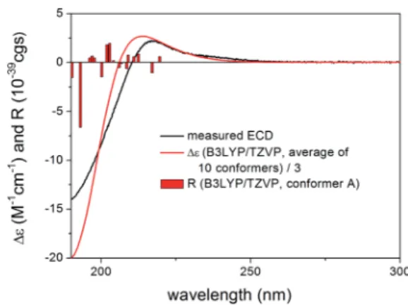
Re-optimization of the initial 51 MMFF conformers of (1R,2S,3R,4S,5S,9S,10R,13R,17R,30R)-3 resulted in 15 and 24 low-energy conformers over 1% Boltzmann-population in gas- phase and solvent model calculations, respectively. In therst
ve low-energy CAM-B3LYP/TZVP PCM/MeCN conformers, there were only minor difference in orientations of the two side- chains and the lactone ring (Fig. 10). Similarly to the lowest- energy conformer of 1, the C-32 carbonyl oxygen of conformers A–E was syn coplanarwith the C-33 methyl group (uO],C-32,C-33,C-36 ¼ 34.8–37.8 range for confs A–E, s-trans enone) and the d-lactone ring had a distorted half-chair conformation even with the sp2-hybridized C-14. For therst
ve low-energy CAM-B3LYP/TZVP PCM/MeCN conformers, the computed ECD spectra had similar prole for the main transi- tions (Fig. 11). The different ECD proles of compounds1and3 derived from the different distributions of conformers differing in the C-3 side-chain conformation.
Boltzmann-weighted ECD spectra obtained at all combina- tions of the applied levels reproduced well the two major tran- sitions at 220 and 199 nm while underestimated the positive
shoulder at 286 nm (Fig. 12). Reproduction of the major tran- sitions allowed verication of the absolute conguration of3as (1R,2S,3R,4S,5S,9S,10R,13R,17R,30R).
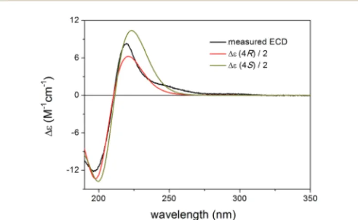
Similarly to compound1,4had a chirality center at C-6 and the only difference from1was the position of theD8,14double bond. The absolute conguration of C-6 has not been deter- mined independently in the original paper16 thus we used TDDFT-ECD and13C NMR DFT calculations of the C-6 epimers to assign this chirality center.
Although chiroptical methods are usually applied for the elucidation of absolute conguration and conformation by exploiting the mirror image ECD prole of the enantiomers, there are reports where they were utilized successfully to distinguish diastereomers or more than two stereoisomers.34–38 MMFF conformational search of the epimeric (1R,2S,3R,4S,5S,6R,9S,10R,13R,17R)-4 and (1R,2S,3R,4S,5S,6S,9S,10R,13R,17R)-4 produced 39 and 120 conformers, respectively, in a 21 kJ mol1 energy window indicating considerable exibility difference between the two Fig. 9 Experimental ECD spectrum of2in MeCN compared with the
Boltzmann-weighted B3LYP/TZVP PCM/MeCN ECD spectrum of (1R,2S,3R,4S,5S,9S,10R,13R,14S,17R)-2. Level of optimization: CAM- B3LYP/TZVP PCM/MeCN. Bars represent the rotatory strength values of the lowest-energy conformer.
Fig. 10 Overlapped geometries of the five lowest-energy CAM-
B3LYP/TZVP PCM/MeCN conformers of
(1R,2S,3R,4S,5S,9S,10R,13R,17R,30R)-3(left: conformers A and D; right:
conformers B, C and E).
Fig. 11 Experimental ECD spectrum of3in MeCN compared with the B3LYP/TZVP PCM/MeCN ECD spectra of the individualfive lowest- energy CAM-B3LYP/TZVP PCM/MeCN conformers of (1R,2S,3R,4S,5S,9S,10R,13R,17R,30R)-3.
Fig. 12 Experimental ECD spectrum of3in MeCN compared with the Boltzmann-weighted B3LYP/TZVP PCM/MeCN ECD spectrum of (1R,2S,3R,4S,5S,9S,10R,13R,17R,30R)-3. Level of optimization: CAM- B3LYP/TZVP PCM/MeCN. Bars represent the rotatory strength values of the lowest-energy conformer.
Open Access Article. Published on 01 September 2020. Downloaded on 9/1/2020 1:59:47 PM. This article is licensed under a Creative Commons Attribution-NonCommercial 3.0 Unported Licence.
and thus DFT C NMR calculation was utilized to distinguish the epimers.
The DFT-NMR methods were validatedrst on compounds 1–3 and 6. The widely applied mPW1PW91/6-311+G(2d,p)//
B3LYP/6-31+G(d,p) level27,39was tested on1–3and6giving good agreement for most carbons (see ESI Tables S1–S4†). Then13C chemical shis were computed for the epimeric (1R,2S,3R,4S,5S,6R,9S,10R,13R,17R)-4 and (1R,2S,3R,4S,5S,6S,9S,10R,13R,17R)-4. The corrected mean absolute error (CMAE)40values were found rather similar (1.80 vs.1.89) and the (6R) epimer appeared to be only slightly better in average (see Table S5†). A strong indication could be observed, however, for the C-5 carbon adjacent to the C-6 chirality center with more than 3 ppm difference suggesting (6R) conguration. Application of the DP4+ statistical method41,42gave a 95.23% condence for the (6R) epimer which is above the empirical limit suggested by the developers42of the method for a solid determination of relative conguration.
In order to conrm further this result, the mPW1PW91/6- 311+G(2d,p) SMD/CHCl3//mPW1PW91/6-311+G(2d,p) SMD/
CHCl3 level40 was also tested on 1. This method including solvent model both at the DFT optimization and the NMR calculation step was not recommended by the CHESHIRE database,43 since it was developed from a very limited test
Table S7†). The DP4+ analysis showed a 99.81% condence for the (6R) epimer. The two13C chemical shiDFT-NMR calcula- tions produced the same conclusion for the AC of C-6 and together with the TDDFT-ECD calculations allowed elucidating
the absolute conguration of 4 as
(1R,2S,3R,4S,5S,6R,9S,10R,13R,17R).
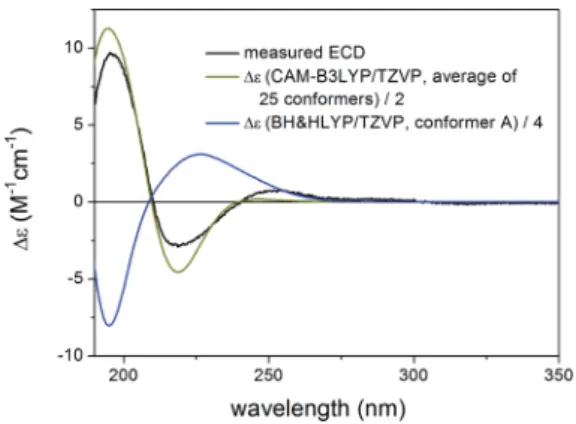
The structure of compound5contained aD8,9double bond instead of theD8,14double bond of3, while their rings A–C had identical substitution pattern. Despite their closely related structures, compound5had oppositely signed CEs to the cor- responding ones of 3 and 4, which resulted in near mirror image experimental ECD spectra. The geometrical background for the mirror image ECD spectra of these homochiral deriva- tives was explored by TDDFT-ECD calculations.
Re-optimization of the 120 MMFF conformers of (1R,2S,3R,4S,5S,10S,13R,14R,17R,30R)-5yielded 19 and 25 low- energy conformers over 1% Boltzmann-population in the gas- phase and the solvent model calculations, respectively. In all the low-energy CAM-B3LYP/TZVP PCM/MeCN conformers, the d-lactone ring adopted a distorted boat conformation with C-15 and C-17 being out of plane (Fig. 14). This conformation of the d-lactone ring was different from the preferred half-chair conformation of 1–4, and it was the reason of the mirror image ECD curves. The ECD spectra computed for the low- energy CAM-B3LYP/TZVP PCM/MeCN conformers reproduced well the experimental ECD curve of5(Fig. 15 and S10†), which conrmed that the geometries of the conformers were predicted properly. Interestingly, the B3LYP/6-31+G(d,p) gas-phase re- optimization provided different low-energy conformers, in
Fig. 13 Experimental ECD spectrum of4in MeCN compared with the Boltzmann-weighted B3LYP/TZVP PCM/MeCN ECD spectrum of (1R,2S,3R,4S,5S,6R,9S,10R,13R,17R)-4(average of 23 conformers) and the Boltzmann-weighted CAM-B3LYP/TZVP PCM/MeCN ECD spec- trum of (1R,2S,3R,4S,5S,6S,9S,10R,13R,17R)-4 (average of 28
conformers). Level of optimization: CAM-B3LYP/TZVP PCM/MeCN. Fig. 14 Comparison of the lowest-energy CAM-B3LYP/TZVP PCM/
MeCN (12.7%) and B3LYP/6-31+G(d,p) gas-phase (10.8%) conformers of (1R,2S,3R,4S,5S,10S,13R,14R,17R,30R)-5.
Open Access Article. Published on 01 September 2020. Downloaded on 9/1/2020 1:59:47 PM. This article is licensed under a Creative Commons Attribution-NonCommercial 3.0 Unported Licence.
which the d-lactone ring had the half-chair conformation (Fig. 14) and the computed ECD gave mirror image agreement with the two high-energy ECD transitions (Fig. 15 and S11†).
This result conrmed that the shiof the position of the double bond in 3 and 5 (from D8,14 to D8,9) changed the preferred conformation of the d-lactone ring from half-chair to boat, which is manifested in near mirror ECD spectra, while the corresponding chirality centers had identical absolute conguration.
This is an instructive example how chiroptical properties of structurally related compounds can differ and why it is impor- tant to apply more than one functional in both the DFT opti- mization and the ECD calculation steps.1,2,45,46 The solvent model results reproduced the experimental ECD spectrum allowing verication of the homochiral nature of5with the AC of (1R,2S,3R,4S,5S,10S,13R,14R,17R,30R) despite its nearly mirror image experimental ECD.
Compared to the structure of 1, compound 6 does not contain the C-6 chirality center and since this center does not have signicant contribution to the ECD spectrum, the
experimental ECD spectra of 1 and 6 were very similar. The
initial 34 MMFF conformers of
(1R,2S,3R,4S,5S,9S,10R,13R,14S,17R)-6were re-optimized at the B3LYP/6-31+G(d,p) and the CAM-B3LYP/TZVP PCM/MeCN levels resulting in 4 and 12 low-energy conformers, respec- tively. Thed-lactone ring adopted half-chair conformation in all the low-energy CAM-B3LYP/TZVP PCM/MeCN conformers of6, while the a,b-unsaturated ester moiety had s-transconforma- tion in conformers A and C and s-cisin conformers B and D (Fig. 16). The computed ECD spectra of conformers A and C had a weak positive CE at 220 nm, which did not match the exper- imental curve (Fig. 17). However, the intense negative computed ECD bands of conformers B and D below 225 nm overrode the positive CE of conformers A and C.14The negative CEs of the experimental ECD spectra below 220 nm are mainly determined by the contribution of the higher-energy conformers B and D containing an s-cisenone chromophore.
All combinations of the applied levels reproduced nicely the experimental ECD spectrum (Fig. 18) allowing verication of the Fig. 15 Experimental ECD spectrum of5in MeCN compared with the
Boltzmann-weighted CAM-B3LYP/TZVP PCM/MeCN ECD spectrum of (1R,2S,3R,4S,5S,10S,13R,14R,17R,30R)-5 (level of optimization:
CAM-B3LYP/TZVP PCM/MeCN) and the lowest-energy BH&HLYP/
TZVP ECD spectrum of (1R,2S,3R,4S,5S,10S,13R,14R,17R,30R)-5[level of optimization: B3LYP/6-31+G(d,p)].
Fig. 16 Overlapped geometries and selected torsional angle of the
four lowest-energy conformers of
(1R,2S,3R,4S,5S,9S,10R,13R,14S,17R)-6. Level of optimization: CAM- B3LYP/TZVP PCM/MeCN.
Fig. 17 Experimental ECD spectrum of6in MeCN compared with the BH&HLYP/TZVP PCM/MeCN ECD spectra of the individual four lowest-energy CAM-B3LYP/TZVP PCM/MeCN conformers of (1R,2S,3R,4S,5S,9S,10R,13R,14S,17R)-6. Experimental ECD spectrum of 6was reproduced with permission from ref. 16.
Fig. 18 Experimental ECD spectrum of6in MeCN compared with the Boltzmann-weighted BH&HLYP/TZVP PCM/MeCN ECD spectrum of (1R,2S,3R,4S,5S,9S,10R,13R,14S,17R)-6. Level of optimization: CAM- B3LYP/TZVP PCM/MeCN. Bars represent the rotatory strength values of the lowest-energy conformer.
Open Access Article. Published on 01 September 2020. Downloaded on 9/1/2020 1:59:47 PM. This article is licensed under a Creative Commons Attribution-NonCommercial 3.0 Unported Licence.
spectra compared to those of 3 and 4, the low-energy conformers of which had half-chair conformation for the d- lactone ring. The different conformations were induced by different positions of the double bond (D8,14orD8,9) in3–5. The s-cisor s-transconformation of the planara,b-unsaturated ester chromophore was found responsible for spectral differences in 1–4 and 6, while the C-5 ester substituent and C-6 chirality center did not have signicant contribution. The (6R) AC of thaigranatin D (4) could be determined by the13C NMR chem- ical shicalculations of the C-6 epimers by two methods. The results may contribute to understand better the geometrical parameters governing the ECD spectra of these limonoids and thus to use ECD more efficiently for their stereochemical analysis.
Experimental
General information
Isolation and characterization of1–6was described in ref. 15 and 16. ECD spectra were measured on a Jasco J-810 spec- tropolarimeter (JASCO Corporation, Tokyo, Japan) in MeCN.
Computational section
Mixed torsional/low-frequency mode conformational searches were carried out by means of the Macromodel 10.8.011 soware by using the Merck Molecular Force Field (MMFF) with an implicit solvent model for CHCl3.47Geometry reoptimizations were carried out at the B3LYP/6-31+G(d,p) levelin vacuo, the CAM-B3LYP/TZVP level with the PCM solvent model for MeCN, the mPW1PW91/6-311+G(2d,p)39 SMD/CHCl3 and the SOGGA11-X/TZVP SMD/MeCN levels. TDDFT-ECD calculations were run with various functionals (B3LYP, BH&HLYP, CAM- B3LYP, and PBE0) and the TZVP basis set as implemented in the Gaussian 09 package with the same or no solvent model as in the preceding DFT optimization step.48 ECD spectra were generated as sums of Gaussians with 3000–6000 cm1width at half-height, using dipole-velocity-computed rotational strength values.49NMR calculations were performed at the mPW1PW91/
6-311+G(2d,p) and the mPW1PW91/6-311+G(2d,p) SMD/CHCl3 levels on the B3LYP/6-31+G(d,p) and the mPW1PW91/6- 311+G(2d,p) SMD/CHCl3 levels, respectively. Computed NMR shidata were corrected withI¼185.4855 andS¼ 1.0306 in the gas-phase27andI¼187.9864 andS¼ 1.0358 in the solvent model calculations.40Boltzmann distributions were estimated from the B3LYP, CAM-B3LYP, mPW1PW91 and SOGGA11-X
GINOP-2.3.2-15-2016-00008. T. K. thanks the National Research, Development and Innovation Office (K120181) and A. M. the J´anos Bolyai Research Scholarship of the Hungarian Academy of Sciences and the ´UNKP-19-4 New National Excellence Program of the Ministry for Innovation and Technology. The Govern- mental Information-Technology Development Agency (KIF¨U) is acknowledged for CPU time.
Notes and references
1 S. Superchi, P. Scafato, M. G´orecki and G. Pescitelli,Curr.
Med. Chem., 2018,25, 287–320.
2 A. M´andi and T. Kurt´an,Nat. Prod. Rep., 2019,36, 889–918.
3 L. Grauso, R. Teta, G. Esposito, M. Menna and A. Mangoni, Nat. Prod. Rep., 2019,36, 1005–1030.
4 I. Kock, S. Draeger, B. Schulz, B. Els¨asser, T. Kurt´an, A.´ Ken´ez, S. Antus, G. Pescitelli, P. Salvadori, J. B. Speakman, J. Rheinheimer and K. Krohn,Eur. J. Org.
Chem., 2009,2009, 1427–1434.
5 P. Moosmann, R. Ueoka, L. Grauso, A. Mangoni, B. I. Morinaka, M. Gugger and J. Piel, Angew. Chem., Int.
Ed., 2017,56, 4987–4990.
6 C. Y. An, X. M. Li, H. Luo, C. S. Li, M. H. Wang, G. M. Xu and B. G. Wang,J. Nat. Prod., 2013,76, 1896–1901.
7 D. H. El-Kashef, G. Daletos, M. Plenker, R. Hartmann, A. M´andi, T. Kurt´an, H. Weber, W. Lin, E. Ancheeva and P. Proksch,J. Nat. Prod., 2019,82, 2460–2469.
8 J. E. Rode, M. G´orecki, S. Witkowski and J. Frelek, Phys.
Chem. Chem. Phys., 2018,20, 22525–22536.
9 S. Kun, N. K´anya, N. Gal´o, A. P´ahi, A. M´andi, T. Kurt´an, P. Makleit, Sz. Veres, ´A. Sipos, T. Docsa and L. Soms´ak,J.
Agric. Food Chem., 2019,67, 6884–6891.
10 H. Zhou, L. Li, C. Wu, T. Kurt´an, A. M´andi, Y. Liu, Q. Gu, T. Zhu, P. Guo and D. Li,J. Nat. Prod., 2016,79, 1783–1790.
11 S. McLean,Can. J. Chem., 1964,42, 191–195.
12 W. Klyne and W. M. Stokes,J. Chem. Soc., 1954, 1979–1988.
13 Y. Z. Sun, T. Kurt´an, A. M´andi, H. Tang, Y. Chou, K. Soong, L. Su, P. Sun, C. L. Zhuang and W. Zhang,J. Nat. Prod., 2017, 80, 2930–2940.
14 A. M´andi, I. W. Mudianta, T. Kurt´an and M. J. Garson,J. Nat.
Prod., 2015,78, 2051–2056.
15 M. Y. Li, Q. Xiao, T. Satyanandamurty and J. Wu, Chem.
Biodiversity, 2014,11, 262–275.
16 J. L. Ren, X. P. Zou, W. S. Li, L. Shen and J. Wu,Mar. Drugs, 2018,16, 434.
Open Access Article. Published on 01 September 2020. Downloaded on 9/1/2020 1:59:47 PM. This article is licensed under a Creative Commons Attribution-NonCommercial 3.0 Unported Licence.
17 R. X. Liu, Q. Liao, L. Shen and J. Wu,Fitoterapia, 2018,131, 96–104.
18 Y. B. Wu, Y. Z. Wang, Z. Y. Ni, X. Qing, Q. W. Shi, F. Sauriol, C. J. Vavricka, Y. C. Gu and H. Kiyota,J. Nat. Prod., 2017,80, 2547–2550.
19 M. Liao, P. Pedpradab and J. Wu,Phytochem. Lett., 2017,19, 126–131.
20 Z. F. Zhou, T. Kurt´an, A. M´andi, Y. C. Gu, L. G. Yao, G. R. Xin, X. W. Li and Y. W. Guo,Sci. Rep., 2016,6, 33908.
21 J. Li, M. Y. Li, G. Feng, J. Zhang, M. Karonen, J. Sinkkonen, T. Satyanandamurty and J. Wu, J. Nat. Prod., 2012, 75, 1277–1283.
22 J. Li, M. Y. Li, T. Satyanandamurty and J. Wu,Helv. Chim.
Acta, 2011,94, 1651–1656.
23 I. A. Najmuldeen, A. H. A. Hadi, K. Awang, K. Mohamad, K. A. Ketuly, M. R. Mukhtar, S. L. Chong, G. Chan, M. A. Naah, N. S. Weng, O. Shirota, T. Hosoya, A. E. Nugroho and H. Morita,J. Nat. Prod., 2011,74, 1313–
1317.
24 K. Awang, C. S. Lim, K. Mohamad, H. Morita, Y. Hirasawa, K. Takeya, O. Thoison and A. H. A. Hadi, Bioorg. Med.
Chem., 2007,15, 5997–6002.
25 S. Vardhan and S. K. Sahoo,Comput. Biol. Med., 2020,124, 103936.
26 S. Fu and B. Liu,Org. Chem. Front., 2020,7, 1903–1947.
27 M. W. Lodewyk, M. R. Siebert and D. J. Tantillo,Chem. Rev., 2012,112, 1839–1862.
28 Y. M. Ren, C. Q. Ke, A. M´andi, T. Kurt´an, C. Tang, S. Yao and Y. Ye,Tetrahedron, 2017,73, 3213–3219.
29 M. Kics´ak, A. M´andi, Sz. Varga, M. Herczeg, Gy. Batta, A. B´enyei, A. Borb´as and P. Herczegh,Org. Biomol. Chem., 2018,16, 393–401.
30 T. Yanai, D. Tew and N. Handy,Chem. Phys. Lett., 2004,393, 51–57.
31 G. Pescitelli, L. di Bari and N. Berova,Chem. Soc. Rev., 2011, 40, 4603–4625.
32 R. Peverati and D. G. Truhlar, J. Chem. Phys., 2011, 135, 191102.
33 ´E. Br´emond, M. Savarese, N. Q. Su, ´A. J. P´erez-Jim´enez, X. Xu, J. C. Sancho-Garc´ıa and C. Adamo,J. Chem. Theory Comput., 2016,12, 459–465.
34 E. Ancheeva, L. K¨uppers, S. H. Akone, W. Ebrahim, Z. Liu, A. M´andi, T. Kurt´an, W. Lin, R. Orfali, N. Rehberg, R. Kalscheuer, G. Daletos and P. Proksch, Eur. J. Org.
Chem., 2017,2017, 3256–3264.
35 Y. Liu, T. Kurt´an, A. M´andi, H. Weber, C. Wang, R. Hartmann, W. Lin, G. Daletos and P. Proksch, Tetrahedron Lett., 2018,59, 632–636.
36 P. Zhang, L. H. Meng, A. M´andi, T. Kurt´an, X. M. Li, Y. Liu, X. Li, C. S. Li and B. G. Wang,Eur. J. Org. Chem., 2014,2014, 4029–4036.
37 L. F. Liang, T. Kurt´an, A. M´andi, L. G. Yao, J. Li, L. F. Lan and Y. W. Guo,Tetrahedron, 2018,74, 1933–1941.
38 J. L. Johnson, D. S. Nair, S. M. Pillai, D. Johnson, Z. Kallingathodi, I. Ibnusaud and P. L. Polavarapu, ACS Omega, 2019,4, 6154–6164.
39 C. Adamo and V. Barone,J. Chem. Phys., 1998,108, 664–675.
40 S. Qiu, E. de Gussem, K. A. Tehrani, S. Sergeyev, P. Bultinck and W. Herrebout,J. Med. Chem., 2013,56, 8903–8914.
41 S. G. Smith and J. M. Goodman,J. Am. Chem. Soc., 2010,132, 12946–12959.
42 N. Grimblat, M. M. Zanardi and A. M. Sarotti,J. Org. Chem., 2015,80, 12526–12534.
43 CHESHIRE CCAT,The Chemical ShiRepository for computed NMR scaling factors, http://cheshirenmr.info/index.htm.
44 N. M. Tran-Cong, A. M´andi, T. Kurt´an, W. E. G. M¨uller, R. Kalscheuer, W. Lin, Z. Liu and P. Proksch, RSC Adv., 2019,9, 27279–27288.
45 P. Sun, D. X. Xu, A. M´andi, T. Kurt´an, T. J. Li, B. Schulz and W. Zhang,J. Org. Chem., 2013,78, 7030–7047.
46 V. F. Ximenes, N. H. Morgon and A. R. de Souza,Chirality, 2018,30, 1049–1053.
47 MacroModel. Schr¨odinger LLC, 2015. http://
www.schrodinger.com/MacroModel.
48 M. J. Frisch, G. W. Trucks, H. B. Schlegel, G. E. Scuseria, M. A. Robb, J. R. Cheeseman, G. Scalmani, V. Barone, B. Mennucci, G. A. Petersson, H. Nakatsuji, M. Caricato, X. Li, H. P. Hratchian, A. F. Izmaylov, J. Bloino, G. Zheng, J. L. Sonnenberg, M. Hada, M. Ehara, K. Toyota, R. Fukuda, J. Hasegawa, M. Ishida, T. Nakajima, Y. Honda, O. Kitao, H. Nakai, T. Vreven, J. A. Montgomery Jr, J. E. Peralta, F. Ogliaro, M. Bearpark, J. J. Heyd, E. Brothers, K. N. Kudin, V. N. Staroverov, R. Kobayashi, J. Normand, K. Raghavachari, A. Rendell, J. C. Burant, S. S. Iyengar, J. Tomasi, M. Cossi, N. Rega, J. M. Millam, M. Klene, J. E. Knox, J. B. Cross, V. Bakken, C. Adamo, J. Jaramillo, R. Gomperts, R. E. Stratmann, O. Yazyev, A. J. Austin, R. Cammi, C. Pomelli, J. W. Ochterski, R. L. Martin, K. Morokuma, V. G. Zakrzewski, G. A. Voth, P. Salvador, J. J. Dannenberg, S. Dapprich, A. D. Daniels, O. Farkas, J. B. Foresman, J. V. Ortiz, J. Cioslowski and¨ D. J. Fox, Gaussian 09 (Revision E.01), Gaussian, Inc., Wallingford, CT, 2013.
49 P. J. Stephens and N. Harada,Chirality, 2010,22, 229–233.
50 U. Varetto, MOLEKEL 5.4, Swiss National Supercomputing Centre, Manno, Switzerland, 2009.
Open Access Article. Published on 01 September 2020. Downloaded on 9/1/2020 1:59:47 PM. This article is licensed under a Creative Commons Attribution-NonCommercial 3.0 Unported Licence.