Human Electrophysiological and Pharmacological Properties of XEN-D0101: A Novel Atrial-Selective
Kv1.5/I Kur Inhibitor
John Ford, PhD,* James Milnes, PhD,* Erich Wettwer, PhD, † Torsten Christ, MD, † Marc Rogers, PhD,*
Kathy Sutton, PhD,* David Madge, PhD,* Laszlo Virag, PhD, ‡ Norbert Jost, PhD, ‡ § Zoltan Horvath, PhD,§ Klaus Matschke, MD,¶ Andras Varro, MD, ‡ § and Ursula Ravens, MD †
Abstract: The human electrophysiological and pharmacological properties of XEN-D0101 were evaluated to assess its usefulness for treating atrialfibrillation (AF). XEN-D0101 inhibited Kv1.5 with an IC50of 241 nM and is selective over non-target cardiac ion channels (IC50Kv4.3, 4.2mM; hERG, 13mM; activated Nav1.5,.100mM;
inactivated Nav1.5, 34mM; Kir3.1/3.4, 17mM; Kir2.1,..100mM).
In atrial myocytes from patients in sinus rhythm (SR) and chronic AF, XEN-D0101 inhibited non-inactivating outward currents (Ilate) with IC50 of 410 and 280 nM, respectively, and peak outward currents (Ipeak) with IC50of 806 and 240 nM, respectively. Whereas Ilate is mainly composed of IKur, Ipeakconsists of IKurand Ito. Therefore, the effects on Itoalone were estimated from a double-pulse protocol where IKurwas inactivated (3.5mM IC50 in SR and 1mM in AF). Thus, inhibition of Ipeakis because of IKurreduction and not Ito. XEN-D0101 significantly prolonged the atrial action potential duration at 20%, 50%, and 90% of repolarization (AF tissue only) and significantly elevated the atrial action potential plateau phase and increased con- tractility (SR and AF tissues) while having no effect on human ven- tricular action potentials. In healthy volunteers, XEN-D0101 did not significantly increase baseline- and placebo-adjusted QTc up to a max-
imum oral dose of 300 mg. XEN-D0101 is a Kv1.5/IKurinhibitor with an attractive atrial-selective profile.
Key Words:XEN-D0101, atrialfibrillation, Kv1.5, IKur (J Cardiovasc PharmacolÔ2013;61:408–415)
INTRODUCTION
Atrialfibrillation (AF) contributes to overall cardiovas- cular morbidity and mortality and occurs in 1%–2% of the general population. Patients with AF have a 5-fold increased risk for stroke, and approximately one-fifth of all strokes can be attributed to AF.1
AF can be triggered by ectopic activity, single circuit reentry, or multiple circuit reentry.2 Current strategies to control AF involve either sinus rhythm (SR) maintenance or heart rate control. AF termination and SR maintenance can be achieved by inhibiting K+ channels responsible for atrial repolarization. Although SR control may be the pre- ferred and most effective treatment of AF, none of the SR control drugs currently available are able to maintain SR without significant side effect risk.3 Furthermore, the cur- rently used class 3 antiarrhythmic agents lack atrial specific- ity, producing highly undesirable proarrhythmic liability in the ventricles that can lead totorsade de pointesand sudden cardiac death.4Thus, there is a clear unmet medical need for new pharmacological AF therapies with improved efficacy and safety.5
A putative atrial-selective drug target is Kv1.5, which underlies the cardiac ultrarapidly activating outward potas- sium current (IKur) in humans6–9 and importantly displays atrial-specific expression.10–12 Inhibition of IKur extends the repolarization phase of the atrial cardiac action potential in AF patients13to provide desirable antiarrhythmic effects with- out unfavorably affecting the ventricles. Selective Kv1.5/IKur
inhibitors thus represent a potentially safer pharmacological intervention strategy.14In addition to antiarrhythmic proper- ties, Kv1.5/IKurinhibitors may also improve atrial contractility and thereby reduce thromboembolic risk.15
Herein we report that XEN-D0101 alters action poten- tials recorded from human atrial tissue, but not human ventricular tissue, and that this is because of the selective Kv1.5/IKur modulation and possibly Kv4.3/Itomodulation at higher test concentrations. These observations reinforce the
Received for publication August 3, 2012; accepted January 11, 2013.
From the *Xention Ltd, Iconix Park, Pampisford, Cambridge, United Kingdom;
†Department of Pharmacology and Toxicology, Medical Faculty Carl Gustav Carus, Dresden University of Technology, Dresden, Germany;
T. Christ is now with the Institut für Experimentelle Pharmakologie und Toxikologie, Universitätsklinikum Hamburg-Eppendorf, Martinistrasse 52, 20246 Hamburg;‡Department of Pharmacology and Pharmacotherapy;
and §Division of Cardiovascular Pharmacology, Hungarian Academy of Sciences, University of Szeged, Szeged, Hungary; and ¶Department of Cardiac Surgery, Heart Center Dresden, Dresden University of Technology, Dresden, Germany.
Supported by Xention Ltd, the National Development Agency (TÁMOP-4.2.2/
B-10/1-2010-0012; the National Office for Research and Technology— National Technology Programmes (NKFP_07_01—RYT07_AF and REG-DA-09-2-2009-0115); EU FP7-Health-2010-single-stage "EU- TRAF" (European Network for Translational Research in Atrial Fibrilla- tion), #261057; the HU-RO Cross Border Cooperation Programmes (HURO/0802/011_AF-HURO_CARDIOPOL); and by the Hungarian Scientific Research Fund (OTKA K-82079 and OTKA CNK-77855).
J. Ford, D. Madge, K. Sutton, M. Rogers, and J. Milnes hold options in Xention Ltd. U. Ravens and E. Wettwer both consult to Xention Ltd.
Supplemental digital content is available for this article. Direct URL citations appear in the printed text and are provided in the HTML and PDF versions of this article on the journal’s Web site (www.jcvp.org).
Reprints: John Ford, PhD, Xention Ltd, Iconix Park, London Road, Pampisford, Cambridge CB22 3EG, United Kingdom (e-mail: john.ford@xention.com).
Copyright © 2013 by Lippincott Williams & Wilkins
408
J Cardiovasc Pharmacolänotion that selective Kv1.5/IKur inhibitors represent a new class of atrial-selective antiarrhythmic agents.
METHODS
For in vitro studies, stock solutions of XEN-D0101 (a thienopyrimidine) formulated in dimethylsulfoxide (DMSO) were frozen and stored as aliquots at approximately 2208C until use. Serial dilution of stock solutions in DMSO were performed in glass vials before dilution in experimental solution to achieve the desiredfinal perfusion concentrations, with a vehicle concentration of typically 0.1% vol/vol DMSO.
Studies reported here conform to the principles outlined in the Declaration of Helsinki were reviewed and approved by relevant Ethics Committees, and human subjects gave written informed consent. Atrial trabecular muscle from the right atrium appendage was received from SR and AF patients undergoing open heart surgery for coronary artery or valve disease (ethical no. 15.1/01031/006/2008 and EK790799). Human ventricular tissue was obtained from general organ donors whose hearts were explanted to obtain pulmonary and aortic valves for transplantation (ethical no. 339/PI/10, 4991-0/2010-1018EKU).
Preparations from patients previously receiving antiarrhythmic drugs were excluded. Additional donor information is provided inSupplemental Digital Content 3(seeSupplementary Meth- ods, http://links.lww.com/JCVP/A107).
Cloned Cardiac Ion Channel
Electrophysiology (Performed by Xention)
Cell lines stably expressing human Kv1.5, Kv4.3, or Nav1.5 in CHO cells and hERG, Kir3.1/3.4, or Kir2.1 in HEK293 cells were maintained in media containing 10% fetal calf serum and appropriate selection antibiotic. Cells were grown either in suspension or in T-flasks and routinely passaged. Cells for patch clamping experiments were plated onto glass cover slips before use. Cells for automated patch clamping experiments (Nav1.5) were freshly prepared on each experimental day. Standard gigaseal whole-cell patch clamp techniques were performed at room temperature using glass pipettes (2–4 MV). Experimental solutions are given in Supplemental Digital Content 3(seeSupplementary Methods, http://links.lww.com/JCVP/A107). Patch clamp amplifier (EPC) and Pulse software from HEKA Electronics (Chester, Nova Scotia, Canada) were used. Series resistance was compen- sated by greater than 70%. Standard voltage protocols for Kv1.5 (VHold 280 mV, 0 mV/900 ms, 240 mV/100 ms, 0.2 Hz), Kv4.3 (VHold 280 mV, +60 mV/1 s, 0.1 Hz), Kir2.1 (VHold260 mV,2110 mV/400 ms, 0.2 Hz), Kir3.1/3.4 (VHold280 mV,2140 mV/100 ms, ramp +60 mV/500 ms,280 mV, 0.1 Hz), and hERG (VHold280 mV, +20 mV/5 s,240 mV/5 s, 0.067 Hz) were employed. For Nav1.5, a 4- pulse voltage clamp protocol was used to elicit current after whole-cell access was achieved using a QPatch16. From a holding potential of290 mV, an initial test pulse to210 mV (10 ms) was applied to elicit an inward Na+current (P1).
Further test pulses to 210 mV (all 10 ms duration) were applied after conditioning steps to either 290 mV (P2) for 500 ms to relieve channel inactivation or 5 seconds to270
mV to promote inactivation (P3). Finally, the membrane was repolarized back to290 mV for 100 ms before afinal test pulse to210 mV was delivered to measure recovery from inactivated block (P4). The command voltage was applied every 15 sec- onds. After a stabilization period, a cumulative concentration response experiment was performed. Peak amplitude of the inward Nav1.5 current of P2 and P3 was measured at the end of each drug application period.
Isolation of Myocytes and Recording of I
peakand I
lateOutward Currents From SR and AF Human Atrial Tissue at 37
8C (Performed by Dresden University of Technology)
SeeSupplemental Digital Content 3(seeSupplementary Methods, http://links.lww.com/JCVP/A107) for atrial myocyte isolation procedures. Experiments were performed at physio- logical temperature (378C). Myocytes held at 260 mV were subject to a double-pulse protocol (0.2 Hz) to +50 mV (500 ms each) separated by an intermediate 25 ms step to 260 mV.
Peak outward currents (Ipeak) of thefirst test pulse were ana- lyzed to determine mixed effects on Itoand IKur. Late outward current mainly represented effects on slowly inactivating IKur. The area under the curve of the initial 50 ms of the second clamp step is a measure of charge transfer and was analyzed as indicator of Ito.
In Vitro Human Action Potential Studies at 378C (Performed by Dresden University of Technology and University of Szeged)
Tissue preparations were stimulated at 1 Hz with a 2-ms square wave stimulus. Measured parameters were resting membrane potential (RMP), action potential amplitude (APA), action potential duration at 20%, 50%, 75%, and 90% repolarization (APD20, APD50, APD75, and APD90), maximum depolarization rate, dV/dtmax, conduction time (ct), and“plateau potential”(PLT) defined as the mean mem- brane potential between 20% and 30% of APD (PLT20).
In Vitro Human Atrial Contractility Studies (Performed by Dresden University
of Technology)
The study included 14 preparations from 8 patients in SR group, and 9 preparations came from 5 patients with chronic AF. Preparations had a mean length 4.660.3 mm and a diam- eter of 1.460.1 mm (n = 23). Resting tension of each prep- aration was adjusted to yield half maximum active force development. The trabeculae were mounted in an organ bath that contained 50 mL of oxygenated Tyrodes solution circulat- ing via the gas stream. The composition of the bath solution was 126.7 mM NaCl, 0.42 mM NaH2PO4, 22 mM NaHCO3, 5.4 mM KCl, 1.8 mM CaCl2, and 1.5 mM MgCl2, pH 7.4, when equilibrated with 5% CO2 in O2 at 37 6 0.38C.
The preparations were stimulated regularly at a frequency of 1 Hz. After a stabilization period of 60–90 minutes, XEN- D0101 was added in cumulatively increasing concentrations allowing at least 15 minutes for the effect to develop before the next increment in concentration. At the end of each
experiment, the preparations were exposed to 8 mM CaCl2for assessment of maximum force development.
QTc Evaluation in Healthy Volunteers (Performed by Xention)
A double-blind, randomized, placebo-controlled phase 1 study was performed to determine safety and tolerability of XEN-D0101 and to carefully examine the drug plasma concentration–QTc relationship. Healthy male volunteers were recruited into the study, and volunteers were specifically excluded if they had a clinically significant electrocardio- graphic (ECG) abnormality, cardiac arrhythmia, heart dis- ease, hypokalemia, hyperkalemia, structural heart disease, bradycardia, myocardial channelopathy, or mean QTc (Fri- dericia correction) greater than 450 ms. Subjects remained resident in the Clinical Pharmacology Unit throughout the study period. Six groups of 8 subjects (fasted overnight) took a single dose of XEN-D0101 or placebo. At each dose level, 6 subjects were randomized to receive active treatment and 2 subjects received placebo. Dosages investigated were 15, 30, 45, 75, 150, and 300 mg XEN-D0101. Blood samples for assay of XEN-D0101 were taken before and frequently up to 48 hours after each single dose. Assay of plasma
concentrations of XEN-D0101 was performed using a high- performance liquid chromatography, tandem mass spectros- copy method. The lower limit of quantification for the assay was 0.5 ng/mL. Safety ECG telemetry (1-lead ECG) was monitored continuously from dosing until 24 hours afterward, using a SpaceLabs system to monitor heart function and detect proarrhythmic events in real time. To accurately detect drug-related changes in the QTc interval, continuous 12-lead ECGs were recorded using SEER MC recorders (GE Health- care) with Mason–Likar electrodes. A continuous 12-lead ECG was recorded for 14 hours, starting at 1 hour before dosing and finishing at 13 hours after dosing (and at corre- sponding times on Day 1). Recordings were also made on Day 2 (23237 hours after dosing). ECG analysis was per- formed on approximately 30,000 ECGs using a semiautomatic method with sufficient precision and reproducibility as described elsewhere.16 As ECGs were measured automati- cally, 20% of the measured ECGs were selected (either ran- domly or targeting the extreme QT- and RR-interval (QT, RR) measurements of each participant) and reviewed by an experienced and medically qualified observer. The QT inter- val was corrected for heart rate using individual regression equations. In each subject, the regression model that provided FIGURE 1. Effect of a range of con-
centrations of XEN-D0101 on cloned cardiac ion channel expressed in mammalian cells. A, Representative steady state Kv1.5 current traces in control and after exposure to XEN- D0101. Current inhibition at the end of the depolarizing step to 0 mV was 77% for this cell. B, Kv4.3 current traces in the absence and presence of 3 mM XEN-D0101. Peak Kv4.3 current inhibition for the cell shown was 43.7%. C, Mean Kv1.5 and Kv4.3 current inhibition (6SEM) data are shown plotted as a function of XEN-D0101 concentration and fitted with a sigmoidal function (constrained at 0% and 100%) yielding a Kv1.5 IC50 of 241 nM [95% confidence interval (CI), 152–
383 nM, n = 6–12 cells at each concentration] and a Hill coefficient (nH) of 1.36 0.2 and a Kv4.3 IC50 of 4.2 mM (95% CI, 2.8–6.4 mM, n = 5 cells at each concentration,P, 0.0001, 2-tailedttest, cf. Kv1.5) and
nHof 0.660.1. D, Mean current inhibition by XEN-D0101 is plotted as a function of concentration and fitted with a sigmoidal function (constrained at 0% and 100%) to yield a hERG IC50of 13mM (95% CI, 11.3–15.1mM, n = 4–6 cells at each concen tration,P,0.0001, 2-tailedttest, cf. Kv1.5) and an nHof 1.360.2. Effect of XEN-D0101 on Nav1.5 current was evaluated on the QPatch16 using a 4-pulse protocol described in the Methods. Mean Nav1.5 current inhibition data are shown plotted as a function of XEN-D0101 concentration yielding IC50values for Peak 3 (Nav1.5 inactivated-state block): 34.4mM (95% CI, 21.9–53.9mM,P,0.0001, 2-tailedttest, cf. Kv1.5) and nHof 1.460.2 and Peak 2 (Nav1.5 activated-state block):.100mM (n = 6 cells). Mean inhibition data for Kir3.1/3.4 are shown plotted as a function of XEN-D0101 concentration yielding IC50value of 17mM (95% CI, 12–24mM, n = 4–6 cells,P,0.0001, 2-tailedttest, cf. Kv1.5) and an nHof 0.760.1. Mean inhibition data for Kir2.1 are shown plotted as a function of XEN-D0101, nominal inhibition of current was observed over a range of concentrations up to 100mM, and no meaningful fit to these data was possible. Kv1.5 fit data are shown as a dashed line for comparison.
the lowest regression residuals was converted into an individ- ualized heart rate correction, which yielded a QT corrected for heart rate (QTc). The means of the repeated measurements on Day 1 were used to derive time-matched changes from baseline (ΔQTc). The subjects who received placebo in indi- vidual cohorts were pooled (n = 12), and their time-matched DQTc readings on days 1 and 2 were averaged at each time point to obtain aDQTc placebo profile of the study. The on- treatmentDQTc profiles were corrected for placebo to obtain DDQTc profiles for each treatment group.
Statistical Analysis
Data are typically presented as mean 6 SEM, and where appropriate, 95% confidence intervals are given.
Student t test or 1-way analysis of variance (ANOVA) with multiple measurements were used as appropriate. Differences were considered statistically significant for *P,0.05, §P, 0.01, and #P , 0.001. Concentration effect modeling was performed for QTc evaluation in healthy volunteers with ΔΔQTc intervals being regressed onto the plasma concentra- tions of XEN-D0101 at the exact times when the continuous 12-lead ECGs were recorded. The effect of XEN-D0101 plasma concentration on ΔΔQTc was investigated using a regression model and ANOVA. At each time of stable supine rest, XEN-D0101 plasma concentration was interpo- lated using nonparametric superposition (seeFigure 1, Supple- mental Digital Content 1, http://links.lww.com/JCVP/A105).
A mixed-effects model, which included plasma concentration, FIGURE 2. Effect of a range of con-
centrations of XEN-D0101 on Ipeak, Ilate, and Ito currents recorded from human atrial myocytes from SR and AF donors at 378C. A and B, Original current recordings, control and in the presence of 10mM XEN-D0101, from SR and AF are shown. Double- pulse protocol—holding potential:
260 mV, 2 voltage steps to +50 mV, duration 500 ms, intermediate pulse to 260 mV, duration 25 ms (see inset in B). Effects on Itowere quan- tified by the area under the curve (AUC) of the inactivating current of the second pulse (inset in A). C and D, Concentration-dependent effects of XEN-D0101 on peak current (Ipeak) and late current (Ilate) in SR and AF are shown. Dashed lines represent adapted Hill equations assuming a Hill slope of 1 to the mean data with IC50values for SR of 806 nM [562–1152 nM, 95% confi- dence interval (CI)] and 413 nM (308–554 nM, 95% CI) for Ipeakand Ilate, respectively, and for AF 241 nM (136–429 nM, 95% CI) and 281 nM (112–708 nM, 95% CI) for Ipeakand Ilate, respectively. E and F, Concen- tration-dependent effects of XEN- D0101 on Ito measured as AUC (in pC/pF) of inactivating current com- ponent during the initial 50 ms of second voltage step (see inset in A) for SR and AF are shown. Two alter- native curve adaptations were con- ducted assuming the maximum value = mean control, Hill coefficient
= 1, and either (1) a minimum value of 0.03 pC/pF or (2) full block (0 pC/pF). The respective IC50values were 3.5mM (2.4–5.1mM, CI) and 8.3mM (4.3–16.2mM, CI). For AF, the IC50was 1.0mM (0.56–1.9mM, CI). n = numbers (cells per patients).
Mean data6SEM.
treatment group, and interaction between plasma concentration and treatment group, asfixed factors, and subject as a random factor, was evaluated.
RESULTS
Effect of XEN-D0101 on Cloned Cardiac Ionic Channels
The pharmacological profile of XEN-D0101 against several ion channels known to be expressed and functionally important in the human heart is reported in Figure 1 (original current traces are shown inFigure 2, Supplemental Digital Content 2, http://links.lww.com/JCVP/A106). XEN-D0101 inhibited Kv1.5 (241 nM IC50) and was shown to be 17-fold selective over Kv4.3, greater than 50-fold selective over hERG, greater than 70-fold selective over Kir3.1/3.4, and greater than 100-fold selective over Nav1.5 and Kir2.1.
Although only observed at very high test concentrations, XEN-D0101 preferentially blocked inactivated-Nav1.5 over open-Nav1.5 channels.
Effect of XEN-D0101 on I
peakand I
lateCurrents Recorded From Human Atrial Myocytes
At physiological temperature, XEN-D0101’s ability to inhibit Ipeak(peak outward currents = Itoand IKur) and Ilate(late outward currents = slowly inactivating IKurand other unchar- acterized currents, also called Isus in previous studies6) was determined in human atrial myocytes from SR and AF donors.
Additionally, the charge carried during the fast inactivating current component of the second voltage step (Fig. 2A, inset) was used as surrogate to estimate the effect on Ito (mainly conducted by Kv4.3). XEN-D0101 concentration dependently reduced all current components, although with different
sub-micromolar potencies in both SR and AF atrial myocytes (Fig. 2). In SR, the IC50 for Ipeak was approximately 4-fold greater than the corresponding value in AF. Inhibition of Ilate, largely representing noninactivating IKur, is similar in SR and AF tissues (Figs. 2C, D). Inhibition of Itobased on area under the curve/charge of the inactivating peak current during the second voltage step is pronounced in SR but small and per- haps not biologically relevant in AF because of reduced contributions of Itoin AF (Figs. 2E, F). The IC50selectivity ratio of Ilateover Ito in SR tissue calculates between 8- and 20-fold depending on the assumption for IC50 determination (Fig. 2E).
Effect of XEN-D0101 on Human Cardiac Action Potentials
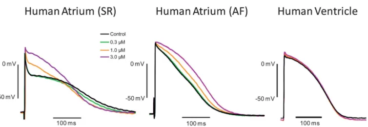
Effects on human atrial and ventricular action potentials are shown in Figure 3 and Table 1. The most striking concentration-dependent effects of 1 and 3mM XEN-D0101 in AF atrial trabeculae were robust elevation in plateau poten- tial (.16 mV) and prolongation in APD20(.47 ms), APD50
(.47 ms), and APD90 (.30 ms). In SR atrial trabeculae, XEN-D0101 also significantly elevated the plateau potential (.26 mV) but in contrast to AF preparations significantly reduced APD90 (,84 ms). Resting potential became signifi- cantly less negative (by 3 mV) in SR atrial tissue. All effects were at least partially reversible upon drug washout with the exception of APD90 shortening in SR tissue. Action potential amplitude, dV/dtmax, and conduction time were not altered.
XEN-D0101 up to 3mM did not significantly alter any action potential parameter in human ventricular tissue. Consistent with what has been reported elsewhere in the literature under control conditions, the resting membrane potential of SR and AF atrial cells was more depolarized than observed in ventricular cells and SR atrial cells were more depolarized than AF atrial cells.17
FIGURE 3. Effects of XEN-D0101 on cardiac action potentials. Example of individual tracings recorded in the presence of increasing concentrations of XEN-D0101 shown at the end of the exposure period of 20 minutes. The effects of XEN-D0101 on human action potentials were examined in right atrial trabeculae from patients in SR and AF and human ventricle at 1 Hz.
The compound concentration dependently elevated the plateau phase of the action potential in atrial tissue only. In SR, this effect was associated with shortening of APD90, whereas prolongation of APD90 was observed in AF. XEN-D0101 did not change action potential parameters recorded from human ventricular tissue. AP measurements and number (n) are provided in Table 1.
Effect of XEN-D0101 on Human Atrial Tissue Contractility
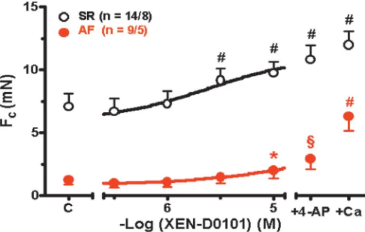
In human SR atrial tissue, XEN-D0101 significantly increased contractility at 3mM (.30%) and 10mM (.38%) but only at 10mM (.63%) in human AF atrial tissue. The force of contraction was greater in the presence of both XEN- D0101 and 3 mM 4-action potential compared with XEN- D0101 alone, but this did not match the maximum force of contraction observed in the presence of 8 mM Ca2+. These results are reported in Figure 4.
Effect of XEN-D0101 on QTc Interval and Drug-induced Proarrhythmia in Healthy Human Subjects
Comprehensive ECG recordings and ECG analysis were performed in a phase 1 clinical trial to determine whether XEN-D0101 prolongs the QTc interval in human. The quality of the ECG data from this study was high with 90% of subjects having a regression residual below 6 ms. XEN-D0101 plasma
concentration plotted againstΔΔQTc is given in Figure 5, and most notably, XEN-D0101 plasma concentrations exceeded 3000 ng/mL (;10mM drug concentration), in some volunteers without increasing ΔΔQTc. The regression coefficients for plasma concentration versus ΔΔQTc derived from the mixed- effects model were not significantly different from zero (P = 0.1990). The ANOVA analysis of the individual model com- ponents showed that XEN-D0101 plasma concentration did not significantly affect ΔΔQTc (P= 0.6719). Categorical analysis of QT and QTc was performed, and no subjects on active treatment had a QT or QTc greater than 450 ms or increase in QTc from baseline greater than 30 ms. In addition to 12-lead Holter monitoring, single-lead ECG telemetry monitoring was performed continually throughout the clinical trial. All results of ECG telemetry were considered by the investigator to be normal. There were no clinically significant changes, including induction of AF or ventricular proarrhythmia after XEN-D0101 dosing.
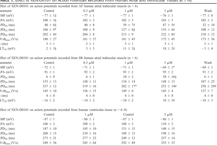
TABLE 1.Effect of XEN-D0101 on Action Potentials Recorded From Human Atrial and Ventricular Tissues at 1 Hz Effect of XEN-D0101 on action potentials recorded from AF human atrial trabecular muscle (n = 6)
Parameter Control 0.3mM 1mM 3mM Wash
RMP (mV) 27761§ 27761 27761 27661 27760
APA (mV) 10063§ 10263 10263 10363 10263
APD20(ms) 4068§ 4068 5967# 8765# 32610
APD50(ms) 10669* 10869 12768§ 15366# 108612
APD90(ms) 20268# 20468 21369 23268# 210612
dV/dtmax(V/s) 180627 181637 181645 175645 173636
ct (ms) 561 561 561 561 561
PLT20(mV) 263§ 363 1163§ 1862# 2364
Effect of XEN-D0101 on action potentials recorded from SR human atrial trabecular muscle (n = 6)
Parameter Control 0.3mM 1mM 3mM Wash
RMP (mV) 27261 27161 27161 26961* 26961
APA (mV) 9161 9362 9362 9562 9162
APD20(ms) 660 661 1062 58616§ 661
APD50(ms) 153613 148611 134614 148613 107625
APD90(ms) 337612 319616 282617* 253614# 258629#
dV/dtmax(V/s) 145610 156615 14966 14564 13767
ct (ms) 460 460 460 460 460
PLT20(mV) 21662 21462 21062 1065# 21863
Effect of XEN-D0101 on action potentials recorded from human ventricular tissue (n = 6–8)
Control 1mM Control 3mM
RMP (mV) 28761 28861 28761 28661
APA (mV) 10662 10862 10863 11062
APD25(ms) 147610 145610 131615 140615
APD50(ms) 208615 210616 188613 198616
APD90(ms) 273621 277623 249612 257614
dV/dtmax(V/s) 249636 245664 292649 255633
XEN-D0101 concentration dependently elevated the plateau phase of the action potential in SR and AF atrial trabecular muscle. In SR, this effect was associated with shortening of APD90, whereas prolongation of APD50and APD90was observed in AF. XEN-D0101 significantly increased the RMP by 3 mV in SR atrial trabecular muscle. Multiple measures ANOVA with Bonferroni postcomparison test was used for statistical analysis of drug effects versus control values for each AP parameter. Studentttest was used to compare control action potential parameters in SR and AF.
*P,0.05, §P,0.01, #P,0.001.
APA, action potential amplitude; ct, conduction time.
DISCUSSION
Ion channel electrophysiology studies performed against native and cloned cardiac ion channels demonstrated that XEN-D0101 preferentially targets Kv1.5/Ilate (ie, IKur) with moderate selectivity over Kv4.3/Ito (between 8- and 20-fold). XEN-D0101 is selective for Kv1.5 over hERG (.50-fold), Kir3.1/3.4 (.70-fold), and Nav1.5 and Kir2.1 (.100-fold). Inhibition of Ilate was demonstrated in both SR and AF atrial tissues, whereas Itowas significantly down- regulated in AF atrial tissue and therefore difficult to measure.
Comparing recent studies on IKur remodeling in AF, down- regulation of IKuris less pronounced than that of Ito.13Also in
this investigation, the late component of outward current is not significantly different from SR. There are indications that besides IKur, other currents may contribute to the late current.13
Findings reported here for human cardiac action potential studies are consistent with the ion channel pharmacology of XEN-D0101. Action potential recordings were performed with atrial tissue from patients who either were in SR or had a history of chronic AF for at least 6 months duration. Action potentials from patients in SR showed a characteristic spike and dome configuration, whereas those from patients in AF had a triangulated shape.18The triangular shape of action potentials from AF preparations is the result of electrical remodeling driven by altered ion channel expression and regulation.19 In this study, XEN-D0101 evoked changes similar to previously reported effects of 4-AP, which at concentrations selective for IKurblock elevated the plateau potential in SR and AF prepa- rations and significantly shortened APD90in SR but prolonged APD90 in AF trabecula.18However, in the presence of XEN- D0101, the reduction in APD90in SR tissue was not reversible in the washout period, unlike plateau elevation in SR and AF tissues and APD90 increase in AF tissue, suggesting that this effect may not be drug related. A possible explanation for this is that Ca2+current rundown is occurring in SR tissue, and this effect is absent or much smaller in AF tissue because ICa is already downregulated. The concentrations of XEN- D0101 required to change action potential parameters were 1 and 3mM, which is higher than the Kv1.5/IlateIC50value. The reason for this difference could relate to drug accessibility to the Kv1.5 channels being much greater in single dissociated myocytes compared with large multicellular preparations, per- haps because of permeability/diffusion problems. Bioanalysis to measure XEN-D0101 levels in cardiac tissue was not per- formed to confirm this hypothesis. Additionally, because the action potential is a multichannel event, the effect that a con- ductance change in a single-channel type has on the overall shape of the action potential depends on the relative contribu- tion to the momentary overall sum of conductances. Similar differences in potencies on current and action potential param- eters are regularly observed. Data from recombinant Kir2.1 and Kir3.1/3.4 channels suggest that the small depolarization in resting membrane potential observed in SR tissue, which is unlikely to be biologically relevant, is not because of inhibition of native IK1or IKACh.
XEN-D0101 increased contractile force in human SR and AF atrial trabeculae in a concentration-dependent man- ner. Similarly, elevation of the plateau phase of the human atrial action potential by 4-AP or via the nonselective IKurand Itoblocker AVE0118 leads to a concentration-dependent pos- itive inotropic effect.15,18The increase in force of contraction after elevation of the plateau phase has been explained by an indirect enhancement of Ca2+ influx via voltage-dependent L-type Ca2+channels18or via Na+–Ca2+exchanger operating in its reverse mode.20AF is associated with marked contrac- tile dysfunction that can be partially counteracted by 4-AP;
however, as demonstrated here, the positive inotropic effect is less robust than in SR. The positive inotropic effects of XEN- D0101 at 10mM are unlikely to be because of only Kv1.5/Ilate
inhibition due to lack of positive inotropic effects at 1 mM, FIGURE 4. Effects of XEN-D0101 on atrial tissue contractility.
Effects of XEN-D0101 and 3 mM 4-AP in addition to XEN- D0101 on force of contraction of isolated human right atrial trabeculae from patients in SR (black symbols) and AF (red symbols). At the end of each experiment, 8 mM CaCl2(+Ca) was added for the assessment of maximum force of contrac- tion. C, pre-drug control. n = Number of trabeculae/number of patients.*P,0.05, §P,0.01, #P,0.001 versus pre-drug control.
FIGURE 5. XEN-D0101 plasma concentration plotted against baseline- and placebo-adjusted QTc (ΔΔQTc) values derived from healthy volunteers (n = 63). Concentration effect mod- eling showed no evidence of a consistent relationship between plasma concentration of XEN-D0101 andΔΔQTc in healthy volunteers orally dosed with XEN-D0101.
which is known to significantly alter atrial action potential parameters and selectively inhibit Kv1.5/Ilate.
The absence of XEN-D0101–induced QTc prolonga- tion and proarrhythmia in healthy volunteers in the clinical study referenced herein is an important safety milestone that has not previously been reported for selective Kv1.5 drugs.
Indeed, it has previously been reported that a“loss of func- tion”KCNA5 polymorphism may induce susceptibility to AF in human, although in vivo causality was not demonstrated in this study.21 The ability of XEN-D0101 to induce AF was carefully evaluated in this clinical trial with continuous ECG monitoring, and no incidents of atrial tachycardia or AF were reported. The lack of drug-induced QTc prolongation in healthy volunteers is consistent with the human ventricular action potential recordings performed in the presence of XEN-D0101. However, it remains to be determined whether the same safety conclusions can be made with respect to the AF patient population, who often experience electrical and structural remodeling in the heart because of cardiovascular disease and ageing. The total drug plasma concentrations in some of the healthy volunteers in the phase 1 clinical study reached;10mM (1mM unbound drug concentration), a con- centration that maximally modulates Kv1.5.
CONCLUSIONS
Studies reported herein demonstrate that XEN-D0101 is selective for Kv1.5/Ilate over nontarget cardiac ion channels and in accordance with these pharmacological properties selectively influences various action potential and contractility parameters in human atrial AF tissue but not human ventric- ular tissue. It remains to be demonstrated that these atrial ex vivofindings translate to atrial-selective antiarrhythmic prop- erties in AF patients.
STUDY LIMITATIONS
It has to be admitted that the number of tissue samples from patients is relatively low. The variability of electrophys- iological parameters in human ex vivo preparations is on the other hand large. Because AF preparations contract much less than SR preparations, action potentials are generally more stable in the AF group. Studying Ca2+handling is beyond the scope of the study; however, effects on force of contraction were added. We interpret that the increase in force of contrac- tion with the profound elevation of plateau phase by XEN- D0101, especially in SR which indirectly enhances Ca2+
entry,18and an inhibitory effect on Ca2+release seems unlikely.
Although a very high number of ECGs were analyzed in this XEN-D0101 clinical study to provide excellent assay sensitivity for detecting drug-related changes in the QTc interval, this particular study was not performed according to ICH E14 regulatory guidance. In particular, a positive phar- macological control known to prolong the heart rate–corrected QTc interval to an expected level was not included and ECG
monitoring covering the pharmacokinetic profile of both the parent compound and the metabolites was not performed.
REFERENCES
1. Camm AJ, Kirchhof P, Lip GY, et al. Guidelines for the management of atrialfibrillation: the Task Force for the Management of Atrial Fibrillation of the European Society of Cardiology (ESC).Europace. 2010;12:1360–1420.
2. Nattel S. New ideas about atrialfibrillation 50 years on.Nature. 2002;
415:219–226.
3. Lafuente-Lafuente C, Mouly S, Longas-Tejero MA, et al. Antiarrhythmic drugs for maintaining sinus rhythm after cardioversion of atrialfibrilla- tion: a systematic review of randomized controlled trials.Arch Intern Med.2006;166:719–728.
4. Camm AJ, Savelieva I. Advances in antiarrhythmic drug treatment of atrial fibrillation: where do we stand now?Heart Rhythm. 2004;1:244–246.
5. Waldo AL. A perspective on antiarrhythmic drug therapy to treat atrial fibrillation: there remains an unmet need.Am Heart J.2006;151:771–778.
6. Wang Z, Fermini B, Nattel S. Sustained depolarization-induced outward current in human atrial myocytes. Evidence for a novel delayed rectifier K+current similar to Kv1.5 cloned channel currents.Circ Res.1993;73:
1061–1076.
7. Fedida D, Wible B, Wang Z, et al. Identity of a novel delayed rectifier current from human heart with a cloned K+channel current.Circ Res.
1993;73:210–216.
8. Feng J, Wible B, Li GR, et al. Antisense oligodeoxynucleotides directed against Kv1.5 mRNA specifically inhibit ultrarapid delayed rectifier K+ current in cultured adult human atrial myocytes.Circ Res.1997;80:572–579.
9. Ravens U, Wettwer E. Ultra-rapid delayed rectifier channels: molecular basis and therapeutic implications.Cardiovasc Res.2011;89:776–785.
10. Amos GJ, Wettwer E, Metzger F, et al. Differences between outward currents of human atrial and subepicardial ventricular myocytes.J Physiol.
1996;491(pt 1):31–50.
11. Li GR, Feng J, Yue L, et al. Evidence for two components of delayed rectifier K+current in human ventricular myocytes.Circ Res.1996;78:
689–696.
12. Nattel S. Therapeutic implications of atrialfibrillation mechanisms: can mechanistic insights be used to improve AF management?Cardiovasc Res.2002;54:347–360.
13. Christ T, Wettwer E, Voigt N, et al. Pathology-specific effects of the IKur/Ito/IK,AChblocker AVE0118 on ion channels in human chronic atrial fibrillation.Br J Pharmacol.2008;154:1619–1630.
14. Ford JW, Milnes JT. New drugs targeting the cardiac ultra-rapid delayed- rectifier current (IKur): rationale, pharmacology and evidence for potential therapeutic value.J Cardiovasc Pharmacol.2008;52:105–120.
15. de Haan S, Greiser M, Harks E, et al. AVE0118, blocker of the transient outward current (Ito) and ultrarapid delayed rectifier current (IKur), fully restores atrial contractility after cardioversion of atrialfibrillation in the goat.Circulation. 2006;114:1234–1242.
16. Malik M, Hnatkova K, Ford J, et al. Near-thorough QT study as part of afirst-in-man study.J Clin Pharmacol.2008;48:1146–1157.
17. Dobrev D, Graf E, Wettwer E, et al. Molecular basis of down-regulation of G-protein-coupled inward rectifying K+current IK, ACh in chronic human atrial fibrillation: decrease in GIRK4 mRNA correlates with reduced IK, ACh and muscarinic receptor-mediated shortening of action potentials.Circulation. 2001;104:2551–2557.
18. Wettwer E, Hála O, Christ T, et al. Role of IKurin controlling action potential shape and contractility in the human atrium: influence of chronic atrialfibrillation.Circulation. 2004;110:2299–2306.
19. Dobrev D, Ravens U. Electrical remodeling of cardiomyocyte ion chan- nels in human atrialfibrillation.Basic Res Cardiol.2003;98:137–148.
20. Schotten U, de Haan S, Verheule S, et al. Blockade of atrial-specific K+- currents increases atrial but not ventricular contractility by enhancing reverse mode Na+/Ca2+-exchange.Cardiovasc Res.2007;73:37–47.
21. Olson TM, Alekseev AE, Liu XK, et al. Kv1.5 channelopathy due to KCNA5 loss-of-function mutation causes human atrialfibrillation.Hum Mol Genet.2006;15:2185–2191.