X. NATURALLY OCCURRING GLYCOSIDES AND GLYCOSIDASES*
HELMUT BAUMANN AND WARD PIGMAN
From a biological standpoint, the natural glycosides comprise one of the important groups of the carbohydrates. Bourquelot states that of 281 species of phanerogams investigated in his laboratories glycosides were found to be present in 205 species (1). Many of the colored pigments of flowers, the naturally occurring dyestuffs and aromatic principles, and drugs such as the heart-stimulating (cardiac) glycosides are of glycosidic nature.
Numerous conjectures of the function of glycosides in plants have been made (#). Glycosides may serve as reserve deposits for sugars, particularly in seeds. Other possible functions are as controls for osmotic pressure and for the stabilization of labile aglycons. By analogy with the use of glu- cosides, or particularly glucuronides, for detoxification by animals (see p. 599), it has been suggested that plant glycosides play a similar role by removing toxic materials or end-products of metabolic processes. More- over, in a few cases, glycosides appear to perform an important function in the regulation of the general plant metabolism in the role of the so-called
"plant hormones."
Plant glycosides are known to be localized in the cell vacuoles. Frey- Wyssling suggests that the aglycons are end-products of metabolic proces- ses. The aglycons usually are lipophilic in character, but they also contain hydrophilic groups and are surface-active. These characteristics lead to the supposition that the aglycons will tend to be deposited along with the lipides in the surface of the border layer of the cytoplasm next to the vacuoles.
Because their accumulation in this layer might be detrimental to the func- tions of the cell, it is likely that glycosidation occurs and the glycosides, now hydrophilic, pass into the aqueous phase of the vacuoles, which act as a disposal ground for the waste products of metabolism.
Although most common in plant materials, glycosides are also found in
* Translated in part by Joe Clayton.
1. Quoted by C. Béguin, Pharm. Acta Helv. 1, 90 (1926).
2. For a discussion of the subject see A. Frey-Wyssling, Naturwissenschaften 33, 500 (1942).
536
substances of animal origin; among the best-known animal sources are brain tissue (cerebrosides) and urine. Naturally occurring oligosaccharides, a type of glycoside, are discussed in a separate chapter (Chapter IX).
Because the principal interest in the glycosides resides in the chemistry of the aglycon (the nonsugar portion), the naturally occurring glycosides are considered in the present chapter along with the enzymes (glycosidases) which act as catalysts for their hydrolysis and synthesis and which often accompany the glycosides in natural products. General methods of synthesis and the definitions and properties of glycosides are considered in an earlier chapter (Chapter IV). The so-called "nitrogen glycosides' ' (glycosylamines) are covered elsewhere (Chapter VIII).
538 H. BAUMANN AND W. PIGMAN
Part I GLYCOSIDES (3)
1. ANTHOCYANIDIN, FLAVANOL, AND CAROTENOID GLYCOSIDES U)
Many plant pigments occur as glycosides which on acid or enzymic hydrolysis yield a sugar, usually glucose, or a mixture of sugars, and the anthoxanthins which include the flavones, the flavonols, the flavanones, the isoflavones, and the xanthones. The soluble red, violet, and blue pigments of flowers, fruits, and leaves which have been termed anthocyanins give a sugar and an anthocyanieZm (the aglycon) on hydrolysis. Before the wide- spread adoption of coal-tar base dyes, these substances had considerable value as dyestuffs. The subject is well reviewed, and the methods for the determination of the structures of these glycosides and their aglycons are given adequately elsewhere (4). The synthesis of peonin (V), the antho- cyanin pigment of the dark-red peony, is outlined below (5) :
HO-
S\
,—OH HO-V
-CHO+ tetra-O-acetylglucosyl bromide
CHO
o c - ^
CH2
OH (I)
OCH3
jj>—OAc + (in)
(I
HC1 ethyl acetate
OGl(Ac)4
(III)
Cl- OCH3
OGl(Ac)4 OGl(Ac)4
(IV) OGl(Ac)4
KOH
ci- OCH3
^ — O H
( HHCl 2SO*
H o
H O W I o-
OCHOH 3OG1
OG1 I H OG1 GIO
(V) Peonin
(Gl(Ac4) = tetra-O-acetylglucosyl group; Gl = glucosyl group.)
In many types of plant tissue, colorless materials are present which, upon treatment with mineral acids, are converted to red substances ex- hibiting many of the properties of anthocyanidins. The colorless compounds were given the names of leucoanthocyanins and leucoanthocyanidins by Rosenheim (6). These compounds probably represent a heterogeneous class.
They may have an important part in bleaching and color-stabilization problems, particularly in the western areas of the United States, where many woods have a reddish rather than the more usual yellowish cast.
Although work has been done, particularly by the Robinsons, the structures of the leucoanthocyanins have not been established definitely.
Quercitrin, a flavonol glycoside from the bark of certain oak trees, is used for the preparation of L-rhamnose (see under this sugar). It has the structure:
OH
H O
li^
?frw
O HT JT 0—Rhamnosyl HO u
Quercitrin
The aglycon is known as quercetin. A number of natural glycosides yield quercitin derivatives on hydrolysis. Thus, xanthorhamnin obtained from the ripe fruit of Rhamnus infectoria yields one mole of rhamnetin, two moles of L-rhamnose, and one of D-galactose. Rhamnetin is the 7-methyl ether of quercetin. Glycosides of the 3'-methyl ether of quercetin, isorham- netin glycosides, also are naturally occurring. From crocus pollen, the 3,4'- diglucoside of isorhamnetin has been isolated (7). Rhamnazin is the 3', 7-dimethyl ether.
One of the flavanone glycosides, hesperidin, has been called vitamin P, which may be concerned with the regulation of capillary permeability and
S. General references: E. F. Armstrong and K. F. Armstrong, "The Glycosides."
Longmans, Green, New York, 1931; J. J. L. van Rijn and H. Dieterle, "Die Glyco- side, " 2nd ed. Borntraeger, Berlin, 1931 ; K. Paech and M. V. Tracey, "Modern Meth- ods of Plant Analysis," Vol. 2. Springer, Berlin, 1955; R. J. Mcllroy, "The Plant Glycosides." Longmans, Green, New York, 1951.
4. K. P. Link, in "Organic Chemistry" (H. Gilman, ed.), 2nd ed., p. 1315. Wiley, New York, 1943; E. F. Armstrong and K. F. Armstrong, "The Glycosides." Long- mans, Green, New York, 1931; R. Robinson, Ber. 67A, 85 (1934).
5. R. Robinson and A. R. Todd, J. Chem. Soc. p. 2488 (1932); for a discussion of the structure of flavylium chlorides of the type of (V), see R. L. Shriner and R. B.
Moffett, J. Am. Chem. Soc. 63, 1694 (1941).
6. SeeW. W. Pigman, E. Anderson, R. Fischer, M. A. Buchanan, andB. L. Brown- ing, Tappi36, 4 (1953).
7. See R. Kuhn, I. Loew, and H. Trischmann, Ber. 77, 196, 202, 211 (1944).
540 H. BAUMANN AND W. PIGMAN
fragility (£). The glycoside is found in the peels of citrus fruits and on hydrolysis yields one mole each of glucose, rhamnose, and the aglycon hesperitin. The sugar units in the hesperidin are united in disaccharide fashion to form 6-0-(ß-L-rhamnosyl)-D-glucose (rutinose), and the hesperidin is hesperitin ß-rutinoside (9). The flavanone exists in equilibrium with its chalcone isomer, the chalcone being formed in alkaline solution and the hesperidin existing in acid solution:
Rutinose
Rutinose - f ^ V - O H ,—(
y!c~CH=CH^3~
OCH,HO 0
Hesperidin chalcone
The chalcone takes up hydrogen readily and the reduced form loses hydro- gen when shaken in air. The reduced chalcone also gives up hydrogen to the well-known oxidation-reduction coenzymes (see Cozymase) and may play such a role in biological systems.
The hesperidin frequently is accompanied by another glycoside, erio- dictyol glycoside. This substance has been shown to be closely related to hesperidin. The methoxyl group (see above formula) is replaced by a hydroxyl group, and the sugar component is L-rhamnose rather than rutinose (10). The glycoside rutin is 3-(3,5,7,3',4'-pentahydroxyflavone) rutinoside.
Several of the glycosides of this group have shown the ability to reduce capillary fragility and permeability. In vitamin C-deficient states, a response to the vitamin may be improved if its injection is accompanied by one of these glycosides. Earlier it was thought that the active material
8. C. Z. Wawra and J. L. Webb, Science 96,302 (1942) ; A. Szent-Györgyi, Z. physiol Chem. 255, 126 (1938); G. J. Martin, Ann. N. Y. Acad. Sei. 61, 637 (1955); H. Scar- borough and A. L. Bacharach, Vitamins and Hormones 7, 1 (1949).
9. G. Zemplén and A. K. Tettamanti, Ber. 71, 2511 (1938); G. Zemplén and R.
Bognâr, Bet. 76, 773 (1943).
10. A. Mager, Z. physiol. Chem. 274, 109 (1942).
was hesperidin, so-called vitamin P, and rutin was also active in many cases.
More recent investigations indicate that the active principle is quercetin, the aglycon of rutin. The activity of the glycosides is more equivocal be- cause of the necessity of in vivo hydrolysis of the glycosidic linkages for the production of the active material. Griffith (11) reported a high degree of improvement in hypertensive patients treated with quercetin, even for cases showing no previous response to rutin. The subject is highly contro- versial (8).
Some of the above and similar compounds exhibit a powerful influence on the sexual processes of the green alga Chlamydomonas. Thus the glucoside from crocus pollen (see above) in dilutions as low as 1 mg. in 6 X 10⁹ ml.
of water (about 80 molecules per cell) immobilizes the gametes of the alga, and the cilia drop off. The yellow aglycon acts on the bisexual cells and imparts to them the property of being able to conjugate only after the addition of male gametes (7).
The coloring principle of saffron, crocin, which was isolated from the hila of Crocus sauvas, is the ester of a carotenoid pigment (crocetin), and has the structure (12) :
O—Gentiobiose Gentiobiose—0
\ / C—C=CH—CH=CH—C=CH—CH=CH—CH=C—CH=CH—CH=C—C
^ I I I I \
O CH3 CH3 CH3 CH3 O
Although crocin is an acyl derivative and not a true glycoside, it appears to be derived from a glycoside pro-crocin (see below).
The sexual processes of green algae, which are inhibited by quercitrin derivatives, can be promoted by crocin and its "aglycon" crocetin. Inter- estingly, the crocin and crocetin were found to act at different times in the fertilization processes of Chlamydomonas eugametos. On exposure of the algae cultures to light, the gametes became motile (formation of cilia) and copulated (13). Kuhn and co-workers showed that a substance which is liberated upon exposure to light and affects these sexual processes of the gametes can be replaced by crocin. Moreover, crocin produced cilia forma- tion and crocetin the copulation. The eis- and irans-isomers of crocetin behaved differently, cis-crocetin influencing the female and irans-crocetin the male gametes. Crocin itself arises in the plant upon exposure to light;
a pro-crocin is assumed to be an intermediate from which picrocrocin and crocin are formed.
11. See J. Q. Griffith, Jr., J. Am. Pharm. Assoc. Sei. Ed. 42, 68 (1953) ; J. Nashski and C. F. Krewson, ibid. 42, 66 (1953).
12. P. Karrer, F. Benz, and M. Stoll, Helv. Chim. Ada 16, 297 (1933); P. Karrer and K. Mizi, ibid. 12, 985 (1929).
IS. R. Kuhn, F. Moewus, and D. Jerchel, Ber. 71, 1541 (1938).
542 H. BAUMANN AND W. PIGMAN
H0C CH, 0—Gentiobiose
CH=C-C=CH-CH=CH-C=CH-CH+ I 0 ' I
Glucose
CH0 CH0
Pro-crocin
CHe
light
H3C CH0
CHO
CH, + Crocin Glucose
Picrocrocin
The glucose in picrocrocin has a β-glucosidic linkage, since picrocrocin is hydrolyzed to the corresponding alcohol by almond emulsin. Upon treat- ment with acids and alkalies, safranal, a doubly unsaturated aldehyde, is formed.
3 w 3
[ l|—CHO emulsin
H - y - C H3 * HO
Aglycon
H3CCH3
i i T
C H 00 1
Glucose Picrocrocin
H„C CH,
3 w 3
-£- CCca
Safranal
The sexual substances of the green algae Chlamydomonas eugametos show the same effects as their alkaline and acid hydrolytic products. Actually these substances can be replaced by picrocrocin, the aglycon, and safranal (14)- Upon addition of picrocrocin, the gametes become all female. Safranal and the aglycon, which has a ten times enhanced activity, permit only male cells to be formed. Quantitative studies showed that the natural material of the green alg»ae is closely related to picrocrocin, and differs only in the sugar component. The investigation of the natural sexual substance has not been completed, since it occurs in very small amounts. Only 1.2 molecules of crocin per gamete suffice to produce these effects.
These compounds comprise a new class of substances which, analogous to the steroids in the animal kingdom, serve as sexual hormones. The close relationship to carotene and vitamin A is interesting.
14. R. Kuhn, F. Moewus, and G. Wendt, Ber. 72, 1702 (1939) ; R, Kuhn and I.
Loew, ibid. 74, 219 (1941).
2. INDICAN
In addition to the anthocyanins, another important source of dyes is the naturally occurring indican which on hydrolysis yields indoxyl and glucose.
The indoxyl upon oxidation is converted to the dye indigo. The preparation of indigo from plants involves the extraction of the glucoside, its enzymic hydrolysis by microorganisms, and the oxidation of the indoxyl to indigo by air. The synthesis by Robertson {15) of indican, illustrated below, fur-
f ^ j i jpOH Tetra-O-acetylglucosyl ^ f ^ r-OGl(Ac)4
l ^ J kNJ L c 02C H3 + bromide k ^ A ^ J LCo2C H3
H H
ΚΟΗ
a
—ir-OGl^OH! f ^ l rO G l ( A c )4 KftOAe r ^ V — ^ - O G l f H H (-CO,) H Indican(Gl(Ac)4= tetra-O-acetylglucosyl group; Gl= glucosyl group)
nishes the final evidence needed for the structural determination and demonstrates the glycoside to be 3-hydroxyindole ß-D-glucoside.
Normal metabolism of tryptophan in animals leads to the production of indoxyl glucosiduronic acids (and sulfate) in the urine (2).
3. AGLYCONS RELATED TO PHENANTHRENE
A. CARDIAC GLYCOSIDES (16)
These glycosides are of considerable medical interest because of their stimulatory action on the heart. Some of the many plant families in which the presence of these glycosides has been demonstrated are: Liliaceae, Ranunculaceae, Scrophulariaceae, and Apocynaceae. The most important sources are certain species of Strophanthus and Digitalis (foxglove), the latter providing most of the drugs of therapeutic value. Many if not all of the cardiac glycosides have a deoxysugar as one of the component sugars and usually this sugar is attached directly to the aglycon. The known sugar components are listed later in this chapter (p. 552). The general formula of
15. A. Robertson, J. Chem. Soc. p. 1937 (1927).
16. W. H. Strain, in "Organic Chemistry" (H. Gilman, ed.), 2nd ed., p. 1427.
Wiley, New York, 1943; R. Tschesche, Fortschr. Chem. org. Naturstoffe 1, 1 (1945);
H. Heusser, ibid. 7, 87 (1950); L. F. Fieser and M. Fieser, "Natural Products Related to Phenanthrene," 3rd ed., p. 507. Reinhold, New York, 1949. See series of more than 134 articles by T. Reichstein and co-workers "Glycoside und Aglycone" in Helv.
Chim. Ada (1938 to 1954).
544 H. BAUMANN AND W. PIGMAN
a cardiac glycoside may be expressed as follows (17):
Aglycon- (deoxysugar) m- (glucose) n
Acids hydrolyze the linkage between the aglycon and the sugars in the cardiac glycosides, and di- and trisaccharides can be isolated. It is stated that this linkage is not hydrolyzed by the enzymes in the same plant, but instead only the glycosidic bonds between the sugars are split. Many of the
H3C
Rhamnose-/3-glucose Proscillaridin A Scillabiose
Scillaren A, principal glycoside of Scilla maritimaa8)
Digitoxose-digitoxose-digitoxose-/?-gIucose
» -^ >
Acetyl-digoxin
- * Lanata-glycoside C,
glycoside from Digitalis lanata
products which have been obtained from the above-mentioned plants are undoubtedly partially degraded due to the loss of glucose by enzymic action during the process of preparation. The structures of several typical cardiac glycosides and of the degradation products are given in the accompanying formulas. It will be noted that the aglycons are lactones all very similar in structure. The aglycon ring structures are similar to those of the sterols, vitamin D, sex hormones, bile acids, neutral saponins, and the adrenal corticosteroids, all being derived from the hydrocarbon cyclopentanoperhy- drophenanthrene.
When administered to persons with impaired heart function, cardiac
Cymarose-/3-glucose-a-glucose Strophanthobiose
/ S u
Cymarin Strophanthotriose
&-Strophanthin-/3
À>Strophanthoside from Strophanthus kombé 17. A. Stoll and J. Renz, Enzymologia 7, 362 (1939).
TABLE I
BIOLOGICAL ACTIVITY OP STROPHANTHIDIN GLYCOSIDES
Substance
Strophanthidin
Strophanthidin 0-glucoside
Strophanthidin 0-glucoside tetraacetate Strophanthidin 0-xyloside
Strophanthidin 0-xyloside triacetate Strophanthidin L-arabinoside
Strophanthidin L-arabinoside triacetate Strophanthidin ß-galactoside tetraacetate Strophanthidin cymaroside (cymarin)
Mean lethal dose (cat assay)
(micrograms/
kg.) 306.2
91.3 1166
109.5 591.6 94.5 1230 1692 110.1
Mean systolic dose (frog as-
say) (micro- grams/kg.)
2.71 0.583 18.77
0.64 8.07 0.308 6.33 11.29 0.60 glycosides produce an increased intensity of heart beat and a decreased rate. Some of these glycosides have been used as arrow poisons.
The influence of the nature of the glycosidic group of the strophanthidin glycosides on their biological activity is illustrated in Table I. The mean lethal dose (cat assay) and the mean systolic dose (frog assay) for a number of natural and synthetic glycosides are compared with those for the un- substituted aglycon (strophanthidin) (19). It is of interest that the glucoside and L-arabinoside are more active than the natural product (the cymaro- side).
Several synthetic glycosides of adrenal corticosteroids have been pre- pared {20). The deoxycorticosterone glucoside is more soluble in water than the aglycon and exhibits full physiological activity in maintaining the life of adrenalectomized rats (21). Synthetic glucosides of cholestanol and epi- cholestanol (22) and the methyl esters of several sterol galactosiduronic acids (28) are reported.
B. SAPONINS (24)
The saponins are an important class of glycosides widely distributed in plants. Saponin solutions foam easily. Given intravenously they are poison-
18. A. Stoll, Helv. Chim. Ada 35, 1934 (1952).
19. F. C. Uhle and R. C. Elderfield, / . Org. Chem. 8, 162 (1943).
20. W. S. Johnson, J. Am. Chem. Soc. 63, 3238 (1941).
21. K. Miescher, W. H. Fischer, and C. Meystre, Helv. Chim. Ada 25, 40 (1942).
22. R. P. Linstead, / . Am. Chem. Soc. 62, 1766 (1940).
28. H. Sell and K. P. Link, J. Biol. Chem. 125, 235 (1938).
24. W. H. Strain, in "Organic Chemistry" (H. Gilman, ed.), 2nd ed., p. 1454.
Wiley, New York, 1943; L. Kofler, "Die Saponine" Springer, Vienna, 1927; R.
Tschesche, Fortschr. Chem. org. Naturstoffe 1, 1 (1945); L. F. Fieser and M. Fieser,
"Natural Products Related to Phenanthrene," 3rd ed. Reinhold, New York, 1949.
546 H. BAUMANN AND W. PIGMAN
ous and possess hemolytic action. They are nontoxic, as a rule, when administered orally, probably because of lack of absorption from the intes- tine. Their use as fish poisons depends on this fact, and fish which have been killed by the addition of plant extracts containing saponins to fish-bearing waters may be safely consumed by humans.
Two classes are recognized, the so-called neutral saponins (digitalis saponins), which have as aglycons substances derived from cyclopentano- perhydrophenanthrene, and the acid saponins, which have as aglycons substances derived from triterpene. The aglycons are called sapogenins.
Most of the investigations have been devoted to the determination of the structures of the neutral saponins.
The formulas of a typical neutral saponin, sarsasaponin from Radix sarsaparillae (25), and the acid saponin of sugar beets (Beta vulgaris), the
"glucuronide" of oleanolic acid (26), are given.
CH, CH,
r^y^o^^
Glucose, glucose, rhamnose
CH,
S ar sa sap o nil.
HQC CH, 0 I
Glucuronic acid
Oleanolic acid D-glucosiduronic acid
COOH
C. SOLANUM ALKALOIDS
The solanum alkaloids occurring in species of Solanum, including pota- toes, "Dead Sea Apple," and "poro-poro," also contain the steroid nucleus
CH3 CH3
CH3
G lucose-galactose-rhamnose Solanin
25. R. E. Marker and E. Rohrmann, J. Am. Chem. Soc. 61, 846 (1939); F. C. Uhle and W. A. Jacobs, J. Biol. Chem. 160, 342 (1945).
26. K. Rehorst, Ber. 62, 519 (1929); O. Jeger, Fortschr. Chem. org. Naturstoffe 7, 1 (1950).
and exist as glycosides {27). An alkaloid, whose structure shows a certain similarity to the saponins, is obtained by the total hydrolysis of solanin, the glycosidic alkaloid of Solatium sodomaeum, along with sugars {28).
Dihydrosolagenin (demissidin), combined with a tetrasaccharide, was found in Solarium demissum {29). This tetrasaccharide, also found in tomatin, the glycosidic alkaloid of tomato plants, is a xylosyl-glucosyl- glucosyl-galactose {SO).
Plants containing demissin are not affected by the larva of the potato beetle {29). This doubtlessly arises from the presence of the alkaloid, since, normally, potato leaves are avoided when they have a gelatinous layer containing demissin. Crossbreeding between species containing solanin and demissin does not lead to the expected resistance, because the demissin content decreases in the hybridization {31).
4. SUBSTITUTED-PHENYL GLYCOSIDES
Many substituted phenols are found as the aglycons of naturally occur- ring glycosides. Arbutin (hydroquinone /3-D-glucoside) and methylarbutin (p-methoxyphenyl /3-D-glucoside) are extracted from the leaves of the bearberry {Arctostaphylos uva-ursi) and frequently occur together in other plants, particularly those of the family Ericaceae. The substances have been synthesized by the reaction between the corresponding salt of the phenol and tetra-O-acetylglucosyl halide {82). The leaves of certain varieties of Pyrus (pear family) turn black when they fall, but others assume an inter- mediate yellow color. These differences are believed to be due to variations in the arbutin and methylarbutin content. Leaves with considerable amounts of arbutin form hydroquinone by enzymic hydrolysis, and the hydroquinone is then oxidized by the air directly to a black product. The hydroquinone methyl ether obtained from the methylarbutin oxidizes first to a transient yellow substance before the black color develops.
Salicin is a glucoside found in willow bark {Salix) and in poplar bark {Populus). Particularly in the poplar bark, it is found accompanied by a second glucoside, populin. Salicin is o-hydroxymethylphenyl 0-D-glucoside,
27. G. Oddo and G. Caronna, Ber. 67, 446 (1934); L. H. Briggs, R. Newbold, and N. E. Stace, J. Chem. Soc. p. 3 (1942) ; L. H. Briggs and J. J. Carroll, ibid. p. 17 (1942).
28. V. Prelog and S. Szpilfogel, Helv. Chim. Ada 25, 1306 (1942); ibid. 27, 390 (1944); F. C. Uhle and W. A. Jacobs, J. Biol. Chem. 160, 243 (1945).
29. R. Kuhn and I. Loew, Chem. Ber. 80, 406 (1947).
50. R. Kuhn and I. Loew, Chem. Ber. 86, 1027 (1953). For later work on solanins see: R. Kuhn et al, Chem. Ber. 88, 1492 (1955).
51. S. M. Prokoshew, E. T. Petochenko, G. S. II'in, V. Z. Baranova, and N. A.
Lebedva, Chem. Abstr. 46, 8201, 8722 (1952).
52. A. Michael, Ber. 14, 2097 (1881); C. Mannich, Arch. Pharm. 250, 547 (1912).
548 H. BAUMANN AND W. PIGMAN
while populin is the 6-benzoyl derivative (83). The salicin is .hydrolyzed easily by enzymes in almond emulsin, but populin is unaffected (34).
However, it is claimed that an enzyme present in Populus monilifera hydro- lyzes populin to salicin and benzoic acid (3δ). These substances have had some medicinal application as remedies for fever and acute rheumatism, and probably are metabolized to the widely used salicylates.
5. VANILLIN AND COUMARIN GLUCOSIDES
Vanillin, the aromatic principle of vanilla extract, occurs in many plants as the glycoside and, in particular, in the vanilla bean (Vanilla planifolia Andrews) of commerce. The curing of the beans is essentially an enzymic hydrolysis with the formation of the free vanillin. The glucoside and related glucosides have been synthesized (36), and the vanillin 0-D-glucoside is the most easily hydrolyzed of all the glycosides cleaved by almond emulsin.
An old but now little-used method for the production of vanillin depends on the oxidation of the glucoside, coniferin, which occurs in the sap of fir trees.
: H = C H - C H2O H HÇ=O
HCr04
OH + Glucose
Vanillin Vanillin iS-glucoside
(Glucovanillin)
The significance of coniferin itself and the other glycosides of hydroxycin- namic alcohols, which have been known for a long time, has been indicated only recently (37). Freudenberg showed that the phenols (coniferyl and sinapin alcohols) liberated in the hydrolysis are very easily dehydrogenated by enzymes and in the process polymerize to ligninlike materials (dehy- drogenative polymerization). As the first step the following compounds
88. P. Piria, Ann. 56, 35 (1845); N. K. Richtmyer and E. Yeakel, / . Am. Chem.
Soc. 56, 2495 (1934).
84. W. W. Pigman and N. K. Richtmyer, / . Am. Chem. Soc. 64, 374 (1942).
85. T. Weevers, Koninkl. Akad. Wetenschappen Amsterdam 12, 193 (1909).
86. See B. Helferich, H. E. Scheiber, R. Streeck, and F. Vorsatz, Ann. 618, 211 (1935).
87. K. Freudenberg, H. Reznik, H. Boesenberg, and D. Rasenack, Ber. 85, 641 (1952); K. Freudenberg and D. Rasenack, Ber. 86, 756 (1953).
were isolated:
OH
Coniferyl alcohol
\ CH2OH
2 H V 2 H |
OCH3 ^ \ CH
Af°
H9
HH C ^ C H ^
Η2γ γΗ HOH2C
HC CH
I I / V H \ / C H 2
>v
H(T
OCH3 ÔH
Pinoresinol Dehydrodiconiferyl alcohol Since the ß-glucosidases have been demonstrated in the cambium layer of conifer stems and the dehydrogenating enzymes have been found in an inner layer, an increase in the lignification progressing from the outer layers to the inner layers may be explained.
It is usually considered that the aromatic principle coumarin, which is found in many plants, occurs as the glucoside. Since it has no free hydroxyl group, it is probably present in the wood as the glucoside of o-coumaric acid (trans) or o-coumarinic acid (eis). In the recovery from hydrolyzed plant extracts, both acids form the lactone coumarin, the o-coumaric acid being isomerized into the eis form, o-coumarinic acid. The glycoside of the unsubstituted o-coumaric acid has been isolated only from the blossoms of Melilotus altissima (38). On the other hand glycosides of hydroxy- and methoxy-substituted coumarin are widely abundant. These cis-trans re- arrangements have been studied especially with the naturally occurring glucosides of furocoumarinic acid (39).
S8. C. Charaux, Bull soc. chim. biol. 7, 1056 (1925).
39. A. Stoll, A. Perreira, and J. Rentz, Helv. Chim. Ada 33, 1637 (1950).
550 H. BAUMANN AND W. PIGMAN
çXX
0—Glucose COOHheat (H20) cis-lrans
, r ^ N ^ ^C-COOH OH
^o^Aoi
GlucoseFurocoumarinic acid glucoside
H+or almond emulsin
çCO-°
Furocoumarin (Psoralen)
Furocoumaric acid glucoside
almond emulsin cis-lrans
CH I
^C-COOH OH H
Furocoumaric acid (Psoralic acid)
6. CYANOGENETIC GLYCOSIDES
One of the earliest known glycosides is amygdalin, the effective principle of "oil of bitter almonds." Most plants belonging to Rosaceae (apricot,
Hepta-O-acetylgentiobiosyl bromide
Ag2o
+ D L - C , H6- C H ( O H ) - C O O C2H6 ( K o e D^K n o r r >
reaction) OCOC2H5 H
I I
HC O—C- C6H5 HCOAc
AcOCH
I
OC—NH2 H
I I
HC O—C-
N H i CH30H
o
CeH6 HCOH
I
HOCH
I
CN
I H H C — O — C —
(Ac)gQ; pyridine (reacetylation)
O
resolved Ρ2Ο6
into stereoisomers
by recrystallization xylene C6H5 HCOAc
I
AcOCH
I
N H j
O
CHsOH (deacetylation)
CH20H
H OH Amygdalin
bitter almond, plum, peach, etc.) contain considerable quantities of the glycoside or other cyanogenetic glycosides in the kernels, leaves, and woody portions. Acid or enzymic hydrolysis converts amygdalin to one mole each of benzaldehyde and hydrogen cyanide and two moles of glucose. The enzymic hydrolysis of amygdalin is of particular interest because it was one of the earliest-observed instances of enzymic action {40). The respon- sible substance was named "emulsin," a term now suggested for mixtures of enzymes (see p. 563).
Since the amygdalin may be synthesized (41) from hepta-O-acetyl- gentiobiosyl bromide and ethyl DL-mandelate by the procedure outlined above, the glycoside is undoubtedly D(levo)-mandelonitrile ß-D-gentio- bioside (42).
The enzymic hydrolysis by almond emulsin (ß-glucosidase component) requires the preliminary hydrolysis of the gentiobiose into glucose and mandelonitrile ß-D-glucoside before the aglycon group is removed (48), but the hydrolysis by other enzymes may follow a different course.
It is claimed that yeast extracts will hydrolyze amygdalin to man- delonitrile 0-glucoside which is identical with the glucoside prunasin found in wild cherry bark (Prunus serotina). A glucoside isomeric with prunasin and called sambunigrin is found in elder leaves (Sambucus nicer). These glucosides differ only in the nature of the aglycon group; that for prunasin is D(levo)-mandelonitrile 0-glucoside whereas that for sambunigrin is L(dex- tro)-mandelonitrile ß-glucoside. In alkaline solution these two isomeric glucosides are racemized to a mixture of the two which is called prulaurasin (44). The synthesis of these substances from the synthetic ethyl mandelates and tetra-O-acetylglucosyl bromide has been accomplished (4δ).
7. HYDROXYANTHRAQUINONE GLYCOSIDES
These important substances are found in many plant materials, and the aglycons have had considerable value as dyestuffs. Ruberythric acid, the principal constituent of madder (the ground root of Rubia tinctoria), is hydrolyzed by the enzymes of Primula officinalis and P. vulgaris emulsin to alizarin (1,2-dihydroxyanthraquinone) and primeverose (6-glucose β- xyloside). Since methylation of the ruberythric acid and acid hydrolysis yields alizarin 1-methyl ether, the glycoside is 2-alizarin ß-D-primevero-
40. F. Wöhler and J. Liebig, Ann. 22, 1 (1837).
41. R. Campbell and W. N. Haworth, J. Chem. Soc. p. 1337 (1924); G. Zemplén and A. Kunz, Ber. 57, 1357 (1924); R. Kuhn and H. Sobotka, ibid. 57, 1767 (1924).
42. For the configuration of mandelic acid see K. Freudenberg, F. Brauns, and H. Siegel, Ber. 56, 193 (1923).
48. R. Weidenhagen, Ergeh. Enzymforsch. 1, 197 (1932).
44. See R. J. Caldwell and S. L. Courtauld, J. Chem. Soc. 91, 666, 671 (1907).
45. E. Fischer and M. Bergmann, Ber. 50, 1047 (1917).
552 H. BAUMANN AND W. PIGMAN
side (46). This structure has received a final proof in the synthesis of the glycoside from alizarin and hexa-O-acetylprimeverosyl bromide in an aqueous acetone-KOH solution (47). Other glycosides present in madder are: purpurin (1,2,4-trihydroxyanthraquinone D-glucoside) and rubiadin glucoside (48) (3-(l ,3-dihydroxy-2-methylanthraquinone) D-glucoside).
T A B L E II
COMMON AND SYSTEMATIC N A M E S OF T H E D E O X Y S U G A B S O F T H E CARDIAC GLYCOSIDES
Common name D-Allomethylose (49) L-Talomethylose (50) L-Acovenose (51) L-Rhamnose L-Acofriose (52) D- and L-Fucose D-Digitalose (58) D-Antiarose (54) D-Quinovose L-Thevetose (55) D-Digitoxose (56) D-Cymarose (57) D-Diginose (58) L-Oleandrose (59) D-Boivinose (60) D-Sarmentose (61)
Systematic name 6-Deoxy-D-allose
6-Deoxy-L-talose
6-Deoxy-3-0-methyl-L-talose 6-Deoxy-L-mannose (Chapter II) 6-Deoxy-3-0-methyl-L-mannose
6-Deoxy-D- and L-galactose (Chapter II) 6-Deoxy-3-0-methyl-D-galactose
6-Deoxy-D-gulose
6-Deoxy-D-glucose (Chapter II) 6-Deoxy-4-0-methyl-L-glucose 2,6-Dideoxy-D-n&o-hexose
2,6-Dideoxy-3-0-methyl-D-n'6o-hexose 2,6-Dideoxy-3-0-methyl-D-h/a;o-hexose 2,6-Dideoxy-3-0 -methyl -L-ara&o-hexose 2,6-Dideoxy-D-xt/Zo-hexose
2,6-Dideoxy-3-0-methyl-D-Z2/ïo-hexose 46. D . Richter, / . Chem. Soc. p . 1701 (1936).
47. G. Zemplén and R. Bognar, Ber. 72, 913 (1939).
48. E. T . Jones and A. Robertson, J. Chem. Soc. p . 1699 (1930).
49. F . Micheel, Ber. 63, 347 (1930); A. Hunger and T . Reichstein, Helv. Chim. Ada 35, 1073 (1952).
50. J. Schmutz and T . Reichstein, Helv. Chim. Ada 34, 1264 (1951); J. Schmutz, ibid. 31, 1719 (1948).
51. C. T a m m and T . Reichstein, Helv. Chim. Ada 34, 1224 (1951).
52. H . Muhr, A. Hunger, and T . Reichstein, Helv. Chim. Ada 37, 403 (1954).
53. O. T . Schmidt and E . Wernicke, Ann. 556, 179 (1944) ; F . Reber and T . Reich- stein, Helv. Chim. Ada 29, 343 (1946).
54. K. Doebel, E . Schütter, and T . Reichstein, ibid. 31, 688 (1948); P . A. Levene and J. Compton, / . Biol. Chem. I l l , 325, 335 (1935).
55. M. Frèrejacque and V. Hasenfratz, Compt. rend. 222, 815 (1946); F . Blinden- bacher and T . Reichstein, Helv. Chim. Ada 31, 1669 (1948).
56. F . Micheel, Ber. 63, 347 (1930); B . Iselin and T . Reichstein, Helv. Chim. Ada 27, 1203 (1944).
57. R. C. Elderfield, J. Biol. Chem. I l l , 527 (1935); D . A. Prins, Helv. Chim. Ada 29, 378 (1946).
The presence of these glucosides along with the ruberythric acid in madder is the probable explanation for the well-known color difference between the natural and the synthetic alizarin.
8. SUGAR COMPONENTS OF NATURAL PLANT GLYCOSIDES The most frequently encountered sugar component of glycosides is D- glucose, but practically all of the naturally occurring sugars are found in plant glycosides. Peculiarly enough, three of the most common sugars (D-galactose, D-mannose, and D-fructose) are only rarely encountered.
Galactose is reported to occur in certain saponins and trisaccharide glyco- sides (robinose and rhamninose). A mannoside is found in some seaweeds.
Fructose is reported to be the sugar of certain saponins. In contrast, the 6-deoxy-L-mannosides and 6-deoxy-D- and L-galactosides (L-rhamnosides and D- and L-fucosides) are often found in plant products. Of the pentoses, L-arabinose and D-xylose are frequent and D-arabinose infrequent constit- uents of glycosides. Glucuronic acid is often a constituent.
In addition to the sugars mentioned, a group of deoxysugars and branched-chain sugars is found as constituents of the digitalis glycosides.
The deoxysugars found in the digitalis glycosides are all derived from 6-deoxyaldoses. In addition, either the hydroxyl group on carbon atom 2 is replaced by a hydrogen atom (dideoxysugars) or the hydroxyl group on carbon 3 is methylated. In Table II the known deoxysugars of the cardiac glycosides are listed. The structures of acovenose and acofriose are still doubtful, but all the others have been proven through synthesis.
A sugar interesting because of its unusual branched-chain structure is apiose, which occurs conjugated with the 5,7,4'-trihydroxyflavone as the glycoside apiin in the leaves and seed of parsley {62), Other branched- chain sugars known are hamamelose, streptose, cordecypose, and mycarose (Chapter II). The structure of apiose is shown by the reduction of apionic acid with phosphorus and hydrogen iodide to 3-methylbutyric acid (63).
The single asymmetric carbon of apiose probably has the configuration of D(levo)-lactic acid.
68. C. W. Shoppee and T. Reichstein, Helv. Chim. Ada 25, 1611 (1942); C. Tamm and T. Reichstein, ibid. 31, 1630 (1948).
69. G. Hesse, Ber. 70, 2264 (1937); F. Bliiidenbacher and T. Reichstein, Helv.
Chim. Ada 31, 2061 (1948).
60. 0. Schindler and T. Reichstein, Helv. Chim. Ada 35,730 (1952) ; H. R. Bellinger and T. Reichstem,"ibid. 36, 302 (1953).
61. W. A. Jacobs and N. M. Bigelow, J. Biol. Chem. 96, 355 (1932) ; H. Hauenstein and T. Reichstein, Helv. Chim. Ada 33, 446 (1950).
62. See C. S. Hudson, Advances in Carbohydrate Chem. 4, 57 (1949).
68. O. Th. Schmidt, Ann. 483, 115 (1930).
554 H. BAIJMANN AND W. PIGMAN HOCH2
I
C(OH)—CH(OH)—CHO
I
HOCH2
Apiose
In the plant materials from which the glycosides are obtained by extrac- tion, the corresponding enzymes are often to be found. If care is not taken to destroy the enzyme, hydrolysis of the glycoside may take place and erroneous conclusions be drawn concerning the sugars present. Hydrolysis is prevented by rapid heating of the aqueous or alcoholic solutions to the boiling temperature. Because this precaution has not always been taken, many reported monosaccharide constituents may really be disaccharides or trisaccharides. In many instances it is known that the sugar portion of the glycoside is a di- or trisaccharide. In other instances an oligosaccharide structure is assumed if several molecules of a sugar are formed from a single mole of the glycoside although, if the aglycon is a polyhydroxyphenol, the glycoside may be a di- or triglycoside. The structures of some di- and tri- saccharides found as constituents of glycosides are described in the chapter on oligosaccharides.
The naturally occurring glucosides have almost exclusively the beta configuration for the glucosidic carbon and as a result are levorotatory.
Dextrorotating phillyrin from Forsythia suspensa and Olea fragrans is believed (64) to be an α-D-glucoside, for it is hydrolyzed by yeast a-glu- cosidase and not by almond emulsin. Alkyl glycosides are rare, but the ethyl a-D-galactoside has been obtained (65) by the extraction of the phosphatides of yellow sweet lupines; floridoside, a crystalline glycoside found in red algae (Florideae), is considered to be 2-glycerol «-D-galactoside (66). Methyl ß-D-glucoside has been isolated from the fresh leaves of Scabiosa succisa (67). A D-glyceric acid α-mannoside occurs in algae of the genus Poly- siphonia (68).
9. THIOGLYCOSIDES AND THIOSUGARS (69)
Black mustard (Brassica nigra Koch) contains a glucoside, sinigrin, which is hydrolyzed by enzymes present in the plant to allyl isothiocyanate,
64. F. Kolle and T. Hjerlow, Pharm. Zentr. 71, 705 (1930).
65. E. Nottbohm and F. Mayer, Vorratspflege u. Lebensmittelforsch. 1, 243 (1938).
66. H. Colin, Bull. soc. chim. France [5] 4, 277 (1937).
67. N. Wattiez, Chem. Abstr. 19, 3284 (1925).
68. H. Colin and J. Augier, Compt. rend. 208, 1450 (1939).
69. For reviews see J. Gadamer, Arch. Pharm. 235, 44 (1897); H. Will and W.
Körner, Ann. 125, 257 (1863); A. L. Raymond, Advances in Carbohydrate Chem. 1, 129 (1945).
B a ( I 0 )* ) p1 > (CH3)2CH—CH2—COOH
potassium hydrogen sulfate, and glucose. The presence of this glucoside explains the odor of allyl isothiocyanate which develops when the seed is bruised and moistened. According to J. Gadamer, the structure is:
OSO3K
CH2=CH—CH2—N=C—S—(CeHiiO*)
I
Almond emulsin does not hydrolyze the glucoside, but mustard-seed emulsin contains an enzyme, myrosin, which catalyzes the hydrolysis. It seems probable that the linkage is beta and indeed enzymic hydrolysis produces jo-glucose. However, silver nitrate and silver carbonate produce a-glucose, presumably as a result of a Waiden inversion (70). The hydrolytic action of sodium hydroxide leads to "thioses" (1-thiosugars).
Many of the plants of Cruciferae produce sinigrin and other sulfur- containing glycosides. These compounds comprise the so-called mustard-oil glycosides and are described in more detail elsewhere (3,69).
Synthetic thioglucosides are prepared by the action of thiophenol on acetylglycosyl bromides in the presence of sodium hydroxide (71,72). They are extremely resistant to acid hydrolysis, and this resistance probably explains the lack of hydrolysis by almond emulsin (71, 73). By treatment of sugar thiols with mercuric chloride, thioglycosides also may be synthesized (Chapter V):
HC(SC2Hs)2 HC—SC2H5 I H g C h I I HCOH H*o HCOH O
I I I
Many aromatic l-thio-0-D-glucosides have been tested as antimalarials.
Some show a slight positive action, but the effect is too small to be of value (74).
The sulfur analogs of the sugars are known as thiosugars (75). When the sulfur replaces the oxygen of the anomeric hydroxyl, the compounds are sometimes distinguished as "thioses," e.g., "glucothiose" and "cello- biothiose." In addition to the thioglucosides discussed above, the only naturally occurring derivative of a thiosugar is the 5-thiomethylribose
70. W. Schneider, H. Fischer, and W. Specht, Ber. 63, 2787 (1930).
71. E. Fischer and K. Delbrück, Ber. 42, 1476 (1909); C. B. Purves, / . Am. Chem.
Soc. 51,3627 (1929).
72. W. Schneider, J. Sepp, and O. Stiehler, Ber. 51, 220 (1918).
78. W. W. Pigman, J. Research Natl. Bur. Standards 26, 197 (1941).
74. E. M. Montgomery, N. K. Richtmyer, and C. S. Hudson, J. Org. Chem. 11, 301 (1946).
75. W. Schneider, R. Gille, and K. Eisfeld, Ber. 61, 1244 (1928).
Ox
Synthesis of Streptidine CH2OH
ΗθΝΕ_
H
1
(I) j / O H
NH2
0 = C H ^ \ H C H ^ ~ \ H H \ | _ - / S I SEt
H NHAc (VII) (not isolated)
H yfc?
H2°# SEt
Pb(0Ac)4
CH2OH .0X HOH
HgCl2
HgO
Η Ο Ο Η / ν \ Η
CH2OH
H O C H ^ /0 >V . SEt
£ NHAc H NHA, SEt
NHAc (III)
(IV) (OH=OAc)
CH3N02
NaOMe
CH2N02
H O C H / ° " \ H
ICOH ny\
+H NHAc (VIII)
CH2N02
H C O H / ° \ ^ H H
i r
g1?fH NHAc (IX)
H H NHAc (V) (not isolated) (VI) (OH~QAc>
£
d 3
HOH HgCl2
Ba(OH)2
HO
H NHAc (X) (not isolated)
HO H HO \ l _ J / r OH H NHAc H NHAc
(XI) (not separated) (XII)
OH H
H2+Ni + HCl
H +
H<N!_
NH.C1
îL
H
H NHAc H NHAc (XIII) (not isolated) (XV) (not isolated)
(XIV) (OH=OAc) (XVI) (OH=OAc)
(NH3Cl=NHAc) (NH3Cl=NHAc) (probable configuration)
NaOH
OH H
H,SO,
Ba(OH)2
•H2S04 ►
558 H. BAUMANN AND W. PIGMAN
reported to occur, combined with adenosine, in yeast extracts (see under Nucleosides).
10. STREPTOMYCIN (76)
Streptomycin, an important antibiotic, was first prepared as a crude concentrate by Waksman and co-workers from cultures of Streptomyces griseusy a soil organism. Acid hydrolysis of streptomycin yields streptidine and streptobiosamine. Wolfrom and co-workers (77) have synthesized streptidine (XVIII) from D-glucosamine (I) by the series of reactions shown on pages 556 and 557. This synthesis establishes the configuration of five of the asymmetric centers (C-2 to C-6 inclusive) as a completely trans relation. From the nature of the cyclization reactions, the remain- ing center is very probably trans to the adjacent nitrogen atoms. On this basis, streptidine is a diguanidino-sct/Mo-inositol.
Streptobiosamine, upon hydrolysis, yields iV-methyl-L-glucosamine and streptose. The structure of iV-methyl-L-glucosamine was confirmed by synthesis (78). In 1948, Wolfrom (79) and Folkers and their oo-workers (80) independently established the structure of streptose as 5-deoxy-3- formyl-L-lyxose. The high resistance toward acetylation of the free hydroxyl group in tetra-O-acetyldideoxydihydrostreptobiosamine suggested that it was tertiary in nature. On this basis, iV-methyl-L-glucosamine was assigned to the C-2 position of streptose (81).
Although the position of the linkage between the streptose and strep- tidine units has not been completely settled, the most probable structure of streptomycin is given in the accompanying formula (XIX) (79, 80, 82, 83).
Another antibiotic (streptomycin B, mannosylstreptomycin), isolated 76. For a review of the subject and detailed references see R. U. Lemieux and M. L. Wolfrom, Advances in Carbohydrate Chem. 3, 337 (1947).
77. M. L. Wolfrom, S. M. Olin, and W. J. Polglase, J. Am. Chem. Soc. 72, 1724 (1950).
78. F. A. Kuehl, Jr., E. H. Flynn, F. W. Holly, R. Mozingo, and K. Folkers, / . Am. Chem. Soc. 68, 546 (1946); 69, 3032 (1947).
79. M. L. Wolfrom and C. W. DeWalt, J. Am. Chem. Soc. 70, 3148 (1948).
80. F. A. Kuehl, Jr., M. N. Bishop, E. H. Flynn, and K. Folkers, J. Am. Chem.
Soc. 70, 2613 (1948).
81. N. G. Brink, F. A. Kuehl, Jr., E. H. Flynn, and K. Folkers, / . Am. Chem.
Soc. 68, 2405 (1946).
82. M. L. Wolfrom, M. J. Cron, C. W. DeWalt, and R. M. Husband, J. Am. Chem.
Soc. 76, 3675 (1954).
88. F. A. Kuehl, Jr., R. L. Peck, C. E. Hoffhine, Jr., E. W. Peel, and K. Folkers, J. Am. Chem. Soc. 69, 1234 (1947); F. A. Kuehl, Jr., R. L. Peck, D. E. Hoffhine, Jr., and K. Folkers, / . Am. Chem. Soc. 70, 2325 (1948).
HN Il H H2N - C - N
H II NH N - C - N H2
Streptose iV-Methyl-L-glucosamine
GS-L) HCO r.n—I HC I 0=C-COH H
CH I CH, I
(«-L) 0
H I CH CHQ-N-CH
HCOH HOCH I I
CH CH2OH
Streptidine Streptobiosamine
(XIX) Streptomycin
from streptomycin concentrates by Fried and co-workers (84), has been assigned (85) the structure (XX).
HO
CH9OH
OH
(XX) Streptomycin B
Several derivatives of streptomycin have been prepared (86). These have
84. J. Friedend E. Titus, J. Biol. Chem. 168, 391 (1947).
85. J. Friedend H. E. Stavely, / . Am. Chem. Soc. 74, 5461 (1952).
86. I. A. Solomons and P. P. Regna, J. Am. Chem. Soc. 72, 2974 (1950); W.. A Winsten, C. I. Jarowski, F. X. Murphy, and W. A. Lazier, «/. Am. Chem. Soc. 72, 3969 (1950).
560 H. BAUMANN AND W. PIGMAN
less activity or more toxicity than the parent compound, except for dihydro- streptomycin, which also has widespread clinical use. In dihydrostrep- tomycin, the aldehyde group of the streptose unit is reduced to CH2OH.
11. CEREBROSIDES (87)
A group of poorly defined substances known as cerebrosides, glycolipides, and gangliosides has been found in the lipide ("Protagon") fraction of brain, nerve tissue, spleen, and the stroma of erythrocytes. The investi- gations of Thudichum, Thierfelder, Levene, Klenk, and others have estab- lished that the cerebrosides normally consist of one mole each of a nitroge- nous base (sphingosine), of a fatty acid, and of a sugar. The cerebrosides are separated from the accompanying gangliosides on the basis of the greater solubility of the gangliosides in water. In gangliosides, more than one mole of monosaccharide is present (88).
The fatty acid components are usually lignoceric acid, CH3(CH2)22—
COOH (of kerasin), 2-hydroxylignoceric acid (of phrenosin), A15-lignoceric acid (of nervone), behenic acid, or stearic acid.
Sphingosine has been shown to be 2-amino-l,3-dihydroxy-4-2rans-D- ertfiro-octadecene (89).
The sugar components are D-galactose and D-glucose in glycosidic linkage with the primary hydroxyl of the sphingosine (90). Hexosamines have also been found, and probably exist partially as a component of an attached acid termed neuraminic acid (see under Sialic acid, Chapter XII).
The presence of a neuraminic acid residue may be characteristic of gangliosides. Some evidence exists that in some pathological conditions, e.g., Gaucher's disease, the hexose component may be D-glucose rather than the more common D-galactose (91). The glycosidic nature is indicated by the hydrolysis of cerebrosides by almond emulsin (92, 98). Methylation of a cerebroside from ox brain has shown that a galactopyranose ring is present (93).
87. J. C. Cowan and H. E. Carter, in "Organic Chemistry" (H. Gilman, ed.), Vol. 3, p. 236. Wiley, New York, 1953; H. J. Deuel, Jr., "The Lipids." Interscience, New York, 1951, 1955; H. Thierfelder and E. Klenk, "Die Chemie der Cerebroside und Phosphatide." Springer, Berlin, 1930.
88. E. Klenk, Z. physiol. Chem. 273, 76 (1942); E. Klenk and F. Rennkamp, ibid.
273, 253 (1942); E. Klenk and K. Lauenstein, ibid. 295, 164 (1953).
89. H. E. Carter, F. J. Glick, W. P. Norris, and G. E. Phillips, J. Biol. Chem.
170, 285 (1947); G. Fodor and D. Banfi, Helv. Chim. Ada 37, 1471 (1954).
90. H. E. CarterandF. L. Greenwood, J. Biol. Chem. 199,283 (1952) ; T. Nakayama, J. Biochem. (Japan) 37, 309 (1950).
91. N. Halliday, H. J. Deuel, Jr., L. J. Tragerman and W. E. Ward, / . Biol. Chem.
132, 171 (1940); A. W. Devor, Arch. Biochem. and Biophys. 50, 217 (1954).
92. B. Helferich, H. Appel, and R. Gootz, Z. physiol. Chem. 215, 277 (1933).
98. J. PrvdP and R. W. Humphreys, Biochem. J. 20, 825 (1926).
The formulas of a typical ganglioside and cerebroside as proposed by Klenk (88) are given in the accompanying formulas:
Kerasin (a cerebroside from cattle spleen) : CH8(CH2)22—CO
NH
I
Ci6H29—CHOH—CH—CH2—O—Galactosyl Ganglioside of brain and spleen:
CH3—(CH2)22—CO NH
OC—(CH2)22—CH8
I
NH Ci6H29—CHOH—CH—CH2—O
(Neuraminic acid)-
Hexose
I
-Hexose
I
Hexose-
O—CH2—CH—CHOH—Ci6H29
I
Hexose
I
Hexose—(Neuraminic acid)
I
-Hexosamine (JV-acetyl)
562 H. BAUMANN AND W. PIGMAN
Part II GLYCOSIDASES (7)
1. INTRODUCTION AND CLASSIFICATION
Many materials of biological origin contain enzymes capable of catalyzing the hydrolysis of naturally occurring glycosides and oligosaccharides.
These enzymes usually are associated with their substrates, and to obtain the latter it is necessary to destroy the enzymes before hydrolysis takes place. Enzymes which hydrolyze glycosides and oligosaccharides are known as glycosidases; those which hydrolyze polysaccharides are known as
polysaccharidases. As is also true for the substrates, no sharp distinction can be drawn between the glycosidases and the polysaccharidases, and it is possible that each group of enzymes may exhibit some but usually slight action on the substrates of the other group. Thus, 0-fructofuranosidase (invertase) may hydrolyze inulin as well as its natural substrate sucrose since both compounds have /3-fructofuranosidic linkages. The glycosidases and polysaccharidases comprise the carbohydrases. Polysaccharidases are discussed under the corresponding polysaccharides. (See under Starch, Cellulose, etc.)
From the historical standpoint the glycosidases are of particular interest since they were the first enzymes to be known. Planche (1810,1820) showed that the extracts of certain plants will cause guaiacum tincture to become blue. It was then demonstrated that extracts of the bitter almond hydrolyze the glycoside amygdalin also found in the bitter almond (Robiquet and Boutron-Chalard, 1830). The active principle of the bitter almond was further investigated and named emulsin (Liebig and Wöhler, 1837; Robi- quet, 1838). Although a diastase in germinated barley was described in 1815 by Kirchoff, salivary amylase by Leuchs in 1831, and other cereal diastases in 1833 (Payen and Persoz), the first of the proteases (pepsin) was not reported until 1836 by Schwann.
The glycosidases are not of great industrial importance at present al- though the polysaccharidases are of the utmost interest from this stand- point. The amylases play important roles in the fermentation and baking industries and are of considerable biological importance. Other polysac- charidases such as cellulases and inulases are of considerable potential im- portance, for the corresponding polysaccharides are widely distributed. Of the glycosidases, 0-fructofuranosidase or invertase, which hydrolyzes
1. For more detailed information see W. W. Pigman, Advances in Enzymol. 4, 41 (1944); J. B. Sumner and K. Myrbäck, eds., "The Enzymes." Academic Press, New York, 1950, especially Vol. I, Part I; S. P. Colowick and N. O. Kaplan, eds.,
"Methods in Enzymology." Academic Press, New York, 1955. (In 4 vols.)
sucrose, is the most important although a-glucosidase (maltase) should be of considerable interest because of the common occurrence of maltose in commercial products. Most of the research carried out with these enzymes has centered around the amylases, the ß-glucosidase and other enzymes of almond emulsin, and yeast invertase. The glycosidases provide excellent enzymes for specificity studies since the number of substrates which may be synthesized is practically limitless and since the compounds are well defined, easily prepared, and usually crystalline. Although the glycosidases offer these advantages to the investigator, studies in this field suffer from the lack of purity of the enzymes and particularly in not having crystalline enzymes.
The historical name "emulsin" as applied to the active principle of the preparation from almonds has gradually assumed the meaning of a crude mixture of glycosidases from any source. Helferich and Vorsatz (ß) have used the term "emulsin" in this sense, but the definition has been broadened to include other enzymes. It is suggested that the partially purified enzyme mixtures obtained from seeds, microorganisms, and animal organs and tissues be termed emulsins. Commercial "enzymes" are known as emulsins according to this definition. Almond emulsin is a mixture of enzymes pre- pared from almonds and not the ß-glucosidase therein. Commercial invertase is a yeast emulsin and Takadiastase is an Aspergillus oryzae emulsin.
The individual glycosidases of the emulsins are named according to the a- or ß-hexoside which they hydrolyze, as a- or ß-hexosidases. Thus, ß- glucosidases (earlier emulsin or prunasin) catalyze the cleavage of ß-gluco- sides, and a-glucosidases the cleavage of a-glucosides.
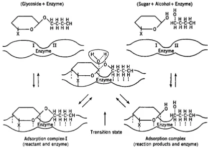
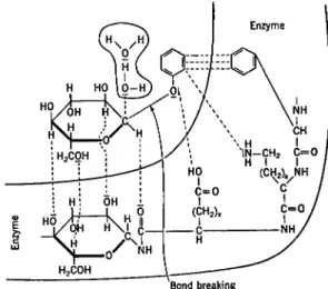
2. MECHANISM OF ACTION
The most widely accepted theory of enzyme action is based on the formation of an intermediate compound or adsorption complex between enzyme and substrate (Brown, 1902; Henri, 1903). Since both conceptions of the nature of the enzyme-substrate complex can lead to the same kinetic equations, the distinction seems unimportant at present. In the following development of the kinetic equations, the original scheme of Michaelis and Menten (1913) will be followed and compound formation will be considered to take place. However, in later discussions, the process will be considered as a type of adsorption.
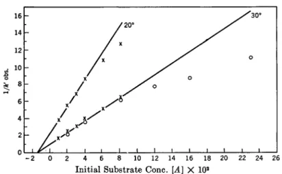
A. KINETIC.EQUATIONS AND EFFECT OF SUBSTRATE CONCENTRATION
In order to develop the-kinetic equations, consider the hydrolysis of a glucoside (S) by an enzyme (E) to an alcohol or phenol (ROH) and glucose.
2. B . Helferich and F . Vorsatz, Z. physiol. Chem. 237, 254 (1935); W. W. Pigman, J. Research Nail. Bur. Standards 30, 159 (1943).