Supported by
Accepted Article
Title: Title Synthesis of α-L-fucopyranoside-presenting glycoclusters and investigation of their interaction with recombinant
Photorhabdus asymbiotica lectin (PHL)
Authors: Jančaříková Gita, Mihály Herczeg, Eva Fujdiarová, Josef Houser, Katalin E. Kövér, Anikó Borbás, Michaela Wimmerová, and Magdolna Csávás
This manuscript has been accepted after peer review and appears as an Accepted Article online prior to editing, proofing, and formal publication of the final Version of Record (VoR). This work is currently citable by using the Digital Object Identifier (DOI) given below. The VoR will be published online in Early View as soon as possible and may be different to this Accepted Article as a result of editing. Readers should obtain the VoR from the journal website shown below when it is published to ensure accuracy of information. The authors are responsible for the content of this Accepted Article.
To be cited as: Chem. Eur. J. 10.1002/chem.201705853
Link to VoR: http://dx.doi.org/10.1002/chem.201705853
Synthesis of a- L -fucopyranoside-presenting glycoclusters and investigation of their interaction with Photorhabdus asymbiotica lectin (PHL)
Gita Jančaříková,
[a,b]Mihály Herczeg,
[c]Eva Fujdiarová,
[b]Josef Houser,
[a,b]Katalin E. Kövér,
[d]Anikó Borbás,*
[c]Michaela Wimmerová*
[a,b,e]and Magdolna Csávás*
[c]Dedicated to Professor Pál Herczegh on the occasion of his 70th birthday.
Abstract: Photorhabdus asymbiotica is a gram-negative bacterium that is not only as effective an insect pathogen as other members of the genus, but it also causes serious diseases in humans. The recently identified lectin PHL from P. asymbiotica verifiably modulates an immune response of humans and insects, which supports the idea that the lectin might play an important role in the host-pathogen interaction. Dimeric PHL contains up to seven L- fucose specific binding sites per monomer, and in order to target multiple binding sites of PHL, a-L-fucoside-containing di-, tri- and tetravalent glycoclusters were synthesized. Methyl gallate and pentaerythritol were chosen as multivalent scaffolds, and the fucoclusters were built from the above-mentioned cores by coupling with different oligoethylene bridges and propargyl a-L-fucosides using 1,3-dipolar azide-alkyne cycloaddition. The interaction between fucoside derivates and PHL was investigated by several biophysical and biological methods, ITC and SPR measurements, hemagglutination inhibition assay and an investigation of bacterial aggregation properties were carried out. Moreover, details of the interaction between PHL and propargyl a-L-fucoside as a monomer unit were revealed using X-ray crystallography. Besides this, the interaction with multivalent compounds was studied by NMR techniques. The newly synthesized multivalent fucoclusters proved to be up to several orders of magnitude better ligands than the
natural ligand, L-fucose.
Introduction
Photorhabdus asymbiotica together with another three species (P. luminescens, P. temperata and P. heterorhabditis) belongs to the Photorhabdus genus – entomopathogenic bacteria forming a symbiotic relationship with nematodes from the genus Heterorhabditis. Unlike other Photorhabdus species, P.
asymbiotica is unique in its ability to act as an emerging human pathogen. It causes invasive soft tissue infections that are difficult to treat locally, and disseminated bacteraemic disease characterized by multifocal skin and soft tissue abscesses.[1–4]
Analysis of the P. asymbiotica genome identified the fucose/galactose-binding lectin PHL.[5] It was revealed that PHL has the ability to act as a host-cell recognizing agent through interacting with the haemolymph of Galleria mellonella (order Lepidoptera, family Pyralidae) and human blood components.
Therefore, PHL could be considered to be a usable therapeutic target.
The soluble lectin PHL forms a seven-bladed b-propeller assembling into a homo-dimer with an inter-subunit disulphide bridge. The crystal structure of the complex with different ligands demonstrated the existence of two sets of binding sites per monomer with preferences for L-fucose and D-galactose, respectively. Biophysical studies show an affinity of PHL mainly for a-L-fucosides over other tested ligands. Recent analysis of PHL/Me-a-L-Fuc complexes revealed up to seven potential fucose-binding sites per PHL monomer, forming a circle.[5] On the basis of biological tests, a specific agglutination of type O human red blood cells (RBCs) was observed as well as interaction with all types of RBCs through a-fucoside. These outcomes suggest that this lectin could be also directly involved in the adhesion to host cells. This leads to the need for glyco- inhibitors which can strongly bind to the lectin to compete with and disrupt host-lectin interaction during the first step of the infection process.
PHL as a multivalent lectin displays an avidity effect, which significantly increases affinity towards its ligands. For that reason, compounds containing multiple carbohydrate moieties could be the most potent inhibitors. A new set of tri- and tetravalent glycoclusters with different architectures and valences were designed to provide an opportunity to evaluate the impact of topology and valency on binding properties toward [a] G. Jančaříková, Dr. J. Houser, Prof. M. Wimmerová
Central European Institute of Technology Masaryk University
Kamenice 5, 625 00 Brno, Czech Republic E-mail: michaw@chemi.muni.cz
[b] G. Jančaříková, E. Fujdiarová, Dr. J. Houser, Prof. M. Wimmerová National Centre for Biomolecular Research, Faculty of Science Masaryk University
Kotlářská 2, 611 37 Brno, Czech Republic [c] Dr. M. Herczeg, Prof. A. Borbás, Dr. M. Csávás
Department of Pharmaceutical Chemistry University of Debrecen
Egyetem tér 1, H-4032, Debrecen, Hungary E-mail: borbas.aniko@pharm.unideb.hu,
csavas.magdolna@science.unideb.hu [d] Prof. K. E. Kövér
Department of Inorganic and Analytical Chemistry University of Debrecen
Egyetem tér 1, H-4032, Debrecen, Hungary [e] Prof. M. Wimmerová
Department of Biochemistry, Faculty of Science Masaryk University
Accepted Manuscript
fucoclusters and study their interaction with the recombinant PHL lectin from P. asymbiotica.
Results and Discussion
Synthesis
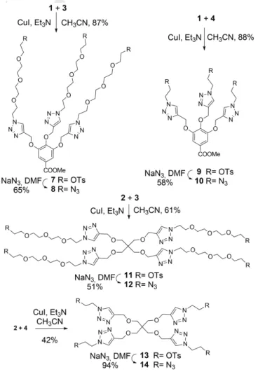
Similarly to our recent works[6,7], propargylated methyl gallate 1[8]
and pentaerythritol 2[9] were chosen as multivalent scaffolds, and heterobifunctionalized tetraethylene glycol 3[10] and ethylene glycol 4[11] were used as linkers with different lengths. Based on the known X-ray structure of PHL, the average distances between the fucose-binding sites is approximately 19 Å, so these linkers are long enough that the a-L-fucose-units of the fucoclusters are able to bind to more than one binding pocket of the lectin.
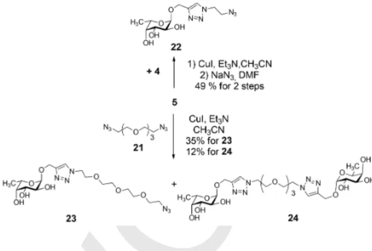
We efficiently prepared propargyl a-L-fucoside 5 by Fischer glycosylation using sulfamic acid[12] and utilized it in both unprotected and acetylated form 6[13] to create multivalent structures (Figure 1).
Figure 1. Building blocks for the fucoclusters.
The fucoclusters were built from the above structural elements from the inside out starting from the central cores applying two consecutive 1,3-dipolar azide-alkyne cycloaddition click reactions. First, the tri- and tetravalent cores 1 and 2 were coupled with bridges 3 and 4, respectively, via the copper(I)- catalyzed azide-alkyne cycloaddition click reaction (CuAAC) to obtain tosylated compounds 7, 9, 11 and 13, in good yields. The tosyl groups of all scaffolds were converted into azido functional groups using sodium azide to result in compounds 8, 10, 12 and 14, which were also suitable for further click reactions (Scheme 1).
Scaffolds containing tetraethylene glycol bridges 8 and 12 were coupled by CuAAC reaction with propargyl 2,3,4-tri-O-acetyl-a-L- fucopyranoside 6 to produce the protected fucoclusters 15 and 17, respectively, while click reaction between the same scaffolds and propargyl a-L-fucopyranoside 5 provided the final products 16 and 18 directly (Scheme 2). The latter two compounds were also obtained by the Zemplén deacetylation of 15 and 17, respectively. Comparing the above syntheses with the unprotected and protected sugars 5 and 6, using the free sugar was not only a shorter, but also a more efficient procedure, although the isolation and purification were more demanding.
(While the overall yields of compounds 16 and 18 were 60% and 65% respectively using free fucoside, these yields decreased to 38% and 32% when using the protected fucoside.) For this
reason, we chose the shorter reaction pathway for further synthesis.
Based on the same strategy, scaffolds 10 and 14 equipped with an ethylene glycol bridge were reacted with propargyl a-L- fucopyranoside 5 by copper(I)-catalyzed cycloaddition to result in the final products 19 and 20 (Scheme 3).
Our further aim was the examination of the role and effect of the linker and the triazole-ring in the lectin-carbohydrate interactions.
For this reason, compounds 5, 22 and 23 were also provided for biological investigations. Compound 5 (propargyl alpha-L- fucoside) is suitable for investigations to define the role of the monomer L-fucose. Moreover, compound 5 was reacted with azido-tetraethylene glycol derivative 4 under the condition of click reaction, then tosyl group was subsequently replaced with azide, to yield compound 22 (49% for two steps), which can provide information about the potency of the ethylene glycol linker-armed monomer. Finally, compound 5 was coupled with diazido-tetraethylene glycol 21[14] via click reaction, to yield compounds 23 (35%) and 24 (12%), which can serve information about the binding properties of the tetraethylene glycol linker-armed monomer and dimer (Scheme 4).
Scheme 1. Synthesis of multivalent scaffolds having functionalized ethylene glycol (10 and 14) and tetraethylene glycol (8 and 12) linker arms.
Accepted Manuscript
The interaction of mono- and multivalent fucosylated ligands with PHL was investigated by a few methods. At first hemagglutination inhibition assay was utilized as a simple microscopy method to preselect the most efficacious inhibitors.
Subsequently two biophysical techniques - isothermal titration calorimetry (ITC) and surface plasmon resonance (SPR) - were employed. The presence of fucose-binding lectins on the surface of P. asymbiotica subsp. australis was indirectly tested through the cross-linking of bacterial cells caused by multivalent ligands.
In summary, these methods provide information about the binding affinity, inhibition potency and effect of the compounds on cell cross-linking. For a better structural characterization of the interaction of PHL with ligands, ligand-soaking of PHL crystals and STD-NMR experiments were carried out.
Scheme 2. Synthesis of tri- and tetravalent fucodendrons 16 and 18 containing TEG-linkers.
Scheme 3. Synthesis of tri- and tetravalent a-L-fucoside-containing dendrons 19 and 20 having short linker arms.
Scheme 4. Synthesis of the linker-armed monomers 22 and 23.
Hemagglutination inhibition assay
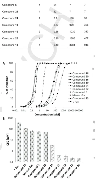
PHL, being a fucose-specific lectin, was shown to agglutinate papain-treated RBCs of blood group O[5] with surface-exposed terminal trisaccharide a-L-Fucp-(1®2)-b-D-Galp-(1®3/4)-D- GlcpNAc. This property was also used to assess the inhibition potency of the set of a-L-fucoside molecules on hemagglutination by PHL (Figure 2). All of them were able to inhibit the hemagglutination with at least four times higher potency than L-fucose (Table 1). Tetravalent compound 18 was shown to be the most efficient inhibitor followed by compounds 16, 19 and 20, all of them being half as efficient as 18.
Compound 20 is also a tetravalent fucocluster, but contains shorter linkers, which may be not able to connect binding sites, in contrast to compound 18.
Figure 2. Representatives of mono-, di-, tri-, and tetrafucosylated compounds and their influence on hemagglutination of RBC O caused by PHL. Two-fold serially diluted carbohydrate compounds 5, 24, 16 and 18 were able to inhibit hemagglutination at minimal concentrations 200 µM, 100 µM, 50 µM and 25 µM, respectively. Hemagglutination of RBC O caused by PHL without inhibitor is shown on the picture in the left down corner.
Accepted Manuscript
Table 1. Hemagglutination inhibition assay with PHL. Minimal inhibitory concentrations (MIC) of synthetized inhibitors and their potency towards L- fucose was determined. To assess the contribution of valency to the MIC, an affinity improvement factor β was calculated as the relationship MICL- fucose/(valency × MICligand).
Ligand Valency MIC [µM] Potency b
L-Fuc 1 3,200 1 1
Compound 23 1 800 4 4
Me-a-L-Fuc 1 400 8 8
Compound 5 1 200 16 16
Compound 22 1 200 16 16
Compound 24 2 100 32 16
Compound 19 3 50 64 21
Compound 16 3 50 64 21
Compound 20 4 50 64 16
Compound 18 4 25 128 32
Surface plasmon resonance
The surface plasmon resonance (SPR) technique was employed to analyse the competitive inhibition of PHL binding to a multivalent chip surface. The experiments were performed using a constant concentration of PHL and increasing concentration of the studied compounds to enable the determination of IC50. Tetravalent compounds 18 and 20 appeared to be the most efficient inhibitors, with IC50 reaching 200 nM and 96 nM, respectively (Table 2). The trivalent compounds 16 and 19 had a weaker inhibitory potential, but were still in the nanomolar range, which is three orders of magnitude lower than for free L-fucose.
The monovalent ligands proved to be at least three times more efficient than L-fucose. Logarithmic plots of IC50 and the inhibition curves from SPR measurements displayed in Figure 3 nicely demonstrate the growing inhibitory effect through monovalent, divalent and tri-/tetravalent a-L-fucoside compounds.
Table 2. Inhibitory effect of representative carbohydrates on PHL binding to immobilized L-fucoside. IC50 was determined from a plot of serial dilutions vs.
% inhibition, and potency was relative to L-Fuc. To assess the contribution of valency to the IC50, an affinity improvement factor β was calculated as the relationship IC50,L-fucose/(valency × IC50,ligand).
Ligand Valency IC50 [µM] Potency b
L-Fuc 1 361 1 1
Compound 23 1 108 3 3
Me-a-L-Fuc 1 68 5 5
Compound 5 1 54 7 7
Compound 22 1 52 7 7
Compound 24 2 3.2 118 59
Compound 19 3 0.37 979 326
Compound 16 3 0.35 1030 343
Compound 20 4 0.20 1806 452
Compound 18 4 0.10 3784 946
Figure 3. IC50 determination of fucoclusters towards PHL from SPR measurements. (A) A logarithmic plot of inhibition curves determined from SPR experiments. Individual types of line correspond to monovalent (dotted), divalent (dashed), trivalent (dashdotted) and tetravalent (densely dashed) ligands. (B) A logarithmic plot of IC50 for individual mono-/multivalent a-L- fucoside compounds. Standard deviations were calculated from three independent measurements. Individual type of pattern corresponds to monovalent (dotted), divalent (dashed), trivalent (dashdotted) and tetravalent (densely dashed) ligands.
Isothermal titration calorimetry
The binding of PHL to multivalent a-L-fucoside compounds was further characterized by isothermal titration calorimetry (ITC) to
Accepted Manuscript
determine the complete thermodynamic profile of the molecular interactions. As for monovalent fucoclusters, all of them were proved to be low-affinity ligands for PHL. The data for compound 23 were impossible to evaluate. The other two monovalent ligands provided better results. In both cases, the ITC curves indicated a low affinity toward PHL, as usually observed for lectin/saccharide interactions (Figure 4). The stoichiometry value n was assessed to be around 4 and the dissociation constants were in the submilimolar range (Table 3). As expected, the enthalpy of binding for shorter compounds was less negative than for longer chains. Shorter and more rigid compounds 5 and 22 also exhibited a positive entropy contribution, in contrast to other studied molecules.
The ITC data for multivalent fucosylated compounds provided a more interesting set of results (Table 3 and Figure 4). The values indicate a strong interaction between PHL and multivalent fucoclusters, with an affinity in the low micromolar range. The stoichiometry varies between 2 and 3 ligand molecules per PHL monomer. Generally, tetravalent ligands exhibited a higher affinity than tri- or divalent ones, however, an
interesting effect of linker length was observed. Short linkers seem to disfavour ligand binding. Thus, compounds with shorter linkages (tetravalent 20 and trivalent 19) bind with considerably lower affinity than longer linker-containing tetravalent 18 and trivalent 16, respectively. Unusually high enthalpy and entropy contributions were observed for compound 19. This suggests the possibility of a different binding mode being employed.
The highest affinity was observed towards tetravalent compound 18, with a KD twice as good as the second best tetravalent compound 20, and more than a thousand times better than free L-Fuc. It is interesting that trivalent compound 16 containing longer linkers has a comparable KD to tetrafucosylated compound 20 with a shorter spacer – the spacer length plays an important role in affinity. Likewise, dimeric fucocluster 24 is a little bit better than trivalent compound 19 with short linkers (Figure 4B).
During the ITC measurements, we also observed an additional effect – the cross-linking of PHL caused by multivalent fucoclusters that was accompanied by the formation of visible precipitates.
Table 3. Thermodynamic profiles for interaction between PHL and carbohydrate ligands determined by isothermal titration calorimetry using one site model fitting procedure at 25 °C (standard deviations were calculated from two independent measurements). To assess the contribution of valency to the affinity increase, an affinity improvement factor β was calculated as the relationship KD,L-fucose/(valency × KD,ligand).
Ligand Valency n KD [µM] DH [kJ mol-1] -TDS [kJ mol-1] DG [kJ mol-1] b
Compound 23 1 ND[a] ND[a] ND[a] ND[a] ND[a] ND[a]
L-Fuc 1 3[b] 1,395 ± 34 -59.5 ± 1.0 43.2 -16.3 ± 0.3 1
Me-a-L-Fuc 1 2.9 ± 0.2 267 ± 11 -59.0 ± 4.2 38.6 -20.4 ± 1.5 5
Compound 22 1 4.0 ± 0.3 175 ± 18 -14.6 ± 1.4 -6.9 -21.5 ± 2.1 8
Compound 5 1 4.6 ± 0.3 149 ± 16 -8.8 ± 0.7 -13.1 -21.9 ± 1.8 9
Compound 19 3 2.3 ± 0.0 8.5 ± 0.5 -65.4 ± 0.7 36.5 -28.9 ± 0.3 55
Compound 24 2 2.5 ± 0.0 6.6 ± 0.7 -42.2 ± 0.8 12.5 -29.7 ± 0.6 106
Compound 16 3 2.9 ± 0.1 3.6 ± 0.8 -54.4 ± 1.4 23.4 -31.0 ± 0.8 129
Compound 20 4 2.1 ± 0.1 3.1 ± 1.0 -56.9 ± 2.3 25.5 -31.4 ± 1.2 113
Compound 18 4 1.9 ± 0.0 1.2 ± 0.4 -77.5 ± 2.6 43.6 -33.9 ± 1.1 290
[a] Values not determined because of low affinity interaction. One-site model for compound 23 was unable to reach a meaningful fit. [b] The stoichiometry value of
L-fucose was fixed during fitting procedure because of low-affinity interaction. The value was derived from the X-ray crystal structure of the PHL/Me-a-L-Fuc complex.
Accepted Manuscript
Figure 4. (A) Thermodynamic profiles for interaction between PHL and mono/multivalent a-L-fucoside compounds determined by isothermal titration calorimetry at 25 °C (standard deviations were calculated from two independent measurements). (B) Selected ITC curves of PHL (50 µM) titration by compound 5 (5 mM), 24 (5 mM) and 19 (1.5 mM). 20 injections of 2.0 µl of fucoclusters were added every 240 s to a PHL-containing cell. Lower plots show the total heat released as a function of total ligand concentration for the titration shown in the upper panels.
Aggregation of PHL lectin monitored by 1H NMR spectroscopy
The aggregation behaviour of the PHL lectin upon titration with the five a-L-fucoside-containing derivatives (monomer 5, trivalent 16 and 19 and tetravalent 18 and 20) has been subsequently investigated and compared by 1H NMR spectroscopy, monitoring the change (decrease) in protein signal intensity as a function of the actual ligand concentration (Figure 5). For a fair comparison of the extent of PHL aggregation, each tested fucoside ligand was added to a similar (ca. 40-50 μM) solution of PHL in concentrations ranging from 6 to 25 μM. Before each titration assay, a regular 1H NMR spectrum was recorded on the sample containing PHL alone, providing the reference protein signal intensity for quantification of protein aggregation during the subsequent titration steps. As expected, the monovalent ligand 5 did not lead to any noticeable aggregation of PHL, even when its concentration was increased up to 0.1 mM. In contrast, when titrating the multivalent ligands into the sample containing PHL a significant reduction (drop) in the protein signal envelope was observed, suggesting the formation of large, non-soluble aggregates via possible cross-linking between the multivalent ligands and PHL. The level of aggregation, however, varied
according to the type and concentration of the multivalent ligand tested. The tri- and tetravalent fucodendron derivatives containing TEG-linkers (16 and 18, respectively) led to the formation of smaller amount of non-soluble aggregates than the ones (19, 20) with the shorter linker arm. Specifically, at 6.2 μM of fucodendrons with a TEG-linker, about 30 % of total protein aggregated (precipitated), while upon adding the ligands with a shorter linker at the same concentration, only ca. 50 % of the protein remained in solution.
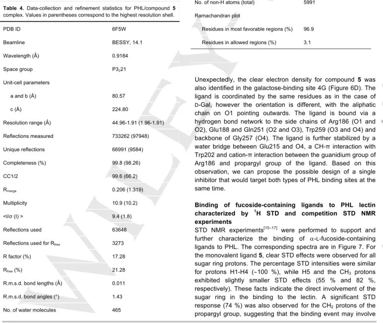
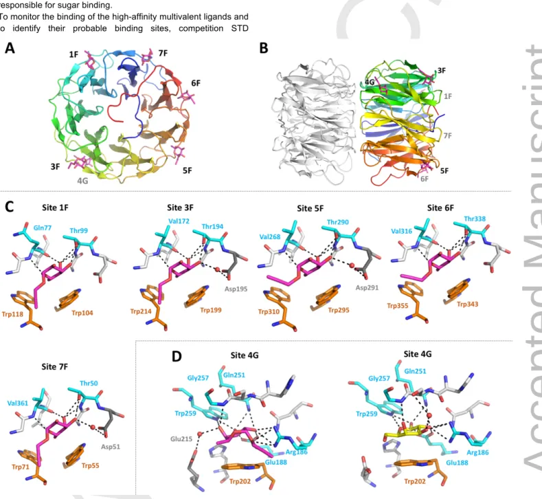
X-ray structure of PHL/compound 5 complex
The PHL lectin was shown to form a dimeric structure consisting of two seven-bladed β-propellers with two types of binding sites situated between the blades.[5] Both types of binding sites have different amino acid composition and binding preferences. Also, the non-identity of individual potential binding sites within the same type was proposed. The direct study of complexes with branched fucosylated structures was not possible, as mixing PHL with the multivalent compound leads to protein precipitation, preventing the co-crystallization. Soaking ligand-free PHL crystals with a low concentration of individual branched compounds did not result in complex formation, while soaking with their higher concentrations led to a crystal decomposition.
Therefore, to determine the potential PHL binding sites for studied molecules, we soaked the crystals with monomeric compound 5.
The structure of the PHL/compound 5 complex was determined using X-ray diffraction (Table 4). The lectin in the complex retains the seven-bladed β-propeller fold (Figure 6A, B) identical to the ligand-free form.[6] The differential electron density map was clear enough to identify compound 5 monomers in five of the seven potential fucose binding sites of monomer A – site 1F, 3F, 5F, 6F and 7F (Figure 6C). In monomer B, the ligand binding in site 5F was affected by intermonomeric contacts in the crystal, and it was not possible to determine the precise position of the ligand. Compound 5 is bound via its fucosylated part, which adopts the same orientation as Me-a-L-Fuc in the previously determined PHL/Me-a-L-Fuc complex.[5] Thus, the O3, O4 and O5 of the fucosyl part is coordinated to the protein backbone of the Gly/Gln and Val residues on one side of the pocket and backbone and side chain of the Thr residue on the other side of the pocket. The C6 methyl group and hydrophobic surface of Fuc C3, C4 and C5 are stabilized through CH-π interactions with two Trp residues, and O3 may be further stabilized by a water bridge to the neighbouring Asp residue (Figure 6C – sites 3F, 5F, 7F). Compared to the PHL/Me-a-L-Fuc complex, more binding sites were occupied by compound 5, which agrees with the higher affinity towards compound 5 than towards Me-a-L-Fuc, as was determined by ITC and hemagglutination inhibition
experiments.
Accepted Manuscript
Figure 5. 1H NMR experiments (500 MHz, T = 298 K) for monitoring the aggregation of the PHL lectin in the presence of increasing concentration of the ligands (5, 16, 18, 19 and 20). 40-50 μM solution of PHL in D2O (PBS) was titrated by increasing amount of ligands. The relevant ligand concentrations together with the percentage of PHL remaining in solution (referenced to pure PHL solution) are given above the spectra. All NMR spectra were recorded with 512 number of scans, allowing 1 s for relaxation between subsequent transients.
Table 4. Data-collection and refinement statistics for PHL/compound 5 complex. Values in parentheses correspond to the highest resolution shell.
PDB ID 6F5W
Beamline BESSY, 14.1
Wavelength (Ǻ) 0.9184
Space group P3221
Unit-cell parameters
a and b (Å) 80.57
c (Å) 224.80
Resolution range (Ǻ) 44.96-1.91 (1.96-1.91)
Reflections measured 733262 (97948)
Unique reflections 66991 (9584)
Completeness (%) 99.8 (98.26)
CC1/2 99.6 (66.2)
Rmerge 0.206 (1.319)
Multiplicity 10.9 (10.2)
<I/σ (I) > 9.4 (1.8)
Reflections used 63648
Reflections used for Rfree 3273
R factor (%) 17.28
Rfree (%) 21.28
R.m.s.d. bond lengths (Ǻ) 0.011
R.m.s.d. bond angles (°) 1.43
No. of water molecules 465
No. of non-H atoms (total) 5991
Ramachandran plot
Residues in most favorable regions (%) 96.9 Residues in allowed regions (%) 3.1
Unexpectedly, the clear electron density for compound 5 was also identified in the galactose-binding site 4G (Figure 6D). The ligand is coordinated by the same residues as in the case of D-Gal, however the orientation is different, with the aliphatic chain on O1 pointing outwards. The ligand is bound via a hydrogen bond network to the side chains of Arg186 (O1 and O2), Glu188 and Gln251 (O2 and O3), Trp259 (O3 and O4) and backbone of Gly257 (O4). The ligand is further stabilized by a water bridge between Glu215 and O4, a CH-π interaction with Trp202 and cation-π interaction between the guanidium group of Arg186 and propargyl group of the ligand. Based on this observation, we can propose the possible design of a single inhibitor that would target both types of PHL binding sites at the same time.
Binding of fucoside-containing ligands to PHL lectin characterized by 1H STD and competition STD NMR experiments
STD NMR experiments[15–17] were performed to support and further characterize the binding of a-L-fucoside-containing ligands to PHL. The corresponding spectra are in Figure 7. For the monovalent ligand 5, clear STD effects were observed for all sugar ring protons. The percentage STD intensities were similar for protons H1-H4 (~100 %), while H5 and the CH3 protons exhibited slightly smaller STD effects (55 % and 82 %, respectively). These facts indicate the direct involvement of the sugar ring in the binding to the lectin. A significant STD response (74 %) was also observed for the CH2 protons of the propargyl group, suggesting that the binding event may involve
Accepted Manuscript
this group as well. It should be noted, however, that the intensity of the propargyl CH-proton is significantly reduced due to partial deuteration in D2O, therefore its STD response is likely also attenuated and so that not quantified in %. It should be also mentioned that the irradiation of the protein in the aromatic signal region led to a significantly lower saturation of all ligand protons with correspondingly smaller STD effects. This fact may suggest that the binding pocket of the PHL lectin probably involves more aliphatic than aromatic amino acid residues responsible for sugar binding.
To monitor the binding of the high-affinity multivalent ligands and to identify their probable binding sites, competition STD
experiments were also carried out. The corresponding STD spectra are shown in Figure 7B. As can be seen, all STD signals decreased considerably upon adding the multivalent ligands (first trivalent, 16, lastly tetravalent, 18) to the sample initially containing the monovalent ligand alone. These data may confirm that both multivalent ligands compete with the monovalent ligand for the same or at least partially the same (overlapping) binding site of the PHL lectin.
Figure 6. Structure of PHL complex with compound 5. (A) PHL monomer (chain A) overall architecture with compound 5 shown as magenta sticks. Individual binding sites are labelled in black (front plane) or grey (back plane) (B) Side view of PHL dimer with chain B shown in grey and without ligands. (C) Individual PHL fucose-type binding sites with compound 5 (magenta) bound. Amino acids responsible in ligand binding are highlighted and labelled. (D) PHL galactose-type binding site with compound 5 (magenta) bound and PHL galactose-type binding site with D-Gal (yellow) bound (PDB ID: 5MXH). Colour code for panels C/D:
amino acids involved in ligand-binding through H-bond – cyan, CH-π interaction – orange or water bridge – grey.
Accepted Manuscript
Figure 7. (A) STD experiments for the monovalent fucoside ligand 5 complexed with the PHL lectin. The estimated molar ratio of sugar/lectin is ca. 100/1. i.-ii.) The corresponding STD spectra (up-scaled by a factor of 64) depicted after saturating for 3 s at δ -1.5 and 10.5 ppm, respectively. The percentage STD intensities are referenced to the STD response of H-1 proton (100 %) obtained upon irradiation at the aliphatic signal region of PHL at -1.5 ppm. On- and off- resonance data were acquired alternately up to total of 1424 scans recorded for each FID; iii.) The regular 500 MHz 1D 1H NMR spectrum with the assignment of resonances. (B) Competition STD experiments for characterizing the binding of the high affinity ligands (16 and 18) to PHL. The bottom spectrum corresponds to the regular 1H NMR spectrum of ligand 5. The other spectra are the corresponding STD responses recorded on samples containing the ligand 5 at constant 2 mM concentration, while the multivalent ligands were subsequently added in increasing concentrations. The top (reference) STD spectrum corresponds to the sample containing only the monovalent ligand. The other spectra were recorded on samples containing trivalent ligand 16 at 70 μM; at 100 μM; and lastly, the mixture of trivalent 16 at 100 μM and tetravalent 18 at 60 μM. For better visualization of the STD signal drop due to competition, an overlay of the STD responses of H1 is shown in the inset at larger scale. Note that during titration no aggregation of protein was observed.
P. asymbiotica cross-linking - Bacterial aggregation properties
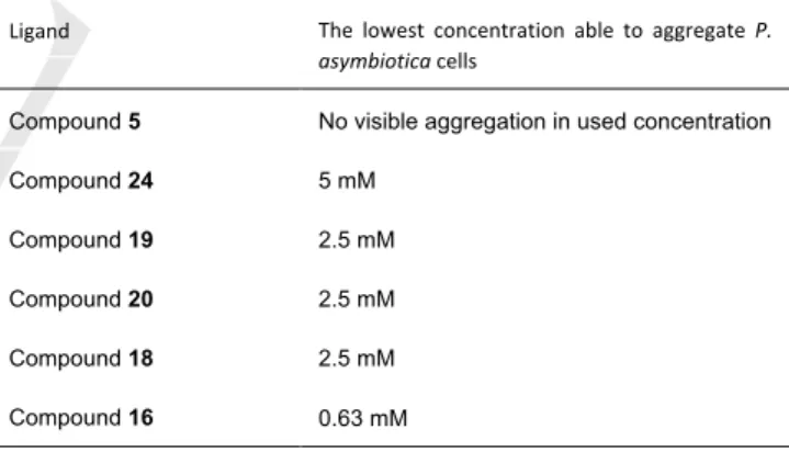
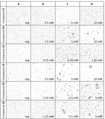
The aggregation of PHL by multivalent fucoclusters was observed in many experiments and therefore we decided to test it at a higher level. We performed a series of in vitro aggregation assays to reveal the aggregation properties of fucosylated compounds towards P. asymbiotica subsp. australis. Prior to each assay, we confirmed the absence of aggregates in the negative control – bacterial cells in 50% DMSO (Figure 8A). We studied mono-, di-, tri- and tetravalent fucoclusters as ligands (Table 5, Figure 8). The monovalent compound 5 did not create any aggregates, as expected. With all multivalent compounds, we observed bacterial aggregates of spherical shape and variable size that confirmed that all multivalent compounds are capable of interacting with a cell surface’s receptors. The concentration of the first appearance of aggregates varied according to the nature of the ligand used (Figure 8C). The highest efficiency (0.63 mM) was observed for the trifucosylated compound 16. Against our expectations, it was higher than for tetrameric fucoclusters (compounds 18 and 20) that aggregated cells at a concentration of 5 mM. A similar result was obtained for the other trivalent compound 19. This could be caused by the structure of compound 16, its longer linker arms allow a higher flexibility of the compound and easier reach on different cells.
Table 5. The determination of the minimal concentration of multivalent fucoclusters able to aggregate P. asymbiotica cells.
Ligand The lowest concentration able to aggregate P.
asymbiotica cells
Compound 5 No visible aggregation in used concentration
Compound 24 5 mM
Compound 19 2.5 mM
Compound 20 2.5 mM
Compound 18 2.5 mM
Compound 16 0.63 mM
Accepted Manuscript
Figure 8. Representative images of optical microscopy observation (200x) of bacterial cell aggregation for Photorhabdus asymbiotica subsp. australis with different fucosylated clusters. Mixture with 50% DMSO was used as negative control (A). Concentrations of the first appearance of aggregates are presented (C) as well as one dilution step lower (B) and higher (D). No aggregates were observed for a monovalent fucocluster (compound 5).
Background of all pictures was subtracted in GIMP software.
Conclusions
P. asymbiotica has proved to be not only an entomopathogenic bacteria, but also a dangerous human pathogen that causes a difficult-to-treat disease. Suitable inhibitors preventing the host- pathogen interaction should be helpful in the early steps of P.
asymbiotica infection. For this purpose, monovalent fucosides and di-, tri-, and tetravalent glycoclusters functionalized with a-L- fucosides were designed and prepared using click chemistry.
Their binding properties toward the PHL lectin from P.
asymbiotica have been evaluated by hemagglutination, ITC, SPR, NMR, X-ray crystallography, and cell cross-linking.
We have demonstrated that all of the compounds were able to inhibit the PHL binding activity and the affinity for multivalent ligands reached nanomolar values, and there is an obvious relation between the increasing number of fucoses per cluster and increasing affinity. The potency of ligands depended on the valency, architecture and flexibility of the fucocluster, but all ligands proved to be up to several orders of magnitude better ligands than L-fucose. Moreover, only multivalent ligands were able to cross-link PHL, resulting in the formation of visible precipitation. We used this property and investigated the agglutination of P. asymbiotica bacteria via the surface lectins that are likely present, as was proved for other cells (e.g. P.
aeruginosa, E. coli, T cell leukemia line).[18–20] We observed nice cell clumps at different concentrations of multivalent compounds, showing the specificity of the interaction and proving the presence of lectins on the bacterial surface. In conclusion, we provide complete assays of the action of mono- and multivalent fucoclusters on the P. asymbiotica lectin PHL and useful information for their possible employment in medical treatments.
Experimental Section
Synthesis - General methods
Optical rotations were measured at room temperature with a Perkin- Elmer 241 automatic polarimeter. TLC analysis was performed on Kieselgel 60 F254 (Merck, Germany) silica gel plates with visualization by immersing in a sulfuric-acid solution (5% in EtOH) followed by heating.
Column chromatography was performed on silica gel 60 (0.063–0.200 mm, Merck, Germany), flash column chromatography was performed on silica gel 60 (Merck 0.040–0.063 mm). Gel filtration was performed on Sephadex G-25 (Sigma-Aldrich, USA), using methanol as the eluent.
Organic solutions were dried over MgSO4 and concentrated under vacuum. The 1H (400 MHz) and 13C NMR (100.28 MHz) spectra were recorded with Bruker Avance II 400 spectrometer (Bruker, Germany).
Chemical shifts are referenced to Me4Si or DSS (0.00 ppm for 1H) and to solvent signals (CDCl3: 77.00 ppm, CD3OD: 49.15 ppm, DMSO-d6: 39.51 ppm for 13C). MS (MALDI-TOF) analysis was carried out in positive reflection mode with a BIFLEX III mass spectrometer (Bruker, Germany) with delayed-ion extraction. The matrix solution was a saturated solution of 2,5-dihidroxy-benzoic acid (DHB) in DMF. Elemental analysis was performed on an Elementar Vario MicroCube instrument (Elementar UK Ltd., UK).
General method A for azide-alkyne click reaction. Et3N (1 equiv./alkyne) and copper(I)-iodide (0.1 equiv./alkyne) were added to a stirred solution of alkyne (0.2 mmol) and azide (1.3 equiv./alkyne) in CH3CN (3 mL) under an argon atmosphere and stirred overnight at room temperature.
The reaction mixture was evaporated, and the crude product was purified by flash column chromatography to give the desired compound.
General method B for nucleophilic substitution of tosylates to azide. NaN3
(2 equiv.) was added to a stirred solution of tosylate (1 mmol) in DMF (5 mL) under an argon atmosphere and stirred overnight at room temperature. The reaction mixture was diluted with water dropwise, stirred for further 5 minutes and evaporated. The residue was dissolved in CH2Cl2 (200 mL) and extracted with water (2 x 50 mL) and brine (50 mL), dried over MgSO4, filtered and evaporated. The crude product was purified by flash column chromatography to give the desired compound.
General method C for Zemplén-deacetylation. The catalytic amount of NaOMe (pH ~ 9) was added to a stirred solution of ester (0.2 mmol) in dry MeOH (5 mL) and stirred overnight at room temperature. The reaction mixture was neutralized with Amberlite IR-120 H+ ion-exchange resin, filtered and evaporated, and then the crude product was purified by flash column chromatography and gel filtration to give the desired compound.
Compound 8. Azide compound 3 (637 mg, 1.71 mmol) and alkyne 1 (131 mg, 0.44 mmol) were reacted in CH3CN according to general method A. The crude product was purified by flash column chromatography (CH2Cl2/MeOH 98:2) to give compound 7 (540 mg, 87%) as a colourless syrup. Rf = 0.50 (CH2Cl2/MeOH 97:3). The product
Accepted Manuscript
7 (540 mg, 0.38 mmol) was converted into azide according to general method B. The crude product was purified by flash column chromatography (CH2Cl2/MeOH 95:5) to give compound 8 (254 mg, 65%) as a colourless syrup. Rf = 0.39 (CH2Cl2/MeOH 95:5); 1H NMR (400 MHz, CDCl3): δ = 7.96, 7.89 (2 x s, 3H, 3 x CH triazole), 7.44 (s, 2H, arom), 5.24 (d, J = 1.8 Hz, 6H, 3 x CH2 propargyl), 4.58 (t, J = 5.1 Hz, 4H, 2 x NCH2 TEG), 4.51 (t, J = 5.3 Hz, 2H, NCH2 TEG), 3.92-3.85 (m, 10H, 5 x OCH2 TEG), 3.65-3.58 (m, 29H, 13 x OCH2 TEG, COOCH3), 3.37- 3.35 (m, 6H, 3 x CH2 TEG) ppm; 13C NMR (100 MHz, CDCl3): δ = 166.3 (1C, COOCH3), 152.0 (2C, Cq arom.), 144.0, 143.1 (3C, 3 x Cq triazole), 141.6 (1C, Cq arom.), 125.5 (1C, Cq arom.), 124.8, 124.4 (3C, 3 x CH triazole), 109.1 (2C, arom.), 70.6, 70.5, 69.9, 69.3, 69.2 (18C, 18 x CH2
TEG), 66.4, 63.1 (3C, 3 x CH2 propargyl), 52.3 (1C, COOCH3), 50.6, 50.2, 50.0 (6C, 6 x NCH2 TEG) ppm. MS (MALDI-TOF): m/z calcd for C41H62N18NaO14: 1054.05 [M+Na]+; found: 1053.79; elemental analysis calcd (%) for C41H62N18O14: C 47.76, H 6.06; found: C 47.81, H 6.12.
Compound 10. Azide compound 4 (626 mg, 2.59 mmol) and alkyne 1 (199 mg, 0.67 mmol) were reacted in CH3CN according to general method A. The crude product was purified by flash column chromatography (CH2Cl2/MeOH 97:3) to give compound 9 (700 mg, 88%) as a colourless syrup. Rf = 0.18 (CH2Cl2/MeOH 97:3). Compound 9 (474 mg, 0.5 mmol) was converted into azide according to general method B. The crude product was purified by flash column chromatography (CH2Cl2/MeOH 97:3) to give compound 10 (175 mg, 58%) as a colourless syrup. [α]24D +1.3 (c = 0.15, CHCl3); Rf = 0.48 (CH2Cl2/MeOH 95:5); 1H NMR (400 MHz, CDCl3): δ = 7.97 (s, 3H, 3 x CH triazole), 7.41 (s, 2H, arom.), 5.31 (d, J = 1.3 Hz, 2H, CH2 propargyl), 5.25 (s, 4H, 2 x CH2 propargyl), 4.55-4.51 (m, 6H, 3 x NCH2 ethylene glycol), 3.88-3.80 (m, 9H, 3 x CH2 ethylene glycol, COOCH3) ppm; 13C NMR (100 MHz, CDCl3): δ = 166.0 (1C, COOCH3), 151.6 (1C, Cq arom.), 144.2, 143.4 (3C, 3 x Cq triazole), 141.1 (1C, Cq arom.), 125.5 (1C, Cq
arom.), 124.6, 124.1 (3C, 3 x CH triazole), 108.8 (2C, arom.), 66.0, 62.7 (3C, 3 x CH2 propargyl), 52.1 (1C, COOCH3), 50.3, 49.2, 49.0 (6C, 6 x NCH2 ethylene glycol) ppm. MS (MALDI-TOF): m/z calcd for C23H26N18NaO5: 657.22 [M+Na]+; found: 657.14; elemental analysis calcd (%) for C23H26N18O5: C 43.53, H 4.13; found: C 43.59, H 4.21.
Compound 12. Azide compound 3 (850 mg, 2.27 mmol) and alkyne 2 (126mg, 0.44 mmol) were reacted in CH3CN according to general method A. The crude product was purified by flash column chromatography (CH2Cl2/MeOH 97:3) to give compound 11 (474 mg, 61%) as a colourless syrup. Rf = 0.29 (CH2Cl2/MeOH 97:3). Compound 11 (474 mg, 0.27 mmol) was converted into azide according to general method B. The crude product was purified by flash column chromatography (CH2Cl2/MeOH 95:5) to give compound 12 (337 mg, 51%) as a colourless syrup. Rf = 0.12 (CH2Cl2/MeOH 95:5); 1H NMR (400 MHz, CDCl3): δ = 7.73 (s, 4H, 4 x CH triazole), 4.55-4.53 (m, 16H, 4 x CH2 propargyl, 4 x NCH2 TEG), 3.89 (t, J = 5.2 Hz, 8H, 4 x OCH2 TEG), 3.68-3.62 (m, 40H, 20 x OCH2 TEG), 3.48 (s, 8H, 4 x CH2 pentaerythrit), 3.39-3.37 (m, 8H, 4 x CH2 TEG) ppm; 13C NMR (100 MHz, CDCl3): δ = 145.1 (4C, 4 x Cq triazole), 123.7 (4C, 4 x CH triazole), 70.7, 70.6, 70.5, 70.0, 69.5 (24C, 24 x OCH2 TEG), 69.2 (4C, 4 x CH2 pentaerythrit), 64.9 (4C, 4 x CH2 propargyl), 50.6, 50.1 (8C, 8 x NCH2 TEG), 45.3 (1C, Cq
pentaerythrit) ppm. MS (MALDI-TOF): m/z calcd for C49H84N24NaO16: 1287.64 [M+Na]+; found: 1287.91; elemental analysis calcd (%) for C49H84N24O16: C 46.51, H 6.69; found: C 46.56, H 6.75.
Compound 14. Azide compound 4 (834 mg, 3.46 mmol) and alkyne 2 (192 mg, 0.67 mmol) were reacted in CH3CN according to general method A. The crude product was purified by flash column chromatography (CH2Cl2/MeOH 95:5) to give compound 13 (430 mg, 42%) as a colourless syrup. Rf = 0.22 (CH2Cl2/MeOH 95:5). Compound 13 (420 mg, 0.34 mmol) was converted into azide according to general
method B. The crude product was purified by flash column chromatography (CH2Cl2/MeOH 95:5) to give compound 14 (238 mg, 94%) as a colourless syrup. [α]24D +16.1 (c = 0.18, CHCl3); Rf = 0.24 (CH2Cl2/MeOH 95:5); 1H NMR (400 MHz, CDCl3): δ = 7.77 (s, 4H, 4 x CH triazole), 4.59 (s, 8H, 4 x CH2 propargyl), 4.56-4.53 (m, 8H, 4 x NCH2
TEG), 3.84-3.81 (m, 8H, 4 x CH2 TEG), 3.48 (s, 8H, 4 x CH2
pentaerythrit) ppm; 13C NMR (100 MHz, CDCl3): δ = 145.4 (4C, 4 x Cq
triazole), 123.4 (4C, 4 x CH triazole), 69.0 (4C, 4 x CH2 pentaerythrit), 64.7 (4C, 4 x CH2 propargyl), 50.5, 49.2 (8C, 8 x NCH2 TEG), 45.1 (1C, Cq pentaerythrit) ppm. MS (MALDI-TOF): m/z calcd for C25H36N24NaO4: 759.32 [M+Na]+; found: 759.53; elemental analysis calcd (%) for C25H36N24O4: C 40.76, H 4.93; found: C 40.82, H 5.03.
Compound 15. Azide compound 8 (100 mg, 0.10 mmol) and alkyne 6 (143 mg, 0.44 mmol) were reacted in CH3CN according to general method A. The crude product was purified by flash column chromatography (CH2Cl2/MeOH 95:5) to give compound 15 (96 mg, 49%) as a colourless syrup. [α]24D –77.9 (c = 0.05, CHCl3); Rf = 0.28 (CH2Cl2/MeOH 95:5); 1H NMR (400 MHz, CDCl3): δ = 7.95, 7.72, 7.71 (3 x s, 6H, 6 x CH triazole), 7.44 (s, 2H, arom.), 5.36-5.26 (m, 12H, 3 x CH2
propargyl, 3 x H-3, 3 x H-4), 5.18 (d, J = 3.6 Hz, 3H, 3 x H-1), 5.12 (dd, J
= 10.8 Hz, J = 3.7 Hz, 3H, 3 x H-2), 4.82 (d, J = 12.4 Hz, 3H, 3 x CH2a
propargyl), 4.66 (d, J = 12.4 Hz, 3H, 3 x CH2b propargyl), 4.53-4.51 (m, 12H, 6 x NCH2 TEG), 4.21 (q, J = 6.4 Hz, 3H, 3 x H-5), 3.90-3.84 (m, 15H, 6 x OCH2 TEG, COOCH3), 3.61-3.56 (m, 24H, 12 x OCH2 TEG), 2.16, 2.02, 1.96 (3 x s, 27H, 9 x CH3 Ac), 1.13 (d, J = 6.5 Hz, 9H, 3 x CH3) ppm; 13C NMR (100 MHz, CDCl3): δ = 170.6, 170.4, 170.0 (9C, 9 x CO Ac), 166.3 (1C, COOCH3), 152.0 (2C, 2 x Cq arom.), 143.8, 142.9 (6C, 6 x Cq triazole), 141.6 (1C, Cq arom.), 125.7 (1C, Cq arom.), 124.6, 124.0 (6C, 6 x CH triazole), 109.2 (2C, arom.), 95.7 (3C, 3 x C-1), 71.2, 68.0, 67.9, 64.7 (12C, skeleton carbons), 70.5, 70.4, 69.4, 69.3 (18C, 18 x OCH2 TEG), 66.3, 62.8, 61.3 (6C, 6 x CH2 propargyl), 52.4 (1C, COOCH3), 50.4, 50.3 (6C, 6 x NCH2 TEG), 20.8, 20.7, 20.6 (9C, 9 x CH3
Ac), 15.9 (3C, 3 x CH3); ppm. MS (MALDI-TOF) m/z calcd for C86H122N18NaO38: 2037.81 [M+Na]+ found: 2037.80; elemental analysis calcd (%) for C86H122N18O38: C 51.24, H 6.10; found: C 51.31, H 6.19.
Compound 16. Method I.: Azide compound 8 (57 mg, 0.06 mmol) and alkyne 5 (50 mg, 0.25 mmol) were reacted in CH3CN according to general method A. The crude product was purified by flash column chromatography (CH3CN/H2O 7:3) to give compound 16 (54 mg, 60%) as a colourless syrup. Method II.: Compound 15 (74 mg, 0.04 mmol) was deacetylated according to general method C. The crude product was purified by flash column chromatography (CH2Cl2/MeOH 7:3) to give compound 16 (47 mg, 78%) as a colourless syrup. [α]24D +173.0 (c = 0.10, MeOH); Rf = 0.42 (CH2Cl2/MeOH 7:3); 1H NMR (400 MHz, D2O): δ = 8.06, 7.94, 7.80 (3 x s, 6H, 6 x CH triazole), 7.38 (s, 2H, arom.), 5.14, 5.08 (2 x s, 6H, 3 x CH2 propargyl), 4.89 (d, J = 2.9 Hz, 3H, 3 x H-1), 4.61 (dd, J = 22.7 Hz, J = 13.3 Hz, 6H, 3 x CH2 propargyl), 4.57-4.44 (m, 12H, 6 x NCH2 TEG), 3.85-3.75 (m, 18H, 3 x H-5, 6 x OCH2 TEG, COOCH3), 3.71-3.70 (m, 6H, 3 x H-2, 3 x H-3), 3.66 (s, 3H, 3 x H-4), 3.46-3.42 (m, 12H, 6 x OCH2 TEG), 3.41-3.39 (m, 12H, 6 x OCH2 TEG), 1.02 (d, J = 6.5 Hz, 9H, 3 x CH3); 13C NMR (100 MHz, D2O): δ = 168.7 (1C, COOCH3), 152.5 (2C, 2 x Cq arom.), 145.0, 143.8 (6C, 6 x Cq triazole), 141.3 (1C, Cq arom.), 126.7 (1C, Cq arom.), 126.6, 126.1 (6C, 6 x CH triazole), 110.2 (2C, arom.), 99.5 (3C, 3 x C-1), 72.8 (3C, 3 x C-4), 70.6 (3C, 3 x C-2), 70.7, 70.5, 69.7, 69.6 (18C, 18 x OCH2 TEG), 69.0 (3C, 3 x C-3), 67.7 (3C, 3 x C-5), 66.0, 62.9, 61.5 (6C, 6 x CH2 propargyl), 53.8 (1C, COOCH3), 51.1, 51.0, 50.9 (6C, 6 x NCH2 TEG), 16.2 (3C, 3 x CH3).
MS (MALDI-TOF): m/z calcd for C68H104N18NaO29: 1659.71 [M+Na]+; found: 1659.80; elemental analysis calcd (%) for C68H104N18O29: C 49.87, H 6.40; found: C 49.92, H 6.50.
Accepted Manuscript
Compound 17. Azide compound 12 (85 mg, 0.05 mmol) and alkyne 6 (94 mg, 0.28 mmol) were reacted in CH3CN according to general method A. The crude product was purified by flash column chromatography (CH2Cl2/MeOH 95:5) to give compound 17 (80 mg, 54%) as a colourless syrup. [α]24D –70.0 (c = 0.31, CHCl3); Rf = 0.22 (CH2Cl2/MeOH 95:5); 1H NMR (400 MHz, CDCl3): δ = 7.74, 7.73 (2 x s, 8H, 8 x CH triazole), 5.34 (dd, J = 10.8 Hz, J = 3.3 Hz, 4H, 4 x H-3), 5.28 (d, J = 2.7 Hz, 4H, 4 x H- 4), 5.18 (d, J = 3.7 Hz, 4H, 4 x H-1), 5.12 (dd, J = 10.8 Hz, J = 3.7 Hz, 4H, 4 x H-2), 4.82 (d, J = 12.4 Hz, 4H, 4 x CH2a propargyl), 4.66 (d, J = 12.4 Hz, 4H, 4 x CH2b propargyl), 4.55-4.52 (m, 24H, 8 x NCH2 TEG, 4 x CH2
propargyl), 4.22 (q, J = 6.4 Hz, 4H, 4 x H-5), 3.88 (t, J = 5.1 Hz, 16H, 8 x OCH2 TEG), 3.61-3.57 (m, 32H, 16 x OCH2TEG), 3.48 (s, 8H, 4 x CH2
pentaerythrit), 2.16, 2.03, 2.01, 1.97 (4 x s, 36H, 12 x CH3 Ac), 1.13 (d, J
= 6.5 Hz, 12H, 4 x CH3); 13C NMR (100 MHz, CDCl3): δ = 170.4, 170.2, 169.8 (9C, 9 x CO Ac), 144.8, 143.6 (8C, 8 x Cq triazole), 123.8 (8C, 8 x CH triazole), 95.5 (4C, 4 x C-1), 71.0, 67.8, 67.7, 64.5 (16C, 4 x skeleton carbons), 70.3, 69.2, (24C, 24 x OCH2 TEG), 69.0 (4C, 4 x CH2
pentaerythrit), 64.6 (4C, 4 x CH2 propargyl), 61.1 (4C, 4 x CH2 propargyl), 50.1, 50.0 (8C, 8 x NCH2 TEG), 45.3 (1C, Cq pentaerythrit), 20.6, 20.5 (12C, 12 x CH3 Ac), 15.7 (4C, 4 x CH3). MS (MALDI-TOF): m/z calcd for C109H164N24NaO48: 2600.10 [M+Na]+; found: 2600.13; elemental analysis calcd (%) for C109H164N24O48: C 50.77, H 6.41; found: C 50.83, H 6.49.
Compound 18. Method I.: Azide compound 12 (85 mg, 0.07 mmol) and alkyne 5 (82 mg, 0.40 mmol) were reacted in CH3CN according to general method A. The crude product was purified by flash column chromatography (CH3CN/H2O 7:3) to give compound 18 (91 mg, 65%) as a colourless syrup. Method II.: Compound 17 (82 mg, 0.03 mmol) was deacetylated according to general method C. The crude product was purified by flash column chromatography (CH3CN/H2O 7:3) to give compound 18 (37 mg, 60%) as a colourless syrup. [α]24D –91.1 (c = 0.09, MeOH); Rf = 0.18 (CH3CN/H2O 7:3); 1H NMR (400 MHz, D2O): δ = 7.98, 7.90 (2 x s, 8H, 8 x CH triazole), 4.91 (d, J = 3.4 Hz, 4H, 4 x H-1), 4.65 (q, J = 12.8 Hz, 8H, 4 x CH2 propargyl), 4.54-4.49 (m, 16H, 8 x NCH2 TEG), 4.41 (s, 8H, 4 x CH2 propargyl), 3.87-3.83 (m, 20H, 4 x H-5, 8 x OCH2
TEG), 3.72 (dd, J = 7.4 Hz, J = 3.3 Hz, 8H, 4 x H-2, 4 x H-3), 3.69-3.68 (m, 4H, 4 x H-4), 3.51-3.49 (m, 16H, 8 x OCH2 TEG), 3.44-3.43 (m, 16H, 8 x OCH2 TEG), 3.30 (s, 8H, 4 x CH2 pentaerythrit), 1.03 (d, J = 6.6 Hz, 12H, 4 x CH3); 13C NMR (100 MHz, D2O): δ = 145.1, 145.0 (8C, 8 x Cq
triazole), 126.2, 126.1 (8C, 8 x CH triazole), 99.5 (4C, 4 x C-1), 72.7 (4C, 4 x C-4), 70.5 (4C, 4 x C-2), 70.6, 70.5, 70.4, 69.7 (24C, 24 x OCH2 TEG), 69.2 (4C, 4 x CH2 pentaerythrit), 68.9 (4C, 4 x C-3), 67.7 (4C, 4 x C-5), 64.5 (4C, 4 x CH2 propargyl), 61.6 (4C, 4 x CH2 propargyl), 51.0 (8C, 8 x NCH2 TEG), 45.6 (1C, Cq pentaerythrit), 16.2 (4C, 4 x CH3). MS (MALDI- TOF): m/z calcd for C85H140N24NaO36: 2097.15 [M+Na]+; found: 2097.24;
elemental analysis calcd (%) for C85H140N24O36: C 49.22, H 6.80; found:
C 49.31, H 6.94.
Compound 19. Azide compound 10 (50 mg, 0.08 mmol) and alkyne 5 (72 mg, 0.36 mmol) were reacted in CH3CN according to general method A. The crude product was purified by flash column chromatography (CH3CN/H2O 7:3) to give compound 19 (70 mg, 71%) as a colourless syrup. [α]24D –67.5 (c = 0.16, H2O); Rf = 0.54 (CH3CN/H2O 7:3); 1H NMR (400 MHz, D2O): δ = 7.87, 7.75, 7.69 (3 x s, 6H, 6 x CH triazole), 7.16 (s, 2H, arom.), 4.99, 4.93 (2 x s, 6H, 3 x CH2 propargyl), 4.88 (s, 8H, 4 x NCH2 ethylene glycol), 4.80 (s, 7H, 2 x NCH2 ethylene glycol, 3 x H-1), 4.47 (dd, J = 27.0 Hz, J = 12.1 Hz, 6H, 3 x CH2 propargyl), 3.77 (s, 3H, COOCH3), 3.76-3.61 (m, 12H, 3 x H-5, 3 x H-2, 3 x H-3, 3 x H-4), 0.95 (d, J = 6.4 Hz, 9H, 3 x CH3) ppm; 13C NMR (100 MHz, D2O): δ = 166.6 (1C, COOCH3), 150.3 (2C, 2 x Cq arom.), 143.4 (6C, 6 x Cq triazole), 139.2 (1C, Cq arom.), 124.6 (1C, Cq arom.), 124.0 (6C, 6 x CH triazole), 108.1 (2C, arom.), 97.5 (3C, 3 x C-1), 70.9 (3C, 3 x C-4), 67.1 (3C, 3 x C-2), 65.9 (3C, 3 x C-3), 64.1 (3C, 3 x C-5), 63.8, 60.9, 59.4 (6C, 6 x CH2
propargyl), 52.1 (1C, COOCH3), 49.0 (6C, 6 x NCH2 ethylene glycol),
14.4 (3C, 3 x CH3) ppm. MS (MALDI-TOF): m/z calcd for C50H68N18NaO20: 1263.48 [M+Na]+; found: 1263.50; elemental analysis calcd (%) for C50H68N18O20: C 48.38, H 5.52; found: C 48.44, H 5.61.
Compound 20. Azide compound 14 (105 mg, 0.14 mmol) and alkyne 5 (170 mg, 0.84 mmol) were reacted in CH3CN according to general method A. The crude product was purified by flash column chromatography (CH3CN/H2O 7:3) to give compound 20 (102 mg, 46%) as a colourless syrup. [α]24D –64.6 (c = 0.11, H2O); Rf = 0.29 (CH3CN/H2O 7:3); 1H NMR (400 MHz, D2O): δ = 7.81, 7.75 (2 x s, 8H, 8 x CH triazole), 4.84 (s, 16H, 8 x NCH2 ethylene glycol), 4.78 (s, 4H, 4 x H- 1), 4.52 (dd, J = 28.9 Hz, J = 12.6 Hz, 8H, 4 x CH2 propargyl), 4.33 (s, 8H, 4 x CH2 propargyl), 3.76 (d, J = 6.4 Hz, 4H, 4 x H-5), 3.64 (s, 8H, 4 x H-2, 4 x H-3), 3.60 (s, 4H, 4 x H-4), 3.19 (s, 8H, 4 x CH2 pentaerythrit), 0.95 (d, J = 6.4 Hz, 12H, 4 x CH3) ppm; 13C NMR (100 MHz, D2O): δ = 145.4, 145.2 (8C, 8 x Cq triazole), 126.1, 126.0 (8C, 8 x CH triazole), 99.4 (4C, 4 x C-1), 72.8 (4C, 4 x C-4), 70.6 (4C, 4 x C-2), 69.4 (4C, 4 x CH2
pentaerythrit), 69.0 (4C, 4 x C-3), 67.7 (4C, 4 x C-5), 64.4 (4C, 4 x CH2
propargyl), 61.2 (4C, 4 x CH2 propargyl), 50.8 (8C, 8 x NCH2 ethylene glycol), 45.6 (1C, Cq pentaerythrit), 16.3 (4C, 4 x CH3) ppm. MS (MALDI- TOF): m/z calcd for C61H92N24NaO24: 1567.66 [M+Na]+; found: 1567.65;
elemental analysis calcd (%) for C61H92N24O24: C 47.41, H 6.00; found: C 47.49, H 6.08.
Compound 22. Azide compound 4 (180 mg, 0.75 mmol) and alkyne 5 (101 mg, 0.50 mmol) were reacted in CH3CN according to general method A. The crude product was purified by flash column chromatography (CH2Cl2/MeOH 9:1) and the product was converted into azide according to general method B. The crude product was purified by flash column chromatography (CH2Cl2/MeOH 9:1) to give compound 22 (77 mg, 49%) as a colourless syrup. [α]24D –132.6 (c = 0.12, H2O); Rf = 0.38 (CH2Cl2/MeOH 85:15); 1H NMR (400 MHz, CDCl3+CD3OD): δ = 7.91 (s, 1H, CH triazole), 4.94 (s, 1H, H-1), 4.83 (d, J = 12.6 Hz, 1H, CH2a propargyl), 4.67 (d, J = 12.5 Hz, 1H, CH2b propargyl), 4.57-4.54 (m, 2H, NCH2 ethylene glycol), 3.98 (q, J = 6.5 Hz, 1H, H-5), 3.86-3.83 (m, 2H, N3CH2 ethylene glycol), 3.78 (s, 2H, H-2, H-3), 3.71 (s, 1H, H-4), 1.26 (d, J = 6.6 Hz, 3H, CH3) ppm; 13C NMR (100 MHz, CDCl3+CD3OD):
δ = 144.2 (1C, Cq triazole), 123.7 (1C, CH triazole), 98.2 (1C, C-1), 71.6, 70.0, 68.4, 66.1 (4C, C-2, C-3, C-4, C-5), 60.2 (1C, CH2 propargyl), 50.1, 49.1 (2C, 2 x NCH2 ethylene glycol), 15.4 (1C, CH3) ppm. MS (MALDI- TOF): m/z calcd for C11H18N6NaO5: 337.12 [M+Na]+; found: 337.26;
elemental analysis calcd (%) for C11H18N6O5: C 42.04, H 5.77; found: C 42.11, H 5.83.
Compound 23. Diazide compound 21 (312 mg, 1.28 mmol) and alkyne 5 (72 mg, 0.36 mmol) were reacted in CH3CN according to general method A. The crude product was purified by flash column chromatography (CH2Cl2/MeOH 9:1à7:3) to give compound 23 (56 mg, 35%) as a colourless syrup. (The dimer compound 24 was also observed). [α]24D – 87.1 (c = 0.16, H2O); Rf = 0.42 (CH2Cl2/MeOH 7:3); 1H NMR (400 MHz, CDCl3): δ = 7.83 (s, 1H, CH triazole), 4.94 (s, 1H, H-1), 4.79 (d, J = 12.3 Hz, 1H, CH2a propargyl), 4.64 (d, J = 12.5 Hz, 1H, CH2b propargyl), 4.54 (s, 2H, NCH2 TEG), 3.96-3.94 (m, 1H, H-5), 3.88-3.84 (m, 4H, OCH2
TEG, H-2, H-3), 3.75 (s, 1H, H-4), 3.68-3.62 (m, 10H, 5 x OCH2 TEG), 3.40-3.37 (m, 2H, N3CH2 TEG), 1.23 (d J = 6.2 Hz, 3H, CH3) ppm; 13C NMR (100 MHz, CDCl3): δ = 144.1 (1C, Cq triazole), 124.2 (1C, CH triazole), 98.5 (1C, C-1), 72.1, 70.7, 68.9, 66.5 (4C, C-2, C-3, C-4, C-5), 70.6, 70.5, 70.1, 69.4 (6C, 6 x OCH2 TEG), 60.8 (1C, CH2 propargyl), 50.7, 50.3 (2C, 2 x NCH2 TEG), 16.3 (1C, CH3) ppm. MS (MALDI-TOF):
m/z calcd for C17H30N6NaO8: 469.20 [M+Na]+; found: 469.24; elemental analysis calcd (%) for C17H30N6O8: C 45.73, H 6.77; found: C 45.81, H 6.88.
Ligand preparation