Type I IFN induces protein ISGylation to enhance cytokine expression and augments
colonic inflammation
Jun-Bao Fana, Sayuri Miyauchi-Ishidaa, Kei-ichiro Arimotoa, Dan Liua, Ming Yana, Chang-Wei Liub, Balázs Gy}orffyc,d,e, and Dong-Er Zhanga,f,1
aMoores Cancer Center, University of California, San Diego, La Jolla, CA 92093;bDepartment of Biochemistry and Molecular Genetics, University of Colorado School of Medicine, Aurora, CO 80045;cHungarian Academy of Sciences, Research Centre for Natural Sciences, Momentum Cancer Biomarker Research Group, Budapest, H-1117, Hungary;dSecond Department of Pediatrics, Semmelweis University, Budapest, H-1094, Hungary;eHungarian Academy of Sciences and Semmelweis University, Pediatrics and Nephrology Research Group, Budapest, H-1085, Hungary; andfDepartment of Pathology and Division of Biological Sciences, University of California, San Diego, La Jolla, CA 92093
Edited by Kenya Honda, RIKEN Center for Integrative Medical Sciences, Yokohama, Japan, and accepted by the Editorial Board October 5, 2015 (received for review March 25, 2015)
Type I IFNs have broad activity in tissue inflammation and malignant progression that depends on the expression of IFN-stimulated genes (ISGs). ISG15, one such ISG, can form covalent conjugates to many cellular proteins, a process termed“protein ISGylation.”Although type I IFNs are involved in multiple inflammatory disorders, the role of protein ISGylation during inflammation has not been evaluated.
Here we report that protein ISGylation exacerbates intestinal in- flammation and colitis-associated colon cancer in mice. Mechanisti- cally, we demonstrate that protein ISGylation negatively regulates the ubiquitin–proteasome system, leading to increased production of IFN-induced reactive oxygen species (ROS). The increased cellular ROS then enhances LPS-induced activation of p38 MAP kinase and the expression of inflammation-related cytokines in macrophages.
Thus our studies reveal a regulatory role for protein ISGylation in colonic inflammation and its related malignant progression, indicat- ing that targeting ubiquitin-activating enzyme E1 homolog has ther- apeutic potential in treating inflammatory diseases.
ISGylation
|
ubiquitylation|
cytokine|
inflammation|
cancerC
hronic inflammation is a major pathologic basis for a number of malignancies, such as esophageal, gastric, hepatic, and colorectal cancer (CRC) (1). Specifically, ulcerative colitis (UC), a form of human inflammatory bowel disease (IBD), is associ- ated with an increased risk for the development of CRC. Ex- tensive experimental and clinical studies strongly suggest that the initiation and progression of human IBD involve complicated interactions among genetic, environmental, and immune factors, which result in aberrant immune responses in the intestinal mucosa (2, 3). IFNs are crucial regulators of cell proliferation, differentiation, survival, and death. They are categorized further as type I (IFN-α, -β, -ω, or -τ), type II (IFN-γ), and type III (IFN-λ) based on their structure (4). Type I IFNs are associated with multiple autoimmune and inflammatory disorders, including IBD (5). Early studies demonstrated beneficial effects of type I IFNs in regulating intestinal homeostasis in experimental colitis models (6); however, most later studies failed to demonstrate a beneficial therapeutic effect of type I IFNs in patients who have IBD (7, 8). Interestingly, recent studies also uncovered a proin- flammatory role for type I IFNs in an experimental colitis model (9). In clinical practice, some patients who were treated with type I IFN for hepatitis C infection or multiple sclerosis either experi- enced an exacerbation of existing UC or developed UC (5, 10).These studies in animal models and clinical observations indicate a complicated role for type I IFNs during intestinal inflammation.
However, the role of specific IFN-stimulated genes (ISGs) in co- lonic inflammation is poorly understood.
ISG15 is strongly up-regulated by type I IFN (11). As a ubiq- uitin-like modifier, ISG15 can form covalent conjugates to many
cellular proteins, a process termed“protein ISGylation”(12, 13). A series of distinct enzymes are involved in the process of protein ISGylation, including ubiquitin-activating enzyme E1 homolog (UBE1L) (14, 15), a conjugating enzyme, ubiquitin carrier protein L6 (UBCH8) (E2) (16, 17), protein ligases (E3) (18–21), and an ISG15-specific protease USP18 (22, 23), as well as some viral proteins (24). Interestingly, ISG15 and the majority of its modi- fication enzymes are encoded by ISGs (25), indicating that protein ISGylation is a tightly regulated process and plays important roles in immune responses (26). Protein ISGylation is strongly en- hanced upon pathogen infection and cellular stress related o IFN induction (25). Several in vitro studies using cell lines and human cancer samples have begun to reveal anti- and protumorigenic roles of ISGylation (27–29). However, in vivo studies of protein ISGylation in tumorigenesis are relatively rare and have become a pressing need for understanding the role of ISG15 in malignant progression. In this report we demonstrate, for the first time to our knowledge, that protein ISGylation is linked to intestinal in- flammation and colitis-associated CRC in mouse models.
Results
Protein ISGylation Is Up-Regulated in Inflamed Mouse Large Intestine.
We first examined ISG15 expression and protein ISGylation in uninflamed and inflamed mouse colons. ISG15 protein is expressed
Significance
This study demonstrates that the formation of covalent conju- gates of IFN-stimulated gene 15 (ISG15) to many cellular proteins, a process known as “protein ISGylation,” exacerbates experi- mental colitis and colitis-associated cancer in mouse models. Ad- ditional mechanistic studies uncovered a previously unidentified regulation of macrophage activity by protein ISGylation through the ubiquitin proteasome system–reactive oxygen species–p38 axis. Using available gene-expression data, we show that higher expression of ubiquitin-activating enzyme E1 homolog is significantly correlated with worse survival in human patients with colorectal cancer. Thus, our work identified, for the first time to our knowledge, an important role of protein ISGylation in regulating colonic inflammation and its related malignant progression.
Author contributions: J.-B.F. and D.-E.Z. designed research; J.-B. F., S.M.-I., K.-i.A., D.L., and M.Y. performed research; C.-W.L. contributed new reagents/analytic tools; J.-B.F., B.G., and D.-E.Z. analyzed data; and J.-B.F. and D.-E.Z. wrote the paper.
The authors declare no conflict of interest.
This article is a PNAS Direct Submission. K.H. is a guest editor invited by the Editorial Board.
1To whom correspondence should be addressed. Email: d7zhang@ucsd.edu.
This article contains supporting information online atwww.pnas.org/lookup/suppl/doi:10.
1073/pnas.1505690112/-/DCSupplemental.
IMMUNOLOGYAND INFLAMMATION
in proximal, middle, and distal parts of the colon. In WT mice, the highest level of protein ISGylation was observed in the proximal colon (Fig. S1A). Expression of free ISG15 was detected in all three parts of the colon. High levels of ISGylation and free ISG15 in the proximal colon may be caused by active bacteria-related fermen- tation events in this area. In protein ISGylation-deficient Ube1L- KO mice free ISG15 was increased. We next examined whether protein ISGylation was modulated during the development of ex- perimental colitis by exposing mice to dextran sodium sulfate (DSS). As shown in Fig. 1A, DSS-treated WT mice had much stronger protein ISGylation in all three parts of the colon than did untreated WT mice. No clear increase in intracellular free ISG15 was observed (Fig. 1A). Furthermore, DSS administration did not significantly alter serum levels of free ISG15 in either WT or Ube1L-KO mice (Fig. S1B). These results indicate that protein ISGylation is enhanced in inflamed mouse colon. In line with our findings in experimental colitis models, the expression of both the ISG15 gene and the UBE1L geneUBA7is significantly higher in
inflamed colons from patients with UC than in uninflamed colons from healthy persons (Fig. S1C).
Protein ISGylation Leads to More Severe Colonic Inflammation in DSS- Induced Colitis.We next compared the colonic inflammation in WT and Ube1L-KO littermates in an experimental colitis model.
Starting from day 8 of DSS administration in the drinking water, WT mice exhibited significantly more weight loss than did Ube1L- KO (Fig. 1B) or ISG15-KO (Fig. S2A) mice. At day 10, the average body weight loss of these WT mice was∼12% compared with∼4%
in Ube1L-KO mice. The greater weight loss in WT mice correlated with increased disease severity as measured by fecal consistency and rectal bleeding in addition to weight loss (Fig. S2B). At day 10 after the initiation of DSS treatment, both groups of mice showed colonic shortening (Fig. 1CandD). However, this symptom was more severe in WT mice (Fig. 1D). Furthermore, at day 10 after DSS treatment, significantly higher levels of two major inflamma- tory cytokines, TNF-α and IFN-γ, were detected in the colon of WT mice (Fig. S2CandD). These data suggest that exaggerated inflammation is associated with protein ISGylation. Histological studies were conducted to examine the response in WT and Ube1L-KO mice. At day 10 after DSS treatment, sections from colons of DSS-treated Ube1L-KO mice revealed damage to the epithelial barrier and part of the crypts structure and also the presence of a limited amount of inflammatory cells (Fig. 1Eand Fig. S2E). In contrast, tissue sections from DSS-treated WT mice exhibited extensive ulcerations characterized by epithelial cell sloughing, increased inflammatory cell infiltrate, and loss of crypt architecture (Fig. 1E and Fig. S2E). Collectively, our results demonstrate that protein ISGylation enhances experimental colitis.
Protein ISGylation Exacerbates Colitis-Associated Cancer in Mice.We further assessed whether protein ISGylation affected colitis-asso- ciated cancer (CAC) in mice using a well-established CAC mouse model (30).The mice were injected with the procarcinogen azoxymethane (AOM) followed by three rounds of DSS exposure to elicit colitis (Fig. 2A). When these mice were killed at day 71, tumors were observed in the large intestine of both WT and Ube1L-KO mice, but the Ube1L-KO mice exhibited significantly fewer tumors than did WT mice (Fig. 2B). No obvious differences between two cohorts were observed in average tumor size (Fig.
S2F) or in the distribution of tumor size (Fig. S2G). Corre- spondingly, the average tumor load, a sum of the diameters of all tumors in a given mouse, was significantly lower in Ube1L-KO
O K T
W
Mock DSS Mock DSS
0.8 0.9 1
1 2 3 4 5 6 7 8 9 10
** **
WT KO
Relative body weight
Day
30 40 50 60 70 80 90
p<0.01
WT KO
Mock DSS
Colon length (mm)
WT DSS KO
WT
KO Mock
ISG15 DSS
Prox Mid Dis
Prox Mid Dis IB:
ISG15
IB:£-actin
- ISG15 Conj.
Mock
10 17 26 43 72 170
A
C
E
D B
Fig. 1. Protein ISGylation leads to more severe intestinal inflammation in DSS-induced colitis. (A) Mice were mock-treated or treated with DSS for 5 d.
Then colon tissue lysates were prepared and immunoblotted for ISG15.β-Actin was used as a loading control. (B) WT and Ube1L-KO Mice (∼12 wk) were treated with 2% (wt/vol) DSS in drinking water for 7 d and were monitored and weighed on a daily basis. Data are presented as mean+SEM (n=8 or 9) and are a representative set of five independent experiments. **P≤0.01, student’st-test. (CandD) Examples of colons (C) and pooled colon-length data (D) for WT and Ube1L-KO mice from two independent experiments (n=8–10) analyzing at least four or five mice per group. The measurement was conducted 10 d after the initial DSS treatment.P, student’st-test. (Scale bars, 10 mm.) (E) Representative H&E staining in healthy (mock-treated) colons and colons fol- lowing DSS treatment. Colon tissue was harvested 10 d after the initial DSS treatment. (Scale bars, 200 nm.) Representative examples shown are from one of two independent experiments analyzing four or five mice in each group.
AOM DSS water DSS water DSS water
Day 1 Day 8 Day 15 Day 29 Day 36 Day 50 Day 57 Day 71
A
B
WT KO
p<0.01
2 4 6 8 10 12 14
Tumor number
C
WT KO
p<0.01
5 10 15 20 25 30 35
Tumor load (mm)
Fig. 2. Protein ISGylation exacerbates CAC in mice. (A) Schematic of the AOM/DSS protocol. WT and Ube1L-KO mice were subjected to an AOM-based CAC induction protocol using three cycles of 2% (wt/vol) DSS in drinking water.
(B) Tumors numbers were counted at day 71 in the CAC induction regimen (n= 10–11).P, student’st-test. (C) Average tumor load was determined by sum- ming all tumor diameters for a given animal (n=10 or 11).P, student’st-test.
mice (Fig. 2C). The distribution of tumor location is similar in both cohorts (Fig. S2H):∼80% of tumors were in the distal colon, and the rest were in the middle colon; no tumors were found in the proximal colon. Taken together, these data indicate that pro- tein ISGylation enhances colonic inflammation-associated tumor development in this CAC mouse model.
Protein ISGylation Is Highly Associated with Macrophages in the Large Intestine.Intestinal inflammation involves a complex interplay of innate and adaptive immune mechanisms. Intestinal dendritic cells and macrophages sense pathogen-associated molecular patterns on microbes through pattern-recognition receptors and play im- portant roles in the initiation of colonic inflammation (31). ISG15 and protein ISGylation have been found in multiple cell types (25). However, no studies describing the distribution of ISG15 and protein ISGylation in multiple cell lineages from the gastrointes- tinal tract are available. Therefore we evaluated the expression of UBA7 in human colorectal adenocarcinoma samples from The Cancer Genome Atlas (TCGA) dataset against genes associated with the presence of different cells types (32). Our results showed that the expression of bothUBA7andISG15is highly correlated with genes associated with the presence of macrophages (CSF1R, CD14, andCD68) (Fig. S3A) and is less significantly correlated with genes associated with the presence of CD4+and CD8+T cells (Fig. S3B). No correlations with the presence of macrophages were found for two other typical IFN-inducible genes,OAS1andMX1, (Fig. S4A), suggesting that these observations are relatively specific to UBA7and ISG15. We further studied UBA7 andISG15 ex- pression using tissue samples from nontreated or DSS-treated mouse colon. ISG15 protein has been detected mainly in F4/80+ macrophages (Fig. S4B), and ISG15 mRNA is highly expressed in CD11b+myeloid cells (Fig. S4C). In contrast, Ube1L mRNA ex- pression is detected similarly across different cell types in the mouse colon. In summary, these observations indicate regulation of macrophage activity by protein ISGylation in the large intestine.
Protein ISGylation Enhances Cytokine Production Through the Reactive Oxygen Species–p38 Axis in Macrophages.We next examined whether protein ISGylation modulates inflammation-related cytokine pro- duction in macrophages. In the absence of type I IFN priming, both WT and Ube1L-KO macrophages are able to respond to stimu- lation by the bacterial toxin LPS. Similar levels of cytokine ex- pression were observed in WT and Ube1L-KO macrophages after treatment with LPS (Fig. 3A). When cells were primed with type I IFN for 24 h, RNA analysis showed that WT macrophages, but not Ube1L-KO cells, had increased expression of multiple in- flammation-related cytokines, including TNF-α, IL-10, IL-12, and IL-23 (Fig. 3A). In addition, after LPS stimulation, IFN-primed WT macrophages secreted more TNF-α in the culture medium than did Ube1L-KO cells (Fig. S5A). These findings demonstrate that the presence of protein ISGylation enhances the generation of inflammatory-related cytokines in macrophages activated by type I IFN.
We also performed experiments to define which molecular pathways are involved in the production of protein ISGylation- regulated cytokines in macrophages. In type I IFN-primed WT and Ube1L-KO cells, LPS induced a similar degradation of IkBα (Fig. S5B). No difference in JNK phosphorylation was detected.
However, maximum phosphorylation of p38 (at 30 min after LPS stimulation) was about twofold that seen in Ube1L-KO cells (Fig.
S5BandC). Together, these results show that protein ISGylation regulates p38 phosphorylation in macrophages, likely mediating the cellular expression of multiple cytokines. It is well documented that cellular reactive oxygen species (ROS) strongly regulate p38 activation in macrophages and affect the expression of in- flammatory cytokines (33). In addition, type I IFN induces cellular ROS in parallel with its activation of the ISG15 conjugation sys- tem (34). Therefore we investigated whether protein ISGylation affects the production of cellular ROS induced by type I IFN.
As shown inFig. S5D, WT cells with an intact protein ISGylation system had significantly sustained ROS production at 24 h after IFN treatment, but Ube1L-KO macrophages did not. Simi- lar results were observed in IFN-stimulated mouse embryonic
0 600 1200 1800
0 400 800 1200
0 400 800 1200
0 20 40 60 80
No IFN IFN 24h
LPS - - + +
TNF¢mRNAIL10 mRNA
N.S
N.S
p=0.05
p=0.03
No IFN IFN 24h LPS
IL23 p19 mRNAIL12 p35 mRNA
N.S
N.S
p=0.02 p=0.08 WT
KO
WT
KO
WT
KO
F4/80 FK1 Merge
F4/80 P-p38 Merge MG132, NAC
or SB203580 Time0
(h) 23
IFN
24
LPS
C B
A
TNF¢relative mRNA
p=0.09
p=0.13 p<0.01
WT KO
p=0.30
IFN
IFN+MG132
IFN+SB203 580 IFN+NAC 1
2 3 4 5
0
- - + + - - + + - - + +
Fig. 3. Protein ISGylation enhances type I IFN-mediated cytokine production. (A) Protein ISGylation promoted cytokine production in the presence of type I IFN treatment. Bone marrow-derived macrophages (BMDMs) from WT and Ube1L-KO mice were treated with type I IFN (500 U/mL) for 24 h and then with LPS (100 ng/mL) for 2 h. The expression of several cytokines was measured by RT-quantitative PCR (qPCR). The mRNA levels in WT cells without any treatment were set as 1, and the relative mRNA levels in other conditions were normalized accordingly. Results are shown as mean+SEM from three independent experiments.P, student’st-test. (B) TNF-αexpression in BMDMs in the presence of different inhibitors. BMDMs from WT and Ube1L-KO mice were primed with type I IFN at time 0 and were treated with the chemical inhibitors MG132 (0.5μM), NAC (20 mM), or SB203580 (40μM), respectively, for 1 h beginning 23 h after IFN treatment. Then the cells were stimulated by LPS (100 ng/mL) for another 2 h. Expression of TNF-αwas measured by RT-qPCR. Data are shown as mean±SD (n=2 or 3).P, student’st-test. (C) Immunofluorescence staining for FK1 (red), F4/80 (green), and p-p38 (red) in mouse colon tissue treated with 2% (wt/vol) DSS. Representative images from one of four or five mouse samples are shown. (Original magnification, 40×.)
IMMUNOLOGYAND INFLAMMATION
fibroblasts (MEF) (Fig. S5EandF). Together, these data support the notion that protein ISGylation enhances inflammatory- related cytokine expression through the ROS–p38 MAPK axis.
Protein ISGylation Regulates the Ubiquitin Proteasome System to Increase Cellular ROS. We further examined the mechanism by which protein ISGylation contributes to enhanced ROS production.
ISGylation is a protein-modification process similar to ubiq- uitylation and is likely to interfere with ubiquitin modification.
Furthermore, both severe and moderate proteasome inhibition contribute to ROS production (35).Therefore, we questioned whether the effect of protein ISGylation on ROS production was the result of its modulation of the ubiquitin proteasome system (UPS) in macrophages. Accumulation of ubiquitylated proteins often results in the increased formation of cellular ubiquitin- containing inclusions, which are also called “aggresome-like in- duced structures”(ALISs). Besides activating protein ISGylation, type I IFNs are known to promote the formation of ALISs (34). If protein ISGylation decreases the UPS-mediated protein degra- dation, we can expect to detect fewer cellular ubiquitin-containing inclusions in Ube1L-KO cells than in WT cells upon IFN stimulation. In agreement with our hypothesis, higher numbers of inclusions were observed in IFN-treated WT cells than in IFN-treated Ube1L-KO cells (Fig. S6 A–C). Accordingly, poly- ubiquitylated proteins have a slower turnover rate in WT macrophages than in Ube1L-KO macrophages, although the presence of protein ISGylation decreased the total cellular level of polyubiquitylated proteins slightly (Fig. S6D). These data indicate that type I IFN-induced protein ISGylation im- pairs the cleanup of cellular ubiquitylated proteins, possibly leading to the formation of cellular inclusions.
Additional biochemical analysis using purified ubiquitylated proteins demonstrated a direct conjugation of ISG15 to ubiq- uitylated proteins in macrophages (Fig. S6E). Because of diffi- culties in obtaining large amount of polyubiquitylated proteins from macrophages, we used MEFs to characterize further the ISGylation of cellular ubiquitylated proteins by in vitro analysis. In MEFs, ISGylation does not affect cellular protein ubiquitylation significantly (Fig. S7A); however a higher amount of ubiquitylated protein was isolated from Ube1L-KO cells than from WT cells with an equal amount of the ubiquitin chain-binding protein HR23A (Fig. S7B). Therefore the ubiquitin chains formed in the presence of protein ISGylation have impaired binding affinity to HR23A. Because HR23A has a higher binding affinity with longer ubiquitin chains (36), our findings indirectly suggest that the presence of protein ISGylation may interfere with the formation of longer ubiquitin chains. Moreover, in agreement with our findings in macrophages, the ubiquitylated proteins with protein ISGylation have significantly reduced turnover against 26s pro- teasomes (Fig. S7 C and D). These data indicate that protein ISGylation reduces the efficiency of UPS-mediated processing of ubiquitylated proteins through direct conjugation of these proteins and by interfering with the formation of ubiquitin chains.
To establish further the relationship between ISGylation-regu- lated UPS function with cytokine expression in macrophages, we examined the effects of specific inhibitors of ROS production, proteasomal degradation, and p38 activation on TNF-αexpression in WT and Ube1L-KO macrophages. Our results showed that a mild proteasome inhibition enhances TNF-αexpression in Ube1L- KO macrophages but not in WT macrophages, whereas both ROS inhibition and p38 inactivation diminish the differences in TNF-α expression between WT and Ube1L-KO macrophages (Fig. 3B).
In addition, strong signals for polyubiquitylated proteins were lo- cated in F4/80+ macrophages, and p38 activation was found mainly in F4/80+macrophages using samples from colons of DSS- treated mice (Fig. 3C). Moreover, higher p38 activation was de- tected in colons from DSS-treated WT mice than in similarly treated colons from Ube1L-KO mice (Fig. S7 E and F).
Collectively, these data indicate that protein ISGylation regulates cytokine expression through the UPS–ROS–p38 axis and that protein ISGylation-regulated macrophage activity contributes to colonic inflammation in vivo.
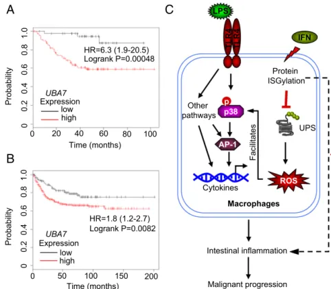
Protein ISGylation Is Correlated with Patient Survival in Human Colorectal Cancer Patients.Using the CAC model, we demonstrated that protein ISGylation leads to more tumor development in mice, suggesting that protein ISGylation may be involved in human CAC. CAC is a subtype of human CRC. There are similarities between CAC and other types of CRC without any signs of overt IBD. Both CAC and CRC are influenced by intestinal microflora and display robust in- flammatory infiltration and increased expression of proinflammatory cytokines (37). Therefore, we examined the possible involvement of protein ISGylation in human CRC using currently available gene- expression microarray data.
We first compared UBA7 and ISG15 expression in healthy persons and CRC patients based on data from ONCOMINE. In a dataset with an adequate number of patients (38), no signifi- cant differences inUBA7andISG15expression were observed in CRC tumor biopsies from the healthy and patient groups (Fig.
S8A andB, Left). However, a recent study suggested that the expression ofISG15was uprated significantly in a group of pa- tients with rectal adenocarcinoma as compared with a healthy group (Fig. S8B,Right) (39). Expression ofUBA7varied widely among individuals in the group of patients with CRC (Fig. S8A).
These data suggest thatUBA7 per se is not an initiating onco- gene in human CRC. To address whether UBE1L has any reg- ulatory function in human CRC, we used two recently published datasets (40, 41) to compare clinical outcomes in groups of CRC patients with low or highUBA7expression. Results from the two datasets revealed that higher expression levels of the UBE1L geneUBA7correlated significantly with worse survival (Fig. 4A and B). As with UBA7, higher expression levels of ISG15 ex- pression are correlated with worse outcomes in these patients (Fig. S8C). In contrast, two other typical IFN-inducible genes (MX1andOAS1) did not show such a correlation with clinical outcome (Fig. S8DandE). These analyses encourage the future study of protein ISGylation in human CRC.
Discussion
Genome-wide association studies and candidate gene studies have identified multiple susceptibility loci in IBD, including the gene locus 3p21 (42). There are six candidate genes in this identified locus, including MST1 and UBA7 (42). MST1 pre- viously has been suggested to be the main gene related to the susceptibility loci in IBD (43). Here we report, for the first time to our knowledge, that theUBA7 gene, via its protein product, ISG15-activating enzyme UBE1L, is also related to colonic in- flammation and the development of CAC. These data suggest a proinflammatory function of protein ISGylation in the large in- testine. Because of the complicated involvement of type I IFN signaling in colonic inflammation, it is reasonable to speculate that certain type I IFN-regulated events are detrimental in IBD but some other types of I IFN-regulated events may be beneficial.
So far, little is known about the role of specific ISGs in colonic inflammation. Therefore, our studies provide insights into how protein ISGylation, a specific IFN-induced cellular event, affects disease progression in mouse models.
In vitro cell line-based studies have revealed contrary roles for protein ISGylation in tumorigenesis (27–29). However, these con- clusions depend largely on cell-autonomous events, such as cell proliferation, cell death, and cell evasion. Nearly all tumors in cancer patients contain immune cells, including macrophages, dendritic cells, and lymphocytes. All these cells produce cytokines and other factors that control inflammatory responses and malig- nant progression (44). In inflammation-associated cancer, immune cells play a more important role in tumor development at sites of
chronic inflammation. We observed that macrophages with protein ISGylation exhibit higher expression of multiple inflammatory cy- tokines, suggesting that UBE1L indirectly stimulates the malignant transition of intestinal epithelial cells by enhancing the production of inflammatory cytokines in lamina propria immune cells. In the current report we focused on macrophages largely because of the high association of protein ISGylation with macrophages in the large intestine. Furthermore, protein ISGylation in epithelia- derived tumor cells may have different roles in cancer development in other contexts; for example, Desai et al. (45) have reported that protein ISGylation promotes motility in human breast cancer cells.
Our earlier studies used two lung cancer mouse models, K-rasLA2and p53-deficient mice, to address the role of protein ISGylation in cancer development by breeding these mice with Ube1L-KO mice (46, 47). However, neither tumor-promoting nor -suppressing effects of protein ISGylation were observed.
The initiation of lung tumors was observed as early as 2 wk after birth in the K-rasLA2lung mouse model, suggesting that K-rasLA2 is extremely tumor promoting. In a p53-deficient mouse cancer model, an alteration of tumor spectrum by protein ISGylation was observed. Using a CAC model, we demonstrated that pro- tein ISGylation significantly exacerbates tumor formation. These results suggest that protein ISGylation may contribute to the regulation of the cellular environment that affects tumor devel- opment but does not play an initiating role in tumorigenesis, at least not in the murine p53-deficient and K-rasLA2cancer mod- els. Additional work with other mouse cancer models should provide more insights into the role of protein ISGylation in the development of specific types of cancer.
One major question regarding the role of protein ISGylation is how the modification of a small fraction of any individual target protein can have a significant effect on the overall activity of that protein. Here we did not focus on the functions of any individual ISG15 target protein; rather, we characterized the negative effect of protein ISGylation on UPS activity in macrophages. We dem- onstrated that regulation of UPS by protein ISGylation contrib- utes to IFN-induced ALIS formation in macrophages. More
importantly, we showed that the direct modification of ubiquity- lated proteins by ISG15 impaired proteasome processing of ubiquitylated proteins. Therefore, this pool of ubiquitylated pro- teins with additional ISG15 incorporation may be a good source of initiators for the formation of IFN-induced cellular inclusions.
Because nucleation limits the rate of aggregate formation, and the presence of a small number of seeds dramatically promotes the process of protein aggregation (48), a few aggregates formed by these ISG15/ubiquitin-containing proteins may affect cellular protein aggregation significantly. In agreement with this notion, ISG15/ubiquitin double-positive inclusions have been observed in brain sections obtained from patients with ataxia telangiec- tasia (49).
To our knowledge, this is the first report ofUBA7and protein ISGylation in colitis and CAC. A proposed model of how protein ISGylation contributes to exacerbated intestinal inflammation is illustrated in Fig. 4C. Protein ISGylation may interfere with the UPS function in multiple arms, such as the competition of E1/E2/E3s for ubiquitylation (50) and the competition of ubiq- uitylation sites on substrates (51). One particular effect may be through ISGylation of ubiquitylated proteins, which disrupts the formation of long ubiquitin chains (52). All these different arms in combination convey a general inhibitory effect on cellular UPS function that can lead to the enhanced production of IFN- induced cellular ROS. Such IFN-induced ROS augments LPS- induced p38 activation, increasing the production of inflammatory cytokines in macrophages. Therefore, our data suggest crosstalk between type I IFN-triggered protein ISGylation and LPS- induced p38 activation in colonic macrophages and the contri- bution of such synergized events to colonic inflammation and malignant progression. Importantly, we observed an interesting correlation of UBE1L expression with clinical outcomes in hu- man CRC. Thus, the enhanced inflammation in the presence of an active ISG15 conjugation system also may affect human CRC.
Future studies focused on protein ISGylation in human IBD or CRC may pave the way for the potential application of inhibitors
0 50 100 150 200
Time (months)
00.20.40.60.81.0
Probability Expression
low high
HR=1.8 (1.2-2.7) Logrank P=0.0082 UBA7
00.20.40.60.81.0
Expression low high
HR=6.3 (1.9-20.5) Logrank P=0.00048
Probability
Time (months)
0 20 40 60 80 100
UBA7
C A
B
Other pathways
Protein ISGylation
UPS
Macrophages
Intestinal inflammation
Malignant progression ROS LPS
TLR4
p38 p
IFN
AP-1
Cytokines TLR4 Facilitates
Fig. 4. Protein ISGylation is correlated with patient survival in patients with CRC. (AandB) Kaplan–Meier curves of patients with CRC from two independent datasets (A: GSE37892,n=130;B: GSE39582,n=519) showing the correlation between relapse-free sur- vival of individuals with UBE1L gene expression (Affymetrix ID 203281_atUBA7). The cutoff value for defining higher/lower expression was 359 for GSE37892 (A) and 311 for GSE39582 (B), at the lower quartile of the expression (maximum value 1,000) in each dataset. (C) Schematic overview of protein ISGylation in regulating the convergence of IFN- and LPS-related pathways and its contribution to in- testinal inflammation. Protein ISGylation conveys a general inhibitory effect on cellular UPS, resulting in an increase in IFN-induced ROS production. The en- hanced cellular ROS further facilitates LPS-induced p38 activation and production of inflammatory cyto- kines in macrophages. These effects together con- tribute to intestinal inflammation and malignant progression in vivo.
IMMUNOLOGYAND INFLAMMATION
of the ISG15-activating enzyme UBE1L in the therapeutic treatment of these human diseases.
Materials and Methods
Animals.All experimental protocols were approved by the Institutional Animal Care and Use Committee of the University of California, San Diego (UCSD). All mice were housed and bred at the vivarium of the Moores Cancer Center at UCSD.
DSS-Induced Intestinal Inflammation and CAC Induction.DSS (MP Biomedicals, molecular weight=∼40,000) was added to drinking water (2%, wt/vol) for 7 d;
regular water was given thereafter. Colons were removed quickly at the in- dicated time points. Gender-matched littermates (age∼12 wk) were monitored
for weight loss, rectal bleeding, and stool consistency. For details, seeSI Materials and Methods.
Further Details. Additional information about the experimental methods maybe found inSI Materials and Methods.Table S1lists the sequences of primers used in this study.
ACKNOWLEDGMENTS.We thank Samuel Stoner for critical editing of the manuscript; Dr. Xiang-dong Fu and all the members of the D.-E.Z. laboratory for discussions and help; Dr. Yu Zhou (College of Life Sciences, Wuhan Uni- versity) for guidance in analyzing the TCGA dataset; and Drs. Herbert Virgin and Debora Lenschow (Washington University School of Medicine) for providing antibodies for ISG15. This work was funded by NIH Grant R01CA177305 (to D.-E.Z.).
1. Grivennikov SI, Greten FR, Karin M (2010) Immunity, inflammation, and cancer.Cell 140(6):883–899.
2. Cho JH, Brant SR (2011) Recent insights into the genetics of inflammatory bowel disease.Gastroenterology140(6):1704–1712.
3. Xavier RJ, Podolsky DK (2007) Unravelling the pathogenesis of inflammatory bowel disease.Nature448(7152):427–434.
4. Sadler AJ, Williams BR (2008) Interferon-inducible antiviral effectors. Nat Rev Immunol8(7):559–568.
5. González-Navajas JM, Lee J, David M, Raz E (2012) Immunomodulatory functions of type I interferons.Nat Rev Immunol12(2):125–135.
6. Katakura K, et al. (2005) Toll-like receptor 9-induced type I IFN protects mice from experimental colitis.J Clin Invest115(3):695–702.
7. Pena Rossi C, et al. (2009) Interferon beta-1a for the maintenance of remission in patients with Crohn’s disease: Results of a phase II dose-finding study. BMC Gastroenterol9:22.
8. Pena-Rossi C, et al. (2008) Clinical trial: A multicentre, randomized, double-blind, placebo-controlled, dose-finding, phase II study of subcutaneous interferon-beta-la in moderately active ulcerative colitis.Aliment Pharmacol Ther28(6):758–767.
9. Rauch I, et al. (2014) Type I interferons have opposing effects during the emergence and recovery phases of colitis.Eur J Immunol44(9):2749–2760.
10. Schott E, et al. (2007) Development of ulcerative colitis in a patient with multiple sclerosis following treatment with interferon beta 1a.World J Gastroenterol13(26):
3638–3640.
11. Farrell PJ, Broeze RJ, Lengyel P (1979) Accumulation of an mRNA and protein in in- terferon-treated Ehrlich ascites tumour cells.Nature279(5713):523–525.
12. Malakhova OA, et al. (2003) Protein ISGylation modulates the JAK-STAT signaling pathway.Genes Dev17(4):455–460.
13. Loeb KR, Haas AL (1992) The interferon-inducible 15-kDa ubiquitin homolog conju- gates to intracellular proteins.J Biol Chem267(11):7806–7813.
14. Kok K, et al. (1993) A gene in the chromosomal region 3p21 with greatly reduced expression in lung cancer is similar to the gene for ubiquitin-activating enzyme.Proc Natl Acad Sci USA90(13):6071–6075.
15. Yuan W, Krug RM (2001) Influenza B virus NS1 protein inhibits conjugation of the interferon (IFN)-induced ubiquitin-like ISG15 protein.EMBO J20(3):362–371.
16. Zhao C, et al. (2004) The UbcH8 ubiquitin E2 enzyme is also the E2 enzyme for ISG15, an IFN-alpha/beta-induced ubiquitin-like protein.Proc Natl Acad Sci USA101(20):
7578–7582.
17. Kim KI, Giannakopoulos NV, Virgin HW, Zhang DE (2004) Interferon-inducible ubiq- uitin E2, Ubc8, is a conjugating enzyme for protein ISGylation.Mol Cell Biol24(21):
9592–9600.
18. Okumura F, Zou W, Zhang DE (2007) ISG15 modification of the eIF4E cognate 4EHP enhances cap structure-binding activity of 4EHP.Genes Dev21(3):255–260.
19. Zou W, Zhang DE (2006) The interferon-inducible ubiquitin-protein isopeptide ligase (E3) EFP also functions as an ISG15 E3 ligase.J Biol Chem281(7):3989–3994.
20. Dastur A, Beaudenon S, Kelley M, Krug RM, Huibregtse JM (2006) Herc5, an in- terferon-induced HECT E3 enzyme, is required for conjugation of ISG15 in human cells.J Biol Chem281(7):4334–4338.
21. Wong JJ, Pung YF, Sze NS, Chin KC (2006) HERC5 is an IFN-induced HECT-type E3 protein ligase that mediates type I IFN-induced ISGylation of protein targets.Proc Natl Acad Sci USA103(28):10735–10740.
22. Malakhov MP, Malakhova OA, Kim KI, Ritchie KJ, Zhang DE (2002) UBP43 (USP18) specifically removes ISG15 from conjugated proteins.J Biol Chem277(12):9976–9981.
23. Liu LQ, et al. (1999) A novel ubiquitin-specific protease, UBP43, cloned from leukemia fusion protein AML1-ETO-expressing mice, functions in hematopoietic cell differen- tiation.Mol Cell Biol19(4):3029–3038.
24. Morales DJ, Lenschow DJ (2013) The antiviral activities of ISG15.J Mol Biol425(24):
4995–5008.
25. Zhang D, Zhang DE (2011) Interferon-stimulated gene 15 and the protein ISGylation system.J Interferon Cytokine Res31(1):119–130.
26. Bogunovic D, Boisson-Dupuis S, Casanova JL (2013) ISG15: Leading a double life as a secreted molecule.Exp Mol Med45:e18.
27. Forys JT, et al. (2014) ARF and p53 coordinate tumor suppression of an oncogenic IFN-β-STAT1- ISG15 signaling axis.Cell Reports7(2):514–526.
28. Burks J, Reed RE, Desai SD (2014) ISGylation governs the oncogenic function of Ki-Ras in breast cancer.Oncogene33(6):794–803.
29. Kiessling A, et al. (2009) Expression, regulation and function of the ISGylation system in prostate cancer.Oncogene28(28):2606–2620.
30. Neufert C, Becker C, Neurath MF (2007) An inducible mouse model of colon carci- nogenesis for the analysis of sporadic and inflammation-driven tumor progression.
Nat Protoc2(8):1998–2004.
31. Neurath MF (2014) Cytokines in inflammatory bowel disease.Nat Rev Immunol14(5):
329–342.
32. Cancer Genome Atlas Network (2012) Comprehensive molecular characterization of human colon and rectal cancer.Nature487(7407):330–337.
33. Hsu HY, Wen MH (2002) Lipopolysaccharide-mediated reactive oxygen species and signal transduction in the regulation of interleukin-1 gene expression.J Biol Chem 277(25):22131–22139.
34. Seifert U, et al. (2010) Immunoproteasomes preserve protein homeostasis upon in- terferon-induced oxidative stress.Cell142(4):613–624.
35. Wu HM, Chi KH, Lin WW (2002) Proteasome inhibitors stimulate activator protein-1 pathway via reactive oxygen species production.FEBS Lett526(1-3):101–105.
36. Raasi S, Orlov I, Fleming KG, Pickart CM (2004) Binding of polyubiquitin chains to ubiquitin-associated (UBA) domains of HHR23A.J Mol Biol341(5):1367–1379.
37. Terzic J, Grivennikov S, Karin E, Karin M (2010) Inflammation and colon cancer.
Gastroenterology138(6):2101–2114.
38. Ki DH, et al. (2007) Whole genome analysis for liver metastasis gene signatures in colorectal cancer.Int J Cancer121(9):2005–2012.
39. Gaedcke J, et al. (2010) Mutated KRAS results in overexpression of DUSP4, a MAP- kinase phosphatase, and SMYD3, a histone methyltransferase, in rectal carcinomas.
Genes Chromosomes Cancer49(11):1024–1034.
40. Marisa L, et al. (2013) Gene expression classification of colon cancer into molecular subtypes: Characterization, validation, and prognostic value.PLoS Med10(5):e1001453.
41. Laibe S, et al.; COL2 Project (2012) A seven-gene signature aggregates a subgroup of stage II colon cancers with stage III.OMICS16(10):560–565.
42. Anderson CA, et al. (2011) Meta-analysis identifies 29 additional ulcerative colitis risk loci, increasing the number of confirmed associations to 47.Nat Genet43(3):246–252.
43. Skieceviciene J, et al. (2013) Replication study of ulcerative colitis risk loci in a Lithuanian- Latvian case-control sample.Inflamm Bowel Dis19(11):2349–2355.
44. Mantovani A, Allavena P, Sica A, Balkwill F (2008) Cancer-related inflammation.
Nature454(7203):436–444.
45. Desai SD, et al. (2012) ISG15 disrupts cytoskeletal architecture and promotes motility in human breast cancer cells.Exp Biol Med (Maywood)237(1):38–49.
46. Yin X, Cong X, Yan M, Zhang DE (2009) Deficiency of a potential 3p21.3 tumor suppressor gene UBE1L (UBA7) does not accelerate lung cancer development in K-rasLA2 mice.Lung Cancer63(2):194–200.
47. Yin X, Cong X, Yan M, Zhang DE (2009) Alteration of tumor spectrum by ISGylation in p53-deficient mice.Cancer Biol Ther8(12):1167–1172.
48. Morales R, Moreno-Gonzalez I, Soto C (2013) Cross-seeding of misfolded proteins:
Implications for etiology and pathogenesis of protein misfolding diseases.PLoS Pathog9(9):e1003537.
49. Wood LM, et al. (2011) A novel role for ATM in regulating proteasome-mediated protein degradation through suppression of the ISG15 conjugation pathway.PLoS One6(1):e16422.
50. Malakhova OA, Zhang DE (2008) ISG15 inhibits Nedd4 ubiquitin E3 activity and en- hances the innate antiviral response.J Biol Chem283(14):8783–8787.
51. Desai SD, et al. (2006) Elevated expression of ISG15 in tumor cells interferes with the ubiquitin/26S proteasome pathway.Cancer Res66(2):921–928.
52. Fan JB, et al. (2015) Identification and characterization of a novel ISG15-ubiquitin mixed chain and its role in regulating protein homeostasis.Sci Rep5:12704.
53. Weischenfeldt J, Porse B (2008) Bone marrow-derived macrophages (BMM): Isolation and applications.CSH Protoc:pdb prot5080. Available at dx.doi.org/10.1101/pdb.
prot5080. Accessed October 10, 2015.
54. Cooper HS, Murthy SN, Shah RS, Sedergran DJ (1993) Clinicopathologic study of dextran sulfate sodium experimental murine colitis.Lab Invest69(2):238–249.
55. Laroui H, et al. (2012) Dextran sodium sulfate (DSS) induces colitis in mice by forming nano-lipocomplexes with medium-chain-length fatty acids in the colon.PLoS One 7(3):e32084.
56. Liu CW, et al. (2006) ATP binding and ATP hydrolysis play distinct roles in the function of 26S proteasome.Mol Cell24(1):39–50.
57. Gyorffy B, Lánczky A, Szállási Z (2012) Implementing an online tool for genome-wide validation of survival-associated biomarkers in ovarian-cancer using microarray data from 1287 patients.Endocr Relat Cancer19(2):197–208.