Measurement of Inositol 1,4,5-Trisphosphate in Living Cells Using an Improved Set of
Resonance Energy Transfer-Based Biosensors
GergőGulyás1☯, József T. Tóth1☯, Dániel J. Tóth1, István Kurucz2, László Hunyady1, Tamas Balla2, Péter Várnai1*
1Department of Physiology, Faculty of Medicine, Semmelweis University, Budapest 1094, Hungary, 2Section on Molecular Signal Transduction, Program for Developmental Neuroscience, National Institute of Child Health and Human Development, National Institutes of Health, Bethesda, MD 20892, United States of America
☯These authors contributed equally to this work.
*varnai.peter@med.semmelweis-univ.hu
Abstract
Improved versions of inositol-1,4,5-trisphosphate (InsP3) sensors were created to follow in- tracellular InsP3changes in single living cells and in cell populations. Similar to previous InsP3sensors the new sensors are based on the ligand binding domain of the human type-I InsP3receptor (InsP3R-LBD), but contain a mutation of either R265K or R269K to lower their InsP3binding affinity. Tagging the InsP3R-LBD with N-terminal Cerulean and C-termi- nal Venus allowed measurement of InsP3in single-cell FRET experiments. Replacing Ceru- lean with a Luciferase enzyme allowed experiments in multi-cell format by measuring the change in the BRET signal upon stimulation. These sensors faithfully followed the agonist- induced increase in InsP3concentration in HEK 293T cells expressing the Gq-coupled AT1 angiotensin receptor detecting a response to agonist concentration as low as 10 pmol/L.
Compared to the wild type InsP3sensor, the mutant sensors showed an improved off-rate, enabling a more rapid and complete return of the signal to the resting value of InsP3after termination of M3 muscarinic receptor stimulation by atropine. For parallel measurements of intracellular InsP3and Ca2+levels in BRET experiments, the Cameleon D3 Ca2+sensor was modified by replacing its CFP with luciferase. In these experiments depletion of plasma membrane PtdIns(4,5)P2resulted in the fall of InsP3level, followed by the decrease of the Ca2+-signal evoked by the stimulation of the AT1 receptor. In contrast, when type-III PI 4-ki- nases were inhibited with a high concentration of wortmannin or a more specific inhibitor, A1, the decrease of the Ca2+-signal preceded the fall of InsP3level indicating an InsP3-, in- dependent, direct regulation of capacitative Ca2+influx by plasma membrane inositol lipids.
Taken together, our results indicate that the improved InsP3sensor can be used to monitor both the increase and decrease of InsP3levels in live cells suitable for high-throughput BRET applications.
OPEN ACCESS
Citation:Gulyás G, Tóth JT, Tóth DJ, Kurucz I, Hunyady L, Balla T, et al. (2015) Measurement of Inositol 1,4,5-Trisphosphate in Living Cells Using an Improved Set of Resonance Energy Transfer-Based Biosensors. PLoS ONE 10(5): e0125601.
doi:10.1371/journal.pone.0125601
Academic Editor:Mohamed Trebak, Penn State Hershey College of Medicine, UNITED STATES Received:January 16, 2015
Accepted:March 24, 2015 Published:May 1, 2015
Copyright:This is an open access article, free of all copyright, and may be freely reproduced, distributed, transmitted, modified, built upon, or otherwise used by anyone for any lawful purpose. The work is made available under theCreative Commons CC0public domain dedication.
Data Availability Statement:All relevant data are within the paper.
Funding:Funding provided by Hungarian Scientific Research Fund (OTKA) K105006: PV,http://www.
otka.hu/en.
Competing Interests:The authors have declared that no competing interests exist.
Introduction
Inositol-1,4,5-trisphosphate (InsP3) plays a central role in calcium signaling. Its production is catalyzed by phospholipase C enzymes activated through receptor stimulation. Measurements of InsP3kinetics for a long time relied upon isotope labeling of cell populations followed by ex- traction and HPLC-separation of the active (1,4,5) isomer from the inactive (1,3,4) one [1].
Mass measurements of InsP3using radio-receptor assays have also been used [2] to measure absolute mass changes, again from populations of cell, but these methods have been quite cum- bersome and unable to provide very detailed time resolution. Moreover, these methods did not allow detection of kinetic changes in single cells with accurate comparisons with other parame- ters, such as cytoplasmic Ca2+changes. These deficiencies prompted several groups, including ours, to develop InsP3sensor that would be useful for single-cell analysis. With the advent of fluorescent proteins and the development of fluorescence resonance energy transfer (FRET) technology, InsP3probes based on the ligand binding domain of the InsP3receptor have been introduced and used successfully in several single-cell applications [3–7].
In reviewing our experimental data obtained with a sensor that was developed in our group (using the same principles referenced above) and comparing it with published sensors, such as IRIS [3], we noted that the sensors not only distorted InsP3kinetics because of their buffering effects (a complication that is unavoidable with any probe), but they also showed InsP3kinetics suggestive of slow off-rates. To overcome this problem, we designed modified probes to address these kinetic deficiencies. Importantly, we also wanted to take this tool further such that it could be used in cell populations allowing a format amenable to screening applications.
Here we report on the fine-tuning and characterization of our InsP3sensor based on the human type-I InsP3receptor LBD (residues 224–605). Structural studies showed that InsP3
binding leads to a conformational change of this protein domain, which can be translated to a change in FRET signal between two appropriate fluorophores placed at the two ends of the LBD. Similar probes have been introduced and published (seeTable 1). It has been show earlier that deletion of the N-terminal 223 amino acids increases the affinity of the LBD, so the 224– 605 LBD has a higher affinity than the native InsP3receptor channel [8]. Therefore, we decided to engineer slightly lower affinity mutants by mutating the InsP3binding site in order to im- prove its off-rate upon decrease in InsP3but still keep their abilities to detect the increase of InsP3level. In addition, we have demonstrated the ability of these probes to faithfully monitor InsP3concentrations by either FRET or bioluminescence resonance energy transfer applica- tions (BRET).
To analyze and compare our sensors, we needed an experimental system in which an in- crease or decrease in InsP3concentration could be equally established. For this, type-1 angio- tensin receptor (AT1R) or the M3 cholinergic receptor was transiently transfected into HEK 293T cells, so that InsP3concentration could be increased by angiotensin II or carbachol stimu- lation, respectively. Decrease of InsP3was evoked by terminating the muscarinic response by atropine. Under these conditions both the wild-type and the mutant sensors were able to show the rapid rise in InsP3levels, but the mutants were able to detect a more rapid and full decline in InsP3concentration. Our data suggest that these improved mutant sensors are suitable to in- vestigate InsP3signaling more accurately than previous ones either by single-cell imaging or in cell population measurements.
Materials and Methods
Materials
Molecular biology reagents were obtained from Fermentas (Vilnius, Lithuania). Cell culture dishes and plates were purchased from Greiner (Kremsmunster, Austria). Coelenterazineh was purchased from Regis Technologies (Morton Grove, IL). Lipofectamine 2000 was from Invitrogen (Carlsbad, CA). Rapamycin was obtained from Merck (Darmstadt, Germany). Gen- eCellin transfection reagent was from BioCellChallenge (Toulon, France). Atropine was pur- chased from EGIS (Budapest, Hungary). Unless otherwise stated, all other chemicals and reagents were purchased from Sigma (St Louis, MO).
DNA constructs
The R265K, R269K, R568K, R504K and R265,269K mutations were introduced by site-directed mutagenesis (Agilent Technologies, Santa Clara, CA USA) in the previously created
mRFP-InsP3R-LBD (residues 224–605 of human type-1 InsP3receptor S1+) construct used for bacterial expression of the fusion proteins [9]. To create the InsP3sensors first we made a FRET plasmid backbone by cloning the monomeric Venus [10] into the pEYFP-C1 plasmid, in which YFP was already replaced by Cerulean [11], using EcoRI and NotI enzymes. The wild type or mutant InsP3R-LBDs were then inserted between the two fluorophores using XhoI and
Table 1. InsP3sensors in use for single-cell fluorescent energy transfer-based applications.
Sensor InsP3-binding domain KD Fluorescent proteins Reference
LIBRA rat type-III InsP3R (1–604) 404 nM CFP/YFP [4,31]
LIBRA-ΔN* rat type-III InsP3R (227–604) ND CFP/YFP [4,5,31]
LIBRA-vI rat type-I InsP3R (1–604) 269 nM CFP/Venus [32]
LIBRA-vII rat type-II InsP3R (1–604) 234 nM CFP/Venus [32]
LIBRA-vIIS LIBRAvII R440Q 117 nM CFP/Venus [32]
LIBRA-vIII rat type-III InsP3R (1–604) 492 nM CFP/Venus [5,32]
LIBRA-vIIIS LIBRAvIII R440Q 250 nM CFP/Venus [32]
Fretino human type-I InsP3R (224–579) 8 nM CFP/YFP [7]
Fretino-2 Fretino R504Q 190 nM CFP/YFP [7]
Fretino-3** Fretino R508Q ND CFP/YFP [7]
Fretino-4 human type-I InsP3R (1–604) ND CFP/YFP [7]
IRIS-1 mouse type-I InsP3R (224–575) 549 nM (cell lysate), 437 nM (purified) Venus/CFP [3]
IRIS-1-Dmut** IRIS-1 T276A, K508Q ND [3]
IRIS-1.2 IRIS-1 K249Q 3–4μM [3]
mouse type-I InsP3R (224–579) 95 nM Venus/CFP [3]
mouse type-I InsP3R (224–584) 105 nM Venus/CFP [3]
mouse type-I InsP3R (224–604) 107 nM Venus/CFP [3]
FIRE-1 rat type-I InsP3R (1–589) 31 nM CFP/YFP [6]
FIRE-2 rat type-II InsP3R (1–604) ND CFP/YFP [6]
FIRE-3 rat type-III InsP3R (1–604) 36 nM CFP/YFP [6]
KDvalues represent the InsP3concentrations required to reach 50% of the dynamic range of the appropriate sensor. ND means not determined.
*InsP3insensitive mutant of LIBRA
**non-binding mutant for control experiments doi:10.1371/journal.pone.0125601.t001
EcoRI resulting in two short linkers before and after the LBD (NEQRSR and NS). From these FRET sensors the BRET sensors were prepared by replacing Cerulean with superRenillalucif- erase [12]. To improve the optical parameters, another set of BRET sensors were created by re- placing Venus with the Venus cp173-Venus tandem used in other sensors like the Epac cAMP sensor [13].
The Ca2+sensor used in the BRET measurements was created by replacing the InsP3R-LBD with the appropriate sequence derived from Cameleon D3 [14]. For the replacement, first this sequence was amplified using PCR with the sense primer CTCGAGACCAACTGACAGAA GAGCAGATTGCAGAG and antisense primer GAATTCAGTGCCCCGGAGCTGGAGA TCTTC, and then it was cloned into the BRET plasmid using XhoI and EcoRI enzymes.
Wild type human M3 cholinergic receptor (N-terminal 3x-hemagglutinin tagged) was pur- chased from S&T cDNA Resource Center (Rolla, MO). The non-internalizing rat type-I angio- tensin receptor (AT1R-Δ319) was described earlier [15]. The plasma membrane targeted FRB- mRFP and mRFP-FKBP-5-ptase constructs used for rapamycin-induced PtdIns(4,5)P2deple- tion were described earlier [16] with the difference that for plasma membrane targeting of the FRB protein, we used the N-terminal targeting sequence (1–10) of mouse Lck (GenBank acces- sion number: NM_001162433) [17].
Cell culture
HEK 293T and COS-7 cells (ATCC, Manassas, VA) were maintained in Dulbecco’s modified Eagle’s medium (DMEM, Lonza 12–604) supplemented with 10% fetal bovine serum, 50 U/ml penicillin and 50μg/ml streptomycin in a 5% humidified CO2incubator at 37°C in 10 cm tissue culture plastic dishes.
Ins
P3binding experiments
mRFP-tagged wild type and mutant InsP3R-LBD domains, built into the pET-23b bacterial ex- pression vector (Novagen) were used to transform the BL-21 DE3 Star strain ofEscherichia coli (Invitrogen). Bacterial cells were grown to A6000.6–0.9 at 37°C and induced with 300μM iso- propyl-1-thio-β-D-galactopyranoside at 18–20°C for 8 hours. Purification of the recombinant protein as well as InsP3binding assay were performed as described previously [9], using 0.75μCi (1 nM) [3H]Ins(1,4,5)P3(Amersham Biosciences) and 200 ng protein in an incubation volume of 50μl.
Fluorescence Resonance Energy Transfer (FRET) measurements
For FRET measurements HEK 293T cells were trypsinized and plated on poly-lysine-pre- treated (0.001%, 1 hour) No 1.5 glass coverslips in 35 mm plastic dishes at 3x10⁵cells/dish den- sity. After one day the culture medium was changed to 1 ml Opti-MEM (Gibco) medium, and then 200μl transfection solution containing the indicated DNA constructs (1μg total DNA/dish) and 2μl/dish Lipofectamine 2000 was added. After 6 hours 1 ml DMEM containing serum and antibiotics was added. Measurements were performed 24–32 hours after the trans- fection. Before the measurements the coverslips were placed into Attofluor cell chambers (Invi- trogen) and the medium was changed to 800μl of a modified Krebs–Ringer buffer containing 120 mM NaCl, 4.7 mM KCl, 1.2 mM CaCl2, 0.7 mM MgSO4, 10 mM glucose, and 10 mM Na- HEPES, pH 7.4. Measurements were performed at room temperature using an inverted micro- scope (Axio Observer D1, Zeiss, Germany) equipped with a 40x/1.3 oil-immersion objective (Plan-APO, Zeiss) and a Cascade II camera (Photometrics, Tucson, AZ). Excitation wave- lengths (435 nm and 500 nm) were set by a monochromator connected to a 75 W Xenon lamp (DeltaRAM, Photon Technology International, Birmingham, NJ). The emitted light was
separated by a dichroic beamsplitter (Chroma 69008bs), and then detected through the appro- priate emission filters for Cerulean (470/24 nm) and Venus (535/30 nm). Images were acquired every 5 s. The indicated reagents were also dissolved in modified Krebs–Ringer buffer and were added manually in 200μl, and mixed three times. The MetaFluor (Molecular Devices, Down- ingtown, PA) software was used for data acquisition, whereas for further data analysis includ- ing background subtraction, bleed through correction and 535/470 emission ratio calculation the MetaMorph (Molecular Devices) software was applied.
Bioluminescence Resonance Energy Transfer (BRET) measurements
For BRET measurements HEK 293T cells were trypsinized and plated on poly-lysine-pre- treated (0.001%, 1 hour) white 96-well plates at a density of 105cells/well together with the in- dicated DNA constructs (0.24–0.3μg total DNA/well) and the cell transfection reagent (0.5μl/well Lipofectamine 2000 or 1.5μl/well GeneCellin). After 6 hours 100μl/well DMEM contain- ing serum and antibiotics was added. Measurements were performed 24–27 h after transfec- tion. Before measurements the medium of cells was changed to a medium (50μl) containing 120 mM NaCl, 4.7 mM KCl, 1.2 mM CaCl2, 0.7 mM MgSO4, 10 mM glucose, and 10 mM Na- HEPES, pH 7.4. Measurements were performed at 37°C using a Mithras LB 940 multilabel reader (Berthold, Germany). The measurements started with the addition of the cell permeable luciferase substrate, coelenterazineh(40μl, final concentration of 5μM), and counts were re- corded using 485 and 530 nm emission filters. Detection time was 500 ms for each wavelength.
The indicated reagents were also dissolved in modified Krebs–Ringer buffer and were added manually in 10μl. For this, plates were unloaded, which resulted in an interruption in the re- cordings. All measurements were done in triplicates. BRET ratios were calculated by dividing the 530 nm and 485 nm intensities, and normalized to the baseline.
Confocal microscopy
COS-7 cells were cultured on IBIDIμ-Slide 8 Well dishes (IBIDI GmbH, Cat. No.: 80826;
2x10⁴cells/well) and transfected with the indicated constructs (1μg DNA total/dish) using 0.5μl/well Lipofectamine 2000 for 24 h. Confocal measurements were performed at 35°C in a modified Krebs-Ringer buffer described above, using a Zeiss LSM 710 scanning confocal mi- croscope and a 63x/1.4 oil-immersion objective. Post-acquisition picture analysis was per- formed using the Photoshop (Adobe) software to expand to the full dynamic range but only linear changes were allowed.
Statistical analysis
To calculate the half-time values (τ) of the decay phase, a curve fitting procedure was applied to the values in each individual experiment using the 3 parametric exponential decay equation of (y = y0+ae-bx).τvalues were then averaged and subjected to a t-test.
Results
Generation of reduced affinity Ins
P3sensors that can be used in energy transfer measurements
To design an improved InsP3sensor first we decided to perform a moderate modification in the InsP3binding domain (224–605 amino acids) of human type-I InsP3receptor (IP3R-LBD) [18] by replacing specific arginine residues to lysines. Since the side chain of lysine is shorter than that of arginine and lysine is a less basic amino-acid than arginine (pI-values are 9.74 and 10.76, respectively) we reasoned that interaction of the mutant protein with negatively charged
phosphate groups of InsP3will be weaker. We expected that this manipulation would decrease the binding affinity and therefore increase the off-rate performance of the sensor. Based on the crystal structure of the binding domain [19] we selected R265, R269, R504 and R568 residues for mutation (Fig 1A). To investigate the InsP3binding properties of these proteins, first we created constructs for bacterial expression. The mRFP-, and 6xHis-tagged InsP3binding do- mains were purified on Ni2+columns, and in vitro binding assays were performed using [3H]
InsP3as the tracer. As shown inFig 1Bthe binding of R265K, R269K and R568K were weaker compared to the wild type protein, while the R504K and the double mutant R265,269K were very poor InsP3binders. Scatchard analysis of the curves also showed the lower affinity of the mutants: the KDvalue for the R265K, R269K and R568K mutants were 6.06 nM, 10.07 nM and 5.30 nM, respectively compared to 3.04 nM for the wild type LBD. Based on these results, the R265K and R269K mutants were selected for further analysis.
In previous studies we created the InsP3R-LBD with various N-terminal fluorescent tags, such as mRFP or Cerulean, in combination with a C-terminal Venus tag. Application of the protein as InsP3sensor in BRET measurements requires that the N-terminal fluorescent pro- tein tag be replaced with a luciferase enzyme.Fig 1Cshows the final structure of the InsP3sen- sors that contain either Cerulean [11] or Super Luciferase (Sluc) [12] tag on their N-terminus, and Venus on the C-terminus. Studies on the dependence of energy transfer efficiency between fluorescent proteins suggested that introduction of double-acceptor moieties may significantly enhance the FRET efficiency value [13]. Therefore, we also created constructs with tandem yel- low fluorescent tag (cp173 Venus and Venus) on their C-termini [13].
As shown before for the previously developed sensors, expression of these sensors in mam- malian cells (COS-7 and HEK 293T) resulted in uniform cytoplasmic distribution of the fusion proteins and their reduced levels in the nucleus (Fig 1D). Similarly to other published InsP3
sensors (seeTable 1), InsP3binding caused a decrease of energy transfer in both FRET and BRET measurements (Fig 1E and 1F). Notably, duplication of the acceptor did not result in im- provement of the signal in the BRET format (Fig 1F). Since raw BRET ratio values often show a spontaneous increase over time, we included wells that were left unstimulated and used these to correct for baseline drift. From these data the reciprocal of the I/I0values were calculated and plotted, therefore the elevation of cytoplasmic InsP3level corresponds to an increased value on the graphs. Since the BRET application is done in cell suspension and therefore inher- ently reports on averaged cell responses, the subsequent characterization was done using the BRET approach.
Characterization of the InsP
3sensitivity and reversibility of the reduced affinity InsP
3sensors
To compare the InsP3-induced signals of the wild type and low affinity InsP3sensors, HEK 293T cells were transfected with the cDNAs of both the human AT1 receptor and the Sluc/
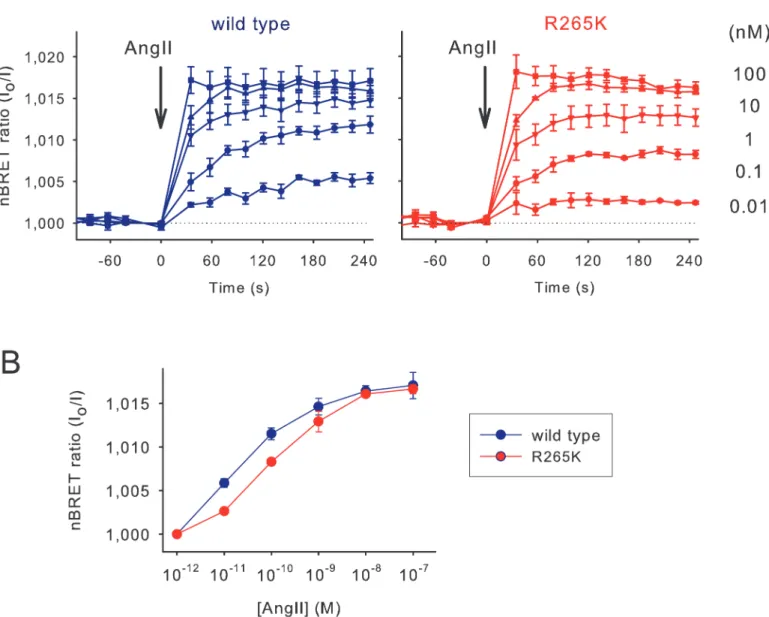
Venus version of the appropriate InsP3sensor. BRET changes were then recorded following angiotensin II (Ang II) stimulation. As shown onFig 2A, increasing concentrations of Ang II from 10–12to 10–7M resulted in an increasing BRET signal in case of both the wild type and the mutant sensors. The signals also showed a kinetic difference as the concentration of Ang II was increased (Fig 2A). While the maximal response was the same, a moderate shift to the right occurred on the dose-response curve in case of the sensor with the R265K mutation re- flecting the lower affinity of this mutant sensor (Fig 2B).
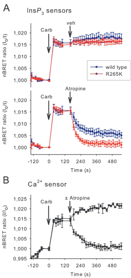
To investigate the reversibility of the InsP3binding of the sensors, HEK 293T cells transient- ly expressing the M3 cholinergic receptor were stimulated first with carbachol (10μM), which activated the Gq signaling pathway and elevated the cytoplasmic InsP3and Ca2+levels. These
responses can be quickly terminated by adding the competitive antagonist atropine (10μM).
As shown inFig 3Athe rising phases of the signals, which reflect the elevation of InsP3, were indistinguishable between the wild-type and mutant InsP3sensors (however, note the low tem- poral resolution of about 3 points per minute). In contrast, the mutant sensor showed a signifi- cantly enhanced off-rate (τ= 23.0±2.3 s) compared to the wild type sensor (τ= 44.9±7.5 s) (Mean ± S.E.M. n = 5, p = 0.024), and a complete return to the baseline. InsP3measurement performed with the wild-type sensor revealed an elevated level of cytoplasmic InsP3concentra- tion after the atropine treatment, which may reflect an incomplete dissociation of InsP3from the high affinity wild-type sensor or a non-complete inhibition of M3R signaling with atropine.
Notably, in control cells, which were not treated with atropine, the InsP3level remains elevated up to 10 minutes consistent with a slow desensitization of the M3R, as demonstrated previously [20].
Parallel measurements of cytoplasmic Ca2+concentration, performed under the same ex- perimental condition (in the same plate, but in cells that expressed the BRET version of a previ- ously described low KDCa2+sensor [14] instead of the InsP3sensors), revealed the termination of the Ca2+signal upon atropine treatment with a kinetic, which was highly similar to the one showed by the mutant InsP3sensor (Fig 3B).
Parallel cytoplasmic InsP
3and Ca
2+measurements suggest a role of phosphoinositides in the generation of capacitative Ca
2+entry
Having performed these control experiments we wanted to apply these new sensors to address some lingering questions in InsP3/Ca2+signaling. It has been widely accepted that a sustained elevation in cytoplasmic Ca2+upon stimulation with a Ca2+-mobilizing agonist is the conse- quence of emptying the intracellular Ca2+stores, with a sequential activation of Ca2+influx into the cells via the Ca2+channel Orai1 [21]. However, the role of phosphoinositides has been raised not only as the source of InsP3but also as possible regulators of the capacitative Ca2+
entry process [22].
To investigate the role of inositol phospholipids and InsP3in this process, we performed parallel measurements of cytoplasmic InsP3and Ca2+levels under conditions, where either the plasma membrane (PM) phosphatidylinositol 4,5-bisphosphate [PtdIns(4,5)P2] level was rap- idly reduced by the rapamycin-inducible PM PtdIns(4,5)P2-depletion system (Fig 4A) or the PtdIns(4)Psupply was decreased by wortmannin pretreatment (Fig 4D). To avoid the compli- cating effects of receptor desensitization, HEK 293T cells expressing the rapamycin-regulated phosphatase system were transfected with the cDNA of a C-terminally tail-deleted non-inter- nalizing AT1 receptor mutant (AT1R-Δ319) [15]. As shown onFig 4B, stimulation with Ang II resulted in a large and sustained increase in cytoplasmic InsP3levels, which was rapidly
Fig 1. Characterization of the newly developed InsP3sensor.(A) Schematic drawing of the inositol 1,4,5-trisphosphate (InsP3)-binding core domain based on its crystal structure [18]. Residues in the alpha helical domain and the beta-trefoil domain are highlighted in green and yellow, respectively. InsP3is highlighted in blue. The hinge region is shown in purple. Arginine to lysine mutations were introduced in the ligand binding residues at positions 265, 269, 504 and 568 (red arrows). (B)In vitrocharacterization of the inositol phosphate binding of recombinant mRFP-tagged InsP3binding domains. The assays were performed using [3H]InsP3(seemethods) in the presence of the indicated concentrations of the respective unlabeled ligand. Two separate experiments in triplicates. (C) Schematic representations of domain structures of the different InsP3biosensors and a modified Cameleon D3 BRET sensor for measuring the changes in cytoplasmic Ca2+concentration. The InsP3sensors contain Cerulean (FRET) or Sluc (BRET) on the N-terminus, the ligand-binding domain of the human type-I InsP3-receptor and either Venus or circularly permuted Venus (cp173) and Venus on the C-terminus. The blue lines show the approximate location of the designed mutations (R265K). The Ca2+sensor contains the MLCK calmodulin binding peptide M13 and the D3 variant of calmodulin. (D) Representative images of InsP3or Ca2+biosensor-containing COS-7 cells. (E) Measurements of FRET in individual HEK 293T cells expressing the FRET InsP3biosensor and the AT1 angiotensin receptor. InsP3production of the cells was triggered by 1μM angiotensin II (Ang II). Note that binding of InsP3
resulted in a decrease of the energy transfer. (F) Measurement of BRET in HEK 293T cells expressing two types of BRET InsP3biosensors containing a single or a tandem fluorescent protein (Venus or cp173-Venus and Venus). InsP3production was induced by ionomycin (10μM). The curves indicate the raw BRET ratios of individual wells of a 96-well white tissue culture plate used in the BRET measurements.
doi:10.1371/journal.pone.0125601.g001
terminated upon PtdIns(4,5)P2depletion evoked by rapamycin addition. The decay of the InsP3signal correlated well with the decline of the cytoplasmic Ca2+level (Fig 4C) measured in parallel (side by side on the same plate) using the BRET version of the original Cameleon D3 Ca2+sensor [14]. This correlation indicates that the newly developed InsP3sensor is suitable for monitoring the rapid termination of this signaling event.
Next we compared InsP3and Ca2+kinetics following Ang II stimulation of cells pretreated with either 10μM wortmannin or 10 nM A1, a recently published potent and more specific PI4KIIIαinhibitor [23]. Wortmannin at this concentration inhibits type-III PI4Ks (bothαand β), thereby limiting the replenishment of the phosphoinositide pools [24]. As shown onFig 4E,
Fig 2. Comparison of the activation properties of the wild type and low-affinity InsP3sensors.(A) Normalized BRET ratio (reciprocal of I/I0) measured in HEK 293T cells transiently transfected with the AT1 angiotensin receptor and the wild type or R265K mutant InsP3biosensors upon Ang II stimulation (10-12-10-7M) added manually. To avoid desensitization of the receptor a non-internalizing receptor was used (AT1R-Δ319). Error bars show standard error values from three independent measurements performed in triplicate. (B) Concentration-response curves for Ang II. InsP3responses were measured 5 minutes after stimulation. The right shift caused by the R265K mutation corresponds to the lower ligand binding affinity of the InsP3biosensor.
doi:10.1371/journal.pone.0125601.g002
Fig 3. Comparison of the reversibility of the wild type and low-affinity InsP3sensors.(A) InsP3
production was evoked in HEK 293T cells expressing the M3 cholinergic receptor and various BRET InsP3
biosensors. Stimulation of the cells with 10μM carbachol (Carb) added manually resulted in a maximal InsP3
both the Ang II-evoked InsP3and Ca2+responses became transient when cells were pretreated with 10 nM A1 or 10μM wortmannin for 10 minutes before Ang II stimulation, whereas the initial increases were very similar. This is in agreement with the earlier observation using iso- tope-based InsP3measurements, in which wortmannin pretreatment greatly reduced resting PtdIns(4)Pbut only slightly affected the prestimulatory PtdIns(4,5)P2level [24]. However, strikingly, the decay in InsP3level was much slower than the decline in the Ca2+levels after the pretreatment with PI4K inhibitors (Fig 4F). This suggested that the rapidly declining Ca2+can- not be simply caused by the depletion of PtdIns(4,5)P2as a cause of declining InsP3and refill- ing of the endoplasmic reticulum (ER) Ca2+stores. These results suggested that PI4K
inhibition had an additional effect on Ca2+signals, most likely on entry via the store-operated pathways.
Discussion
The purpose of the present studies was to develop an InsP3sensor that allows kinetic measure- ment of cytoplasmic InsP3concentration both in singe cells and in cell populations. The con- formational change evoked by InsP3binding to the LBD domain of the InsP3receptor served as a rational basis for the development of various energy transfer-based InsP3sensors, summa- rized inTable 1. The evolution and evaluation of these sensors clearly showed that their InsP3
binding affinity is a crucial factor in their suitability. While using the high affinity wild-type LBD domain in these applications makes the sensors very sensitive, their buffering effects and their slow off-rate has limited their use, especially in experiments where assessment of InsP3
decreases was important. Several approaches have been used to modify the binding affinity of the LBD. For example, keeping the N-terminal inhibitory domain or truncation of the C-termi- nus of the LBD both reduced the binding affinity (Table 1). Mutations of amino acids responsi- ble for the interaction with the InsP3ligand can either increase or decrease the affinity of ligand interaction. Unfortunately, the impact of such modifications often is too drastic and results in unsuitable sensors. In our studies the starting point was the binding domain (amino acids 224– 605) of the human type-I InsP3receptor, which we previously used as a cytoplasmic InsP3buff- er [25].In vitrobinding measurements performed with tritium-labelled InsP3resulted in an IC50value of 4 nM for the ligand binding affinity of the GFP-tagged version of this domain [9], which is in good agreement with the 3 nM, calculated here from the Scatchard analysis for the ligand binding of the mRFP-tagged LBD. Since adding the N-terminal inhibitory domain led to a large drop in binding affinity (about one order of magnitude) [25], we attempted to make a more subtle change in the LBD, replacing key arginines within the InsP3binding site with ly- sines, trying to mimic the affinity close to that of the endogenous receptor. Among several mu- tants tested, the R265K and R269K mutants has proven to be suitable candidates for the development of improved InsP3sensors.
For FRET applications the wild type, R265K and R269K mutant LBDs were tagged with mo- nomeric versions of Cerulean and Venus on their N- and C-termini, respectively. To make the sensor suitable for use in BRET measurements, we replaced Cerulean with Luciferase. The
response sustained for at least 10 minutes (upper graph) as indicated by both the wild type and the R265K mutant InsP3biosensors. To examine how the biosensors can monitor the decrease of the InsP3signal the activation process was stopped by 10μM atropine (lower graph) added manually, which caused the divergence of the curves corresponding to the wild type and R265K mutant InsP3biosensors. (B) Parallel measurement of the cytoplasmic Ca2+level measured with the modified Cameleon D3 BRET sensor.
Experiments were carried out on the same plate in parallel wells containing cells expressing the Ca2+sensor instead of the InsP3sensors. Values are means±SE of three independent experiments performed
in triplicate.
doi:10.1371/journal.pone.0125601.g003
advantage of BRET measurement over FRET is that it can be carried out in simple plate readers and that it shows the average change in a cell population. Since it does not require excitation, there is no crosstalk between the emission channels. Therefore, energy transfer can be followed by simply calculating the emission ratio without any correction. Data processing is reduced only to taking the reciprocal of the ratio values to obtain an increase when InsP3level elevates, and normalizing the data to the resting values. Replacement of Venus with a double Venus construct (Venus cp173-Venus) did not yield further benefits either in the FRET or BRET for- mat. Our mutant InsP3sensors tested in BRET applications showed a slightly reduced but still high sensitivity to agonist stimulation, while they performed better when the decay of InsP3
was followed after termination of InsP3production. This was either achieved by terminating receptor stimulation or rapidly removing PtdIns(4,5)P2, the precursor of InsP3. This allowed us to perform specific experiments to test the utility of these sensors to address questions regard- ing the connection between InsP3and Ca2+entry regulation.
It has been shown previously that pretreatment of cells with concentrations of wortmannin, that inhibit PI3- and PI4-kinases, modifies the shape of the agonist-evoked Ca2+signal by elim- inating its sustained elevation [24,26]. This was initially attributed to the fact that wortmannin limits the replenishment of the plasma membrane phosphoinositide pools by inhibiting type- III PI4Ks, therefore leading to the run-down of these lipids during agonist stimulation [24].
Since Ca2+influx through the capacitative Ca2+entry pathway is regulated by the ER luminal Ca2+concentration, which in turn, is controlled by the opening of the InsP3receptor channels in the ER, it was logical to assume that the falling InsP3levels in wortmannin-treated cells would allow ER Ca2+pools to refill and shut down the store-operated Ca2+entry process. In fact, it has been shown by early studies that Icrac (the current that corresponds to store-regulat- ed Ca2+entry) is inhibited by wortmannin [27]. Subsequent studies performed after the identi- fication of the molecules responsible for SOCE, have also indicated that phosphoinositides in the plasma membrane may directly influence this Ca2+entry route either by affecting the Orai channels or the ER-sensor STIM1 molecule [22,28–30].
Our new ability of monitoring InsP3and Ca2+changes allowed us to address this question in a way that relied upon endogenous levels of STIM1 and Orai1.
We compared the decay kinetics of Ca2+and InsP3either after rapidly removing PtdIns(4,5) P2or limiting phosphoinositide supplies by wortmannin (or now the more specific PI4KIIIa in- hibitor, A1) pretreatment. We found a significant difference between the two manipulations:
while the InsP3and Ca2+decreases were parallel after PtdIns(4,5)P2depletion, the Ca2+de- crease was substantially faster than that of InsP3in the PI4K inhibitor pretreated cells. This suggested that PI4KIIIa inhibition exerted an additional effect on Ca2+signaling. that was dif- ferent from a simple PtdIns(4,5)P2depletion evoked by the PM-recruited 5-ptase.
The most important difference between the two manipulations is the change in the level of plasma membrane PtdIns(4)P. While PtdIns(4,5)P2elimination by the 5-ptase increases PtdIns(4)Plevels, A1 or wortmannin pretreatment selectively depletes PtdIns(4)Peven before
Fig 4. Reduction of plasma membrane PtdIns(4)Pand PtdIns(4,5)P2levels and their effect on the InsP3and Ca2+signals upon hormonal stimulation.(A) Schematic representation of the plasma membrane PtdIns(4,5)P2depletion system [33]. Addition of rapamycin induces the
heterodimerization of the FRB and FKBP domains in PM-FRB and FKBP-5ptase and thus causes the translocation of the latter molecule to the plasma membrane where it degrades PtdIns(4,5)P2. (B and C) HEK 293T cells were transiently transfected with AT1R-Δ319 together with the PtdIns(4,5)P2 depleting system and the indicated BRET sensors. After hormonal stimulation (100 nM Ang II), 300 nM rapamycin was added manually to cause acute depletion of PtdIns(4,5)P2in the PM. (means±SE, n = 4). (D) During the synthesis of PtdIns(4,5)P2, first the PI4K enzymes phosphorylate PI, and then PIP5K produces PtdIns(4,5)P2from PtdIns(4)P. The plasma membrane PtdIns(4)Pcontent can be depleted by preincubation of the cells with the selective PI4KIIIαinhibitor, A1 or a high concentration of wortmannin (Wm). (E and F) HEK 293T cells were transiently transfected with AT1R-Δ319 together with the indicated BRET sensors. A 10-minute preincubation with 10μM wortmannin or 10 nM A1 was used to decrease the PtdIns(4)Plevel of the PM. The curves show the changes in the normalized BRET ratio (means±SE, n = 3) of the indicated sensors upon manual addition of 100 nM Ang II.
doi:10.1371/journal.pone.0125601.g004
agonist stimulation and PtdIns(4,5)P2decreases only occur after agonist addition. All of these argue for a role for PtdIns(4)Por a mechanism sensitive to its level in playing a role in the con- trol of store-operated Ca2+entry. These conclusions were consistent with those of earlier re- ports using different approaches [22,27].
We also observed a peculiar decrease of the Ca2+levels below the prestimulatory values when cells were stimulated after PI4K inhibition. While this may be a feature brought out by our genetically encoded Ca2+sensor, it raised the possibility that Ca2+extrusion is also stimu- lated by agonists and together with inhibition of Ca2+entry due to the simultaneous elimina- tion of both PtdIns(4)Pand PtdIns(4,5)P2manifests in a Ca2+decrease below basal. Further exploration of this possibility, however, was beyond the scope of this study.
In summary, in this paper we described and characterized an improved InsP3sensor which is based on the ligand binding domain of human type-1 InsP3receptor but contains the R265K mutation. While this sensor maintains its sensitivity and can be used to monitor small increases of cytoplasmic InsP3concentration, it also has an improved off-rate to follow the decrease of the InsP3level. While the sensor also works in FRET applications, an important new feature was its adaptation for BRET applications usable in plate readers to support high-throughput measurement of cytoplasmic InsP3in live cells alone, or in combination with measurements of other signaling factors, such as cytoplasmic Ca2+concentration.
Acknowledgments
P. V. was supported by the Hungarian Scientific Research Fund (OTKA K105006). T.B. was supported by the Intramural Research Program of the Eunice Kennedy Shriver National Insti- tute of Child Health and Human Development of the National Institutes of Health. The techni- cal assistance of Kata Szabolcsi is highly appreciated.
Author Contributions
Conceived and designed the experiments: IK TB PV. Performed the experiments: GG JTT PV.
Analyzed the data: GG JTT TB PV. Contributed reagents/materials/analysis tools: LH. Wrote the paper: DJT IK LH TB PV.
References
1. Balla T, Baukal AJ, Guillemette G, Morgan RO, Catt KJ. Angiotensin-stimulated production of inositol trisphosphate isomers and rapid metabolism through inositol 4-monophosphate in adrenal glomerulosa cells. Proceedings of the National Academy of Sciences of the United States of America. 1986; 83 (24):9323–7. Epub 1986/12/01. PMID:3025836; PubMed Central PMCID: PMC387130.
2. Bredt DS, Mourey RJ, Snyder SH. A simple, sensitive, and specific radioreceptor assay for inositol 1,4,5-trisphosphate in biological tissues. Biochemical and biophysical research communications. 1989;
159(3):976–82. Epub 1989/03/31. PMID:2539157.
3. Matsu-ura T, Michikawa T, Inoue T, Miyawaki A, Yoshida M, Mikoshiba K. Cytosolic inositol 1,4,5-tri- sphosphate dynamics during intracellular calcium oscillations in living cells. The Journal of cell biology.
2006; 173(5):755–65. Epub 2006/06/07. doi:10.1083/jcb.200512141PMID:16754959; PubMed Cen- tral PMCID: PMC2063891.
4. Nezu A, Tanimura A, Morita T, Shitara A, Tojyo Y. A novel fluorescent method employing the FRET- based biosensor "LIBRA" for the identification of ligands of the inositol 1,4,5-trisphosphate receptors.
Biochimica et biophysica acta. 2006; 1760(8):1274–80. Epub 2006/06/17. doi:10.1016/j.bbagen.2006.
04.004PMID:16777332.
5. Nezu A, Tanimura A, Morita T, Tojyo Y. Visualization of Ins(1,4,5)P3 dynamics in living cells: two dis- tinct pathways for Ins(1,4,5)P3 generation following mechanical stimulation of HSY-EA1 cells. Journal of cell science. 2010; 123(Pt 13):2292–8. Epub 2010/06/18. doi:10.1242/jcs.064410PMID:20554898.
6. Remus TP, Zima AV, Bossuyt J, Bare DJ, Martin JL, Blatter LA, et al. Biosensors to measure inositol 1,4,5-trisphosphate concentration in living cells with spatiotemporal resolution. The Journal of biological chemistry. 2006; 281(1):608–16. Epub 2005/10/27. doi:10.1074/jbc.M509645200PMID:16249182.
7. Sato M, Ueda Y, Shibuya M, Umezawa Y. Locating inositol 1,4,5-trisphosphate in the nucleus and neu- ronal dendrites with genetically encoded fluorescent indicators. Analytical chemistry. 2005; 77 (15):4751–8. Epub 2005/08/02. doi:10.1021/ac040195jPMID:16053285.
8. Yoshikawa F, Iwasaki H, Michikawa T, Furuichi T, Mikoshiba K. Cooperative formation of the ligand- binding site of the inositol 1,4, 5-trisphosphate receptor by two separable domains. The Journal of bio- logical chemistry. 1999; 274(1):328–34. PMID:9867847.
9. Varnai P, Lin X, Lee SB, Tuymetova G, Bondeva T, Spat A, et al. Inositol lipid binding and membrane lo- calization of isolated pleckstrin homology (PH) domains. Studies on the PH domains of phospholipase C delta 1 and p130. The Journal of biological chemistry. 2002; 277(30):27412–22. doi:10.1074/jbc.
M109672200PMID:12019260.
10. Nagai T, Ibata K, Park ES, Kubota M, Mikoshiba K, Miyawaki A. A variant of yellow fluorescent protein with fast and efficient maturation for cell-biological applications. Nature biotechnology. 2002; 20(1):87– 90. doi:10.1038/nbt0102-87PMID:11753368.
11. Rizzo MA, Springer GH, Granada B, Piston DW. An improved cyan fluorescent protein variant useful for FRET. Nature biotechnology. 2004; 22(4):445–9. doi:10.1038/nbt945PMID:14990965.
12. Woo J, von Arnim AG. Mutational optimization of the coelenterazine-dependent luciferase from Renilla.
Plant methods. 2008; 4:23. doi:10.1186/1746-4811-4-23PMID:18826616; PubMed Central PMCID:
PMC2565673.
13. van der Krogt GN, Ogink J, Ponsioen B, Jalink K. A comparison of donor-acceptor pairs for genetically encoded FRET sensors: application to the Epac cAMP sensor as an example. PloS one. 2008; 3(4):
e1916. doi:10.1371/journal.pone.0001916PMID:18382687; PubMed Central PMCID: PMC2271053.
14. Palmer AE, Giacomello M, Kortemme T, Hires SA, Lev-Ram V, Baker D, et al. Ca2+ indicators based on computationally redesigned calmodulin-peptide pairs. Chemistry & biology. 2006; 13(5):521–30.
doi:10.1016/j.chembiol.2006.03.007PMID:16720273.
15. Hunyady L, Bor M, Balla T, Catt KJ. Identification of a cytoplasmic Ser-Thr-Leu motif that determines agonist-induced internalization of the AT1 angiotensin receptor. The Journal of biological chemistry.
1994; 269(50):31378–82. PMID:7989302.
16. Toth DJ, Toth JT, Gulyas G, Balla A, Balla T, Hunyady L, et al. Acute depletion of plasma membrane phosphatidylinositol 4,5-bisphosphate impairs specific steps in endocytosis of the G-protein-coupled receptor. Journal of cell science. 2012; 125(Pt 9):2185–97. doi:10.1242/jcs.097279PMID:22357943;
PubMed Central PMCID: PMC3367940.
17. Rodgers W. Making membranes green: construction and characterization of GFP-fusion proteins tar- geted to discrete plasma membrane domains. BioTechniques. 2002; 32(5):1044–6, 8, 50–1. PMID:
12019777.
18. Mikoshiba K. IP3 receptor/Ca2+ channel: from discovery to new signaling concepts. Journal of neuro- chemistry. 2007; 102(5):1426–46. Epub 2007/08/19. doi:10.1111/j.1471-4159.2007.04825.xPMID:
17697045.
19. Bosanac I, Alattia JR, Mal TK, Chan J, Talarico S, Tong FK, et al. Structure of the inositol 1,4,5-trispho- sphate receptor binding core in complex with its ligand. Nature. 2002; 420(6916):696–700. Epub 2002/
11/21. doi:10.1038/nature01268PMID:12442173.
20. Torrecilla I, Spragg EJ, Poulin B, McWilliams PJ, Mistry SC, Blaukat A, et al. Phosphorylation and regu- lation of a G protein-coupled receptor by protein kinase CK2. The Journal of cell biology. 2007; 177 (1):127–37. Epub 2007/04/04. doi:10.1083/jcb.200610018PMID:17403928; PubMed Central PMCID:
PMC2064117.
21. Varnai P, Hunyady L, Balla T. STIM and Orai: the long-awaited constituents of store-operated calcium entry. Trends in pharmacological sciences. 2009; 30(3):118–28. doi:10.1016/j.tips.2008.11.005PMID:
19187978; PubMed Central PMCID: PMC3125588.
22. Korzeniowski MK, Popovic MA, Szentpetery Z, Varnai P, Stojilkovic SS, Balla T. Dependence of STIM1/Orai1-mediated calcium entry on plasma membrane phosphoinositides. The Journal of biologi- cal chemistry. 2009; 284(31):21027–35. doi:10.1074/jbc.M109.012252PMID:19483082; PubMed Central PMCID: PMC2742867.
23. Bojjireddy N, Botyanszki J, Hammond G, Creech D, Peterson R, Kemp DC, et al. Pharmacological and genetic targeting of the PI4KA enzyme reveals its important role in maintaining plasma membrane phosphatidylinositol 4-phosphate and phosphatidylinositol 4,5-bisphosphate levels. The Journal of bio- logical chemistry. 2014; 289(9):6120–32. Epub 2014/01/15. doi:10.1074/jbc.M113.531426PMID:
24415756; PubMed Central PMCID: PMC3937678.
24. Nakanishi S, Catt KJ, Balla T. A wortmannin-sensitive phosphatidylinositol 4-kinase that regulates hor- mone-sensitive pools of inositolphospholipids. Proceedings of the National Academy of Sciences of the United States of America. 1995; 92(12):5317–21. Epub 1995/06/06. PMID:7777504; PubMed Central PMCID: PMC41685.
25. Varnai P, Balla A, Hunyady L, Balla T. Targeted expression of the inositol 1,4,5-triphosphate receptor (IP3R) ligand-binding domain releases Ca2+ via endogenous IP3R channels. Proceedings of the Na- tional Academy of Sciences of the United States of America. 2005; 102(22):7859–64. doi:10.1073/
pnas.0407535102PMID:15911776; PubMed Central PMCID: PMC1142351.
26. Balla A, Kim YJ, Varnai P, Szentpetery Z, Knight Z, Shokat KM, et al. Maintenance of hormone-sensi- tive phosphoinositide pools in the plasma membrane requires phosphatidylinositol 4-kinase IIIalpha.
Molecular biology of the cell. 2008; 19(2):711–21. doi:10.1091/mbc.E07-07-0713PMID:18077555;
PubMed Central PMCID: PMC2230591.
27. Broad LM, Braun FJ, Lievremont JP, Bird GS, Kurosaki T, Putney JW Jr. Role of the phospholipase C- inositol 1,4,5-trisphosphate pathway in calcium release-activated calcium current and capacitative cal- cium entry. The Journal of biological chemistry. 2001; 276(19):15945–52. doi:10.1074/jbc.
M011571200PMID:11278938.
28. Calloway N, Owens T, Corwith K, Rodgers W, Holowka D, Baird B. Stimulated association of STIM1 and Orai1 is regulated by the balance of PtdIns(4,5)P(2) between distinct membrane pools. Journal of cell science. 2011; 124(Pt 15):2602–10. doi:10.1242/jcs.084178PMID:21750194; PubMed Central PMCID: PMC3138702.
29. Maleth J, Choi S, Muallem S, Ahuja M. Translocation between PI(4,5)P2-poor and PI(4,5)P2-rich micro- domains during store depletion determines STIM1 conformation and Orai1 gating. Nat Commun. 2014;
5:5843. Epub 2014/12/18. doi:10.1038/ncomms6843PMID:25517631; PubMed Central PMCID:
PMC4270102.
30. Walsh CM, Chvanov M, Haynes LP, Petersen OH, Tepikin AV, Burgoyne RD. Role of phosphoinosi- tides in STIM1 dynamics and store-operated calcium entry. The Biochemical journal. 2010; 425 (1):159–68. Epub 2009/10/22. doi:10.1042/BJ20090884PMID:19843011; PubMed Central PMCID:
PMC2860680.
31. Tanimura A, Nezu A, Morita T, Turner RJ, Tojyo Y. Fluorescent biosensor for quantitative real-time measurements of inositol 1,4,5-trisphosphate in single living cells. The Journal of biological chemistry.
2004; 279(37):38095–8. Epub 2004/07/24. doi:10.1074/jbc.C400312200PMID:15272011.
32. Tanimura A, Morita T, Nezu A, Shitara A, Hashimoto N, Tojyo Y. Use of Fluorescence Resonance En- ergy Transfer-based Biosensors for the Quantitative Analysis of Inositol 1,4,5-Trisphosphate Dynamics in Calcium Oscillations. The Journal of biological chemistry. 2009; 284(13):8910–7. Epub 2009/01/23.
doi:10.1074/jbc.M805865200PMID:19158094; PubMed Central PMCID: PMC2659248.
33. Varnai P, Thyagarajan B, Rohacs T, Balla T. Rapidly inducible changes in phosphatidylinositol 4,5- bisphosphate levels influence multiple regulatory functions of the lipid in intact living cells. The Journal of cell biology. 2006; 175(3):377–82. doi:10.1083/jcb.200607116PMID:17088424; PubMed Central PMCID: PMC2064515.