Monitoring and optimizing antiplatelet therapy in patients undergoing percutaneous coronary
intervention
PhD thesis
Sarolta Leé MD
Basic Medicine Doctoral School Semmelweis University
Supervisor: Róbert Gábor Kiss MD, PhD Official reviewers:
Judit Skopál, PhD Zoltán Járai MD, PhD
Head of the Final Examination Committee:
Kraszimir Kolev MD, DSc
Members of the Final Examination Committee:
Péter Andrássy MD, PhD László Gellér MD, PhD
Budapest, 2016
2
TABLE OF CONTENTS
ABBREVIATIONS ... 4
1. INTRODUCTION ... 6
1.1. Role of platelets in ischemic heart disease ... 6
1.1.1. Central pathophysiological role of platelets in atherosclerosis ... 6
1.1.2. Platelets in atherothrombosis: adhesion, activation and aggregation ... 7
1.2. Antiplatelet therapy in coronary artery disease ... 10
1.2.1. Antiplatelet agents ... 10
1.2.1.1. COX1 inhibitors ... 10
1.2.1.2. P2Y12 receptor inhibitors ... 10
1.2.1.3. GPIIb/IIIa receptor inhibitors ... 13
1.2.1.4. PDE inhibitors ... 13
1.2.1.5. Antiplatelets under development ... 14
1.2.2. Antiplatelet strategies and current clinical guidelines in patients with SCAD ... 15
1.2.3. Antiplatelet strategies and current clinical guidelines in ACS ... 16
1.3. Laboratory monitoring of platelet function in current cardiology practice ... 18
1.3.1. Aggregometry methods ... 19
1.3.1.1. Light transmission aggregometry (Born aggregometry) ... 19
1.3.1.2. VerifyNow method ... 20
1.3.1.3. Multiple electrode aggregometry (Multiplate) ... 20
1.3.1.4. Plateletworks ... 20
1.3.1.5. Platelet function analyzer 100 (PFA-100) ... 21
1.3.1.6. Impact-R (cone and plate(let) analyzer - CAP) ... 21
1.3.2. Other methods to detect platelet activation ... 21
1.3.2.1. Flow cytometry ... 21
1.3.2.2. VASP test ... 22
1.3.2.3. Measurement of soluble activation markers ... 22
1.3.2.4. Thromboelastography (TEG, ROTEM) ... 22
1.4. Definition and clinical significance of high on-treatment platelet reactivity ... 23
1.4.1. Clinical and laboratory approach to the efficacy of antiplatelet therapy ... 23
1.4.2. Clinical significance of measuring aspirin-effect and high on-aspirin platelet reactivity (HAPR) ... 24
1.4.3. High on-clopidogrel platelet reactivity (HCPR) and high platelet reactivity on P2Y12 inhibitor therapy (HPR) ... 25
1.4.4. Poor response to antiplatelet agents - determining factors of HPR ... 27
1.4.5. Tailored antiplatelet therapy ... 28
2. OBJECTIVES ... 32
3. METHODS ... 33
3.1. Monitoring and optimizing antiplatelet therapy in patients with myocardial infarction and stable coronary artery disease ... 33
3.1.1. Patient population and study design ... 33
3.1.2. Aggregometry measurements ... 33
3.1.3. Patient follow-up ... 34
3.2. Determining factors of high on-treatment platelet reactivity in patients with acute coronary syndrome ... 34
3
3.2.1. Patient population and study design ... 34
3.2.2. Platelet function testing ... 35
3.2.3. Other laboratory measurements ... 36
3.3. Statistical methods used in the studies ... 36
4. RESULTS ... 38
4.1. Monitoring and optimizing antiplatelet therapy in patients with myocardial infarction and stable coronary artery disease ... 38
4.1.1. Patient characteristics ... 38
4.1.2. Baseline platelet aggregations in the MI and SCAD patients ... 39
4.1.3. Definition and ratio of high on-clopidogrel platelet reactivity based on LTA results in MI and SCAD patients ... 43
4.1.4. Definition, ratio and management of clopidogrel pseudo and real non- responders ... 44
4.1.5. Functional results of antiplatelet therapy modification ... 46
4.1.6. Long term follow-up of platelet function in MI and SCAD patients ... 47
4.1.7. Incidence of new HCPR during 12 month follow-up in MI and SCAD patients ... 48
4.1.8. Clinical end points during 12 month follow up ... 50
4.2. Determining factors of high on-treatment platelet reactivity in patients with acute coronary syndrome ... 50
4.2.1. Patient characteristics ... 50
4.2.2. Distribution of platelet aggregation values measured by MEA ... 53
4.2.3. Factors related to HCPR, model construction ... 54
4.2.4. Internal validation ... 56
4.2.5. Risk stratification ... 58
5. DISCUSSION ... 61
6. CONCLUSIONS ... 73
7. SUMMARY ... 75
8. ÖSSZEFOGLALÁS ... 76
9. REFERENCES ... 77
10. OWN PUBLICATIONS ... 96
11. ACKNOWLEDGEMENTS ... 98
4
ABBREVIATIONS
5HT – serotonin
ACS – acute coronary syndrome ADP – adenosine-diphosphate ApoE – apolipoprotein E
ARC – Academic Research Consortium ASA – acetylsalicylic-acid
ATP – adenosine-triphosphate AUC – area under the curve CAD – coronary artery disease
CD40L – cluster of differentiation 40 ligand CI – confidence interval
COX – cyclooxygenase CRP – C reactive protein CYP – cytochrome P450
DAPT – dual antiplatelet therapy
ELISA – enzyme linked immunosorbent assay EP1-4 – prostaglandin E2 receptor family ESC – European Society of Cardiology FXIII – factor XIII
GP – glycoprotein HR – hazard ratio
IP – prostacyclin (prostaglandin I2) receptor IQR – interquartile range
LDL – low density lipoprotein
LOWESS – locally weighted scatterplot smoothing LTA – light transmission aggregometry
MEA – multiple electrode aggregometry NO – nitric-oxide
OR – odds ratio
PAD – peripheral artery disease
5 PAI1 – plasminogen activator inhibitor 1 PAR – protease-activated receptor
PCI – percutaneous coronary intervention PDE - phosphodiesterase
PDGF – platelet derived growth factor PF4 – platelet factor 4
PF – platelet function PGE1 – prostaglandin E1
PI3K - phosphatidylinositol-3 kinase PKA – protein kinase A
PKC – protein kinase C PLA2 – phospholipase A2 PLT - platelet
PPP – platelet poor plasma PRP – platelet rich plasma PsNR – pseudo non-responder RCT – randomized controlled trial RNR – real non-responder
SCAD – stable coronary artery disease TIA – transient ischemic attack
TIMI – thrombolysis in myocardial infarction TP – thromboxane protein
TRAP – thrombin receptor activating peptide TXA2 – thromboxane A2
TXB2 – thromboxane B2 U - unit
VASP – vasodilator-stimulated phosphoprotein vWF – von Willebrand factor
6
1. INTRODUCTION
Cardiovascular disease is the principal cause of death in the western societies. Clinical manifestations of coronary artery disease include stable coronary artery disease and acute coronary syndromes. Revascularization by percutaneous coronary intervention became one of the most important therapeutic possibilities in both forms of ischemic heart disease. To maximize its clinical benefit, many procedural and pharmaceutical improvements have been carried out in the past two decades, of which introduction of dual antiplatelet therapy with aspirin and clopidogrel to prevent thrombotic events after percutaneous coronary intervention proved to be essential. Recently, novel, more potent antiplatelet agents turned up and the clinical utility of platelet function testing and tailored antiplatelet therapy became an extensively researched field in cardiology.
1.1. Role of platelets in ischemic heart disease
1.1.1. Central pathophysiological role of platelets in atherosclerosis
Atherosclerosis, the disease of the vascular intima characterized by intimal lipid accumulation and plaque formation, affects principally the large and medium-sized elastic and muscular arteries. Atherogenesis starts with the loss of intact endothelial function due to several different noxae [1]. In its physiological state, endothelium is a substantive organ ensuring normal blood flow conditions, trans-endothelial transports and equilibrium of vasoactive substances and hemostasis. Different forms of endothelial injury increase its adhesiveness, permeability and procoagulant properties and also, result in intensified platelet adherence and aggregation [1]. Platelets play an important role in the initiation of atherosclerosis via several surface glycoproteins (e.g. GPIbα, GPIIb/IIIa, and β3 integrin) which enables platelet “rolling” even on the structurally intact endothelium, then a firm platelet-vessel wall interaction followed by leukocyte accretion and release of several mediators [2-4]. The earliest type of atherosclerotic lesion is the fatty streak, which is a pure inflammatory lesion [5]. Several data supports how platelets contribute to vascular inflammation via interactions with inflammatory cells. Platelet activation results in expression of several inflammatory receptors, enrolment of leukocytes and monocytes, formation of platelet-leukocyte aggregates [6- 11] and release of active biomolecules and chemokines from the platelet granules [12].
7
Permanent activation of inflammatory cells and platelets results in smooth-muscle cell migration into the intima and fatty streaks progress into intermediate and advanced lesions, leading to wall thickening and lumen narrowing of the arteries. With further progression, atherosclerotic lesions tend to form a fibrous cap (mediated by distinct growths factors and decreased connective-tissue degradation) [1] covering a necrotic core containing leukocytes, lipid, and debris.
Stable advanced lesions usually have uniformly dense fibrous caps. In contrast, plaques may become unstable with thinning of the fibrous cap at the shoulder region of the atheroma due to permanent activation of macrophages releasing proteolytic enzymes [13]. The most dangerous consequences of unstable plaques are plaque rupture occurring at thinning of the fibrous cap and plaque erosion [14,15] followed by intra plaque hemorrhage, activation of platelets and the coagulation cascade, resulting in thrombus formation and occlusion of the artery. Active disruption is related to extrinsic factors (e.g. rheological circumstances) and passive disruption is due to several intrinsic factors (large lipid core, high cholesteryl ester content, high density of macrophages and low density of smooth muscle cells, narrowed fibrous cap and high tissue factor concentration [16]) defining the stable or unstable characteristics of the plaque. The potentially dangerous lesions are often non occlusive and thus are difficult to diagnose by angiography. The other mechanism of plaque injury is erosion, which occurs mainly in women [17] and is caused by endothelial denudation. Causative role of erosion can be as high as 40% of acute coronary syndromes and 25% of myocardial infarctions [18].
1.1.2. Platelets in atherothrombosis: adhesion, activation and aggregation
Platelets play central roles in physiologic and pathologic processes of primary and secondary hemostasis. Primary hemostasis is the rapid formation of a platelet plug at the injured/alternated vessel wall. Secondary hemostasis is the parallel activation of the coagulation cascade resulting in formation of a fibrin strand further strengthening the primary thrombus. The initial step in primary hemostasis is the adhesion of platelets to the exposed subendothelial matrix with surface glycoproteins. The adhesive process can be initiated via the collagen receptor GPIa/IIa complex, but under high shear conditions platelet adhesion is mediated by the soluble plasma protein von Willebrand factor through the GPIb/V/IX complex [19]. Primary adhesion activates intracellular signaling
8
pathways (outside-in signaling) leading to intracellular Ca2+ elevation and activation of intracellular kinases (e.g. PKA, PKC, PI3K, see Figure 1) [20]. This results in cytoskeletal and membrane rearrangement, morphological changes (shape change), secretion of several mediators (e.g. TXA2) and also, exhaustion of the α-granules (fibrinogen, plasminogen, fibronectin, vitronectin, thrombospondin, PF4, PAI1, and PDGF) and dense granules (e.g. ADP, ATP, serotonin, epinephrine, Ca2+). The consequent conformational changes of certain membrane glycoproteins (inside-out signaling) lead to exposure of binding sites and enables interaction of soluble adhesive plasma proteins fibrinogen and vWF with the membrane GPIIb/IIIa complex [21]. This results in further conformational change of the receptor with subsequent activation of several intracellular kinases leading to bridge formation between the adjacent platelets leading to formation of aggregates [22]. After initial activation, amplification loops represented by accelerated TXA2 and ADP production ensure recruitment and rapid formation of platelet rich thrombi. The most important platelet activating receptors are thrombin receptors (PAR1 and PAR4), purinergic receptors (P2X1, P2Y1 and P2Y12), collagen receptors (GPIb/V/IX, GPIa/IIa, and GPVI), TXA2 receptors (TP), 5HT2A
receptors, α2-adrenergic receptors, and the prostaglandin E2 receptors (EP1-4) while the most important inhibitory receptors are the prostacyclin (PGI2) and NO receptors. The main platelet activating receptors and intracellular signaling pathways are summarized in Figure 1.
9
Figure 1. Main platelet activating receptors and pathways. Black arrows indicate activation, red arrows denote inhibition. Bent arrows indicate transformation/metabolization. After activation of a stimulatory surface receptor, one of the most important consequences is the elevation of intracellular Ca2+
level, activation of intracellular kinases (eg. PKA, PKC, PI3K) and decrease of intracellular cAMP (which enhances the Ca2+ uptake into the sarcoplasmic reticulum). These processes result in shape change, degranulation, membrane rearrangement and exposure of hidden binding sites of certain membrane glycoproteins enabling the platelets to anchor to the subendothelial collagen and adhere to adjacent platelets. The purinergic P2X1 and P2Y1 receptors mediate mainly the initiation of platelet activation by increasing the intracellular Ca2+ level resulting in shape change of the platelet. The P2Y12 receptor, activated by ADP, represents the major amplification loop of platelet activation leading to the formation of a stable platelet aggregate. 5HT2A: serotonin receptor, AA: arachidonic-acid, AMP: adenosine- monophosphate, cAMP: cyclic-adenosine-monophosphate, COX: cyclooxygenase, EP: prostaglandin receptor, Gi (inhibitory)-, Gq- and Gs (stimulatory): different subtypes of the G protein according to the α subunit, Gp: glycoprotein, IP3: inositol trisphosphate, PAR: protease activated receptor, PDE:
phosphodiesterase, PI3K: phosphatidylinositol-3-kinase, PKA: protein kinase A, PKC: protein kinase C, PLC: phospholipase C, P2X1, P2Y1 and P2Y12: subtypes of the purinergic receptors, TXA2: thromboxane A2, VASP: vasodilator-stimulated phosphoprotein, VASP-P: phosphorylation of vasodilator-stimulated phosphoprotein
10
1.2. Antiplatelet therapy in coronary artery disease 1.2.1. Antiplatelet agents
1.2.1.1. COX1 inhibitors
Synthetized in the late nineteenth century, aspirin (acetylsalicylic-acid) is the oldest antiplatelet drug [23]. Its platelet inhibitory effect was discovered in the mid-1960s, and since then it has become the cornerstone of antiplatelet therapy in primary and secondary prevention of ischemic heart disease [24,25]. ASA covalently binds to and irreversibly inhibits the cyclooxygenase enzymes, exerting 50-100 fold higher affinity for the COX1 than to the COX2 enzyme [26]. COX inhibition leads to impaired TXA2
synthesis from arachidonic-acid, which is released from the membrane constituent phospholipids by PLA2 following cell activation [23]. As TXA2 is a potent vasoconstrictor and platelet activating agent via the platelet TP receptor, inhibiting this pathway profoundly decreases platelet activity and aggregability. Impaired platelet function caused by ASA is obtained during the life span of the platelet (7-10 days).
Though aspirin has a short plasma half-life (15-20 minutes), constant administration of low dose aspirin results in continuous platelet inhibition due to low daily platelet turn- over [27]. Therefore, the platelet inhibitory effect of aspirin is not linearly dose- dependent in contrast to its gastrointestinal toxicity. ASA has several COX independent antithrombotic effects as well, such as inhibition of thrombin generation [28] and enhancement of fibrinolysis [29], contributing to the favorable therapeutic effect of aspirin. The mechanism of action of the currently available and developmental antiplatelet agents are summarized in Figure 3.
1.2.1.2. P2Y12 receptor inhibitors
According to our current understanding, the P2Y12 receptor has a central role in almost all platelet functions, being responsible for the major amplification loop of platelet activation and thus is a very promising target for antithrombotic drugs. Platelet aggregatory response provoked by ADP is initiated by the P2Y1 receptor resulting in shape change and initial activation and aggregation of adjacent platelets. P2Y12 receptor is liable for the ADP-dependent amplification of secretion, procoagulant activity, aggregation and finally for stabilization of the platelet thrombus [30]. Since the results of early studies with ticlopidine [31] and those of the large clinical studies with
11
clopidogrel [32,33], additional use of a P2Y12 blocking agent to ASA has been used to prevent ischemic events after percutaneous coronary intervention .
Thienopyridines. Thienopyridines covalently bind to the P2Y12 receptor leading to irreversible inhibition of platelet function for the life span of the platelet (7-10 days).
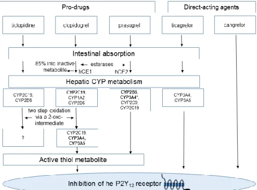
The first generation ticlopidine was soon replaced by the second generation clopidogrel due to ticlopidine’s adverse side effects (i.e neutropenia and thrombotic thrombocytopenic purpura). Clopidogrel’s favorable therapeutic effect is attributed to its antiplatelet and anti-inflammatory profile. Beside irreversible P2Y12 blockade, clopidogrel has also been reported to attenuate platelet–leukocyte aggregate formation [34], periprocedural increase of CRP levels [35], and to reduce P-selectin and CD40L expression [36] and the rate of thrombin formation [37]. However, clopidogrel has wide inter-individual efficacy in terms of platelet inhibition. The potential causative roles in its background included pharmacokinetic, pharmacodynamic, clinical, genetic and cellular mechanisms as well as the patient’s compliance [38] (see also below). Recently, the third generation, more potent prasugrel has been introduced in the clinical practice [39,40]. Prasugrel has a more rapid onset of action and a more extent platelet inhibitory effect and proved to be superior to clopidogrel in large clinical studies, though at a price of increased bleeding risk [41]. All thienopyridines are prodrugs and require metabolization via the hepatic CYP enzymes to pass into its biologically active metabolite. The activating steps and the involved hepatic enzymes somewhat differ at each drug, resulting in different pharmacokinetic and -dynamic features, and explains the distinct drug-drug interference profile of the compounds (Figure 2, Table 1).
Nucleotide/nucleoside analogues. These drugs are active agents not requiring metabolic transformation, thus having immediate antiplatelet effect upon intravenous administration or intestinal absorption. Ticagrelor is an orally administered reversible allosteric inhibitor of the P2Y12 receptor. Ticagrelor proved to be favorable compared to clopidogrel in large clinical studies exerting a more expressed and faster antiplatelet effect [42]. Cangrelor recently got approval based on the results of the CHAMPION- PHOENIX study, where it significantly reduced the rate of ischemic events during PCI, with no significant increase in severe bleeding [43]. This intravenously administered ATP derivative is used to achieve immediate and complete platelet inhibition before
12
PCI and may be used in patients who are unable to swallow and/or in bridging therapy before surgery in patients requiring P2Y12 inhibitor therapy [44]. After cessation of its administration normal platelet function is restored within 1 hour [43,44].
Figure 2. Metabolization of the P2Y12 inhibitors. After absorption, ticlopidine undergoes extensive metabolization by the hepatic CYP enzymes. The active metabolite is formed in a two-step oxidation, through the intermediate 2-oxo-ticlopidine [45]. The CYP enzymes involved in the second oxidation step are not identified. Clopidogrel, after intestinal absorption, is extensively hydrolyzed by plasma esterases into an inactive compound. The remaining clopidogrel fraction undergoes a two-step oxidation process in the liver to turn into its bioactive form via the intermediate 2-oxo-clopidogrel [45]. Prasugrel is also a pro-drug, but its activation process is started by plasma esterases and includes only one CYP-dependent step, which importantly, is mediated mainly by the intestinal CYP3A enzymes (*) [45]. This explains the lack of drug-drug interactions reported with the use of prasugrel. Ticagrelor is an ab ovo active agent, though 30-40% of its action is linked to its active, bioequivalent metabolite transformed by hepatic CYP enzymes. Cangrelor is also an active compound administered intravenously, and its metabolization is independent from the hepatic CYP enzymes. Bold letters indicate major enzymes of the metabolic steps.
hCE1: human carboxylesterase 1 (primarily synthetized by the liver), hCE2: human carboxylesterase 2 (primarily synthetized by the intestine). CYP: cytochrome p450 enzymes.
13
Table 1. Pharmacological profiles of currently approved P2Y12 inhibitors.
ticlopidine clopidogrel prasugrel ticagrelor cangrelor
Group based on chemical structure
1st generation thienopyridine
2nd generation thienopyridine
3rd generation thienopyridine
cyclo- pentyltriazolo-
pyrimidine
ATP-analogue
Way of
administration Oral (bid) Oral Oral Oral (bid) Intravenous
Receptor
inhibition Irreversible Irreversible Irreversible Reversible Reversible
Onset of action 6 h 2-8 h 30 min-4 h 30 min-2 h Seconds
Offset of action 7-10 d 7-10 d 7-10 d 3-5 d cca. 60 min
Main enzymes of CYP
metabolization
CYP2C19, CYP2B6, CYP3A4 (?)
CYP1A2, CYP2B6, CYP2C19, CYP3A4,
CYP3A5
CYP2B6, CYP3A4
CYP3A4,
CYP3A5 -
Reported significant drug- drug interactions
yes yes no yes no
1.2.1.3. GPIIb/IIIa receptor inhibitors
The platelet specific GPIIb/IIIa receptor is the final common pathway of platelet aggregation, being the receptor for fibrinogen with the highest affinity and binding also fibronectin, vitronectin and vWF [46]. Blocking this final step by intravenously administered GPIIb/IIIa inhibitors results in very efficient platelet inhibition, but also in increased risk of bleeding. These antiplatelets have different pharmacological features.
Abciximab is a monoclonal antibody fragment and causes impaired platelet function for several days [47]. In contrast, eptifibatide (a low-molecular weight heptapeptide) and tirofiban (a non-peptide tyrosine derivative) shows more rapid dissociation from the GPIIb/IIIa receptor and restoration of normal hemostatic function after their cessation is expected after 3-4 hours [47]. In current clinical practice, the use of GPIIb/IIIa inhibitors is limited to bail-out situations -in patients with low bleeding and high thrombotic risk- for a short period of time during and immediately before and after PCI in patients with ACS [48].
1.2.1.4. PDE inhibitors
Phosphodiesterase enzyme breaks down the intracellular cAMP resulting in enhanced platelet activity by increasing the intracellular Ca2+ level. By inhibiting PDE, cilostazol and dipyridamole effectively lowers platelet reactivity. Dipyridamole is primarily approved for the secondary prevention of transient ischemic attack. Cilostazol is also a
14
direct arterial vasodilator mainly used in patients with PAD [49]. It was also tested in former studies, whether adding these agents to DAPT could overcome high on- treatment platelet reactivity in PCI treated patients, though due to controversial results, they did not bring any break-through in antithrombotic treatment [50-52].
1.2.1.5. Antiplatelets under development
There are further promising targets for platelet inhibition. Thrombin, the most potent agonist activates platelets through the PAR receptor family. Human platelets express PAR1 and PAR4 receptors. By inhibiting the PAR1 receptor, only the effects of thrombin on platelets are prevented without impairing effects on coagulation or other functions.
The recently approved PAR1 inhibitor in clinical use is vorapaxar [53]. Other antiplatelet agents being under clinical investigation/development include compounds targeting the P2Y1 receptor, the 5-HT receptors, the prostaglandin receptors (IP, EP1-4), the GPVI main collagen receptor, the GPIb-VWF axis, the thromboxane receptors and the phosphatidylinositol-3-kinase intracellular pathway (see Figure 3, indicated with orange color) [54,55].
15
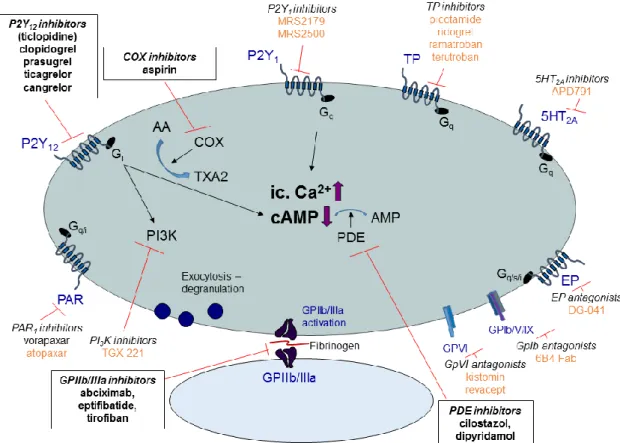
Figure 3. The antiplatelet agents’ sites of action. Currently approved drugs are indicated with black bold text in frame, agents under development are signed with orange color. Black arrows denote stimulation, red lines depict inhibition. Thromboxane protein receptor inhibitors have not been proven to be superior to aspirin up to date [56-58]. The P2Y1 inhibitors MRS2179 and MRS2500 and the PI3K inhibitor TGX-221 are only for research use currently [59,60]. The safety trial of additional use of the serotonin antagonist APD-791 beside clopidogrel/aspirin therapy has not yet started patient recruitment (NCT02034292). The EP3 prostaglandin receptor antagonist DG-041 has only been investigated in Phase I clinical trials [61]. The GPIb antibody 6B4-Fab and the GPVI antagonist kistomin and revacept were evaluated only in preclinical studies up to date [62-64]. The evaluation of PAR-1 receptor antagonist atopaxar was suspended after phase II clinical trials due to safety concerns [65,66] . For abbreviations see legend of Figure 1.
1.2.2. Antiplatelet strategies and current clinical guidelines in patients with SCAD The antiplatelet regimens in SCAD are summarized in Table 2. ASA treatment is indicated before elective stenting and a loading dose is recommended if the patient is not pre-treated. Clopidogrel loading is not recommended routinely, until coronary anatomy is not known. After elective PCI, dual antiplatelet treatment is recommended with aspirin and clopidogrel for at least 1 month after BMS deployment and 6-12 months after DES insertion. General use of GPIIb/IIIa inhibitors and prasugrel or
16
ticagrelor is not recommended in these patients, as they may increase bleeding risk [67].
In SCAD lifelong administration of low dose aspirin (75-150 mg daily) is recommended in all patients. In case of aspirin intolerance or allergy, clopidogrel may be considered.
Use of platelet function testing is recommended only in specific, high risk situations when it may have potential therapeutic consequences [48,67].
Table 2. Recommendations for antiplatelet treatment in patients with SCAD undergoing PCI based on the current ESC guidelines [48,67].
Recommendations for antiplatelet therapy in SCAD patients Class1 Level2
Pretreatment
Clopidogrel 600 mg loading dose is recommended in elective PCI patients once coronary anatomy is known and
decision to proceed with PCI is made (≥2 hours before PCI). I A
Pretreatment with clopidogrel may be considered in patients with high probability for significant CAD. IIb C In patients already on a 75 mg clopidogrel, a new loading dose of 600 mg or more may be considered
once the indication for PCI is confirmed. IIb C
During PCI
ASA is indicated for elective stenting. I B
ASA loading (oral 150-300 mg, i.v. 80-150 mg) is recommended if not pretreated. I C
Clopidogrel is recommended for elective stenting (≥600 mg loading, 75 mg maintenance dose). I A
GPIIb/IIIa inhibitors may be used only for bail-out. IIa C
After stenting
DAPT is indicated for at least 1 month after BMS implantation. I A
DAPT is indicated for 6 months after DES implantation. I B
Shorter DAPT duration (<6 months) may be considered after DES implantation in patients at high bleeding risk. IIb A Longer DAPT duration (>6 months) may be used in patients at high ischemic and low bleeding risk. IIb C
Life-long single antiplatelet therapy, usually ASA, is recommended. I A
Use of PF testing
PF testing may be used in specific or high risk situations (e.g. history stent thrombosis; compliance issue; suspicion
of resistance; high bleeding risk) if results may change the treatment strategy. IIb C Routine platelet function testing to adjust antiplatelet therapy before or after elective stenting is not recommended. III A
1.2.3. Antiplatelet strategies and current clinical guidelines in ACS
In patients with STEMI, P2Y12 inhibitor therapy should be initiated orally as early as possible before angiography, usually at first medical contact (Table 3). Upstream use of
17
GPIIb/IIIa antagonists may also be considered in high risk patients undergoing transfer that delays PCI. ASA loading should be performed in all cases. According to the current guidelines [39,48], the first choice of P2Y12 inhibitors are prasugrel or ticagrelor because of their more rapid onset of action and greater platelet inhibitory potency compared to clopidogrel [41,42]. Clopidogrel should be used only in lack of availability of the newer agents or in case of contraindications.
Table 3. Recommendations for antiplatelet treatment in patients with STEMI undergoing primary PCI based on the actual ESC guidelines [39,48].
Recommendations for antiplatelet therapy in STEMI patients Class1 Level2
Pretreatment
It is recommended to give P2Y12 inhibitors at the time of first medical contact. I B
Upstream use of a GPIIb/IIIa inhibitor (vs. in-lab use) may be considered in high-risk patients undergoing transfer
for primary PCI. IIb B
During and after PCI
ASA is recommended for all patients without contraindications at an initial oral loading dose of 150–300 mg (or
80–150 mg i.v.) and at a maintenance dose of 75–100 mg daily long-term regardless of treatment strategy. I A
A P2Y12 inhibitor is recommended in addition to ASA and maintained over 12 months unless there are
contraindications such as excessive risk of bleeding. Options are: I A
• Prasugrel (60 mg loading dose, 10 mg daily dose) if no contraindication I B
• Ticagrelor (180 mg loading dose, 90 mg twice daily) if no contraindication I B
• Clopidogrel (600 mg loading dose, 75 mg daily dose), only when prasugrel or ticagrelor are not
available or are contraindicated I B
GPIIb/IIIa inhibitors should be considered for bail-out or evidence of no-reflow or a thrombotic complication. IIa C
In NSTE-ACS, recommendations for antiplatelet treatment are similar, with the exception of upstream use of GPIIb/IIIa inhibitors and pre-treatment of prasugrel, which regimens are not recommended in patients in whom coronary artery anatomy is not known (Table 4).
After any type of acute coronary syndrome treated with PCI, fibrinolysis or medically, long term dual antiplatelet treatment is recommended. Regarding aspirin, low dose maintenance therapy should be used indefinitely due to its well established benefits in secondary prevention. In case of aspirin intolerance or allergy, clopidogrel
18
might be used instead. Duration of parallel P2Y12 therapy is usually recommended to last up to 12 month.
Table 4. Recommendations for antiplatelet treatment in patients with NSTE-ACS undergoing PCI according to current ESC guidelines [40,48].
Recommendations for antiplatelet therapy in NSTE-ACS patients Class1 Level2 Pretreatment
Pre-treatment with prasugrel in patients in whom coronary anatomy is not known, is not recommended. III B Pre-treatment with GPIIb/IIIa antagonists in patients in whom coronary anatomy is not known, is not
recommended. III A
During and after PCI
ASA is recommended for all patients without contraindications at an initial oral loading dose of 150–300 mg
(or 80–150 mg i.v.), and at a maintenance dose of 75–100 mg daily long-term regardless of treatment strategy. I A A P2Y12 inhibitor is recommended in addition to ASA, and maintained over 12 months unless there are
contraindications such as excessive risk of bleeding. Options are: I A
• Prasugrel (60 mg loading dose, 10 mg daily dose) in patients in whom coronary anatomy is known and
who are proceeding to PCI if no contraindication I B
• Ticagrelor (180 mg loading dose, 90 mg twice daily) for patients at moderate-to-high risk of ischemic events, regardless of initial treatment strategy including those pre-treated with clopidogrel if no
contraindication I B
• Clopidogrel (600 mg loading dose, 75 mg daily dose), only when prasugrel or ticagrelor are not
available or are contraindicated I B
GPIIb/IIIa antagonists should be considered for bail-out situation or thrombotic complications. IIa C
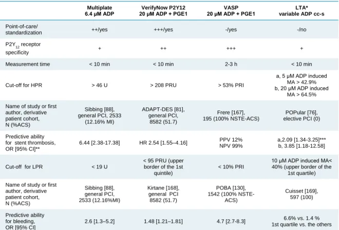
1.3. Laboratory monitoring of platelet function in current cardiology practice There are several laboratory methods to measure platelet function, most of which were primarily developed to screen and detect hereditary and acquired platelet/primary hemostasis disorders. Since the use of dual antiplatelet therapy results in acquired platelet dysfunction, most of these tests were challenged in monitoring the efficacy of antiplatelet therapy, though with various success. The more functional aggregometry tests give more information about the overall platelet reactivity and are less specific for the inhibitory effect of a given drug. In contrast, more specific tests assessing the inhibitory effect of a drug at the subcellular level might be less informative about the overall function of the activation-aggregation cascade. In the next session I will provide a non-exhaustive overview of the most often used platelet function tests in cardiology practice. The most widely applied tests are summarized in Table 7 (see below).
19 1.3.1. Aggregometry methods
Platelet activation finally results in platelet aggregation, which can be detected by several functional aggregometry methods. These tests measure the ability of different platelet agonists to induce platelet activation and aggregation in vitro.
1.3.1.1. Light transmission aggregometry (Born aggregometry)
The gold standard of aggregometry methods is light transmission aggregometry, also referred as the Born-method, introduced by Gustavo Born more than 50 years ago [68,69]. The method detects the decrease in optical density of the platelet rich plasma which occurs when the platelets in it aggregate due to activation with an agonist.
Change in optical density is depicted as a function of time. PRP is prepared by centrifugation and is basically free of other blood cellular elements. The standard used in the measurement is the platelet poor plasma of the same subject (PPP, prepared by further centrifugation), representing the minimum of optical density (or the maximum of light transmittance).
In spite of the wide-spread use of LTA for detecting platelet function, it is often criticized because of comparability difficulties of the obtained results. The method is fairly non-standardized, concerning pre-analytical processes (blood sampling, preparation of PRP, circumstances and time of sample storage until the measurement), agonists of different sources and concentrations, as well as the settings of the recorder [70] or the measurement parameter to be evaluated (e.g. maximal aggregation, final aggregation and disaggregation) [71,72]. The most widely used agonists are collagen, epinephrine, ADP, TRAP, and arachidonic acid.
Most initial data about the association of high on-treatment platelet reactivity and recurrent ischemic events in CAD patients after PCI raised from studies using the LTA method [73-75]. Moreover, in a comparative platelet function study, LTA was able to predict recurrent ischemic events during 1 year follow-up [76]. However, because of the non-standardized, labor-intensive procedure of the LTA method it is not advised for monitoring the efficacy of dual antiplatelet therapy in every-day clinical practice according to the latest consensus document [77].
20 1.3.1.2. VerifyNow method
The VerifyNow is a semi-automated, standardized point-of-care method to measure platelet aggregation in whole blood. Activated platelets aggregate to fibrinogen-coated microbeads, decreasing the optical density of the sample. The P2Y12 assay includes ADP agonist along with prostaglandin E1, whichincreases the sensitivity of the test and can be successfully used to assess P2Y12 therapy [78-80]. The VerifyNow Aspirin test uses arachidonic-acid agonist and assesses the thromboxane A2 mediated activation pathway. VerifyNow has been widely and successfully used to measure the efficacy of DAPT and its association with clinical outcome and the benefit of tailored antiplatelet therapy in patients undergoing PCI [81-84].
1.3.1.3. Multiple electrode aggregometry (Multiplate)
Multiple electrode aggregometry or impedance aggregometry is based on detection of impedance change caused by the adherence of platelets after activation with an agonist to multiple electrode-pairs immersed in the whole blood sample. The change of impedance is depicted as a function of time, and the area under the curve value given in units (U) indicates the measure of platelet reactivity [85]. Multiplate is a semi- automated, standardized point-of-care device, with the following available assays: ADP test and high sensitive ADP test (with use of PGE1 to eliminate false-positive results) to monitor P2Y12 inhibitor therapy, ASPI test (arachidonic-acid agonist) to monitor ASA therapy, and other non-specific assays such as TRAP test (TRAP-6 agonist), and COL test (collagen agonist), RISTO test (ristocetin agonist) to examine the vWF and GpIb dependent aggregation (e.g. to detect von Willebrand’s disease). The Multiplate ADP test was widely used in clinical studies assessing the efficacy of dual antiplatelet therapy [86] and it was found to be capable to predict ischemic [87] or bleeding events [88,89]
in PCI treated patients.
1.3.1.4. Plateletworks
In this standardized, point-of-care method, aggregation testing in whole blood is based on the comparison of a platelet count performed before and after provoking platelet activation, using different agonists. Though, the method is not widely used and/or investigated, it was successfully tested in a comparative platelet function study, where its results were associated with occurrence of ischemic events during 12 months follow-
21
up [76]. Also, it was found to be capable to identify patients at higher risk of myocardial infarction and rehospitalization within 3 months after coronary angiography [90].
1.3.1.5. Platelet function analyzer 100 (PFA-100)
This automated point-of-care method was developed to screen primary hemostatic disorders (e.g. von Willebrand’s disease). In this device, whole blood sample flows at high shear rates through an aperture in a membrane which is coated with distinct agonists and leads to rapid occlusion of the aperture, referred as the closure time. The COX inhibitors usually prolong the closure time of the collagen/epinephrine cartridge, therefore the method has often been used to measure functional aspirin resistance [91].
However, clopidogrel’s efficacy [92] and its association with clinical outcomes cannot be assessed with this method [76].
1.3.1.6. Impact-R (cone and plate(let) analyzer - CAP)
By this method the anticoagulated whole blood sample is added to a polystyrene well.
Platelet activation is induced by application of shear stress and aggregates are visualized and quantified by staining. The results are expressed as percentage of well surface covered by aggregates (SC) as an index of adhesion and average aggregate size (AS) as an index of aggregation [93]. Agonist-induced aggregation was suggested to be capable to monitor antiplatelet therapy [94]. However, in a comparative platelet function testing study, Impact-R failed to discriminate between patients with and without major cardiovascular events at 1 year follow-up after PCI [76].
1.3.2. Other methods to detect platelet activation
There are several other tests to measure platelet activation by detecting appearance of activation markers on the platelets’ surface or soluble activation markers in the plasma (representing cleaved and shed surface glycoprotein fragments), and structural changes of membrane or intracellular regulatory proteins.
1.3.2.1. Flow cytometry
Flow cytometry can measure several aspects of platelet activation [95]. After stimulation of platelets by an agonist, different markers of activation can be measured with the help of fluorescent antibodies (e.g. P-selectin exposure, activated GPIIb/IIIa expression, binding of fibrinogen). In a comparative study, ADP induced aggregometry
22
(VerifyNow, LTA and MEA) and flow cytometry results correlated significantly in PCI treated patients on DAPT [96]. In contrast, after AA induction, correlations were only partially observed. Also, association of platelet activation detected by flow cytometry with clinical outcome is fairly unknown yet. Furthermore, due to expense of the unit and need for labor-intensive sample preparation, flow cytometry is not practical for monitoring DAPT at point-of care.
1.3.2.2. VASP test
Over a decade ago, an important new technique has been introduced by using fixed and permeabilized platelets and specific labeling of the intra-cellular regulatory protein vasodilator-stimulated phosphoprotein [97]. It is phosphorylated through the cAMP biochemical cascade, indicating P2Y12 receptor inhibition and its dephosphorylation indicates P2Y12 receptor activity [98], thus the VASP assay can be used to monitor P2Y12 inhibitor therapy. Results of this assay correlate well with those of distinct platelet aggregometry methods [99-101]. Though VASP assay was described to be capable to predict adverse ischemic events after PCI [102], its discriminative capacity was lower than that of the aggregometry methods [103].
1.3.2.3. Measurement of soluble activation markers
An alternative way to assess platelet function is to find a reliable plasma marker that reflects platelet activation and is a specific marker of the platelet, is not affected by artefacts of sample collection, and is measureable by a reproducible and simple laboratory technique. Possible candidate molecules can be substances that are released from the platelet’s granules, molecules that are expressed on and shed from the platelet’s surface, and secreted metabolic molecules [104]. In spite of high former interest, none of these markers could fulfill the above mentioned criteria, thus their clinical utility is controversial at this point [105].
1.3.2.4. Thromboelastography (TEG, ROTEM)
This is a viscoelastic hemostatic assay, which analyzes clot formation (time to initial fibrin generation), clot elasticity development (clot strength) and the process of fibrinolysis. Elasticity of the clot is influenced by several factors, such as the contractile force of platelets during clot retraction (which is the major determinant), platelet and fibrinogen concentration, hematocrit, FXIII and the thrombin generation during
23
coagulation [106]. TEG is most widely used in surgery and anesthesiology and was less extensively investigated in monitoring DAPT therapy in cardiology patients, however some data suggested that clot strength and rapid fibrin formation beside DAPT were risk factors of recurrent ischemic events in patients after PCI [73].
1.4. Definition and clinical significance of high on-treatment platelet reactivity 1.4.1. Clinical and laboratory approach to the efficacy of antiplatelet therapy
Since the introduction of percutaneous coronary intervention in the therapeutic toolbar of coronary artery disease, many procedural and pharmaceutical improvements have been carried out. The spread of drug eluting stents successfully decreased the occurrence of restenosis, though they brought along a new complication in the form of late and very late stent thrombosis. Regarding antiplatelet therapy, which is a sine qua non in the success of percutaneous coronary intervention and intracoronary stenting, it has been proven, that adding clopidogrel to aspirin was more effective in preventing recurrent ischemic events in the long term compared to aspirin monotherapy [32,33].
Furthermore, DAPT was shown to decrease the long term rate of both ischemic and bleeding events compared to aspirin combined with an anticoagulant (heparin or coumarin) [107,108] or an oral GPIIb/IIIa inhibitor [109]. However, in spite of the application of dual antiplatelet therapy after PCI, ischemic events such as stent thrombosis and myocardial infarction still continued to occur, therefore the terms
“treatment failure” and “aspirin and clopidogrel resistance” emerged. Treatment failure refers to the state, when an adverse clinical event/condition recurs despite the administration of a drug to avoid it. In the literature, “aspirin and clopidogrel resistance”
referred to the condition, when the lack of aspirin/clopidogrel’s effect was justified by a laboratory platelet function test. However, in pharmacology, drug resistance means inability of a drug to hit its therapeutic target (e.g. receptor, enzyme or regulatory protein) either from a pharmacokinetic (e.g. impaired intestinal absorption leading to suboptimal concentration of the drug at the effect site) or from a pharmacodynamic (meaning that the target protein itself is impaired/altered) cause. Usually, platelet function tests are not capable to assess these details of antiplatelet mechanism of action.
Thus, laboratory proven aspirin and clopidogrel “resistance” was replaced by the term
“high on-treatment platelet reactivity” (HPR) or “residual platelet reactivity/activity”
24
(RPR/RPA). With respect to the applied antiplatelet therapy and platelet function test we may differentiate “high on-clopidogrel and high on-aspirin platelet reactivity”
(HCPR and HAPR) or “dual high on-treatment platelet reactivity”.
1.4.2. Clinical significance of measuring aspirin-effect and high on-aspirin platelet reactivity (HAPR)
Measuring the efficacy of aspirin treatment proved to be more complicated and less reliable from the methodological aspect, than that of the P2Y12 inhibitors [110,111].
Methods to monitor aspirin therapy include measuring of serum TXB2 (the stable metabolite of TXA2) concentrations or detecting the urine TXB2 or 11-dehydro- thromboxane-B2 level [111]. However, these measurements are complicated and these metabolites might be generated through COX-1 independent pathways as well, thus they rather reflect an overall inflammatory state, than the measure of aspirin-effect.
Therefore, the inhibitory effect of aspirin most widely is measured indirectly in platelet function tests using arachidonic-acid as an agonist (via the induction of TXA2
generation and consequent platelet activation) [112]. Many non-specific antagonists (ADP, collagen, and epinephrine) are also rely on TXA2 generation as an amplification loop, therefore were used in the laboratory determination of aspirin response. However these tests overestimate the prevalence of true aspirin non-response [110,113].
To date, the predictive value of measuring aspirin-effect in association with clinical outcome is controversial. In early studies, the “aspirin-resistant” phenotype was associated with higher risk of ischemic events, however in these studies patients were on aspirin monotherapy usually and non-aspirin specific platelet function tests were used [114-117]. Therefore, these tests rather identified patients with a “hyper-reactive platelet phenotype”, than measured aspirin’s platelet inhibitory effect via the COX1
inhibition.
Recently, the largest clinical trial investigating high on-treatment platelet reactivity on aspirin and clopidogrel so far found no association between on-aspirin treatment platelet reactivity assessed by VerifyNow Aspirin test and all cause death, stent thrombosis or myocardial infarction, though HAPR was inversely related to bleeding [81]. Similarly, another large scale study failed to link high residual platelet reactivity on aspirin measured by PFA-100 to clinical outcome [118]. On the other
25
hand, recent data from a large scale, one center registry suggested, that patients with HAPR identified by the Multiplate ASPI test (using AA induction) showed a significantly higher risk of death or ST at 1 year and HAPR was an independent predictor of the combined primary outcome [119]. Similarly, dual high on-treatment platelet reactivity to aspirin and clopidogrel was associated with a higher risk for atherothrombotic events and identifies patients at highest thrombotic risk [120,121].
Whether or not HAPR is associated with clinical outcome, the benefit of aspirin dose adjustment based on laboratory efficacy testing is questionable anyway. Several clinical data justified that overcoming HAPR by increasing aspirin dose only increased the risk of bleeding, without further improving ischemic outcomes [122,123]. Since such a therapeutic modification is not recommended, reasonableness of assessing HAPR currently remains debated.
In summary, in contrast to high on-clopidogrel platelet reactivity, the term “high on-aspirin treatment platelet reactivity” and “aspirin resistance” are less clearly defined and have a widely variable prevalence in the literature [124], and though some data suggest, that HAPR along with HCPR might be helpful in thrombosis risk stratification, its clinical significance remains controversial. Consequently, the following part of this thesis will focus on high platelet reactivity on P2Y12 inhibitor therapy.
1.4.3. High on-clopidogrel platelet reactivity (HCPR) and high platelet reactivity on P2Y12 inhibitor therapy (HPR)
In the past decade, extensive research has been focused on the association of laboratory certified HCPR and recurrent ischemic events resulting in growing evidence, that HCPR and HPR are independent risk factors of thrombotic events after PCI [77,81,125]. The largest studies (N>300) investigating the existence and strength of an association between on-treatment platelet reactivity and clinical outcome are summarized in Table 5.
26
Table 5. Major studies linking on-treatment platelet reactivity with clinical outcome
First author (year), Study name
No. of patients and presenting clinical syndrome
Platelet function test(s)/applied P2Y12 inhibitor
Cut-off for HPR
Cut-off for LPR
OR [95% CI] for ischemic event(s)*
OR [95% CI] for bleeding event(s)*
Follow-up time
Sibbing (2010) [88]
308 NSTEMI /STEMI 2245 UAP/SCAD
Multiplate ADP/
clopidogrel >46 U <19 U 6.44 [2.38–17.38] for def ST**
2.6 [1.3–5.2]
for in hospital TIMI major bleeding
30 days
Geisler (2010) [126]
514 SCAD 505 ACS
LTA 20 µM ADP/
clopidogrel
Upper tertile border for FA >42.5%
NR
HR 1.05 [1.01–1.08]
for early (<30 days) stent thrombosis 2.21 [1.31–3.73] for combined CV end point
2.31 [1.1–4.84] for all 3 month ST
NR 3 months
Breet (2010), POPular [76]
1069 elective PCI
LTA 5 µM ADP LTA 20 µM ADP VerifyNow P2Y12 Plateletworks/
clopidogrel
MA ≥42.9%
MA≥ 64.5%
≥ 236 PRU
≥ 80.5% NR
2.09 [1.34-3.25]
2.05 [1.32-3.19]
2.53 [1.63-3.91]
2.22 [1.25-3.93]
for combined CV end point
No association was found between TIMI major or minor bleeding and any of the PF tests.
1 year
Park (2011) [127]
1586 SCAD 1264 ACS
VerifyNow P2Y12/ Clopidogrel
>235 PRU and/or % inhibition <15
<235 PRU
HR 1.33 [0.88–2.01]
for combined CV end point
1.45 [0.27–7.92] for def/prob ST
HR 0.78 [0.35–1.69]
for TIMI major bleeding
2 years
Bonello (2012) [128]
128 STEMI 100 NSTEMI 73 UAP
VASP/
prasugrel ≥53.5 % PRI <16 % PRI
1.44 [1.2–1.72] per 10% increase for def/prob ST
0.75 [0.59–0.96] per 10 % increase for TIMI major and minor bleeding
1 year
Siller- Matula (2012), PEGASUS- PCI [129]
274 elective PCI 67 NSTE-ACS 73 STEMI
Multiplate ADP+PGE
1/ clopidogrel
≥48 U NR 36.9 [4.3–319] for def/prob ST
No predictive ability was found for TIMI major bleeding.
1 year
Cuisset (2013), POBA [130]
1542 ACS
VASP/
25% prasugrel 75% clopidogrel
NR ≤10 %
PRI NR 4.7 [2.7-8.3] for
BARC bleeding 6 months
Stone (2013), ADAPT- DES [81]
4147 SCAD 2373 UAP 1250 NSTEMI 813 STEMI
VerifyNow P2Y
12/
Clopidogrel >208 PRU <95 PRU
HR 2.54 [1.55–4.16]
for def/prob ST, HR 1.42 [1.09–1.86]
for MI
HR 1.52 [1.17-1.97]
for clinically relevant bleeding
1 year
ACS: acute coronary syndrome; ADP: adenosine-diphosphate; BARC: Bleeding Academic Research Consortium; CI: confidence interval; def: definite; FA: final aggregation; HPR: high on-treatment platelet reactivity; HR: hazard ratio; LPR: low platelet reactivity; LTA: light transmission aggregometry; NR: not reported; NSTEMI: non-ST elevation myocardial infarction; OR: odds ratio; PRI: platelet reactivity index; prob: probable; SCAD: stable coronary artery disease; STEMI: ST elevation myocardial infarction;
TIMI: thrombolysis in myocardial infarction; UAP: unstable angina pectoris; VASP: vasodilator- stimulated phosphoprotein.*Odds ratios are given unless otherwise indicated. **All ST are defined according to Academic Research Consortium criteria.
Recognition of an association between HPR (mostly on clopidogrel) and clinical outcome as well as wide inter-individual variability of platelet inhibition and relatively high prevalence of HCPR raised doubt in one-size-fits all dosing method and led to the development of more potent P2Y12 receptor inhibitors. However, the price to pay for
27
more expressed platelet inhibition is an increased bleeding risk in unselected patient populations [41,42]. Consequently, current recommendations exclude patients with stable coronary artery disease and ACS patients at high bleeding risk from the benefit of these novel P2Y12 inhibitors [48,131,132]. These patients remain subjects of clopidogrel treatment. Notably, the prevalence of high on-treatment platelet reactivity (HPR) only diminished but not vanished with the use of novel antiplatelets [133,134].
On the other hand, recently published data from the ADAPT-DES study highlighted further interesting aspects of platelet function testing, verifying that overcoming high on-treatment platelet reactivity at any price might profoundly increase bleeding risk and through that may counter-balance the favorable effects of intense platelet inhibition. The authors discussed it as a potential explanation of the lack of association between HPR and mortality [81]. However, association of low platelet reactivity on dual antiplatelet therapy with higher bleeding risk [88] and the concept of a
“therapeutic window” of P2Y12 inhibitor treatment was already introduced in earlier studies [77,89]. The fact that prognostic significance of bleeding consequences is equally important with that of ischemic events—particularly with the spreading use of more potent P2Y12 inhibitors [77,81]— further expands the space for platelet function testing.
1.4.4. Poor response to antiplatelet agents - determining factors of HPR
Factors associated with high on-clopidogrel platelet reactivity have already been investigated by a large number of studies [38] (Table 6). Although certain genetic polymorphisms (CYP2C19 loss of function, CYP3A4 and CYP3A5, and ABCB1 gene polymorphisms), drug-drug interactions (e.g. CCBs and PPIs) and accompanying clinical risk factors such as higher body mass index (BMI), reduced LVF, diabetes mellitus, chronic kidney disease, female gender and smoking have already been linked to HPR measured by various platelet function tests, there is a lot of discrepancy among study results. This discrepancy might derive from the use of distinct platelet function tests assessing different aspects of platelet activation/aggregation and variable cut-off values as well as from inclusion of platelet reactivity as categorical or continuous parameter. Moreover, there is a gap of knowledge regarding the response variability to newer antiplatelet agents.
28
Table 6. Response variability to P2Y12 therapy
Determinants of HPR References
Clinical factors
Age [135-137]
Gender [137-139]
BMI [103,136,140,141]
ACS [142,143]
Renal failure [137,142,144,145]
Reduced LVF [142,146]
Smoking [139,147,148]
Inflammation [149-151]
Underdosing [152,153]
Compliance [154]
Genetic factors
CYP2C19*2 loss of function [155-157]
CYP3A4, A5 gene variants [158-160]
ABCB1 gene variants [161]
Drug-drug interactions
PPI (mostly omeprazole) [162-164]
CCB [135,165,166]
1.4.5. Tailored antiplatelet therapy
Due to extensive research, several methods for platelet function testing underwent clinical validation beside the gold standard optical aggregometry and clinically determined cut-off values have been established [76,77,87]. Nevertheless, platelet function testing has a fairly variable, but generally low predictive value depending on the applied platelet function test, investigated clinical end point, follow-up time and clinical presentation of coronary artery disease (CAD) [77,125]. The most recent consensus document advices only a few platelet testing method for assessing platelet reactivity in cardiology practice [77]. The most widely used and investigated platelet function tests are summarized in Table 7.

![Table 2. Recommendations for antiplatelet treatment in patients with SCAD undergoing PCI based on the current ESC guidelines [48,67]](https://thumb-eu.123doks.com/thumbv2/9dokorg/1374315.112814/16.892.133.773.344.1019/table-recommendations-antiplatelet-treatment-patients-undergoing-current-guidelines.webp)
![Table 3. Recommendations for antiplatelet treatment in patients with STEMI undergoing primary PCI based on the actual ESC guidelines [39,48]](https://thumb-eu.123doks.com/thumbv2/9dokorg/1374315.112814/17.892.176.778.374.823/table-recommendations-antiplatelet-treatment-patients-undergoing-primary-guidelines.webp)
![Table 4. Recommendations for antiplatelet treatment in patients with NSTE-ACS undergoing PCI according to current ESC guidelines [40,48]](https://thumb-eu.123doks.com/thumbv2/9dokorg/1374315.112814/18.892.131.781.250.639/recommendations-antiplatelet-treatment-patients-undergoing-according-current-guidelines.webp)