Lesson 3 - The biomechanical properties of articular cartilage
This curriculum has been made at the University of Szeged, and supported by the European Union. Project identity number: EFOP-3.4.3-16-2016-00014.
This lesson contains 10 screens teaching text, 4 zoomable figures, and 4 videos. This lesson requires approximately 2 - 4 hours of study but can vary depending on the student.
At synovial joints, the articulating bone surfaces are covered with a protective layer of dense white connective tissue. This thin (1 to 5 mm) specialized connective tissue layer called hyaline articular cartilage. It allows movement between articulating bones at the joint with minimal friction and wear and spreads load at the joint over a wide area (3).
There are two additional types of cartilage in the human musculoskeletal system which are closely related to hyaline cartilage histologically. Fibrocartilage (e.g. menisci, intervertebral discs), and elastic cartilage (e.g. in the external ear, trachea) have greatly different biomechanical properties (6).
Please now watch the following video:
https://www.youtube.com/watch?v=yL_vdJ_uzUY
In this lesson, we focus on the properties and biomechanical behavior of the hyaline articular cartilage.
Composition of articular
cartilage
Articular cartilage can be described as a heterogeneous material, since its structure and composition
exhibit depth dependency.
Mechanically,
articular cartilage is a composite of materials with different properties (6).
Cells (chondrocytes) – account for less than 10% of the cartilage volume. These components produce collagen, proteoglycans, and enzymes. Chondrocytes are inhomogeneously distributed in articular cartilage arranged similarly as collagen fibers, giving the tissue a layered character.
Normal articular cartilage is composed of three zones and the tidemark, zones based on the shape of the chondrocytes and the orientation of the type II collagen.
Superficial zone (10 to 20% of the total thickness):Type II collagen orientation is parallel to joint in this region. This zone has flattened chondrocytes, condensed collagen fibers, and sparse proteoglycans.It has the highest concentration of water.
Intermediate zone (40 to 60% of the total thickness): Is the thickest layer with round chondrocytes, and abundant proteoglycan content. Type II collagen has an oblique or random organization in this zone.
Deep layer (about 30 of the total thickness): Type II collagen is perpendicular to joint and crosses tidemark in this zone. It has the highest concentration of proteoglycans and lowest concentration of water. Chondrocytes are roundly arranged in columns.
Tidemark is deep to the basal layer and separates the true articular cartilage from the deeper cartilage. The tidemark divides the superficial, uncalcified cartilage from the deeper, calcified cartilage.
Calcified cartilage Subchondral bone
The zonal
arrangement of cells and collagen fibers is shown in the following video:
https://www.youtube.com/watch?v=VuFnA8Zl6Q0&t=46s
Extracellular matrix:
Water - makes up 65% to 80% of the mass of the cartilage. It accounts for 80% of the weight near the surface and 65% at the deep zone. About 30% of the water is strongly associated with the collagen fibrils. Most of the water (about 70%) is free to move when the cartilage is loaded. Articular cartilage is devoid of blood vessels, lymph channels, and nerves. The fluid component is essential to permit diffusion of gases, nutrients, and waste products between chondrocytes and synovial fluid. Water as the fluid component of the articular cartilage - with many free mobile cations - greatly responsible for the mechanical behavior of the cartilage. The interstitial fluid flow provides the viscoelastic behavior of the articular cartilage under compressive loading (6).
Type II collagen -makes up 10 to 20% of total cartilage mass. Type II collagen accounts for 90% to 95% of the total collagen content and provides cartilaginous framework and tensile strength. Collagen fibers create a framework that houses the other components of cartilage (6).
Proteoglycans -make up 10 to 15% of cartilage. Proteoglycans are large protein-polysaccharide molecules produced by chondrocytes. Proteoglycans consist of glycosaminoglycan chains and oligosaccharides (6).
Sulfated glycosaminoglycan as keratin sulfate and chondroitin sulfate also found in articular cartilage.
Hyaluronic acid does not contain any sulfate and forms non-covalently linked complexes with proteoglycans in the extracellular matrix (6).
Glycosaminoglycan chains have negative charges that create strong charge repulsive forces. This charge repulsion stiffens proteoglycans in the collagen network, gives cartilage compressive stiffness (6).
Biphasic characteristic of articular cartilage
The biomechanical behavior of articular cartilage is best understood when the tissue is viewed as a biphasic medium. From the mechanical point of view, components of articular cartilage can be divided into two major phases, fluid phase includes water and cations, and solid phase includes chondrocytes, type II collagen, proteoglycans and other glycoproteins (2).
Biomechanical behavior of articular cartilage
Like bone, articular cartilage is anisotropic, it has direction-dependent material properties. Mechanical behavior of articular cartilage is characterized by poroviscoelastic model.
Structural macromolecules (proteoglycans) interact to form a porous matrix possessing all the essential mechanical properties of a solid. Collagen fibers and proteoglycans interaction serve the organization of the extracellular matrix and contribute to the mechanical properties of the tissue (6).
The fiber-reinforced (by collagen fibers) matrix is swollen with water (the analogy of a water saturated sponge may be helpful) (6). The fluid pressure provides a significant component of total load support, thereby reducing the stress
acting upon the solid matrix (2) Articular cartilage exhibits
viscoelastic
behavior in tension and compression.
The viscoelastic properties of articular cartilage
are mainly
attributed to two
mechanisms: the flow-dependent and the flow-independent viscoelasticity.
The interstitial fluid flows slowly due to compressive loading. The motion of interstitial fluid through the matrix causes a high frictional drag and an energy dissipation, which constitutes the flow-dependent viscoelasticity. During the fluid flow, a frictional resistance is applied from solid matrix to the water and this results to a very low permeability of the tissue, which has a direct effect in its compressive properties Furthermore, the macromolecular deformation is known as the flow-independent viscoelastic behavior of the collagen-proteoglycans solid matrix. Flow-independent viscoelastic behavior means that the matrix volume is unchanged and matrix molecules reorient at their own place after compression (6).
Biphasic viscoelastic behavior of articular cartilage
As you remember, the two important responses of a viscoelastic material are creep and stress relaxation.
Slow, progressively increasing deformation (creep) occurs until an equilibrium is reached when the articular cartilage is subjected to the action of a constant load. The initial and rapid application of articular contact forces during joint loading causes an immediate increase in interstitial fluid pressure. This local increase in pressure causes the fluid to flow out of the extracellular matrix. The biphasic creep response is caused by exudation of the interstitial fluid.
Fluid flow and creep discontinue when the compressive stress within the solid matrix is capable to balance the applied stress alone.
During a constant compression, high stress is generated by forced exudation of interstitial fluid. Fluid exudation gives rise the stress and stress relaxation is associated with fluid redistribution within the porous matrix. Biphasic
stress relaxation response ceases
when the
compressive stress within the solid matrix reaches the equilibrium state (6).
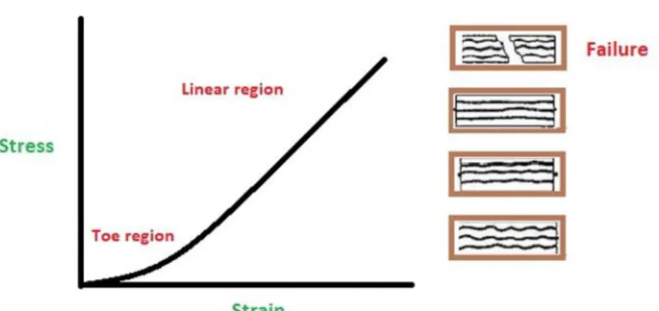
Tensile properties and behavior of the cartilage solid matrix
As with the other soft collagenous tissues, the mechanical behavior of the cartilage solid matrix is determined by the amount and crimp of collagen in the matrix. This matrix follows the classic nonlinear stress-strain curve for soft tissues as shown below:
Fig. 1 Stress-strain curve for articular cartilage.
The initial toe region represents the collagen fibers pull-out during the initial part of the tensile experiment.
The linear region represents the stretching of the aligned collagen fibers. Failure occurs when all the collagen fibers are ruptured (6).
There is an important
difference between tensile behaviors of articular cartilage and bone. In contrast to bone, articular cartilage tends to stiffen with increasing strain.
Lubrication of articular cartilage
Articular cartilage provides a smooth and lubricated surface for articulation while minimizing wear. During the diarthrodial joint function, hyaline cartilage is subjected to an excessive range of loading conditions (6).
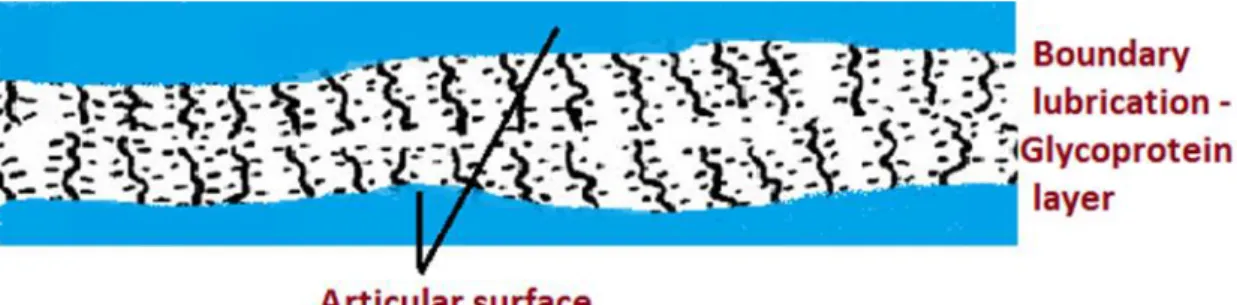
Lubrication is a phenomenon that prevents the direct contact between articular surfaces and eliminates the most of the wear. In other words, lubrication is the process by friction dissipation arises from as two surfaces slide past each other is minimized.
There are two basic types of lubrication (6).
Boundary lubrication is provided by a single monolayer of the specific glycoprotein which is adsorbed to each articulating surface (Fig.2) (6).
Fig. 2 Boundary lubrication.
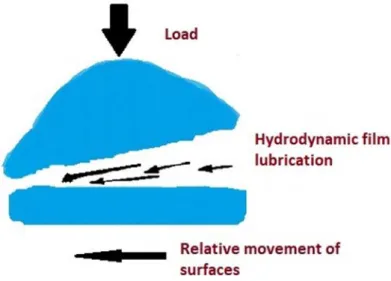
Fluid film lubrication
utilizes a thin film of lubricant that completely
separates sliding surfaces. Fluid film lubrication exists by two different modes (6).
Hydrodynamic lubrication occurs when nonparallel surfaces move
tangentially slide or roll each other, forming a wedge of fluid. During hydrodynamic lubrication, the pressure between the surfaces is generated as a result of the shape and motion of the articulating surfaces (Fig.3) (6).
Fig. 3Hydrodynamic lubrication.
Squeeze film lubrication occurs when the surfaces move perpendicularly toward each other, forced the fluid film out from the gap between the two articulating surfaces (Fig. 4) (6).
Fig. 4 Squeeze film lubrication.
Wear components of articular cartilage
Wear or attrition of articular cartilage is the process when the material is removed from the articular surfaces by mechanical action. The two types of wear are the interfacial and the fatigue wear.
Interfacial wear appears due to direct contact of bearing surfaces. In this case, the amount and function of boundary or fluid lubricant film are insufficient. Interfacial wear occurs in either of two ways: adhesion or abrasion (6).
Adhesive wear occurs when articulating
surfaces come into contact and surface fragments adhere to each other.
During this process, hyaline fragments torn from the surface (6).
Abrasive wear arises typically,
when the two surface materials have a different hardness and the soft surface is scratched by the harder one (6).
Direct contact between cartilage surfaces rarely occurs in healthy articulations, consequently, these types of wear may take play in an impaired or degenerated synovial joint.
Fatigue wear results from the repeated high load deformation of the hyaline surface over a relatively short period or may occur with the repetition of low loads over an extended period.
These mechanisms of wear may be the cause of structural damage observed in articular cartilage (6).
Cartilage degeneration
Regular joint movement and the dynamic load are important to maintain healthy articular cartilage metabolism, normal articular cartilage structure and function. Damaged joint articulation (i.e. intra-articular fractures, excessive joint surface incongruity, and ligament rupture) allows altered, excessive movement and increases the stress acting on the joint surface (6).
Effect of immobilization
Moderate mechanical stress generated by normal joint loading and movement is essential for the maintenance of healthy articular cartilage. Inactivity or immobilization caused by a disorder lead to cartilage degradation that may generate joint pain and reduced range of motion. (1).
Immobilization causes atrophy, primarily due to changes in the
content of
proteoglycans.
Reduced loading by the absence of weight bearing or external fixation lead to a decreased thickness of articular cartilage.
The alteration in articular cartilage become apparent following immobilization, from 4 to 6 weeks.
Articular cartilage has a limited capacity for intrinsic healing and repair. Mechanical disruption of articular cartilage stimulates chondrocyte synthetic activity, but it rarely results in repair of the injury. Disruption of subchondral bone stimulates chondral and bony repair, but it rarely restores an articular surface that duplicates the biologic and mechanical properties of normal articular cartilage. Various treatment strategies have been suggested for cartilage regeneration, but the optimum treatment is yet to be defined (8). Moderate and adapted exercise prevents weakening of the joints and alterations in the articular cartilage (5).
Effect of aging
Articular cartilage undergoes significant structural, matrix composition, and mechanical changes with age (4).
Aging-related changes in articular cartilage increase the risk of articular cartilage degeneration.
Effects of aging on articular cartilage include:
fibrillation of the articular surface,
a decrease in the size and aggregation of proteoglycans,
increased collagen cross-linking,
decreased water content
zonal changes in the distribution of chondrocytes
loss of tensile strength and stiffness.
Aged articular cartilage has a reduced concentration of proteoglycans. Due to altered composition, the water- binding capacity of
the extracellular
matrix is
decreased.
Osteoarthritis is a degenerative joint disease, which is associated with aging and mainly affects the articular cartilage. It is one of the most common causes of pain, disability,
and decreased the quality of life among middle-aged and older adults. Excessive loading, anatomical abnormalities, injuries, repetitive use, and decreased weight bearing in the joint are triggers of this disease (7).
As a matter of curiosity, let us see this video about osteoarthritis:
https://www.youtube.com/watch?v=BBqjltHNOrc
This video below helps to repeat and deepen your knowledge about biomechanical properties of articular cartilage:
https://www.youtube.com/watch?v=56SYy0jcl6I
Please, after watching the video above, answer study questions:
Study questions:
I. Which of the following statements regarding articular cartilage is TRUE?
1. Cartilage is an isotropic material.
2. Most of the water in articular cartilage exists in the deep layer next to the calcified cartilage.
3. Mechanical behavior of articular cartilage is characterized by poroviscoelastic model.
4. Structural macromolecules (proteoglycans) interact to form a viscous matrix.
5. About 70% of the water is strongly associated with the collagen fibrils.
II. Which of the following best describes the appearance of chondrocytes and orientation of collagen fibrils in the superficial zone of articular cartilage?
1. Round chondrocytes oriented parallel with the tidemark and collagen fibrils oriented perpendicular to the tidemark.
2. Round chondrocytes oriented parallel with the tidemark and collagen fibrils oriented parallel to the tidemark.
3. Flattened chondrocytes oriented parallel with the tidemark and collagen fibrils oriented perpendicular to the tidemark.
4. Flattened chondrocytes oriented perpendicular with the tidemark and collagen fibrils oriented perpendicular to the tidemark.
5. Flattened chondrocytes oriented parallel with the tidemark and collagen fibrils oriented parallel to the tidemark.
III. What part of the articular cartilage has the highest concentration of proteoglycans and the lowest concentration of water?
1. Superficial 2. Intermediate 3. Deep
4. Tidemark
5. Calcified cartilage
IV. What is the function of articular cartilage?
1. Articular cartilage is meant to increase load distribution area of the bone and allows movement while reducing friction and wear.
2. Articular cartilage provides a soft layer covered bone ending to produce synovial fluid.
3. The function of articular cartilage is to serve joint stability.
4. Articular cartilage improves nourished of subchondral bone.
5. Articular cartilage helps to maintain the blood-supply of the joint.
V. What happens during compression of articular cartilage?
1. Some water should leave for optimal dispersion.
2. Fixed water can leave the superficial area.
3. Proteoglycans moving away from each other during compression.
4. Less repelling of negative charges.
TRUE/FALSE questions
Read each statement below carefully. Choose the T if you think a statement is TRUE. Choose the F if you think the statement is FALSE.
1. Cartilage is approximately 70% water and has little blood supply. T or F 2. Aged articular cartilage has a reduced concentration of proteoglycans. T or F 3. Immobilization causes cartilage hypertrophy, primarily due to
changes in the content of proteoglycans. T or F 4. Articular cartilage has a good capacity for intrinsic healing and repair. T or F 5. Boundary lubrication is provided by a fluid film layer,
which is adsorbed to each articulating surface. T or F
References
1. Buckwalter JA, Mankin HJ (1998): Articular cartilage: tissue design and chondrocyte-matrix interactions. Instr Course Lect. 47:477-86.
2. Fox AJS, Bedi A, Rodeo SA (2009): The Basic Science of Articular Cartilage: Structure, Composition, and Function. Sports Health. 1(6): 461–468.
3. Hall SJ:Basic Biomechanics 2002.McGrawHill Coll; 4th Edition
4. Martin JA, Buckwalter JA (2001): Roles of Articular Cartilage Aging and Chondrocyte Senescence in the Pathogenesis of Osteoarthritis, Orthop. J. 1–7.
5. Musumeci G (2016): The Effect of Mechanical Loading on Articular Cartilage. J. Funct. Morphol.
Kinesiol. 1(2): 154-161.
6. Nordin M, Frankel VH: Basic Biomechanics of the Musculoskeletal System. 2014. Wolters Kluwer Health; 4th Edition
7. Novelli C, Costa JBV, Souza RR (2012): Effects of aging and physical activity on articular cartilage:
a literature review. J. Morphol. Sci. 1(29): 1-7.
8. Rai V, Dilisio MF, Dietz NE, Agrawal DK (2017): Recent strategies in cartilage repair: A systemic review of the scaffold development and tissue engineering. J Biomed Mater Res A. 105(8):2343- 2354.