ARTICLE
Correlation of neurochemical and imaging markers in
AU1
migraine
PACAP38 and DTI measures
D´anielVer´eb, MD,NikolettaSzab´o, PhD,BernadettTuka, PhD,J´anosTajti, DSc,Andr´asKir´aly, MD,
P´eterFarag´o, MD,Kriszti´anKocsis,EszterT´oth, MD,B´alintKincses, MD,Ter´ezBagoly,ZsuzsannaHelyes, DSc, L´aszl´oV´ecsei, DSc, andZsigmond Tam´asKincses, PhD
Neurology
®
2018;91:1-9. doi:10.1212/WNL.0000000000006201Correspondence Dr. Z.T. Kincses kincsesz@
nepsy.szote.u-szeged.hu
Abstract
Objective
To examine whether interictal plasma pituitary adenylate cyclase-activating peptide 38-like immunoreactivity (PACAP38-LI) shows correlation with the microstructural integrity of the white matter in migraine.
Methods
Interictal plasma PACAP38-LI was measured by radioimmunoassay in 26 patients with mi- graine (24 women) who underwent diffusion tensor imaging afterward using a 1.5-tesla magnetic resonance scanner. Data were analyzed using tract-based spatial statistics included in FMRIB’s Software Library.
Results
Interictal plasma PACAP38-LI showed significant correlation with mean diffusivity (p <
0.0179) mostly in the bilateral occipital white matter spreading into parietal and temporal white matter. Axial and radial diffusivity showed positive correlation with interictal PACAP38-LI (p<
0.0432 and p < 0.0418, respectively) in the left optic radiation and left posterior corpus callosum. Fractional anisotropy did not correlate significantly with PACAP38-LI. With disease duration as a nuisance regressor in the model, PACAP38-LI correlated with axial and mean diffusivity in the left thalamus (p< 0.01).
Conclusion
We report a link between PACAP38, a pathobiologically important neurochemical biomarker, and imaging markers of the disease that may bolster further research into the role of PACAP38 in migraine.
From the Departments of Neurology (D.V., N.S., J.T., A.K., P.F., K.K., E.T., B.K., L.V., Z.T.K.) and Radiology (Z.T.K.), Albert Szent-Gy¨orgyi Clinical Center, University of Szeged, Hungary;
Central European Institute of Technology (N.S., A.K.), Brno, Czech Republic; MTA-SZTE Neuroscience Research Group (B.T., L.V.), Szeged; and Department of Pharmacology and Pharmacotherapy, Faculty of Medicine (T.B., Z.H.), and J´anos Szent´agothai Research Centre & Centre for Neuroscience (Z.H.), University of P´ecs, Hungary.
Go to Neurology.org/N for full disclosures. Funding information and disclosures deemed relevant by the authors, if any, are provided at the end of the article.
Pituitary adenylate cyclase-activating peptide 38 (PACAP38) is a neuropeptide of growing importance in migraine litera- ture. There are consistent reports of its migraine-inducing properties,1,2and its blood levels correspond with headache attacks, with decreased blood levels in the interictal term.3 The exact nature of its connection to migraine, however, remains unclear. Research points to the involvement of PACAP38 in the activation of the trigeminovascular system,4 and it has been affiliated with headache-related photophobia as well.5
Alterations of brain function and structure in migraine were identified with various MRI modalities. Magnetic resonance spectroscopy studies demonstrated neurochemical differ- ences mainly in the cingulate and occipital cortices in patients with migraine.6,7Functional MRI studies found altered acti- vation in regions related to pain processing, and the activity of resting-state networks was also shown to be altered in mi- graine.8Several diffusion tensor imaging (DTI) studies found microstructural abnormalities of the white matter in patients with migraine in pathways related to pain sensation9and vi- sual processing.10 Since PACAP38 turns up as a functional molecule in these systems,11,12the question arises whether the peptide’s assumed role in migraine pathophysiology is linked to alterations of the white matter microstructure. A recent study by Yilmaz et al.13revealed increased ictal levels of S100B (a marker of glial damage) and neuron-specific enolase (a marker of neuronal damage) in migraineurs without aura.
Furthermore, infusion of PACAP38 in migraineurs changed the plasma concentrations of S100B.14Combined with de- creased interictal levels of PACAP38, these results led us to the hypothesis that PACAP38 may induce degenerative changes in migraineurs that might be detectable with DTI.
Alternatively, PACAP38 also exerts neurotrophic and neu- roprotective effects,15,16 and these effects might also be detected by diffusion MRI.
Serum levels of PACAP38 seem to approximately represent intracerebral PACAP38 metabolism in human studies since PACAP38 passes through the blood-brain barrier by way of a saturable transport mechanism,17and there are also reports of increased blood-brain barrier permeability in migraine.18 Since decreased interictal PACAP38 levels could hypotheti- cally be a product of altered PACAP38 metabolism, which might coexist with microstructural changes, we hypothesized that interindividual variation of subnormal interictal PACAP38 levels in patients with migraine might correlate with microstructural characteristics. In this exploratory study,
we investigated this correlation in patients with migraine, as measured by DTI.19
Methods
Participants
We recruited 26 patients with migraine from outpatients of the Headache Outpatient Clinic at the Department of Neurology. Patients were diagnosed according to the In- ternational Headache Society criteria.20Participants were screened for depression using the Hamilton Depression Scale,21 and those with a test result of >8 points were excluded. Apart from migraine, participants did not have any neuropsychiatric illnesses. Of 26 patients, 8 received prophylactic treatment for migraine (2 topiramate, 6 iprazochrome).
Standard protocol approvals, registrations, and patient consents
The study was approved by the local ethics committee (87/
2009), and written consent was provided by all participants.
Acquisition of MRI data
MRI scans took place in the interictal period, at least 1 week after the last migraine attack, using a 1.5T GE Signa Excite HDxt MRI Scanner (GE Healthcare, Milwaukee, WI). We obtained 3-dimensional fast spoiled gradient echo images (echo time = 4.1 milliseconds [ms]; repetition time = 10.276 ms; matrix: 256 × 256;field of view: 25 × 25 cm;flip angle:
15°; in-plane resolution: 1 × 1 mm; slice thickness: 1 mm) and 60 directional diffusion-weighted images with 6 non- diffusion-weighted reference volumes (echo time = 93.8 ms;
repetition time = 16 ms; matrix: 96 × 96;field of view: 23 × 23 cm;flip angle: 90°; in-plane resolution: 2.4 × 2.4 mm; slice thickness: 2.4 mm; b = 1,000 s/mm2; number of excitations = 2; array spatial sensitivity encoding technique factor = 2) for all participants, using similar parameters as published in our recent study.22
PACAP38-like immunoreactivity measurements Blood samples were drawn interictally from the cubital vein just before MRI scans, while patients maintained a sitting position. The samples were collected in cooled glass tubes, which contained 12 mg of EDTA and aprotinin, a protease inhibitor (Trasylol 1,200 IU; Bayer Pharmaceuticals Corp., West Haven, CT). We kept the tubes at 4°C before centri- fugation and stored them at −80°C afterward pending
Glossary
AD= axial diffusivity;DTI= diffusion tensor imaging;FA= fractional anisotropy;FMRIB= Oxford Centre for Functional Magnetic Resonance Imaging of the Brain;FSL= FMRIB’s Software Library;MD= mean diffusivity;PACAP38= pituitary adenylate cyclase-activating peptide 38;PACAP38-LI= pituitary adenylate cyclase-activating peptide 38-like immunoreactivity;
RD= radial diffusivity.
PACAP38-like immunoreactivity (PACAP38-LI) measure- ment with a specific and sensitive radioimmunoassay method published earlier.3
The PACAP38 antiserum “88111-3” was raised against synthetic peptides bound to bovine thyroglobulin or bovine serum albumin in rabbits. The tracers were labeled with mono-125I and prepared in our laboratory. As standards, we used synthetic peptides in concentrations of 0 to 1,000 fmol/mL. We prepared the assay in 1 mL of 0.05 M (pH = 7.4) phosphate buffer that contained 0.1 M sodium
chloride, 0.25% (w/v) bovine serum albumin, and 0.05%
(wt/vol) sodium azide.
Following centrifugation at 2,000 rpm, 4°C, 10 minutes, precipitation with absolute alcohol took place and after an- other centrifugation at 2,000 rpm, 4°C for 10 minutes, we dried the samples under nitrogenflow and resuspended them in 300μL of assay buffer. Afterward, we measured the anti- serum (100 mL, diluted 1:10,000), the tracer (100 mL, 5,000 cpm/tube), and the standard/unknown samples (100 mL) into polypropylene tubes along with the assay buffer.
TableClinical and demographic data of the patients
Patient Age, y Sex
Migraine type
Disease duration, y
Attack frequency, attacks/y
Allodynia
score VAS
Headache side
1 33 F MwoA 15 36 1 7 A
2 34 F MwoA 3 52 8 8 R
3 54 F MwoA 20 12 0 10 R
4 30 F MwA 16 18 2 6 L
5 29 F MwoA 18 36 2 8 L
6 38 F MwoA 30 60 6 9 A
7 53 F MwoA 24 12 10 5 L
8 23 F MwA 8 72 2 7 R
9 21 F MwoA 1 12 0 10 L
10 27 F MwoA 3 52 9 7 L
11 38 F MwoA 13 120 0 9 A
12 24 M MwA 7 1 0 7 R
13 44 F MwoA 32 24 2 9 A
14 37 F MwA 9 3 2 7 A
15 37 F MwoA 27 36 0 9 A
16 33 F MwoA 15 48 0 9 A
17 28 F MwoA 5 120 4 7 A
18 46 F MwoA 31 30 0 8 A
19 29 F MwA 10 6 2 8 A
20 35 F MwA 18 53 4 8 L
21 28 F MwoA 4 60 3 9 A
22 25 F MwoA 7 36 8 8 L
23 47 F MwoA 11 182 6 8 R
24 38 F MwoA 12 30 2 10 A
25 24 M MwA 11 8 0 6 A
26 42 F MwA 31 36 0 7 R
Mean±SD 34 ± 9.05 14.65 ± 9.54 44.42 ± 41.59 Median 2,
mode 0
Median 8, mode 7 Abbreviations: A = alternating; MwA = migraine with aura; MwoA = migraine without aura; VAS = visual analog scale.
Following incubation for 48 to 72 hours at 4°C, we separated peptides bound by antibodies from free ones by way of adding 100 mL of separating solution (containing 10 g charcoal, 1 g dextran, 0.5 g fat-free milk powder dissolved in 100 mL of distilled water). After another centrifugation at 3,000 rpm, 4°C for 15 minutes, we carefully decanted the tubes’contents and measured the radioactivity of the precipitates in a gamma counter (NZ310; Gamma, Budapest, Hungary). Finally, we read the concentration values of PACAP38 in the unknown samples from the calibration curves.
MRI analysis
We performed analysis of MRI data using FMRIB’s Software Library (FSL version 5.0, fmrib.ox.ac.uk/fsl).23 The obtained diffusion-weighted images underwent correction for eddy cur- rents and movement artifacts using a 12 degrees of freedom affine linear registration to thefirst reference volume without diffusion weighting.24Diffusion tensors werefitted in each voxel of the motion- and eddy-corrected diffusion data using the al- gorithm included in FSL’s FDT (FMRIB’s Diffusion Tool- box).25 We calculated fractional anisotropy (FA), mean
diffusivity (MD), and diffusivity parallel (λ1, axial diffusivity [AD]) and perpendicular ([λ2 +λ3]/2, radial diffusivity [RD]) to the main direction of diffusion in the whole brain.
We performed statistical analysis of the FA data in each voxel via tract-based spatial statistics.26 All participants’ FA data were allineated into standard space using nonlinear registra- tion as implemented in FSL’s FNIRT (FMRIB’s nonlinear image registration tool),27which uses a b-spline representa- tion of the warp field utilized during registration.28 Next, a mean FA image was calculated and then thresholded at 0.2 to produce a mean FA skeleton representing the centers of white matter tracts shared by the group. Each participant’s aligned FA data were then projected onto the mean FA skeleton, and the skeletonized images were fed into voxelwise cross-subject statistics.
We calculated linear correlation between diffusion measures (FA, MD, AD, RD) and PACAP38-LI in each voxel using a standard general linear model design with permutation- based cluster analysis as realized in FSL,29with age and sex as
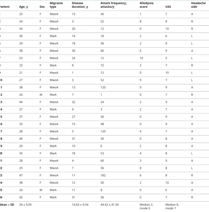
Figure 1Correlation of PACAP38-LI and mean diffusivity
(A) The skeleton is overlaid in green on the mean fractional anisotropy image. Significant correlations are depicted in copper (maximumpvalue MNI coordinates: x = 125, y = 69, z = 75). Clusters are thickened for better visualization. The color bar represents 1−pvalues corrected for multiple comparisons. (B) Scatterplot PACAP38-LI is plotted against average mean diffusivity under the significant voxels. The boxplots stand for mean, 95% confidence interval, and range. Outliers are depicted with open circles. MNI = Montreal Neurological Institute; PACAP38-LI = pituitary adenylate cyclase-activating peptide 38-like immunoreactivity.
nuisance regressors in the model. Although no clinical varia- bles correlated with interictal PACAP38-LI in the studied patients, we also tested another model with disease duration included as an additional nuisance regressor, as it was pre- viously shown to correlate with interictal PACAP38-LI in a larger sample of patients.3Clusters were formed using the threshold-free cluster enhancement method,30and correction for multiple comparisons was performed using FSL’s ran- domise tool31at a threshold ofp< 0.05.
Data availability
Anonymized data will be shared on request through personal correspondence after the approval of the local ethics committee.
Results
Demographic and clinical data of the patients Twenty-six patients with migraine were recruited into the study, 8 of whom had migraine with aura. Aura symptoms were visual except for a male and a female patient who had additional somatosensory symptoms. The demographic and clinical data of the patients are summarized in the
½T1 table.
Correlation of PACAP38-LI and white matter microstructure
Interictal plasma PACAP38-LI showed significant correlation MD (p< 0.0179, corrected for multiple comparisons) in the bilateral occipital white matter reaching into parietal and
temporal white matter (figure 1). ½F1
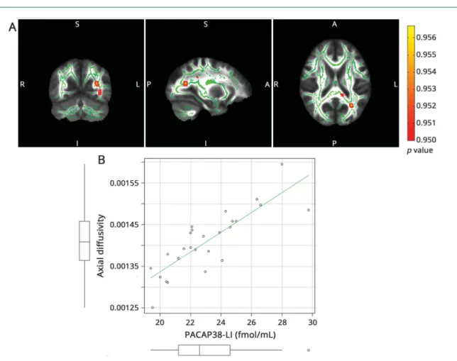
The correlation with AD was significant (p < 0.0432, cor- rected for multiple comparisons) in the left optic radiation and left posterior corpus callosum (figure 2). ½F2
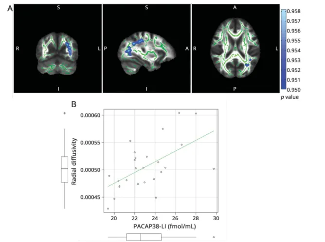
RD correlated with interictal PACAP38-LI (p< 0.0418, cor- rected for multiple comparisons) in the left optic radiation and parietal white matter (figure 3). FA did not show any ½F3
significant correlation with interictal PACAP38-LI.
Correlation of PACAP38-LI and diffusion measures with disease duration as nuisance regressor
With age, sex, and disease duration as nuisance regressors, interictal PACAP38-LI showed significant correlation with MD and AD in the left thalamus (p < 0.01, corrected for
Figure 2Correlation of PACAP38-LI and axial diffusivity
(A) The skeleton is overlaid in green on the mean fractional anisotropy image. Significant correlations are depicted in red-yellow (maximumpvalue MNI coordinates:
x = 125, y = 69, z = 75). Clusters are thickened for better visualization. The color bar represents 1−pvalues corrected for multiple comparisons. (B) Scatterplot PACAP38-LI is plotted against the average axial diffusivity under the significant voxels. The boxplots stand for mean, 95% confidence interval, and range. Outliers are depicted with open circles. MNI = Montreal Neurological Institute; PACAP38-LI = pituitary adenylate cyclase-activating peptide 38-like immunoreactivity.
multiple comparisons) (
½F4 figure 4). We found no significant correlation with FA and RD.
Discussion
Herein, we report significant correlation between diffusion parameters of the white matter and interictal plasma PACAP38-LI in patients with migraine in the occipital white matter. Similar to our former results,3 the interictal PACAP38-LI was lower than in normal healthy controls.
Interpretation of diffusion measures in terms of microstruc- tural characteristics is a much-debated area complicated by the fact that microstructural properties are probed in a few millimeter large elements of the space. However, it is generally accepted that lower FA or higher MD corresponds to reduced white matter integrity. AD and RD are suggested to correlate with the number of axons and the integrity of the myelin, respectively.32,33As such, our results could be interpreted as follows: abnormally lower plasma PACAP38 levels in the
interictal phase are accompanied by decreased values of MD, AD, and RD representing higher axonal density and/or myelination, or generally more compact white matter in the optic radiation, corpus callosum, and temporoparietal regions.
The absence of correlation with FA may be attributable to the fact that FA is a measure that combines AD and RD; if there is a similar change in both AD and RD, FA would change only if the magnitude of change is different in the 2 perpendicular directions.
Considering that interictal hyperexcitability of the cortex in migraineurs has been demonstrated before,8,34 and this in- creased baseline activity might also reveal to be maladaptive plasticity leading to more compact white matter, our results could be an indication of such maladaptive remodeling in response to migraine symptoms that is in connection with PACAPergic signaling. In particular, a functional MRI study of PACAP38-induced migraine-like attacks found increased connectivity of the left visual cortex in the default-mode network.35 This increased connectivity and presumable ac- tivity could also lead to increased structural connections.
Figure 3Correlation of PACAP38-LI and perpendicular diffusivity
(A) The skeleton is overlaid in green on the mean fractional anisotropy image. Significant correlations are depicted in blue/light blue (maximumpvalue MNI coordinates: x = 115, y = 64, z = 102). Clusters are thickened for better visualization. The color bar represents 1−pvalues corrected for multiple comparisons.
(B) Scatterplot PACAP38-LI is plotted against the average perpendicular diffusivity under the significant voxels. The boxplots stand for mean, 95% confidence interval, and range. Outliers are depicted with open circles. MNI = Montreal Neurological Institute; PACAP38-LI = pituitary adenylate cyclase-activating peptide 38-like immunoreactivity.
Of course, it might also be possible that the connection be- tween PACAP38 and white matter integrity is purely co- incidental and decreased interictal plasma PACAP38 levels reflect changes in PACAPergic signaling that contribute to disrupted neurochemical coupling in migraine, as demon- strated by magnetic resonance spectroscopy studies.36In the long term, these functional alterations may prove to be structural changes.
Longitudinal studies might help in understanding the exact nature of these connections. The location of correlating dif- fusion measurements suggests involvement of the visual
system, many aspects of which are affected in migraine.37 Photophobia is a prominent accompanying symptom present in a large percentage of migraineurs, and bright light is known to exacerbate migrainous headache. The phenomenon is thought to originate in part from intrinsically photosensitive retinal ganglionic cells38that express PACAP38 to be used as a cotransmitter in retinohypothalamic projections.12,39,40 Neurons of the suprachiasmatic nucleus on the receiving end of these pathways connect to and modulate light-sensitive neurons in the trigeminal nucleus caudalis,41an area that also contains PACAP38-expressing neurons,42 thus providing possible linkage between the visual and trigeminovascular system. Intrinsically photosensitive retinal ganglionic cells also show direct connections to thalamic pain centers in rats43 and have been thought to be part of a photophobia pathway that involves direct stimulation of trigeminal afferents in the eye without interposition of the optic nerve.44,45
Other animal studies link PACAP38 to behavioral aspects of photophobia: PACAP-deficient mice, after nitroglycerol- induced activation of the trigeminovascular system, exhibit light avoidance to a lesser degree than their wild-type coun- terparts.46 In light of the above, it is possible that reduced interictal PACAP38 levels reflect changes in photophobia- associated signaling that co-occur with alterations of fiber integrity in the optic tract, which would also be corroborated by thefinding that PACAP38 uptake peaks in the occipital cortices after intranasal administration in mice.17
The correlation between the white matter microstructural measures and the PACAP38 immunoreactivity was somewhat lateralized. Formerly, we showed that the diffusion charac- teristics of subcortical structures are asymmetric,47 which might be present in major white matter tracts as well.48Al- though they did not reach significance when corrected for multiple comparisons, there are voxels in the contralateral white matter that mirror significant results at a more liberal statistical threshold.
The change in the pattern of correlating diffusion measure- ments with the inclusion of disease duration as a nuisance regressor suggests that altered PACAP38 metabolism and its effect might develop as a function of disease progression, though it is unclear whether this is due to disease pathology or accumulating effects of allostatic load. In another study, we found that longer disease duration corresponds with de- creased values of AD in the left parietooccipital regions in migraine patients with aura,22 which co-occur with PACAP38-related alterations in the current study. Consid- ering that decreased PACAP38 levels correlate with diffusion measures of the left thalamus irrespective of disease duration, we would suggest that PACAP38 might have a few different roles in the development of white matter alterations, which should be addressed in further studies.
One limitation of our study is the heterogeneity of the pa- tient population because of the inclusion of migraine Figure 4Correlation of pituitary adenylate cyclase-activat-
ing peptide 38-like immunoreactivity and axial and mean diffusivity with disease duration as nuisance regressor
The skeleton is overlaid in green on the mean fractional anisotropy image.
Significant correlations are depicted in red for axial diffusivity (A) and copper for mean diffusivity (B) (maximumpvalue MNI coordinates: mean diffusivity:
x = 98, y = 101, z = 85; axial diffusivity: x = 102, y = 96, z = 82). Clusters are thickened for better visualization. The color bar represents 1−pvalues corrected for multiple comparisons. MNI = Montreal Neurological Institute.
patients both with and without aura. Based on our recent results, there are microstructural differences in the 2 groups that would necessitate regarding them as separate entities.22 Hence, we repeated the analysis separately for patients with and without aura. Since group sizes are considerably smaller, no significant correlation was found in either group with interictal PACAP38-LI. However, looking at the un- corrected results, we can identify similar patterns of corre- lation in both groups (data available from Dryad [Material]:
doi.org/10.5061/dryad.g58811v), which would indicate no major differences in terms of correlating PACAP38-LI and diffusion measures. Still, studies deploying greater sample sizes are needed to assess differences between migraine patients with and without aura.
Also, since PACAPergic signaling seems to be altered in migraine, we decided to focus our study on the in- terindividual variation of PACAP38-LI in patients with migraine. However, information about the relationship be- tween PACAP38-LI and white matter diffusion measures in healthy controls is rare in the literature. Controlled studies are needed to assess possible differences in the relationship between PACAP38 levels and microstructural character- istics in healthy controls and migraineurs.
While various MRI measures are useful biomarkers of mi- graine, direct connection between the disease and MRI fea- tures is still to be established. Providing connection between a pathobiologically important neurochemical biomarker of the disease and MRI alterations found in migraine emphasizes the value of both markers and opens up new directions of investigations.
Author contributions
D´aniel Ver´eb: study concept and design, data acquisition, analysis and interpretation, writing the manuscript. Niko- letta Szab´o: study concept and design, data acquisition, analysis and interpretation, drafting the manuscript. Ber- nadett Tuka: data acquisition, interpretation, drafting the manuscript. J´anos Tajti: contribution to study design, study supervision, revising the manuscript for important in- tellectual content. Andr´as Kir´aly: data acquisition and analysis, revising the manuscript for important intellectual content. P´eter Farag´o: data acquisition and analysis, revising the manuscript for important intellectual content. Kriszti´an Kocsis: data acquisition and analysis, revising the manu- script for important intellectual content. Eszter T´oth: data acquisition and analysis, revising the manuscript for im- portant intellectual content. B´alint Kincses: data analysis, revising the manuscript for important intellectual content.
Ter´ez Bagoly: data acquisition. Zsuzsanna Helyes: contri- bution to study design, data acquisition, revising the man- uscript for important intellectual content. L´aszl´o V´ecsei:
contribution to study design, study supervision, revising the manuscript for important intellectual content. Zsigmond Tam´as Kincses: study concept and design, study supervi- sion, drafting the manuscript.
Study funding
This study was supported by the MTA-SZTE Neuroscience Research Group, the National Brain Research Programs (grant KTIA_13_NAP-A-II/20 and NAP B KTIA_NAP_
13-2014-0022: MTA-PTE NAP B Pain Research Group, identification number 888819 and NAP 2.0 [2017-1.2.1- NKP-2017-00002]), and GINOP grants (GINOP-2.3.2-15- 2016-00034, GINOP-2.3.2-15-2016-00050). This research was supported by the EU-funded Hungarian grant EFOP- 3.6.1-16-2016-00008 and EFOP3.6.2-16-2017. Dr. Szab´o was supported by the Bolyai Scholarship Program of the Hungarian Academy of Sciences. Dr. Ver´eb, Dr. Kir´aly, and Dr. Farag´o were supported by the UNKP-17-3 New Na- tional Excellence Program of the Ministry of Human Capacities.
Disclosure
The authors report no disclosures relevant to the manuscript.
Go to Neurology.org/N for full disclosures.
Received January 24, 2018. Accepted infinal form June 26, 2018.
References
1. Schytz HW, Birk S, Wienecke T, Kruuse C, Olesen J, Ashina M. PACAP38 induces migraine-like attacks in patients with migraine without aura. Brain 2009;132:16–25.
2. Amin FM, Hougaard A, Schytz HW, et al. Investigation of the pathophysiological mechanisms of migraine attacks induced by pituitary adenylate cyclase-activating polypeptide-38. Brain 2014;137:779–794.
3. Tuka B, Helyes Z, Markovics A, et al. Alterations in PACAP-38-like immunoreactivity in the plasma during ictal and interictal periods of migraine patients. Cephalalgia 2013;33:1085–1095.
4. Tajti J, Szok D, Majlath Z, Tuka B, Csati A, Vecsei L. Migraine and neuropeptides.
Neuropeptides 2015;52:19–30.
5. Rossi HL, Recober A. Photophobia in primary headaches. Headache 2015;55:600–604.
6. Gonz´alez de la Aleja J, Ramos A, Mato-Abad V, et al. Higher glutamate to glutamine ratios in occipital regions in women with migraine during the interictal state. Head- ache 2013;53:365–375.
7. Becerra L, Veggeberg R, Prescot A, et al. A“complex”of brain metabolites distinguish altered chemistry in the cingulate cortex of episodic migraine patients. Neuroimage Clin 2016;11:588–594.
8. Farago P, Tuka B, Toth E, et al. Interictal brain activity differs in migraine with and without aura: resting state fMRI study. J Headache Pain 2017;18:8.
9. Szabo N, Kincses ZT, Pardutz A, et al. White matter microstructural alterations in migraine: a diffusion-weighted MRI study. Pain 2012;153:651–656.
10. Rocca MA, Pagani E, Colombo B, et al. Selective diffusion changes of the visual pathways in patients with migraine: a 3-T tractography study. Cephalalgia 2008;28:1061–1068.
11. Zhang Y, Malmberg AB, Sjolund B, Yaksh TL. The effect of pituitary adenylate cyclase activating peptide (PACAP) on the nociceptive formalin test. Neurosci Lett 1996;
207:187–190.
12. Engelund A, Fahrenkrug J, Harrison A, Hannibal J. Vesicular glutamate transporter 2 (VGLUT2) is co-stored with PACAP in projections from the rat melanopsin- containing retinal ganglion cells. Cell Tissue Res 2010;340:243–255.
13. Yilmaz N, Karaali K, Ozdem S, Turkay M, Unal A, Dora B. Elevated S100B and neuron specific enolase levels in patients with migraine-without aura: evidence for neurodegeneration? Cell Mol Neurobiol 2011;31:579–585.
14. Guo S, Vollesen AL, Hansen YB, et al. Part II: biochemical changes after pituitary adenylate cyclase-activating polypeptide-38 infusion in migraine patients. Cephalalgia 2017;37:136–147.
15. Ogata K, Shintani N, Hayata-Takano A, et al. PACAP enhances axon outgrowth in cultured hippocampal neurons to a comparable extent as BDNF. PLoS One 2015;10:
e0120526.
16. Shioda S, Nakamachi T. PACAP as a neuroprotective factor in ischemic neuronal injuries. Peptides 2015;72:202–207.
17. Nonaka N, Farr SA, Nakamachi T, et al. Intranasal administration of PACAP: uptake by brain and regional brain targeting with cyclodextrins. Peptides 2012;36:168–175.
18. Gao HM, Li L, Zhang KL, Chen XH, Tian SQ, Zhang ZL. Impact of migraine attacks on the blood-brain barrier. Chin Med J 2010;123:2559–2561.
19. Beaulieu C. The basis of anisotropic water diffusion in the nervous system: a technical review. NMR Biomed 2002;15:435–455.
20. Lipton RB, Bigal ME, Steiner TJ, Silberstein SD, Olesen J. Classification of primary headaches. Neurology 2004;63:427–435.
21. Hamilton M. A rating scale for depression. J Neurol Neurosurg Psychiatry 1960;23:
56–62.
22. Szabo N, Farago P, Kiraly A, et al. Evidence for plastic processes in migraine with aura:
a diffusion weighted MRI study. Front Neuroanat 2018;11:138.
23. Smith SM, Jenkinson M, Woolrich MW, et al. Advances in functional and structural MR image analysis and implementation as FSL. Neuroimage 2004;23(suppl 1):
S208–S219.
24. Jenkinson M, Smith S. A global optimisation method for robust affine registration of brain images. Med Image Anal 2001;5:143–156.
25. Smith SM. Fast robust automated brain extraction. Hum Brain Mapp 2002;17:
143–155.
26. Smith SM, Jenkinson M, Johansen-Berg H, et al. Tract-based spatial statistics: vox- elwise analysis of multi-subject diffusion data. Neuroimage 2006;31:1487–1505.
27. Andersson JRL, Jenkinson M, Smith SM. Non-Linear Registration, aka Spatial Nor- malization: FMRIB Technical Report TR07JA2. Oxford, UK: FMRIB Centre; 2007.
28. Rueckert D, Sonoda LI, Hayes C, Hill DL, Leach MO, Hawkes DJ. Nonrigid regis- tration using free-form deformations: application to breast MR images. IEEE Trans Med Imaging 1999;18:712–721.
29. Nichols TE, Holmes AP. Nonparametric permutation tests for functional neuro- imaging: a primer with examples. Hum Brain Mapp 2002;15:1–25.
30. Smith SM, Nichols TE. Threshold-free cluster enhancement: addressing problems of smoothing, threshold dependence and localisation in cluster inference. Neuroimage 2009;44:83–98.
31. Winkler AM, Ridgway GR, Webster MA, Smith SM, Nichols TE. Permutation in- ference for the general linear model. Neuroimage 2014;92:381–397.
32. Song SK, Yoshino J, Le TQ, et al. Demyelination increases radial diffusivity in corpus callosum of mouse brain. Neuroimage 2005;26:132–140.
33. Senda J, Watanabe H, Tsuboi T, et al. MRI mean diffusivity detects widespread brain degeneration in multiple sclerosis. J Neurol Sci 2012;319:105–110.
34. Afra J, Proietti Cecchini A, Sandor PS, Schoenen J. Comparison of visual and auditory evoked cortical potentials in migraine patients between attacks. Clin Neurophysiol 2000;111:1124–1129.
35. Amin FM, Hougaard A, Magon S, et al. Change in brain network connectivity during PACAP38-induced migraine attacks: a resting-state functional MRI study. Neurology 2016;86:180–187.
36. Younis S, Hougaard A, Vestergaard MB, Larsson HBW, Ashina M. Migraine and magnetic resonance spectroscopy: a systematic review. Curr Opin Neurol 2017;30:
246–262.
37. Kowacs PA, Utiumi MA, Piovesan EJ. The visual system in migraine: from the bench side to the office. Headache 2015;55(suppl 1):84–98.
38. Noseda R, Kainz V, Jakubowski M, et al. A neural mechanism for exacerbation of headache by light. Nat Neurosci 2010;13:239–245.
39. Hannibal J, Hindersson P, Ostergaard J, et al. Melanopsin is expressed in PACAP- containing retinal ganglion cells of the human retinohypothalamic tract. Invest Ophthalmol Vis Sci 2004;45:4202–4209.
40. Hannibal J, Ding JM, Chen D, et al. Pituitary adenylate cyclase-activating peptide (PACAP) in the retinohypothalamic tract: a potential daytime regulator of the bi- ological clock. J Neurosci 1997;17:2637–2644.
41. Okamoto K, Tashiro A, Chang Z, Bereiter DA. Bright light activates a trigeminal nociceptive pathway. Pain 2010;149:235–242.
42. Uddman R, Tajti J, Hou M, Sundler F, Edvinsson L. Neuropeptide expression in the human trigeminal nucleus caudalis and in the cervical spinal cord C1 and C2.
Cephalalgia 2002;22:112–116.
43. Noseda R, Burstein R. Advances in understanding the mechanisms of migraine-type photophobia. Curr Opin Neurol 2011;24:197–202.
44. Amini A, Digre K, Couldwell WT. Photophobia in a blind patient: an alternate visual pathway. Case report. J Neurosurg 2006;105:765–768.
45. Dolgonos S, Ayyala H, Evinger C. Light-induced trigeminal sensitization without central visual pathways: another mechanism for photophobia. Invest Ophthalmol Vis Sci 2011;52:7852–7858.
46. Markovics A, Kormos V, Gaszner B, et al. Pituitary adenylate cyclase-activating polypeptide plays a key role in nitroglycerol-induced trigeminovascular activation in mice. Neurobiol Dis 2012;45:633–644.
47. Kiraly A, Szabo N, Pardutz A, et al. Macro- and microstructural alterations of the subcortical structures in episodic cluster headache. Cephalalgia 2018;38:
662–673.
48. Wahl M, Li YO, Ng J, et al. Microstructural correlations of white matter tracts in the human brain. Neuroimage 2010;51:531–541.