Review
Nox/Duox Family of NADPH Oxidases: Lessons from
Knockout Mouse Models
Gábor Sirokmány,
1,2Ágnes Donkó,
1,2and Miklós Geiszt
1,*
Nox/Duox NADPH oxidases are now considered the primary, regulated sources of reactive oxygen species (ROS). These enzymes are expressed in diverse cells and tissues, and their products are essential in several physiological settings.
Knockout mouse models are instrumental in identifying the physiological func- tions of Nox/Duox enzymes as well as in exploring the impact of their pharma- cological targeting on disease progression. The currently available data from experiments on knockout animals suggest that the lack of non-phagocytic Nox/
Duox enzymes often modifies the course and phenotype in many disease models. Nevertheless, as illustrated by studies on Nox4-deficient animals, the absence of Nox-derived ROS can also lead to aggravated disease manifes- tation, reinforcing the need for a more balanced view on the role of ROS in health and disease.
NADPH Oxidases
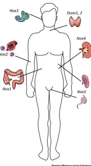
Members of the Nox/Duox family NADPH oxidases are the primary sources of regulated ROS production in a wide range of organisms (Box 1)[1–3]. Once thought to be an exclusive feature of phagocytes, ROS production by NADPH oxidases has now also been described in several different tissues and cells[1]. There are seven members of the Nox/Duox enzyme family in humans: Nox1–5, Duox1, and Duox2[4](Figure 1). Intriguingly, mice and rats lack the Nox5 isoform. Nox/Duox enzymes have been implicated in a vast array of biological settings including host defense, vasoregulation, hormone synthesis, fertilization, cell proliferation, cell differentia- tion, and extracellular matrix formation[1,3].
It is generally accepted that homeostasis normally ensures tight regulation of ROS levels since the highly reactive molecules can easily inflict damage on cellular constituents. By contrast, deficient production of ROS may also have deleterious consequences as illustrated by the serious clinical picture of chronic granulomatous disease, in which phagocytic superoxide production is practically absent[5], or by congenital hypothyroidism caused by Duox2 deficiency [6]. Considering the widespread expression of non-phagocytic Nox/Duox enzymes, and their presence in vital organs, it is reasonable to believe that altered activity of other Nox/Duox enzymes can also contribute to disease pathogenesis (Table 1). In cases where the pathogenic role of certain Nox/Duox isoforms is caused by increased activity, pharmacological inhibition may have therapeutic potential[7]. However, to obtain a fair assessment of the feasibility of therapeutic Nox inhibition, the physiological functions of different isoforms should be identified.
To this end, the phenotypic analysis of knockout animals may provide information about the physiological functions of specific NADPH oxidase isoforms. Studying disease development and progression in knockout animals also offers the possibility of uncovering the role of Nox enzymes
Trends
Members of the Nox/Duox NADPH oxi- dase family produce ROS in a regu- lated manner in several different cells and tissues.
Pharmacological inhibition of non-pha- gocytic Nox/Duox enzymes might have therapeutic potential.
Several studies have described the dis- ease-modifying phenotypes of Nox1 and Nox4 knockouts.
The lack of Nox4-derived ROS can lead to aggravated disease development, which is in contrast to the prevailing dogma that considers ROS to be gen- erally harmful.
1Department of Physiology, Semmelweis University, Faculty of Medicine,“Momentum”Peroxidase Enzyme Research Group of the Semmelweis University and the Hungarian Academy of Sciences, PO Box 259, H-1444 Budapest, Hungary
2These authors contributed equally to this work[2_TD$DIFF].
*Correspondence:geiszt@eok.sote.hu (M. Geiszt).
in disease pathogenesis. This latter experimental approach is particularly important in the process of drug target identification. Genetic models of Nox/Duox deficiency are also crucial tools when expression of these enzymes is examined at the protein level, as using knockout samples in western blot or immunostaining experiments is the ultimate control to prove the specificity of an antibody. In this review, we discuss the results of studies involving mouse models with Nox/Duox deficiency. The complex host defense problem that develops in the absence of a functional phagocytic oxidase and the role of Nox2 in other disease models have been covered in several papers[1,5,8], therefore they are not discussed in this work. Since non-phagocytic Nox/Duox isoforms are expressed in various cells and tissues, this review necessarily includes the discussion offindings from seemingly unrelated experimental models.
Even so, the results discussed here are connected in a way that they all reflect the consequences of the absence of Nox/Duox-derived ROS.
Nox1
Nox1 was thefirst homolog of Nox2 (formerly known as gp91phox[1_TD$DIFF]) to be identified[9]. Nox1 is highly expressed in the colon (Figure 1), where it localizes to epithelial cells. Similar to its phagocytic counterpart, Nox1 functions in a complex with p22phox[10]. To form a superox- ide-producing enzyme complex, Nox1 requires two additional proteins, NOXO1 and NOXA1, which are also present in the colon epithelium[11]. Nox1-deficient mice werefirst created by Matsunoet al., who described no obvious abnormality of the animals[12].
In spite of the fact that the colon has the highest level of Nox1 expression, surprisingly little is known about the function of Nox1 in this organ. The colon is normally inhabited by a huge number of bacteria; thus, the presence of Nox1 and its regulators in the colon, along with the homology between the phagocytic and colon oxidases suggest a role in host defense. However, disturbed antimicrobial function in Nox1 knockout animals has not yet been reported. Coant et al.showed that progenitor cells in the colon of Nox1 knockout mice are converted into goblet cells in higher numbers than that in wild-type animals[13]. The increase in goblet cell number was paralleled by the reduced number of colonocytes. Cell fate in the colon is controlled by Wnt and Notch1 signaling, and ROS produced by the colon oxidase may influence these pathways through the redox regulation of PTEN and NF-kB[13]. Recent data suggest that bacteria may have a significant role in controlling cell proliferation in the colon through Nox1. Joneset al.have shown thatLactobacillistimulate ROS production and proliferation of intestinal epithelial cells in a Nox1-dependent manner[14]. ROS production by the colon oxidase might also contribute to mucosal repair after injury. In an intestinal epithelial-specific Nox1 knockout model, mucosal healing was impaired in Nox1 knockouts[15]. This deficiency is likely explained by a previously unknown signaling pathway, in which the activity of Nox1 is regulated by an endogenous N-formyl peptide receptor ligand, annexin 1. Nox1-derived ROS then stimulate epithelial cell
Box 1. NADPH Oxidases
The story of NADPH oxidases stems from phagocyte research. When exposed to foreign pathogens, phagocytic cells– such as neutrophil granulocytes–produce large amounts of superoxide. Superoxide and its derivatives (ROS) have a crucial role in the killing of the invading pathogens. ROS act like a‘double-edged sword’, meaning that they are essential for killing pathogens; however, they can also be harmful to host tissues. In phagocytic cells, superoxide (O2–) is produced by the NADPH oxidase enzyme, which transfers electrons from NADPH to molecular oxygen. The‘core’of this enzyme is a heme-binding,flavoprotein Nox2 (formerly known as gp91phox), which catalyzes thefinal steps of electron transfer. It was known for some time that besides phagocytes, other types of mammalian cells synthesize and release ROS, which can serve various biological functions. However, the enzymatic background of non-phagocytic ROS production was unknown. Beginning from 1999, several enzymes were identified at the molecular level, which are now proven to be responsible for regulated ROS production observed in diverse tissues. These proteins are highly homologous to the phagocytic NADPH oxidase and are now designated the Nox/Duox family of NADPH oxidases. The Nox family of NADPH oxidases consists of the following members: Nox1, Nox2, Nox3, Nox4, Nox5, Duox1, and Duox2. The discovery of this enzyme family has stimulated ROS research since we can now assign a specific source of ROS in several tissues, including the gastrointestinal tract, kidney, heart, lung, thyroid, inner ear, and blood vessels.
migration and wound repair through the oxidative inactivation of protein phosphatases PTEN and PTP-PEST[15]. Several lines of evidence suggest that ROS contribute to the development of inflammatory bowel disease[16,17]. In this regard, a potential role of Nox1 is particularly interesting since the enzyme is located in the colon epithelial cells themselves while previously only inflammatory cells were considered as a ROS source. Glutathione peroxidase-1 and -2 double knockout mice develop spontaneous ileocolitis, the severity of which is dependent on the genetic background. When these mice were crossed with Nox1 knockout animals, the severity of the colitis was reduced, thus proving a role for Nox1-derived ROS in the inflammatory process [18]. In contrast to thisfinding, the absence of Nox1 in IL-10-deficient animals induced intestinal inflammation, which is probably caused by endoplasmic reticulum stress, developing in double knockout goblet cells[19].
The expression of Nox1 is not restricted to the colon epithelium, as the enzyme has been described in several other cell types, including vascular smooth muscle cells[9]. Angiotensin II
Nox3 Duox1, 2
Nox4 Nox2
Nox1 Nox5
Figure 1. Characteristic Expression of Nox/Duox Isoforms.For each Nox/Duox protein thefirst organ where its expression was originally reported–and where it also shows an accordingly high expression level–is displayed. Nox1 in the colon, Nox2 in phagocytes, Nox3 in the inner ear, Nox4 in renal epithelial cells, Nox5 in the spleen and testis, and Duox 1 and Duox2 in the thyroid gland. For a detailed description of the expression pattern of each isoform, refer to review articles[1–3].
infusion induces increased blood pressure in mice, and this response is attenuated in the genetic absence of Nox1 [12,20]. One study reported decreased basal blood pressure in Nox1 knockout animals[20], while another group did notfind such a difference[12]. The absence of Nox1 might be protective against angiotensin II-induced hypertension because the lack of superoxide increases the bioavailability of nitric oxide. Matsuno et al. found that vascular hypertrophy stimulated by angiotensin II was unchanged in the absence of Nox1[12], while Gavazziet al. reported reduced aorta hypertrophy, characterized by decreased accumulation of extracellular matrix[20]. In Nox1 knockout animals, angiotensin II-elicited aortic dissection is less severe, and this protective effect is unlikely due to lower blood pressure since norepinephrine treatment induces hypertension but fails to induce dissections in wild-type or Nox1 knockout mice[21]. In angiotensin II-infused Nox1 knockout animals, the expression level of tissue inhibitor of metalloproteinase 1 (TIMP-1) is increased when compared with wild-type mice. The elevation of TIMP-1 level in the vascular wall can reduce the activity of matrix metalloproteinases (MMPs), Table 1. Mouse Models of Non-Phagocytic Nox/Duox Deficiency
Gene Genotype Predicted or Proved Change in Pro-
tein Expression
Gross Phenotype
Nox1[12,20] 1. Exons 3–6 deleted[12,20]
2. Exon 13 truncated[15](Cre/
lox-based)
Predicted loss of protein[12,20], predicted truncated protein[15]
None observed
Nox3[30] Spontaneous mutations:
Line hetR96: 441 (T!A) Line het2J: 1282 (G!A) Line hetR542: 1576 (A!G)
Line hetR96: a 147 amino acid long truncated protein, early STOP after third transmembrane domain Line het2J: 428. Conserved Gly!Arg change in the NADPH binding site Line hetR542: 526. Lys!Gln change in putative NADPH binding site
Balance defect (head tilt)
Nox4[28,36,40] 1. Exons 1–2 deleted[36](Cre/
lox-based)
2. Exon 4 deleted[45]
2. Exons 14–15 deleted[28]
3. Exon 9 deleted[40](Cre/lox- based)
Proved loss of protein None observed
Duox1[67] Trapping cassette inserted between exons 20 and 21
Proved loss of protein None observed
Duox2[54] Spontaneous mutation:
2021 (T!G)
674. Val!Gly change after thefirst transmembrane helix, reduced protein expression, and loss of ROS production[55]
Congenital hypothyroidism
p22phox[32] Spontaneous mutation:
361 (T!C)
121. Tyr!His change in the second transmembrane domain, proved loss of protein
Balance defect
Noxo1[31] Spontaneous mutation: A (deoxyadenosine) insertion in exon 1 after the 28th nucleotide
Frameshift after the ninth amino acid that yields a 34 amino acid long truncated protein
Balance defect (head slant)
Noxa1[33] Deletion of exons 3–6 (Cre/lox- based)
Frameshift after the 88th amino acid, then incorporation of 19 novel amino acids before the premature stop codon, predicted loss of protein
None observed
DuoxA1/
DuoxA2[56]
Simultaneous deletion of exons after the exon 5 in both DuoxA genes
246 amino acid long truncated DuoxA1, 232 amino acid long truncated DuoxA2, predicted loss of activators
Congenital hypothyroidism
which may be behind the lower occurrence of aortic dissections in Nox1 knockouts[21]. In a transgenic model of hypertension, where active human renin is produced under the control of the transthyretin promoter, deletion of Nox1 did not reduce blood pressure, while oxidative stress was less prominent in the knockouts[22]. There is also evidence that oxidative stress elicited by hyperglycemia in endothelial cells is mediated by Nox1[23]. Furthermore, in an experimental model of diabetes mellitus, ApoE-deficient mice develop less severe atherosclerosis when the nox1gene is also disrupted[23].
Nox1 mRNA is also present in neurons, although the expression has never been demonstrated at the protein level. We also know little about the expression of NOXO1 and NOXA1 in neurons.
This is an important problem since the mere demonstration of Nox1 mRNA expression does not necessarily prove that a Nox1-based oxidase complex is active in the cell.
Nox1 mRNA expression was detected in dorsal root ganglion (DRG) neurons and Nox1-deficient animals show attenuated response to inflammatory pain[24]. Chemical mediators of inflamma- tory pain augment the capsaicin-induced calcium signal through the activation of TRPV1 channel, and this regulatory effect was found to be altered in Nox1 knockouts. The same group reported that in the absence of Nox1 the analgesic effect of morphine was potentiated, and analgesic tolerance was suppressed[25]. Further experiments that include an attempt to identify each component of the Nox1-based oxidase complex in DRG neurons are clearly needed before we can appreciate the physiological significance of the above-discussed observations.
Working with the same Nox1-deficient mouse line, conflicting results have been published about the role of Nox1 in the development of ischemic injury of the brain. Kahleset al. observed a significant reduction of infarct size at 24 h following a 1 h ischemic period implemented by the occlusion of the middle cerebral artery (MCAO)[26]. The beneficial effect of Nox1 deletion no longer existed if the occlusion lasted 2 h or longer. In a similar study, Jackmanet al.applied MCAO for only 0.5 h and analyzed the injury also after 24 h[27]. They found no difference in total infarct volume; however, a 4-fold greater cortical infarct volume was observed in Nox1 knock- outs, suggesting that the activity of Nox1 could be protective in the cortex. In contrast to these studies, Kleinschnitzet al.reported no difference in lesion size and functional outcome between wild-type and Nox1 knockout animals[28].
Nox1 is also expressed in microglia, where Nox2 appears to be the other dominant Nox isoform [29]. When lipopolysaccharide (LPS) was injected into the striatum of Nox1 knockouts and wild- type animals, nitrotyrosine formation was reduced in the knockouts, implicating Nox1 in the formation of peroxynitrite. This activity may inflict damage on neurons since in the LPS-injected region of Nox1 knockouts, a reduced loss of synapsin (a marker of presynaptic terminals) was detected[29].
Nox3
Among NADPH oxidases, Nox3 seems to have the most restricted expression pattern in the mammalian organism. Nox3 is specifically localized to the inner ear (Figure 1), where it is required for the proper development of otoconia crystals of the vestibular system. This unique function of Nox3 wasfirst revealed by successful positional cloning of several mutant mouse alleles at the head tilt (het) locus on chromosome 17[30]. Mutations at this locus lead to severe balance and spatial orientation defects of adult mice. All these mutants caused loss of function of thenox3 gene[30].
The specific role of Nox3 in vestibular development was further corroborated by a study identifying an NADPH oxidase organizer 1 (Noxo1) mutation responsible for the head slant (hslt) mouse
phenotype[31]. Similarly to the Nox3 mutanthetmice, the formation and crystallization of otoconia in thehsltmice were also severely disturbed, while other morphological structures of the inner ear seemed to be normal[31]. In agreement with these results, another indispensable component of the functioning Nox3 complex, p22phox, was also shown to be necessary for otoconia formation [32]. Thenmf333mutant mouse strain carries missense mutation in the transmembrane region eliminating the expression of the p22phoxprotein[32]. A special staining method (von Kossa) showed the lack of calcium carbonate crystals in the utriculus just like in the case of the Nox3- deficienthead tiltanimals. Along with the balance defects, thenmf333animals also have severe immunodeficiency due to the lack of phagocyte oxidase (Nox2) activity[32].
Although Noxo1 and Noxa1 were shown to interact in the Nox1 complex of colon epithelial cells [11], Noxa1 knockouts do not show balance defects, indicating that Noxa1 is not part of the inner ear NADPH oxidase complex[33].
Nox4
Nox4 wasfirst identified in kidney epithelial cells[34](Figure 1), but in the past 10 years the enzyme has been identified in several different cell types, including endothelial cells, vascular smooth muscle,fibroblasts, cardiomyocytes, skeletal muscle, osteoclasts, adipocytes, neurons, and microglia [1]. Although Nox4 is currently the most extensively studied Nox isoform, surprisingly few knockout studies have been published on Nox4. Research on this Nox isoform has been hampered by the lack of specific antibodies that can detect endogenously expressed protein[35]. Accordingly, the intracellular localization of Nox4 is still unclear and the protein was described at several intracellular locations including the nucleus, endoplasmic reticulum, focal adhesions, plasma membrane, and mitochondria.
In the mouse kidney, Nox4 is mainly expressed in proximal tubules although its mRNA was also detected in other segments of the nephron, including glomeruli and medullary collecting ducts [34]. It is of note that Nox4-deficient animals show no gross abnormality in their kidneys[28,36].
Babelovaet al.studied the course of three different chronic kidney disease models in mice with global or inducible Nox4 deletion[37]. Models included diabetes-induced kidney injury, unilateral ureter ligation, and 5/6 nephrectomy, respectively. The absence of Nox4 was not protective in either of the models, and the course offibrosis induced by unilateral ureter obstruction was even slightly more severe in Nox4-deficient animals[37]. An independent study, using the same model of kidneyfibrosis in a different Nox4-deficient strain, yielded similar results–the absence of Nox4 was found to be associated with more severe interstitialfibrosis[38]. Further analysis of the Nox4-deficient kidney revealed that the absence of the enzyme increased the apoptotic rate of tubular cells and reduced vascularization [38]. The lower interstitial capillary density was probably a consequence of decreased HIF-1/expression. Interestingly, in ApoE-deficient mice the absence of Nox4 offered protection against glomerular damage in diabetic nephropathy[39].
Hopefully in the future, Nox4 knockouts will also assist to elucidate the physiological role of Nox4 in the kidney. The high expression level in proximal tubular cells along with the constitutive ROS-producing activity suggest a yet to be identified physiological function for the enzyme.
Nox4 is also present in the heart, where its precise expression pattern remains to be determined.
Zhanget al.studied the role of Nox4 in cardiac adaptation to pressure overload using Nox4 knockouts and transgenic animals that overexpressed Nox4 in a cardiac-specific manner[36].
Their results suggest that Nox4 has a protective role in chronic load-induced stress[36]. The signaling pathways behind the protective effect of Nox4 include higher induction of HIF-1/ resulting in elevated vascular endothelial growth factor (VEGF) levels and enhanced angiogene- sis[36]. This study was thefirst to demonstrate that the absence of Nox4 might have deleterious consequences. Interestingly, in mice where Nox4 was deleted in a cardiomyocyte-specific
manner, pressure overload evoked less severe pathological changes in Nox4 knockouts, suggesting a pathogenic role for Nox4[40]. Further studies should clarify the reason for these conflictingfindings; however, it should be mentioned that a more severe pressure overload was induced in the latter study. It is also important to note that results obtained with cell-specific knockouts could be misleading when the function of an organ is analyzed as a whole. As for the heart, three different cell types in the heart (cardiomyocytes, endothelial cells,fibroblasts) were described to express Nox4[41]. Consequently, deletion of Nox4 activity in a cardiomyocyte- specific manner does not ensure a complete loss of Nox4 activity in the organ.
Endothelial cells are a particularly rich source of Nox4 and the enzyme seems to have a protective role in blood vessels[42]. In response to ischemia, the formation of new blood vessels was found to be lowered in Nox4-deficient animals and angiotensin II-induced vascular dysfunction was also more severe in Nox4 knockouts. Loss of Nox4 activity was accompanied by reduced expression of endothelial nitric oxide synthase (eNOS) and heme oxygenase leading to decreased formation of protective endothelial mediators[42]. In a recent study, accelerated atherosclerosis was found in Nox4 knockout animals[43]that might be explained by reduced leukocyte adhesion to Nox4-deficient endothelial cells. Overexpression of Nox4 in an endothe- lium-specific manner reduces atherosclerosis in ApoE–/–mice, an observation that also points to an anti-atherosclerotic effect of Nox4-derived ROS[44].
Nox4 is also expressed in the lung, where type II epithelial cells and endothelial cells contain the enzyme. In a bleomycin-inducedfibrosis model, the lungs of Nox4 knockout mice showed less severe pathological changes than that of wild-type animals[45]. The protective effect of Nox4 deficiency was probably due to the reduced apoptotic rate of type II epithelial cells. A previous study, using RNA interference, already suggested that Nox4 might be important in the devel- opment of lungfibrosis. That study, however, implicated reduced formation of myofibroblasts as an explanation for the protective effect of Nox4 deficiency[46].
The possible role of ROS in the pathogenesis of neurodegenerative diseases is currently a subject of intense research, and thus elucidating the function of Nox4 in the central nervous system (CNS) is also of great interest. In an ischemic stroke model induced by transient MCAO, Nox4-deficient mice developed less oxidative stress and were protected from brain damage [28]. In immunostaining experiments, the authors detected an induction of Nox4 in neurons, although Nox4 knockouts were not included as negative controls[28]. VAS2870, a non-specific inhibitor of Nox enzymes mimicked the effect of genetic Nox4 deletion, raising hopes for the development of an anti-stroke therapeutic strategy using Nox inhibitors. Experiments on Nox4 knockout animals revealed that the enzyme is also present in a subset of DRG neurons, where it has a role in nociceptive processing [47]. Although Nox4 was identified in nonmyelinated neurons, Nox4-mediated ROS production induced dysmyelination of a differentfiber population in a mouse model of peripheral nerve injury.
Osteoclasts are among the cell types where Nox4 wasfirst described [48]. Nox4 knockout animals have higher bone density, a phenotype that is likely caused by impaired formation and activation of osteoclasts[49]. In a murine model of postmenopausal osteoporosis, pharmaco- logical or genetic inhibition of Nox4 reduced the loss of trabecular bone[49].
Duox1 and Duox2
Duox1 and Duox2 werefirst cloned from porcine and human thyroid gland (Figure 1)[50,51]. For proper targeting and function, Duox enzymes must interact with Duox activator proteins DuoxA1 and DuoxA2[52]. Homozygous nonsense mutation in Duox2 causing congenital hypothyroidism demonstrated its obligate function in thyroid hormone biosynthesis[6,53]. Mice with a sponta- neously developed missense Duox2 mutation develop the phenotype characteristic for
congenital hypothyroidism [54] due to the loss of Duox2-mediated ROS production [55].
Simultaneous knockout of the two genes encoding DuoxA1 and DuoxA2 led to the development of the same phenotype[56].
Duox expression is not confined to the thyroid gland; the enzyme is present on mucosal surfaces of the respiratory and gastrointestinal tract, where a host defense function was suggested[57].
According to the model introduced by Geisztet al., Duox-produced H2O2supports lactoperox- idase to generate hypothiocyanite, which is an effective antimicrobial compound against a broad range of bacteria[58–60]. In non-vertebrates, such asDrosophila, zebrafish, andCaenorhabditis elegans, a growing cluster of data supports the indispensable role of Duox in gastrointestinal control of commensal, foodborne, or pathogenic microorganisms[61–63]. Recently, an interesting study on DuoxA double knockout mice confirmed the protective role of Duox2-derived H2O2, as it prevented the gastric colonization byHelicobacter felisand attendant inflammatory responses [64]. This study was thefirstin vivogenetic demonstration in a mammalian organism of how Duox can control the colonization of a noninvasive pathogen of the mucus layer. The innate immune modulatory effect of Duox-derived H2O2was further confirmed by a recent study in an ovalbumin- induced allergic asthma model using the Duox1- and Duox2-deficient DuoxA–/–double knockouts, where reduced mucus cell metaplasia, decreased airway resistance, and lower level of Th2 cytokine secretion was found in the DuoxA–/–mice[65]. In a separate study,flagellin-stimulated expression of inflammation and immune response-related genes was attenuated in the nasal mucosa of the Duox2-deficient mice compared with wild-type animals[66]. Importantly, in the latter study Duox2 knockout animals were not hormone-supplemented, thus the profound effect of thyroid hormone deficiency on gene expression should also be considered. Since precise restoration of physiological thyroid hormone levels is probably difficult to achieve, conditional knockouts lacking Duox2 in a tissue-specific manner will be instrumental in the assessment of Duox2 function in mucosal host defense. Another interesting question is whether the specific deletion of Duox2 in colon epithelial cells affects the expression and function of Nox1, which is also highly expressed in the colon. It is possible that the two enzymes have some overlapping functions, although the differences in their regulation may be a sign of distinct biological functions. Never- theless, the presence of two different Nox/Duox isoforms in colon epithelial cells highlights the importance of regulated ROS production in the colon.
Duox1 is also expressed in the thyroid gland, although its function remains unknown. Functional redundancy in hormone synthesis is unlikely since Duox1 cannot substitute for the loss of Duox2, and Duox1 knockout mice are not hypothyroid[67]. We detected Duox1 expression in mouse urothelium and showed inin vivocystometry experiments using knockout mice that Duox1-derived H2O2attenuates urinary bladder contractions[67]. We propose that mechanical stretch of the bladder urothelium triggers Duox1 activation, but further studies are needed to elucidate whether H2O2acts directly or in a paracrine manner, diffusing to neighboring cells and acting on smooth muscle or nerve cells.
Concluding Remarks
There are several approaches to study the function of a particular gene. Although the use of pharmacological inhibitors, and more recently RNA interference, offer a quick way to assess gene function in cell culture orin vivo, it is important to keep in mind that specificity of these approaches is often insufficient. The creation of inheritable mutation in mice offers a more specific way to study gene function. Although the generation of knockout mouse models was generally time-consuming, recently discovered gene-modifying techniques have dramatically reduced the time needed for gene targeting[68]. Knockout mice models are now available for all mammalian Noxs and their regulators, except for Nox5, which is not expressed in mice.
Experiments on gene-deficient animals have helped to understand the physiological function of Nox3 and confirmed the essential role of Duox2 in thyroid hormone synthesis. In the case of
Outstanding Questions
In sharp contrast to the pathology of Nox3 and Duox2 deficiencies, the absence of Nox1, Nox4, or Duox1 does not result in an obvious pheno- type and knockout models for these genes did not help uncover their roles in organs where they were originally discovered. Thus, we still do not know:
What is the physiological function of Nox1 in the colon?
What is the role of Nox4 in kidney epi- thelial cells?
What is the function of epithelial- expressed Duox1 in different organs, including the thyroid?
What is the physiological role of Duox2 outside of the thyroid?
What is the function of Nox5? The absence of Nox5 in mice and rats calls for developing animal models of other species.
Nox1, Nox4, and Duox1, the hunt is still on for their main functions in organs where they are expressed at their highest levels (see Outstanding Questions). This is a major concern when considering the pharmacological targeting of these isoforms, because unexpected side effects may result from the inhibition. Several studies have documented the disease-modifying phe- notypes of Nox1 and Nox4 knockouts, and in some cases the results suggest that pharmaco- logical inhibition of their activity might have therapeutic value. However, several observations that document the deleterious effects of Nox gene deletion on disease progression offer the provocative conclusion that devising pharmacological tools to stimulate their activity would sometimes be more desirable.
The discovery of the Nox/Duox family of NADPH oxidases has dramatically transformed our view of ROS and oxidative stress. The fact that diverse cells and tissues express Nox/Duox enzymes proves that regulated production of ROS is an integral part of maintaining homeostasis. Thus, the traditional dogma that considered ROS as exclusively harmful substances is no longer acceptable and is gradually replaced by a more differentiated view on their role in physiology and pathophysiology. Hopefully, future experiments using Nox/Duox-deficient knockouts will help us to better comprehend their physiological functions and clarify the therapeutic potential of Nox/Duox inhibition.
Acknowledgments
This research is supported by grants from the Hungarian Research Fund (OTKA K106138, PD103960) and by a
‘Momentum[4_TD$DIFF]’grant from the Hungarian Academy of Sciences to the authors[5_TD$DIFF]. We thank Zita Dávid and Anna Pató for help withFigure 1.
References
1. Bedard, K. and Krause, K.H. (2007) The NOX family of ROS- generating NADPH oxidases: physiology and pathophysiology.
Physiol. Rev.87, 245–313
2. Geiszt, M. and Leto, T.L. (2004) The Nox family of NAD(P)H oxidases:
host defense and beyond.J. Biol. Chem.279, 51715–51718 3. Lambeth, J.D. and Neish, A.S. (2014) Nox enzymes and new
thinking on reactive oxygen: a double-edged sword revisited.
Annu. Rev. Pathol.9, 119–145
4. Brandes, R.P.et al.(2014) Nox family NADPH oxidases: molecular mechanisms of activation.Free Radic. Biol. Med.76C, 208–226 5. Segal, B.H.et al.(2000) Genetic, biochemical, and clinical features of chronic granulomatous disease.Med. (Baltimore)79, 170–200 6. Moreno, J.C.et al.(2002) Inactivating mutations in the gene for thyroid oxidase 2 (THOX2) and congenital hypothyroidism.N.
Engl. J. Med.347, 95–102
7. Cifuentes-Pagano, E.et al.(2014) The quest for selective nox inhibitors and therapeutics: challenges, triumphs and pitfalls.Anti- oxid. Redox Signal.20, 2741–2754
8. Kahles, T. and Brandes, R.P. (2013) Which NADPH oxidase isoform is relevant for ischemic stroke? The case for nox 2.
Antioxid. Redox Signal.18, 1400–1417
9. Suh, Y.A.et al.(1999) Cell transformation by the superoxide- generating oxidase Mox1.Nature401, 79–82
10.Ambasta, R.K.et al.(2004) Direct interaction of the novel Nox proteins with p22phox is required for the formation of a functionally active NADPH oxidase.J. Biol. Chem.279, 45935–45941 11.Geiszt, M.et al.(2003) Proteins homologous to p47phoxand
p67phoxsupport superoxide production by NAD(P)H oxidase 1 in colon epithelial cells.J. Biol. Chem.278, 20006–20012 12.Matsuno, K.et al.(2005) Nox1 is involved in angiotensin II-medi-
ated hypertension: a study in Nox1-deficient mice.Circulation112, 2677–2685
13.Coant, N.et al.(2010) NADPH oxidase 1 modulates WNT and NOTCH1 signaling to control the fate of proliferative progenitor cells in the colon.Mol. Cell. Biol.30, 2636–2650
14.Jones, R.M.et al.(2013) Symbiotic lactobacilli stimulate gut epi- thelial proliferation via Nox-mediated generation of reactive oxygen species.EMBO J.32, 3017–3028
15.Leoni, G.et al.(2013) Annexin A1, formyl peptide receptor, and NOX1 orchestrate epithelial repair.J. Clin. Invest.123, 443–454 16.Babbs, C.F. (1992) Oxygen radicals in ulcerative colitis.Free
Radic. Biol. Med.13, 169–181
17.Grisham, M.B. (1994) Oxidants and free radicals in inflammatory bowel disease.Lancet344, 859–861
18.Esworthy, R.S.et al.(2014) Nox1 causes ileocolitis in mice defi- cient in glutathione peroxidase-1 and -2.Free Radic. Biol. Med.
68, 315–325
19.Treton, X.et al.(2014) Combined NADPH oxidase 1 and interleu- kin 10 deficiency induces chronic endoplasmic reticulum stress and causes ulcerative colitis-like disease in mice.PLoS ONE9, e101669
20.Gavazzi, G.et al.(2006) Decreased blood pressure in NOX1- deficient mice.FEBS Lett.580, 497–504
21.Gavazzi, G.et al.(2007) NOX1 deficiency protects from aortic dissection in response to angiotensin II.Hypertension50, 189–196 22.Yogi, A.et al.(2008) Renal redox-sensitive signaling, but not blood pressure, is attenuated by Nox1 knockout in angiotensin II-depen- dent chronic hypertension.Hypertension51, 500–506 23.Gray, S.P.et al.(2013) NADPH oxidase 1 plays a key role in
diabetes mellitus-accelerated atherosclerosis.Circulation127, 1888–1902
24.Ibi, M.et al.(2008) Reactive oxygen species derived from NOX1/
NADPH oxidase enhance inflammatory pain.J. Neurosci.28, 9486–9494
25.Ibi, M.et al.(2011) Involvement of NOX1/NADPH oxidase in morphine-induced analgesia and tolerance. J. Neurosci.31, 18094–18103
26.Kahles, T.et al.(2010) NADPH oxidase Nox1 contributes to ischemic injury in experimental stroke in mice.Neurobiol. Dis.
40, 185–192
27.Jackman, K.A.et al.(2009) Importance of NOX1 for angiotensin II- induced cerebrovascular superoxide production and cortical infarct volume following ischemic stroke.Brain Res.1286, 215–220 28.Kleinschnitz, C.et al.(2010) Post-stroke inhibition of induced
NADPH oxidase type 4 prevents oxidative stress and neurode- generation.PLoS Biol.8, e1000479
29.Cheret, C.et al.(2008) Neurotoxic activation of microglia is pro- moted by a nox1-dependent NADPH oxidase.J. Neurosci.28, 12039–12051
30.Paffenholz, R.et al.(2004) Vestibular defects in head-tilt mice result from mutations in Nox3, encoding an NADPH oxidase.
Genes Dev.18, 486–491
31.Kiss, P.J.et al.(2006) Inactivation of NADPH oxidase organizer 1 results in severe imbalance.Curr. Biol.16, 208–213 32.Nakano, Y.et al.(2008) Mutation of the Cyba gene encoding
p22phox causes vestibular and immune defects in mice.J. Clin.
Invest.118, 1176–1185
33.Flaherty, J.P.et al.(2010) Generation of a conditional null allele of NADPH oxidase activator 1 (NOXA1).Genesis48, 568–575 34.Geiszt, M.et al.(2000) Identification of renox, an NAD(P)H oxidase
in kidney.Proc. Natl. Acad. Sci. U.S.A.97, 8010–8014 35.Altenhofer, S.et al.(2012) The NOX toolbox: validating the role of
NADPH oxidases in physiology and disease.Cell. Mol. Life Sci.69, 2327–2343
36.Zhang, M.et al.(2010) NADPH oxidase-4 mediates protection against chronic load-induced stress in mouse hearts by enhancing angiogenesis.Proc. Natl. Acad. Sci. U.S.A.107, 18121–18126 37.Babelova, A.et al.(2012) Role of Nox4 in murine models of kidney
disease.Free Radic. Biol. Med.53, 842–853
38.Nlandu, K.S.et al.(2012) NADPH-oxidase 4 protects against kidneyfibrosis during chronic renal injury.J. Am. Soc. Nephrol.
23, 1967–1976
39.Jha, J.C.et al.(2014) Genetic targeting or pharmacologic inhibition of NADPH oxidase nox4 provides renoprotection in long-term diabetic nephropathy.J. Am. Soc. Nephrol.25, 1237–1254 40.Kuroda, J.et al.(2010) NADPH oxidase 4 (Nox4) is a major source
of oxidative stress in the failing heart.Proc. Natl. Acad. Sci. U.S.A.
107, 15565–15570
41.Nabeebaccus, A.et al.(2011) NADPH oxidases and cardiac remodelling.Heart Fail. Rev.16, 5–12
42.Schroder, K.et al.(2012) Nox4 is a protective reactive oxygen species generating vascular NADPH oxidase.Circ. Res.110, 1217–1225
43.Schurmann, C.et al.(2015) The NADPH oxidase Nox4 has anti- atherosclerotic functions.Eur. Heart J.36, 3447–3456 44.Craige, S.M.et al.(2015) Endothelial NADPH oxidase 4 protects
ApoE–/–mice from atherosclerotic lesions.Free Radic. Biol. Med.
89, 1–7
45.Carnesecchi, S.et al.(2011) A key role for NOX4 in epithelial cell death during development of lungfibrosis.Antioxid. Redox Signal.
15, 607–619
46.Hecker, L.et al.(2009) NADPH oxidase-4 mediates myofibroblast activation andfibrogenic responses to lung injury.Nat. Med.15, 1077–1081
47.Kallenborn-Gerhardt, W.et al.(2012) NADPH oxidase-4 maintains neuropathic pain after peripheral nerve injury.J. Neurosci.32, 10136–10145
48.Yang, S.et al.(2001) A new superoxide-generating oxidase in murine osteoclasts.J. Biol. Chem.276, 5452–5458 49.Goettsch, C.et al.(2013) NADPH oxidase 4 limits bone mass by
promoting osteoclastogenesis.J. Clin. Invest.123, 4731–4738
50.Dupuy, C.et al.(1999) Purification of a novelflavoprotein involved in the thyroid NADPH oxidase. Cloning of the porcine and human cdnas.J. Biol. Chem.274, 37265–37269
51.De Deken, X.et al.(2000) Cloning of two human thyroid cDNAs encoding new members of the NADPH oxidase family.J. Biol.
Chem.275, 23227–23233
52.Grasberger, H. and Refetoff, S. (2006) Identification of the matu- ration factor for dual oxidase. Evolution of an eukaryotic operon equivalent.J. Biol. Chem.281, 18269–18272
53.Weber, G.et al.(2013) Genetic defects of hydrogen peroxide generation in the thyroid gland.J. Endocrinol. Invest.36, 261–266 54.Johnson, K.R.et al.(2007) Congenital hypothyroidism, dwarfism, and hearing impairment caused by a missense mutation in the mouse dual oxidase 2 gene,Duox2.Mol. Endocrinol.21, 1593–1602 55.Donko, A.et al.(2014) Hypothyroidism-associated missense
mutation impairs NADPH oxidase activity and intracellular traffick- ing of Duox2.Free Radic. Biol. Med.73, 190–200
56.Grasberger, H.et al.(2012) Mice deficient in dual oxidase maturation factors are severely hypothyroid.Mol. Endocrinol.26, 481–492 57.Geiszt, M.et al.(2003) Dual oxidases represent novel hydrogen
peroxide sources supporting mucosal surface host defense.
FASEB J.17, 1502–1504
58.Oram, J.D. and Reiter, B. (1966) The inhibition of streptococci by lactoperoxidase, thiocyanate and hydrogen peroxide. The oxida- tion of thiocyanate and the nature of the inhibitory compound.
Biochem. J.100, 382–388
59.Gerson, C.et al.(2000) The lactoperoxidase system functions in bacterial clearance of airways.Am. J. Respir. Cell Mol. Biol.22, 665–671
60.Clem, W.H. and Klebanoff, S.J. (1966) Inhibitory effect of saliva on glutamic acid accumulation byLactobacillus acidophilusand the role of the lactoperoxidase–thiocyanate system.J. Bacteriol.91, 1848–1853
61.Ha, E.M.et al.(2005) A direct role for dual oxidase inDrosophila gut immunity.Science310, 847–850
62.Chavez, V.et al.(2009) Ce-Duox1/BLI-3 generates reactive oxy- gen species as a protective innate immune mechanism inCaeno- rhabditis elegans.Infect. Immun.77, 4983–4989
63.Flores, M.V.et al.(2010) Dual oxidase in the intestinal epithelium of zebrafish larvae has anti-bacterial properties.Biochem. Biophys.
Res. Commun.400, 164–168
64.Grasberger, H.et al.(2013) Dual oxidases control release of hydrogen peroxide by the gastric epithelium to preventHelico- bacter felisinfection and inflammation in mice.Gastroenterology 145, 1045–1054
65.Chang, S.et al.(2013) Dual oxidase regulates neutrophil recruit- ment in allergic airways.Free Radic. Biol. Med.65, 38–46 66.Joo, J.H.et al.(2012) Dual oxidase 2 is essential for the toll-like
receptor 5-mediated inflammatory response in airway mucosa.
Antioxid. Redox Signal.16, 57–70
67.Donko, A.et al.(2010) Urothelial cells produce hydrogen perox- ide through the activation of Duox1.Free Radic. Biol. Med.49, 2040–2048
68.Wijshake, T.et al.(2014) Endonucleases: new tools to edit the mouse genome.Biochim. Biophys. Acta1842, 1942–1950