Original article
MicroRNA-25-dependent up-regulation of NADPH oxidase 4 (NOX4) mediates hypercholesterolemia-induced oxidative/nitrative stress and subsequent dysfunction in the heart
Zoltán V. Varga
a,b, Krisztina Kupai
a, Gerg ő Sz ű cs
a, Renáta Gáspár
a, János Pálóczi
a, Nóra Faragó
c, Ágnes Zvara
c, László G. Puskás
c, Zsolt Rázga
d, László Tiszlavicz
d, Péter Bencsik
a,e, Anikó Görbe
a,e, Csaba Csonka
a,e, Péter Ferdinandy
a,b,e, Tamás Csont
a,e,⁎
aCardiovascular Research Group, Department of Biochemistry, University of Szeged, Szeged, Hungary
bDepartment of Pharmacology and Pharmacotherapy, Semmelweis University, Budapest, Hungary
cInstitute of Genetics, Biological Research Center, Hungarian Academy of Sciences, Szeged, Hungary
dDepartment of Pathology, University of Szeged, Szeged, Hungary
ePharmahungary Group, Szeged, Hungary
a b s t r a c t a r t i c l e i n f o
Article history:
Received 6 December 2012 Received in revised form 13 May 2013 Accepted 17 May 2013
Available online 27 May 2013 Keywords:
Hyperlipidemia Peroxynitrite Superoxide Metabolic syndrome Cholesterol Nitrosative stress
Diet-induced hypercholesterolemia leads to oxidative/nitrative stress and subsequent myocardial dysfunction.
However, the regulatory role of microRNAs in this phenomenon is unknown. We aimed to investigate, whether hypercholesterolemia-induced myocardial microRNA alterations play a role in the development of oxidative/
nitrative stress and in subsequent cardiac dysfunction. Male Wistar rats were fed with 2% cholesterol/0.25%
cholate-enriched or standard diet for 12 weeks. Serum and tissue cholesterol levels were significantly elevated by cholesterol-enriched diet. Left ventricular end-diastolic pressure was significantly increased in cholesterol- fed rats both in vivo and in isolated perfused hearts, indicating diastolic dysfunction. Myocardial expression of microRNAs was affected by cholesterol-enriched diet as assessed by microarray analysis. MicroRNA-25 showed a significant down-regulation as detected by microarray analysis and QRT-PCR. In silico target prediction revealed NADPH oxidase 4 (NOX4) as a putative target of microRNA-25. NOX4 protein showed significant up-regulation in the hearts of cholesterol-fed rats, while NOX1 and NOX2 remained unaffected. Cholesterol- feeding significantly increased myocardial oxidative/nitrative stress as assessed by dihydroethidium staining, protein oxidation assay, and nitro-tyrosine ELISA, respectively. Direct binding of microRNA-25 mimic to the 3′ UTR region of NOX4 was demonstrated using a luciferase reporter assay. Transfection of a microRNA-25 mimic into primary cardiomyocytes decreased superoxide production, while a microRNA-25 inhibitor resulted in an up-regulation of NOX4 protein and an increase in oxidative stress that was attenuated by the NADPH oxidase inhibitor diphenyleneiodonium. Here we demonstrated for thefirst time that hypercholesterolemia affects myo- cardial microRNA expression, and by down-regulating microRNA-25 increases NOX4 expression and conse- quently oxidative/nitrative stress in the heart. We conclude that hypercholesterolemia-induced microRNA alterations play an important role in the regulation of oxidative/nitrative stress and in consequent myocardial dysfunction.
© 2013 Elsevier Ltd. All rights reserved.
1. Introduction
Incidence of metabolic diseases (including obesity, type II diabe- tes, high blood pressure, and dyslipidemia) leading to severe cardio- vascular complications is constantly growing worldwide. The cost of this both in human lives and in dollars is overwhelming. According to the American Heart Association 2012 Heart Disease and Stroke
Statistics, elevated cholesterol level is still the leading risk factor for heart diseases. Almost 34 million people in the US have elevated total cholesterol level[1]. While metabolic diseases and especially hy- percholesterolemia have been studied extensively for many years, the underlying genetic networks and molecular signaling pathways re- main to be identified.
It is well known, that hypercholesterolemic patients have a much higher morbidity and mortality from cardiovascular diseases, consid- ering the central role of elevated cholesterol levels in the develop- ment of atherosclerosis. However, several studies have shown that hypercholesterolemia exerts direct myocardial effects independent of the development of atherosclerosis both in humans [2–4] and
⁎ Corresponding author at: Cardiovascular Research Group, Department of Biochemistry, University of Szeged, Dóm tér 9, Szeged, H-6720, Hungary. Tel.: +36 62 545 096; fax: +36 62 545 097.
E-mail address:csont.tamas@med.u-szeged.hu(T. Csont).
0022-2828/$–see front matter © 2013 Elsevier Ltd. All rights reserved.
http://dx.doi.org/10.1016/j.yjmcc.2013.05.009
Contents lists available atSciVerse ScienceDirect
Journal of Molecular and Cellular Cardiology
j o u r n a l h o m e p a g e : w w w . e l s e v i e r . c o m / l o c a t e / y j m c c
animal models[5–12]. Hypercholesterolemia negatively influences myocardial performance: impairs systolic as well as diastolic con- tractile function[5–7], aggravates the deleterious effects of ischemia/
reperfusion injury[8], and interferes with the anti-ischemic effect of ischemic pre- [7,9,13] and postconditioning [10]. Moreover, it has been previously shown that hypercholesterolemia-induced myocardial oxidative/nitrative stress significantly contributes to the development of cardiac dysfunction[5,11,12]. However, the exact underlying molec- ular mechanisms in relation to hypercholesterolemia and oxidative stress are still not entirely clear.
Previous works from our group suggested the central role of NADPH oxidase enzymes in hypercholesterolemia-induced oxidative stress[11]as well as in pro-inflammatory cytokine-induced myocar- dial dysfunction[14]. Factors (epigenetic, transcriptional, posttran- scriptional, and posttranslational) that may regulate NADPH oxidase expression and activity are not entirely known. Recently, microRNAs have emerged as powerful posttranscriptional regulators of gene expression[15]. MicroRNAs are known to play important roles in many physiological and pathological processes in the heart, including myocyte contractility, cardiac development, myocardial infarction, cardiacfibrosis, hypertrophic response, and arrhythmogenesis[16].
However, the role of cardiac microRNAs in metabolic disease states, especially in the myocardium in hypercholesterolemia is not known.
In the present study we aimed to investigate whether microRNAs are altered in the heart of hypercholesterolemic rats. We have demonstrated a decrease in the level of microRNA-25 in the heart of cholesterol-fed rats, and identified microRNA-25 as an important reg- ulator of NADPH oxidase 4 expression and thereby cardiac oxidative/
nitrative stress, which is known to be a major contributor of myocar- dial dysfunction induced by hypercholesterolemia.
2. Materials and methods
This investigation conforms to the Guide for the Care and Use of Laboratory Animals published by the US National Institutes of Health (NIH publication No. 85-23, revised 1996) and was approved by local animal ethics committee of the University of Szeged.
2.1. Experimental setup
Six-week-old male Wistar rats were fed normal or 2% cholesterol- and 0.25% cholate-enriched rat chow for 12 weeks. At the end of the diet period, venous blood was taken for determination of serum lipids. Hearts of anesthetized (sodium pentobarbital; 60 mg/kg i.p.) and heparinized (sodium heparin; 500 U/kg i.v.) rats were then iso- lated and perfused according to Langendorff with an oxygenated Krebs–Henseleit buffer at 37 °C for 10 min as described[17]. Tissue samples of the ventricular myocardium (n = 10–12 in each group) were rapidly frozen in liquid nitrogen for further biochemical analy- sis. Some of the hearts werefixed in 4% formaldehyde and were used for histological analysis. In a separate set of experiments, isolat- ed hearts were perfused in a“working”mode according to Neely for 30 min following the 10-min Langendorff perfusion. Hemodynamic parameters including heart rate, coronaryflow, aorticflow and ven- tricular pressure parameters were measured at the end of“working” perfusion, as described earlier[18]. Another set of animals was used for heart catheterization to record ventricular pressure in vivo.
2.2. In vivo heart catheterization
Rats were anesthetized with an intraperitoneal injection of pento- barbital sodium (60 mg/kg) and placed on a heated pad to maintain body temperature. To record left ventricular pressure curve, the right carotid artery was exposed, and a 1.6-Fr pressure catheter (Transonic Scisense Inc., London, ON, Canada) was inserted into the
left ventricle. Left ventricular pressure curves were evaluated using the Labscribe2 software.
2.3. Histology, immunohistochemistry, and transmission electron microscopy
Formaldehyde-fixed myocardial samples were embedded in paraffin, 4-μm sections were placed on silanized slides and, after conventional methods of dewaxing and rehydration, the samples were either stained with hematoxylin–eosin (to determine the development of hypertrophy and number of infiltrating immune cells), stained for collagen (Massons's trichrome), or were subjected to immunohistochemical analysis for NOX4 protein. Rabbit polyclonal anti-NOX4 antibody (Novus Biologicals, Cambridge, UK) was used as primary antibody (1:100; 30 min). The Envision Flex System (DAKO, Denmark) with high pH was used as visu- alization system. The sections were counterstained with hematoxylin (for 1 min) and examined by two independent histologists by means of light microscopy at 40× magnification.
For transmission electron microscopy, the formalin-fixed, paraffin- embedded specimens were re-embedded to plastic (Embed812, EMS, USA). 70 nm-thick sections were cut and placed on oval slot copper grids, and were analyzed under a transmission electron microscope (Philips CM10, 100 KV).
2.4. Measurement of lipid parameters
At the end of the diet period, serum cholesterol and triacylglycerol levels were determined by colorimetric assays, as described earlier [11](Diagnosticum, Budapest, Hungary). To analyzeα(HDL), pre-β (VLDL), andβ-lipoproteins (LDL) in the serum of rats fed cholesterol- enriched or normal diet, lipoproteins were separated on agarose gels, using Paragon Electrophoresis System Lipoprotein Electrophoresis Kit (Beckman Coulter, Fullerton, CA) according to the manufacturer's in- structions. To determine the tissue cholesterol content of normal and cholesterol-fed myocardial samples a gas chromatographic–mass spec- trometric analysis was carried out according to the method published by Luzon-Toro et al.[19]as previously described[9]. Amount of tissue cholesterol determined in each sample was normalized to tissue wet weight and expressed asμg/mg tissue.
2.5. MicroRNA isolation
Heart samples (n = 6) from both diet groups were powdered in liquid nitrogen. Steps of microRNA isolation were done according to the protocol of the microRNA isolation kit (Roche, Germany) with modifications[20]. The quality and quantity were assessed spectropho- tometrically (Nanodrop, USA) and with 2100 Bioanalyzer (Agilent, CA, USA). Random pairs of the RNA extracted from the 6 different samples in each group were pooled, and the obtained 3 samples/group were assayed on the microarrays.
2.6. Microarray analysis of microRNA expression
A total of 50 ng purified microRNA was labeled using Agilent's microRNA Complete Labeling and Hyb kit system (Agilent Technolo- gies Palo Alto, CA, USA). The protocol was briefly the following: 25– 25 ng of two parallel samples were pooled together in afinal volume of 2μl and subjected to a dephosphorylation reaction using Calf Intes- tinal Alkaline Phosphatase (CIP) at 37 °C for 30 min in thefinal vol- ume of 4μl. In the second step, 2.8μl of DMSO was added to each sample for denaturation at 100 °C for 5 min and placed on ice imme- diately. Following this step, a ligation reaction was carried out using T4 RNA Ligase and Cyanine3-pCp in a total volume of 11.3μl for 2 h at 16 °C to label the RNA samples. The labeled samples were completely vacuum dried on medium–high (45 °C) heat setting and hybridized onto the surface of Agilent 8 × 15k Rat microRNA
Microarray (Agilent Technologies, Palo Alto, CA, USA). The microarray contained probes for 350 microRNAs from the Sanger database v 10.1.
Dried and labeled samples were re-suspended in 18μl of nuclease- free water and denatured at 100 °C for 5 min in the presence of 1 × Blocking Agent and 1 × Hi-RPM Hybridization Buffer in afinal volume of 45μl and immediately placed on ice. These mixes were used for the hybridization, which was done in microarray hybridization chambers (Agilent Technologies, Palo Alto, CA), as previously described[21].
After hybridization the slides were washed in Gene Expression Wash buffer 1 containing Triton X-100 from Agilent Technologies at room temperature for 1 min, then in Gene Expression Wash buffer 2 containing Triton X-100 at 37 °C for another 1 min before scanning.
Each array was scanned as described earlier with an Agilent Scanner with 5μm resolution[21]. Output image analysis and feature ex- traction were done using Feature Extraction software of Agilent Technologies.
2.7. Validation of microarray data by QRT-PCR
To confirm microarray results, quantitative real-time PCR (QRT-PCR) was used. The reverse transcription reaction was performed with the TaqMan® MicroRNA Reverse Transcription Kit (Applied Biosystems, CA, USA). 350 ng from each sample was reverse transcribed in the pres- ence of 5× RT TaqMan® MicroRNA Assays (Applied Biosystems, CA, USA). 8μl reaction mixture contained 0.2μl dNTPs, 1.5μl MultiScribe
™Reverse Transcriptase (50 U/μl), 0.8μl 10× RT Buffer, 0.9μl MgCl2, 0.1μl RNase Inhibitor (20 U/μl), 1.5μl 5× RT primer and the template in a total volume of 3μl. Reverse Transcription was carried out with the following cycling parameters in a thermocycler (Bioneer, Daedong, Korea): 16 °C for 2 min, 42 °C for 1 min, 50 °C for 1 s, 45 cycles, then hold the samples on 85 °C for 5 min. After dilution with 64μl of water, 9μl of the diluted reaction mix was used as template in QRT-PCR. Reac- tions were performed on a RotorGene 3000 instrument (Corbett Re- search, Sydney, Australia) with the TaqMan protocol. 20μl reaction mixture contained 10μl TaqMan® Universal PCR Master Mix (Applied Biosystems), 1μl of the TaqMan® MicroRNA Assays and 9μl of the dilut- ed cDNA.
2.8. MicroRNA target prediction
The microRNA databases and target prediction tools TargetScan (www.targetscan.org)[22] and microRNA.org (www.microrna.org) [23]were used to identify potential microRNA-25 targets.
2.9. Quantitative analysis of NOX mRNAs by QRT-PCR
QRT-PCR was performed on a RotorGene 3000 instrument (Corbett Research, Sydney, Australia) with gene-specific primers and SybrGreen protocol to monitor gene expression. 2μg of total RNA was reverse transcribed using the High-Capacity cDNA Archive Kit (Applied Biosystems Foster City, CA, USA) according to the manufacturer's in- structions in afinal volume of 30μl. The temperature profile of the re- verse transcription was the following: 10 min at room temperature, 2 h at 37 °C, 5 min on ice andfinally 10 min at 75 °C for enzyme inac- tivation. These steps were carried out in a Thermal Cycler machine (MJ Research Waltham, MA, USA). After dilution with 30μl of water, 1μl of the diluted reaction mix was used as template in the QRT-PCR.
Reactions were done with FastStart SYBR Green Master mix (Roche Ap- plied Science, Mannheim, Germany) according to the manufacturer's instructions at afinal primer concentration of 250 nM under the follow- ing conditions: 15 min at 95 °C, 45 cycles of 95 °C for 15 s, 60 °C for 25 s and 72 °C for 25 s. Thefluorescence intensity of SybrGreen dye was detected after each amplification step. Relative expressions were calculated as normalized ratios to the average Ct value of rat HPRT and Cyclophyllin genes. Thefinal relative gene expression ratios were calculated as delta–delta Ct values[21].
2.10. Measurement of NOX proteins by western immunoblotting
In order to investigate whether hypercholesterolemia or microRNA- 25 inhibitor transfection leads to an increased expression of NOX4 at the protein level in the heart, western blot analysis was performed.
Frozen heart samples were homogenized with NP-40 lysis buffer (150 mM NaCl, 50 mM Tris, 1%NP-40) or cultured cardiomyocytes were harvested by scraping and then homogenized and concentrated by centrifugation (7500 ×g, 20 min). Protein concentrations were determined by means of bicinchoninic acid method using bovine serum albumin as standard (Pierce, Rockford, USA). Forty (heart) or twenty (cells)μg of protein was loaded from each sample onto 10%
SDS-polyacrylamide gel. After separation by electrophoresis, proteins were transferred (wet transfer, 2.5 h) onto the nitrocellulose mem- brane (Amersham Biosciences, Piscataway, USA). Transfer was con- trolled by using Ponceau dye. The membrane was blocked with 5%
non-fat dry milk in 0.1% TBS-T overnight at 4 °C. After the blocking step, the membrane was cut horizontally and the upper part was incu- bated with a primary antibody (dissolved in 1% non-fat dry milk–TBS-T, 1:1000 dilution) against either NOX4 (Novus Biologicals, Cambridge, United Kingdom)–reported to specifically recognize NOX4 protein [24–27]–, NOX2 (Merck-Millipore, Darmstadt, Germany), or NOX1 (Novus Biologicals, Cambridge, United Kingdom) for 2 h at room tem- perature, followed by washing with TBS-T (3 × 10 min). After washing, the membrane was incubated with a secondary antibody (horseradish peroxidase-conjugated affinity purified goat anti-rabbit, 1/5000 dilu- tion) in 1% non-fat dry milk in TBS-T for 1 h at room temperature.
Then the membrane was washed again 3 times for 10 min. For detec- tion of the bands, the membrane was incubated with ECL-plus reagent (Amersham Biosciences, Piscataway, USA) for 5 min and then visual- ized on X-rayfilms. Band densities were evaluated by using Quantity One software (Bio-Rad Imaging System, Hercules, USA). Loading control was done by determining the GAPDH content of each sample. Briefly, the bottom of the membrane was probed with a primary antibody that recognizes GAPDH (1/10,000 dilution) for 2 h at room tempera- ture, followed by washing with TBS-T. Then the membrane was incu- bated with horseradish peroxidase-conjugated affinity purified goat anti-rabbit antibody (1/20,000 dilution) for 1 h at room temperature.
The membrane was washed again and band visualization and evalua- tion of band densities were done as described above. There was no sig- nificant difference in GAPDH between the groups.
2.11. In situ detection of superoxide
In situ detection of superoxide was performed by fluorescent microscopy using the oxidative fluorescent dye dihydroethidium (DHE). DHE is freely permeable to cell membranes and reacts with superoxide anions forming a redfluorescent product which interca- lates with DNA. Fresh frozen heart sections (30μm) were incubated with 10−6mol/L DHE (Sigma) in PBS (pH 7.4; 37 °C; 30 min) in a dark humidified container. Fluorescence in heart sections was then detected by afluorescent microscope (Nikon, Japan) with a 590-nm long-passfilter[11]. Images of sections treated with saline (negative control) were measuredfirst. Thefluorescence intensity was evaluat- ed with ImageJ 1.44.
2.12. Measurement of protein carbonylation
Total myocardial protein carbonylation was measured using the Oxyblot protein oxidation detection kit (Merck-Millipore, Billerica, USA) according to the manufacturer's protocol. Briefly, carbonyl groups were derivatized with 2,4-dinitrophenylhydrazine for 15 min. Dinitrophenyl- derivatized proteins were resolved by sodium dodecylsulphate– polyacrylamide gel electrophoresis and transferred to nitrocel- lulose membrane. Membranes were incubated overnight with anti-dinitrophenyl primary antibodies and then with goat anti-rabbit/
horseradish peroxidase antibodies. Signals were visualized by chemilu- minescence detection using X-rayfilms.
Myocardial nitrotyrosine was measured from cardiac tissue ho- mogenates by ELISA, as described earlier[10].
2.13. Luciferase reporter assay
To investigate whether microRNA-25 directly regulates NOX4 ex- pression, human NOX4 3′UTR sequence was inserted downstream of a Renilla luciferase open reading frame (GoClone System, SwitchGear Ge- nomics, Menlo Park, CA). The luciferase construct was transfected into HEK293 reporter cells together with either a mimic of microRNA-25 or with an inhibitor of microRNA-25 or with a non-targeting sequence (mimic/inhibitor control), respectively, by using the Dharmafect Duo transfection reagent. In each case, 10 ng plasmid DNA and 100 nM microRNA were used. HEK293 cells were chosen for their high efficiency of transfection. Cells were cultured for 24 h and assayed with the Lucifer- ase Assay Reagent of the manufacturer (SwitchGear Genomics, Menlo Park, CA).
2.14. Primary cardiomyocyte culture
To further confirm the direct link between microRNA-25 down- regulation and NOX4 expression followed by increased oxidative stress, neonatal rat cardiomyocyte cultures were prepared from newborn Wistar rats, as described earlier [28]. Cardiomyocytes were plated at a density of 3 × 10⁴cells/well onto 96-well plates and 5 × 10⁵cells/well onto 6-well plates. The culture was grown for 3 days before experimentation. Culture medium was changed the day after preparation. The cells were maintained at 37 °C (humidified atmosphere; 5% CO2) in a standard CO2 incubator (Sanyo, Japan).
2.15. MicroRNA-25 inhibitor or mimic transfection
To knock-down endogenous expression of microRNA-25, a microRNA-25 inhibitor was used, while to up-regulate microRNA-25, a synthetic microRNA-25 mimic was used (both from Dharmacon, Lafayette, USA). As a negative control, a microRNA inhibitor or mimic control (Dharmacon, Lafayette, USA) was transfected into rat neonatal cardiomyocytes, as recommended by the manufacturer. The transfec- tion was carried out according to the recommendations of the manufac- turer. Briefly, the microRNA-25 inhibitor or the inhibitor control was diluted in the medium at afinal concentration of 100 nM. To con- firm the efficiency of transfection, the same amount of Dy547-labeled positive control (Dharmacon, Lafayette, USA) was transfected in separate experiments. The 100 nM concentration of Dy547-labeled microRNA-inhibitor did not decrease cell viability as compared to 25 and 50 nM, however, it considerably increased efficacy of transfection (data not shown). The morphology and contractile ability of the transfected primary cardiomyocytes were not affected significantly by the transfection process as indicated by spontaneous beating 24 h after transfection (online video supplemental material, showing beating cardiomyocytes transfected with thefluorescent non-targeting negative control microRNA). Cells were incubated with microRNA-25 inhibitor or with the negative control microRNA inhibitor for 24 h and then used for NOX4 western blot analysis or forfluorescent oxidative stress measure- ment. Expression level of microRNA-25 following microRNA-25 mimic and inhibitor transfection was assessed by QRT-PCR, as described in Section 2.7.
2.16. Measurement of oxidative stress in microRNA-25 inhibitor or mimic transfected cells byfluorescent microplate reader
Transfected neonatal cardiomyocytes were loaded either with 2′,7′-dichlorofluorescin-diacetate (DCF-DA) or dihydroethidium
(DHE). Briefly, the cells were washed twice with Dulbecco's PBS (D-PBS) and loaded with DCF-DA (10μM) or DHE (10μM) for 30 min in dark at room temperature. After loading the dyes were removed, and the cells were covered with D-PBS and each well was scanned in a fluorescent microplate reader (FluoStar Optima, BMG Labtech, Ortenberg, Germany) using 495 nm excita- tion/520 nm emissionfilter combination for DCF-DA, and 505 nm exci- tation/620 nm emissionfilter combination for DHE. To test the NADPH oxidase inhibitor diphenyleneiodonium (DPI), the cells were pre- incubated with DPI for 4 h, beforefluorescent staining.
2.17. Measurement of oxidative stress in microRNA-25 inhibitor or mimic transfected cells byfluorescent microscopy
Neonatal cardiomyocytes were plated on glass coverslips, and transfected with a microRNA-25 mimic or inhibitor, or with their corresponding controls. The transfected cells were loaded with dihydroethidium (DHE), as described above. The coverslips were mounted on glass microscope slides covered with a fluorescent mounting medium (Dako, Glostrup, Denmark), to reduce fading of the fluorescent dye. Fluorescence in the transfected cells was detected by afluorescent microscope (Nikon, Japan) with a stan- dard rhodamine filter, using the same settings (exposition time, ISO sensitivity, etc) on each sample.
2.18. Statistical analysis
Values are expressed as mean ± SEM. One way analysis of variance (ANOVA) was used to evaluate differences in cell transfec- tion experiments to determine differences in oxidative stress. Other- wise Student's T-test was used. Statistical analysis of microRNA microarrays was done as described earlier by using the Feature Ex- traction software of Agilent Technologies [29]. All the individual microRNAs were represented by 20 different probes on the array. A microRNA was considered as detected, if at least one probe from all the 20 probes was detected. Total gene signal is equal to the sum of the signals of the individual probes. Expressions of all the 350 microRNAs found in the Sanger miRBase (version 10.1) were checked.
Six heart samples from each diet groups were analyzed by microRNA microarray. Two–two samples were pooled together and a total of 6 hybridization experiments were carried out to gain raw data for statistical analysis. Altogether 9 individual parallel gene activity com- parisons were done to determine the average changes, standard devi- ations and p-values. Using two tailed two sample unequal variance Student t-test, the p-value was determined and used to find the significant gene expression changes. Gene expression ratio with p-valueb0.05 and log2 ratio b−0.6 or log2 ratio > 0.6 (~1.5 fold change) are considered as repression and overexpression, respective- ly. Changes in gene expression were plotted as log2 ratios of signal in- tensity values. MicroRNAs having less than 4 individual parallel gene activity comparisons were excluded.
3. Results
3.1. Cholesterol-fed rats display elevated serum and tissue cholesterol levels
Cholesterol-enriched diet for 12 weeks caused a significant in- crease in cholesterol level in the serum (Fig. 1A) as well as in the myocardial tissue (Fig. 1B) which was accompanied by an altered li- poprotein pattern (Fig. 1C). The 12 week long diet induced a decrease in HDL-fraction. Cholesterol-enriched diet had no influence on serum triacylglycerol level (Fig. 1D).
3.2. Cholesterol-feeding leads to mild myocardial dysfunction without signs of histological alterations
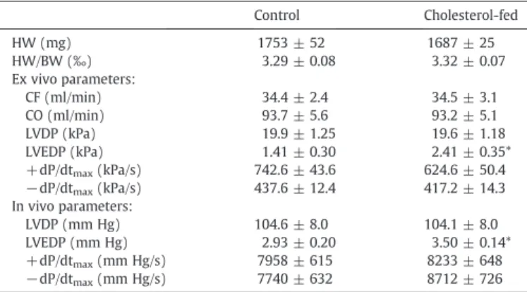
Parameters of myocardial function measured both in vivo and in isolated perfused hearts are shown in Table 1. Left ventricular end-diastolic pressure (LVEDP) showed a significant increase in the cholesterol-fed group as assessed both in vivo and ex vivo, indicating impaired relaxation and diastolic dysfunction. In addition, there was a non-significant (p = 0.096) decrease in + dP/dtmaxin the ex vivo perfused hearts of cholesterol-fed rats. Other examined parameters (heart weight, heart weight/body weight ratio, coronaryflow, cardiac output, left ventricular developed pressure, −dP/dtmax) did not change significantly due to the diet.
Cholesterol feeding had no apparent effect on myocardial histology (hematoxylin/eosin—Figs. 1E–F, Masson's trichrome—Figs. 1G–H) and subcellular ultrastructure (transmission electron microscopy — Figs. 1I–J).
3.3. MicroRNA-25 is down-regulated in hypercholesterolemic hearts
Myocardial microRNAs were isolated from the left ventricles of cholesterol-fed and control rats and were analyzed on a microRNA microarray. Among the assessed 350 microRNAs 120 showed detect- able expression in both groups (Fig. 2A). One of the most pronounced and significant alteration with reasonable inter-sample variation was seen in the case of microRNA-25 (−1.13 log2 down-regulation).
MicroRNA-29c* also showed a significant down-regulation (−2.35 log2 down-regulation), however, a marked inter-sample variation was seen in the case of this microRNA. The cholesterol-enriched diet-induced down-regulation of microRNA-25 was further con- firmed by QRT-PCR (Fig. 2B).
3.4. In silico microRNA-25 target prediction reveals NOX4 as a putative target
To identify putative microRNA-25 targets, we have performed a bio- informatic analysis by searching for potential 3′UTR binding sites in two different databases (Table 2.). Several proteins were listed, which were previously shown to be involved in myocardial physiology and/or path- ophysiology. Out of these hypothetical targets, NADPH oxidase 4 (NOX4) seemed to be a promising one, as it has 3 conserved and 1 poorly con- served 3′UTR sites (Fig. 2C). As we have previously proposed in our stud- ies that NADPH oxidases might be involved in hypercholesterolemia- induced oxidative stress[11], in the present study we have examined, whether hypercholesterolemia leads to microRNA-25-dependent modu- lation of NOX4 expression. To exclude the role of other cardiac NOX isoforms, we also screened microRNA-25 binding sites for NOX1, NOX2, and p22 phox, however, neither of these were predicted to be a potential target of microRNA-25.
3.5. NOX4 is expressed in the heart and is up-regulated by cholesterol-enriched diet
To examine the presence of NOX4 in the heart of cholesterol-fed rats, the expression level of NOX4 protein was determined by western blot. Our analysis revealed the up-regulation of NOX4 after 12 weeks of cholesterol-enriched diet, suggesting its role in
Fig. 1.Effect of 12-week cholesterol-enriched diet on serum cholesterol (A), heart total tissue cholesterol (B), serum lipoprotein distribution (C), serum triacylglycerol (D), and microscopic morphology (representative images E–J). The cholesterol-fed group (Chol) displayed a decrease in HDL concentration as compared to the standard chow-fed group (Cont). There was no sign of any histological alteration as assessed by standard HE (E and F) and Masson's trichrome staining (G and H). Scale bar represents 100μm.
There was no sign of any subcellular ultrastructural alterations (sarcomere or mitochon- drial disorganization) as assessed by transmission electron microscopy (magnification 25,000×, I and J). Results are expressed as mean ± SEM; n = 8-10. *pb0.05 vs. control.
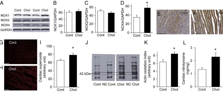
hypercholesterolemia-induced oxidative stress (Figs. 3A and D). To determine whether hypercholesterolemia induces expression alter- ation in other abundant cardiac NOX isoform, we also examined the expression of NOX1 and NOX2, however, there were no alterations in the protein level of cardiac NOX1 or NOX2 due to cholesterol- enriched diet (Figs. 3A, B, and C).
Immunostaining for NOX4 showed a diffuse positive staining in cardiomyocytes in both diet groups (Figs. 3E and F).
QRT-PCR analysis of myocardial samples obtained from cholesterol- fed and control animals showed no alterations in transcript levels of any of the NOX isoforms (NOX1, 2, and 4;Table 3.), suggesting that the up-regulation of NOX4 protein due to cholesterol-enriched diet may occur due to posttranscriptional gene regulation.
3.6. Increased myocardial oxidative and nitrative stress in cholesterol-fed rats
Myocardial oxidative stress was estimated by staining frozen myocardial sections with dihydroethidium. Increased nuclear red fluorescence was detectable in the hearts of cholesterol-fed rats as compared to control rats, indicating enhanced superoxide formation (Figs. 3G, H, and I).
The amount of oxidatively modified myocardial proteins was elevat- ed in the cholesterol-fed group as assessed by dinitrophenylhydrazine assay (Oxyblot;Fig. 3J). The intensity of the band with a molecular weight corresponding to actin (42 kDa) was significantly in- creased (Figs. 3J and K), suggesting that oxidation of contractile proteins is likely involved in hypercholesterolemia-induced myo- cardial dysfunction.
The nitrative stress marker nitrotyrosine was also elevated in the heart due to cholesterol-enriched diet (Fig. 3L).
3.7. NOX4 is a direct target of microRNA-25
Luciferase reporter assay was carried out, in order to prove the direct binding of microRNA-25 to NOX4 3′ UTR (Fig. 4A).
Co-transfection of a mimic of microRNA-25 and the luciferase con- struct (consisting of the 3′UTR region of the NOX4 mRNA) resulted in a decrease in the luciferase signal, indicating a direct binding of microRNA-25 to NOX4 3′UTR. In addition, when the inhibitor of microRNA-25 was co-transfected with the luciferase construct, there was a non-significant increase in the luciferase signal (p = 0.062).
To further investigate, whether binding of microRNA-25 to NOX4 3′ UTR affects oxidative stress, cardiomyocytes were transfected with a
mimic of microRNA-25 resulting in an up-regulation of microRNA-25 (4.14 ± 1.22 log2 expression alteration). Dihydroethidium staining showed a decrease in superoxide level (Fig. 4B), suggesting that the di- rect binding of microRNA-25 to NOX4 3′UTR likely results in decreased NOX4 activity.
Table 1
Hemodynamic parameters at the end of 12-week diet.
Control Cholesterol-fed
HW (mg) 1753 ± 52 1687 ± 25
HW/BW (‰) 3.29 ± 0.08 3.32 ± 0.07
Ex vivo parameters:
CF (ml/min) 34.4 ± 2.4 34.5 ± 3.1
CO (ml/min) 93.7 ± 5.6 93.2 ± 5.1
LVDP (kPa) 19.9 ± 1.25 19.6 ± 1.18
LVEDP (kPa) 1.41 ± 0.30 2.41 ± 0.35*
+dP/dtmax(kPa/s) 742.6 ± 43.6 624.6 ± 50.4
−dP/dtmax(kPa/s) 437.6 ± 12.4 417.2 ± 14.3 In vivo parameters:
LVDP (mm Hg) 104.6 ± 8.0 104.1 ± 8.0
LVEDP (mm Hg) 2.93 ± 0.20 3.50 ± 0.14*
+dP/dtmax(mm Hg/s) 7958 ± 615 8233 ± 648
−dP/dtmax(mm Hg/s) 7740 ± 632 8712 ± 726 Data are presented as means ± SEM, *pb0.05 vs. control, n = 8.
HW: heart weight; HW/BW: heart weight/body weight ratio; CF: coronaryflow; CO: cardiac output; LVDP: left ventricular developed pressure; LVEDP: left ventricular end-diastolic pressure; +dP/dtmax: maximal rate of ventricular pressure rise;−dP/dtmax: maximal rate of ventricular pressure decline.
Fig. 2.Effects of 12-week cholesterol-enriched diet on global cardiac microRNA expres- sion as assessed by microarray analysis (A), each data point represents a log2 ratio of signal intensity values of a certain microRNA, showing myocardial expression alteration in response to cholesterol-enriched diet. The black circle represents, microRNA-25 showing a pronounced and significant down-regulation in the heart of cholesterol-fed rats. MicroRNA-25 expression was further validated by using QRT-PCR (B), results are expressed as mean ± SEM. Sequences of microRNA-25 bind- ing sites in NOX4 3′UTR (C). Complementary nucleotide sequences are indicated by bold letters.
3.8. Effects of microRNA-25 inhibitor on oxidative stress and NOX4 expression
To further investigate if the down-regulation of microRNA-25–as seen in hypercholesterolemic hearts–is responsible for increased ox- idative stress by modulation of NOX4, we have transfected neonatal rat cardiomyocytes with a synthetic microRNA-25 inhibitor which in- duces knock-down of endogenous microRNA-25 level (−6.96 ± 3.07 log2 expression alteration). As a negative control, a non-targeting microRNA inhibitor control was used. Transfection efficiency was an- alyzed by transfecting Dy547-labeled positive control microRNA yielding in an efficient transfection (data not shown).
We have found that microRNA-25 knock-down significantly in- creased thefluorescence intensity of the hydrogen peroxide-sensitive dye dichlorofluorescin-diacetate (DCF-DA) (Fig. 4C) in cultured cardiomyocytes. Similarly, thefluorescence signal of the superoxide- specific dihydroethidium (DHE) was also increased significantly in cells transfected with microRNA-25 inhibitor (Fig. 4D). The non- selective NADPH oxidase inhibitor, diphenileniodonium (DPI), dose- dependently attenuated the microRNA-25 inhibitor-induced oxidative stress (Figs. 4C and D).
Transfection of cardiomyocytes with microRNA-25 inhibitor showed an increased fluorescent signal as assessed by DHE histochemistry, whereas, a reduced signal was detected in the microRNA-25 mimic transfected cells, as shown on representative images (Fig. 4E).
To confirm that the knock-down of microRNA-25 results in increased oxidative stress as a result of NOX4 up-regulation, NOX4 protein level was determined in transfected cells. MicroRNA-25 inhibitor significantly increased NOX4 protein level in primary cardiomyocytes (Fig. 4F).
4. Discussion
In the present study we have confirmed that hypercholesterol- emia leads to cardiac oxidative/nitrative stress and myocardial dys- function. We have demonstrated for thefirst time in the literature that hypercholesterolemia affects myocardial microRNA expression, moreover, hypercholesterolemia by down-regulating microRNA-25 increases NOX4 expression and consequently oxidative/nitrative stress in the heart, thereby leading to diastolic dysfunction (Fig. 5).
Our present results show that microRNA-25 is an important regulator of cardiac NADPH oxidase 4 and thereby myocardial oxidative/
nitrative stress.
Table 2
Predicted microRNA-25 target messenger RNAs selected on their relation to cardiovascular diseases.
Gene symbol Gene name Reference Predicted target (TargetScan) Predicted target (microRNA.org) Seed match (TargetScan)
NOX4 NADPH oxidase 4 [43] Yes Yes 8mer
BGN Biglycan [28] Yes Yes 8mer
SMAD7 SMAD family member 7 [52] Yes Yes 7mer
GATA2 GATA binding protein 2 [53] Yes Yes 8mer
PRKCE Protein kinase C, epsilon [54] Yes No 7mer
AQP2 Aquaporin 2 [55] Yes Yes 7mer
COL1A2 Collagen, type I, alpha 2 [56] Yes Yes 8mer
MEF2D Myocyte-specific enhancer factor 2D [57] Yes No 8mer
IDH1 Isocitrate dehydrogenase [58] Yes No 8mer
The microRNA target prediction tools microRNA.org (http://www.microrna.organd TargetScan (http://www.targetscan.org) were used to identify potential microRNA-25 targets.
Fig. 3.Representative western blots of NOX1, NOX2, NOX4, and GAPDH from cardiac homogenates from cholesterol-fed (Chol) and control (Cont) animals (A). Quantification of western blot analysis of NOX1 (B), NOX2 (C), and NOX4 (D) proteins in hearts of normal and cholesterol-fed rats normalized to the GAPDH control. Data are mean ± SEM;
n = 8–10 in each group. *pb0.05 vs. control rats. Representative ventricular sections from control (E) and cholesterol-fed rats (F) immunostained for NOX4 (scale bar represents100μm). Representative ventricular sections from control (G) and cholesterol-fed rats (H) stained for superoxide by dihydroethidium histochemistry. The representative images show the localization and intensity (redfluorescence) of superoxide in the nucleus (scale bar represents 100μm). Bar chart (I) shows quantification of dihydroethidium fluorescence of hearts from control and cholesterol-fed rats, representing myocardial oxidative stress. Values are mean ± SEM; n = 10 in each group. *pb0.05 vs. control group. Representative western blot of carbonylated myocardial proteins (J) from cardiac homogenates from cholesterol-fed (Chol) and control (Cont) animals (NC represents neg- ative control lane). The 42 kDa band corresponds to actin. Quantification of the intensity of the carbonylated 42 kDa bands (K) from cholesterol-fed (Chol) and control (Cont) an- imals. Values are mean ± SEM; n = 5 in each group. *pb0.05 vs. control group. Quantification of cardiac 3-nitrotyrosine content, a marker of endogenous peroxynitrite-induced nitrative stress (L). Free 3-nitrotyrosine was assayed by enzyme-linked immunosorbent assay (ELISA). Values are mean ± SEM; n = 10–12. *pb0.05 vs. control. (For interpreta- tion of the references to color in thisfigure legend, the reader is referred to the web version of this article.)
Here we report for thefirst time in the literature that diet-induced hypercholesterolemia affects myocardial microRNA expression pat- tern, indicating a possible role of these posttranscriptional regulators
in the hearts of hypercholesterolemic rats. Cholesterol-induced gene regulation is still an enigmatic research area. Cholesterol may regu- late gene expression at both transcriptional and posttranscriptional Table 3
QRT-PCR analysis of NOX isoenzyme (NOX1, NOX2, and NOX4) transcript levels.
Gene Accession number Forward primer Reverse primer Log2 change SEM Fold change
NOX1 NM_053683 ggcatccctttactctgacct tgctgctcgaatatgaatgg −0.25 0.33 −1.19
NOX2 NM_023965 gctgggattggagtcacg gcacagccagtagaagtagatcttt −0.001 0.24 −1.001
NOX4 NM_053524 gaacccaagttccaagctca gcacaaaggtccagaaatcc −0.123 0.16 −1.09
Fig. 4.Demonstration of the direct binding of rat microRNA-25 to human NOX4 3′UTR (A) using HEK293 cells. Values are mean ± SEM; n = 3. *pb0.05 vs. corresponding control.
Quantification of cellular oxidative stress by the superoxide-specific dihydroethidium in microRNA-25 mimic transfected primary rat cardiomyocytes (B). Values are mean ± SEM;
n = 4. *pb0.05 vs. control. Quantification of cellular oxidative stress by the hydrogen peroxide-sensitive dye dichlorofluorescin-diacetate (DCF-DA) and by the superoxide-specific dihydroethidium (DHE), analyzed by afluorescent microplate reader (C and D). Neonatal cardiac myocytes were treated with a microRNA-25 inhibitor or a non-targeting microRNA inhibitor control in the presence or absence of 500 nM or 2μM diphenyleneiodonium (DPI), a non-specific NOX inhibitor (C and D). MicroRNA-25 inhibitor significantly increased the amount of ROS level while DPI abolished both hydrogen peroxide (C) and superoxide (D) generations in a dose-dependent manner. Values are mean ± SEM; n = 5–6.
*pb0.05 vs. control. Representative microscopic images showing the relative amount of superoxide in primary cardiomyocytes transfected with the inhibitor control, mimic con- trol, and inhibitor control treated with LPS (10μg/ml) or with microRNA-25 inhibitor, and microRNA-25 mimic (E). Scale bar represents 20μm. Regulation of NOX4 protein expres- sion by microRNA-25 in neonatal cardiac myocytes transfected with microRNA-25 inhibitor or microRNA inhibitor control (F). Representative NOX4 and GAPDH western blots as well as quantified NOX4/GAPDH ratios show increased NOX4 expression in neonatal cardiac myocytes transfected with microRNA-25 inhibitor (F). Values are mean ± SEM;
n = 5–6. *pb0.05 vs. control.
levels, and in the former, two key transcription factors have been im- plicated so far, sterol-regulatory element binding protein (SREBP) and the liver X receptor (LXR)[30,31]. There are only a few reports showing that cholesterol affects microRNA expression, however, these studies were carried out on the liver in cholesterol fed pigs [32]and baboons[33]. Interestingly, a recent report showed an epige- netic regulation of microRNA-29b expression by oxidized-LDL[34], raising the possibility that serum cholesterol may also control microRNA-25 level by epigenetic regulation.
In our present study, cardiac microRNA-25 showed a pronounced down-regulation due to cholesterol-enriched diet. MicroRNA-25 is encoded in an intronic region of the mini-chromosome maintenance helixase 7. Interestingly, this region has been previously associated with a serum cholesterol QTL (quantitative trait locus) in rats[35].
To date, mainly cancer related reports were published showing the involvement of microRNA-25 in apoptotic signaling[36]and in cell invasion and migration[37]. Here we used bioinformatic analyses to characterize microRNA-25 mRNA targets (targetome) and a luciferase reporter assay to validate the direct binding of microRNA-25 to the 3′ UTR region of the most putative target, NOX4. We have identified and validated NOX4 as a major microRNA-25 target that is responsible for oxidative/nitrative stress in hearts of hypercholesterolemic rats.
However, several additional targets may be involved in the action of microRNA-25, especially in other cardiovascular disease conditions, considering the relevance of the identified hypothetical targets in car- diovascular diseases (Table 2.).
One of the most interestingfinding in this study was to show that hypercholesterolemia-induced myocardial dysfunction is mediated
by a microRNA-dependent regulation of NADPH oxidase activity.
The NADPH oxidase 4 isoenzyme (NOX4,first described as Renox [38]) is highly expressed in the heart[39]including cardiomyocytes (Figs. 3E and F) and localized in different intracellular organelles (e.g. mitochondria[39], ER[40], nucleus[41,42]). In addition, its in- creased expression has been shown to be involved in myocardial dys- function in heart failure after transverse aortic constriction [43].
Nevertheless, some reports showed opposite results, i.e. NOX4 may be protective against pressure overload-induced heart failure[44].
This discrepancy may be due to the use of different knock-out animal models, allowing the expression of active splice variants[45]. Howev- er, there is increasing evidence showing that NOX4 is involved in many pathological cardiac and vascular processes, including diabetic cardiomyopathy[24], TNF alpha-induced endothelial cell apoptosis [25], and stroke associated neurodegeneration by increasing the pro- duction of reactive oxygen species[46]. Our present study also sug- gests that the reactive oxygen species produced by NOX4 may contribute to myocardial dysfunction induced by cholesterol-feeding.
Despite its definite role in the myocardium, the molecular mecha- nisms, which mediate up-regulation of cardiac NOX4 were not iden- tified so far. It has been previously hypothesized, that increased cellular cholesterol level increases NADPH oxidase activity by acting on membrane microdomain assembly to recruit and organize the cy- tosolic NADPH oxidase subunits to form the active NADPH oxidase [47]. This can be true especially for the NOX2 isoenzyme, having sev- eral cytosolic and membrane-associated subunits. However, in case of NOX4, cytosolic regulatory subunits are not required for activation [48]. Moreover, the NOX4 mRNA and protein have a very rapid turn- over (1–2 h is needed for induction and 4–8 h for degradation) in strike contrast to NOX2 (persists for several days) suggesting that NOX4 is likely regulated at the posttranscriptional level [48]. In a recent report, Fu et al. analyzed the potential role of microRNAs in the regulation of NOX4 in renal mesangial cells. They found, that at leastfive different microRNAs (microRNA-363, -92-a, 92-b, -32 and -25) have a predicted binding site to the rat NOX4 3′UTR[49]. This further suggests that microRNA-25 may act as an endogenous regula- tor of NOX4 byfine-tuning its expression in physiologic and patho- logic conditions as well, which was now confirmed in our present study in the hearts of hypercholesterolemic rats as well as in primary cardiomyocyte cultures.
Our present results show, that the microRNA-25 dependent up-regulation of NOX4 is likely a major contributor of increased oxidative/nitrative stress. Oxidative as well as nitrative stress has been shown to play a central role in several cardiovascular diseases, including hyperlipidemia-induced cardiovascular pathologies [50].
Previous studies from our research group showed that diet-induced hyperlipidemia increases cardiac oxidative and nitrative stress and contributes to myocardial dysfunction [5,11]. In addition, recently Canton et al. has reported that oxidation of myocardial contractile proteins (e.g. actin, tropomyosin) in human samples correlates with decreased ejection fraction in heart failure[51]. In line with these reports we have confirmed in our current model that oxidation of myocardial proteins may contribute to hypercholesterolemia- induced cardiac dysfunction. The present study confirms our previous reports, and reveals a previously unknown molecular regulatory pathway (i.e. hypercholesterolemia–microRNA-25–NOX4–oxidative stress–oxidized contractile protein–dysfunction axis, see.Fig. 5), pro- viding more insight into the mechanism of hypercholesterolemia- induced myocardial dysfunction.
Although in the present study the link between microRNA-25, NOX4, oxidative stress, and myocardial dysfunction has been proven by several lines of evidence, further studies would be necessary to confirm ourfindings in vivo. This would require treatment of experi- mental animals with microRNA-25 antagomiR and/or mimicmiR in vivo. In these extensive studies the administration routes of the exog- enous microRNA modulators and their dosing schedule would require Fig. 5.Hypothetical model for the role of microRNA-25-dependent NOX4 up-regulation in
hypercholesterolemia-mediated myocardial dysfunction. Cholesterol feeding induces hy- percholesterolemia, which results in the down-regulation of myocardial microRNA-25.
MicroRNA-25 targets NOX4, which is up-regulated in the absence of its feedback regulator microRNA-25. NOX4 produces reactive oxygen species which negatively affects myocardi- al contractile performance likely due to oxidation of contractile proteins.
further thorough experimentations. Another limitation of our study is that unspecific oxidation of the superoxide- and H2O2-sensitivefluo- rescent dyes in transfected cardiomyocytes cannot be excluded.
5. Conclusion
In conclusion, we have demonstrated that microRNA-25 is an im- portant regulator in the context of hypercholesterolemia-induced ox- idative stress by targeting NADPH oxidase 4. Loss of microRNA-25 expression induced by hypercholesterolemia in the heart may have a pathogenic role in the impaired diastolic function, which is mediat- ed by a microRNA-25-dependent NOX4 up-regulation. Thefindings of this study highlight important clinical implications of microRNA-25 and NOX4 in hypercholesterolemia-associated cardiac complications.
Supplementary data to this article can be found online athttp://
dx.doi.org/10.1016/j.yjmcc.2013.05.009.
Sources of funding
This work was supported by the following grants: National Develop- ment Agency (BAROSS-DA07-DA-TECH-07-2008-0041; MED_FOOD), New Hungary Development Plan (TÁMOP-4.2.2-08/1/2008-0013, TÁMOP-4.2.1/B-09/1/KONV-2010-0005, TÁMOP-4.2.2/B-10/1-2010- 0012, TÁMOP-4.2.2.A-11/1/KONV-2012-0035), Hungarian Scientific Research Fund (OTKA K79167), and Hungary–Romania Cross-Border Co-operation Program 2007–2013 (HU-RO-TRANS-MED HURO/0901/
137/2.2.2). T.C., A.G. and A.Z. hold a“János Bolyai Fellowship”from the Hungarian Academy of Sciences. Z.V.V. was supported by the National Program of Excellence (TÁMOP 4.2.4.A/1-11-1-2012-0001).
Conflict of interest statement
No competingfinancial interest exists.
Acknowledgment
We thank Dr. Miklós Geiszt (Semmelweis University) for his sci- entific advices and we are indebted to Szilvia Török and Tamás Baranyai (University of Szeged) for their skillful technical assistance.
References
[1]Roger VL, Go AS, Lloyd-Jones DM, Benjamin EJ, Berry JD, Borden WB, et al. Execu- tive summary: heart disease and stroke statistics—2012 update: a report from the American Heart Association. Circulation 2012;125:188–97.
[2]Dalen H, Thorstensen A, Romundstad PR, Aase SA, Stoylen A, Vatten LJ. Cardiovas- cular risk factors and systolic and diastolic cardiac function: a tissue Doppler and speckle tracking echocardiographic study.J Am Soc Echocardiogr 2011;24:322 [32.e6].
[3]Horio T, Miyazato J, Kamide K, Takiuchi S, Kawano Y. Influence of low high- density lipoprotein cholesterol on left ventricular hypertrophy and diastolic func- tion in essential hypertension. Am J Hypertens 2003;16:938–44.
[4]Wang TD, Wu CC, Chen WJ, Lee CM, Chen MF, Liau CS, et al. Dyslipidemias have a detrimental effect on left ventricular systolic function in patients with afirst acute myocardial infarction. Am J Cardiol 1998;81:531–7.
[5]Onody A, Csonka C, Giricz Z, Ferdinandy P. Hyperlipidemia induced by a cholesterol-rich diet leads to enhanced peroxynitrite formation in rat hearts.
Cardiovasc Res 2003;58:663–70.
[6]Huang Y, Walker KE, Hanley F, Narula J, Houser SR, Tulenko TN. Cardiac systolic and diastolic dysfunction after a cholesterol-rich diet. Circulation 2004;109:97–102.
[7]Ferdinandy P, Szilvassy Z, Horvath LI, Csont T, Csonka C, Nagy E, et al. Loss of pacing-induced preconditioning in rat hearts: role of nitric oxide and cholesterol-enriched diet. J Mol Cell Cardiol 1997;29:3321–33.
[8]Osipov RM, Bianchi C, Feng J, Clements RT, Liu Y, Robich MP, et al. Effect of hyper- cholesterolemia on myocardial necrosis and apoptosis in the setting of ischemia–
reperfusion. Circulation 2009;120:S22–30.
[9]Gorbe A, Varga ZV, Kupai K, Bencsik P, Kocsis GF, Csont T, et al. Cholesterol diet leads to attenuation of ischemic preconditioning-induced cardiac protection:
the role of connexin 43. Am J Physiol Heart Circ Physiol 2011;300:H1907–13.
[10]Kupai K, Csonka C, Fekete V, Odendaal L, van Rooyen J, Marais de W, et al. Cholesterol diet-induced hyperlipidemia impairs the cardioprotective effect of postconditioning:
role of peroxynitrite. Am J Physiol Heart Circ Physiol 2009;297:H1729–35.
[11]Csont T, Bereczki E, Bencsik P, Fodor G, Gorbe A, Zvara A, et al. Hypercholesterol- emia increases myocardial oxidative and nitrosative stress thereby leading to car- diac dysfunction in apoB-100 transgenic mice. Cardiovasc Res 2007;76:100–9.
[12]Chu LM, Lassaletta AD, Robich MP, Liu Y, Burgess T, Laham RJ, et al. Effects of red wine and vodka on collateral-dependent perfusion and cardiovascular function in hypercholesterolemic swine. Circulation 2012;126:S65–72.
[13]Ungi I, Ungi T, Ruzsa Z, Nagy E, Zimmermann Z, Csont T, et al. Hypercholesterol- emia attenuates the anti-ischemic effect of preconditioning during coronary an- gioplasty. Chest 2005;128:1623–8.
[14]Csont T, Viappiani S, Sawicka J, Slee S, Altarejos JY, Batinic-Haberle I, et al. The in- volvement of superoxide and iNOS-derived NO in cardiac dysfunction induced by pro-inflammatory cytokines. J Mol Cell Cardiol 2005;39:833–40.
[15]Bartel DP. MicroRNAs: genomics, biogenesis, mechanism, and function. Cell 2004;116:
281–97.
[16]Thum T, Catalucci D, Bauersachs J. MicroRNAs: novel regulators in cardiac devel- opment and disease. Cardiovasc Res 2008;79:562–70.
[17]Csonka C, Kupai K, Kocsis GF, Novak G, Fekete V, Bencsik P, et al. Measurement of myocardial infarct size in preclinical studies. J Pharmacol Toxicol Methods 2010;61:163–70.
[18]Csonka C, Szilvassy Z, Fulop F, Pali T, Blasig IE, Tosaki A, et al. Classic pre- conditioning decreases the harmful accumulation of nitric oxide during ischemia and reperfusion in rat hearts. Circulation 1999;100:2260–6.
[19]Luzon-Toro B, Zafra-Gomez A, Ballesteros O. Gas chromatographic–mass spectro- metric determination of brain levels of alpha-cholest-8-en-3beta-ol (lathosterol).
J Chromatogr B Analyt Technol Biomed Life Sci 2007;850:177–82.
[20]Farago N, Zvara A, Varga Z, Ferdinandy P, Puskas LG. Purification of high-quality micro RNA from the heart tissue. Acta Biol Hung 2011;62:413–25.
[21]Farago N, Kocsis GF, Feher LZ, Csont T, Hackler Jr L, Varga-Orvos Z, et al. Gene and protein expression changes in response to normoxic perfusion in mouse hearts.
J Pharmacol Toxicol Methods 2008;57:145–54.
[22]Lewis BP, Burge CB, Bartel DP. Conserved seed pairing, oftenflanked by adenosines, in- dicates that thousands of human genes are microRNA targets. Cell 2005;120:15–20.
[23]Betel D, Koppal A, Agius P, Sander C, Leslie C. Comprehensive modeling of microRNA targets predicts functional non-conserved and non-canonical sites. Ge- nome Biol 2010;11:R90.
[24]Maalouf RM, Eid AA, Gorin YC, Block K, Escobar GP, Bailey S, et al. Nox4-derived reactive oxygen species mediate cardiomyocyte injury in early type 1 diabetes.
Am J Physiol Cell Physiol 2012;302:C597–604.
[25]Basuroy S, Bhattacharya S, Leffler CW, Parfenova H. Nox4 NADPH oxidase medi- ates oxidative stress and apoptosis caused by TNF-alpha in cerebral vascular en- dothelial cells. Am J Physiol Cell Physiol 2009;296:C422–32.
[26]Kim SM, Kim YG, Jeong KH, Lee SH, Lee TW, Ihm CG, et al. Angiotensin II-induced mitochondrial Nox4 is a major endogenous source of oxidative stress in kidney tubular cells. PLoS One 2012;7:e39739.
[27]Siuda D, Zechner U, El Hajj N, Prawitt D, Langer D, Xia N. Transcriptional regula- tion of Nox4 by histone deacetylases in human endothelial cells.Basic Res Cardiol 2012;107 [283,012-0283-3. Epub 2012 Jul 13].
[28]Csont T, Gorbe A, Bereczki E, Szunyog A, Aypar E, Toth ME, et al. Biglycan protects cardiomyocytes against hypoxia/reoxygenation injury: role of nitric oxide. J Mol Cell Cardiol 2010;48:649–52.
[29]Feher LZ, Kalman J, Puskas LG, Gyulveszi G, Kitajka K, Penke B, et al. Impact of hal- operidol and risperidone on gene expression profile in the rat cortex. Neurochem Int 2005;47:271–80.
[30]Brown MS, Goldstein JL. A proteolytic pathway that controls the cholesterol con- tent of membranes, cells, and blood. Proc Natl Acad Sci U S A 1999;96:11041–8.
[31]Repa JJ, Mangelsdorf DJ. The role of orphan nuclear receptors in the regulation of cholesterol homeostasis. Annu Rev Cell Dev Biol 2000;16:459–81.
[32]Cirera S, Birck M, Busk PK, Fredholm M. Expression profiles of miRNA-122 and its target CAT1 in minipigs (Sus scrofa) fed a high-cholesterol diet. Comp Med 2010;60:136–41.
[33]Karere GM, Glenn JP, Vandeberg JL, Cox LA. Differential microRNA response to a high-cholesterol, high-fat diet in livers of low and high LDL-C baboons. BMC Ge- nomics 2012;13:320.
[34]Chen KC, Liao YC, Hsieh IC, Wang YS, Hu CY, Juo SH. OxLDL causes both epigenetic modification and signaling regulation on the microRNA-29b gene: novel mecha- nisms for cardiovascular diseases. J Mol Cell Cardiol 2012;52:587–95.
[35]Dwinell MR, Worthey EA, Shimoyama M, Bakir-Gungor B, DePons J, Laulederkind S, et al. The Rat Genome Database 2009: variation, ontologies and pathways.
Nucleic Acids Res 2009;37:D744–9.
[36]Razumilava N, Bronk SF, Smoot RL, Fingas CD, Werneburg NW, Roberts LR. miR-25 targets TNF-related apoptosis inducing ligand (TRAIL) death receptor-4 and pro- motes apoptosis resistance in cholangiocarcinoma. Hepatology 2012;55:465–75.
[37]Xu X, Chen Z, Zhao X, Wang J, Ding D, Wang Z, et al. MicroRNA-25 promotes cell migration and invasion in esophageal squamous cell carcinoma. Biochem Biophys Res Commun 2012;421:640–5.
[38]Geiszt M, Kopp JB, Varnai P, Leto TL. Identification of renox, an NAD(P)H oxidase in kidney. Proc Natl Acad Sci U S A 2000;97:8010–4.
[39]Ago T, Kuroda J, Pain J, Fu C, Li H, Sadoshima J. Upregulation of Nox4 by hypertro- phic stimuli promotes apoptosis and mitochondrial dysfunction in cardiac myocytes. Circ Res 2010;106:1253–64.
[40]Zhang L, Nguyen MV, Lardy B, Jesaitis AJ, Grichine A, Rousset F, et al. New insight into the Nox4 subcellular localization in HEK293 cells:first monoclonal antibod- ies against Nox4. Biochimie 2011;93:457–68.
[41]Anilkumar N, Jose GS, Sawyer I, Santos CX, Sand C, Brewer AC, et al. A 28-kDa splice variant of NADPH oxidase-4 is nuclear-localized and involved in redox sig- naling in vascular cells. Arterioscler Thromb Vasc Biol 2013;33:e104–12.
[42]Matsushima S, Kuroda J, Ago T, Zhai P, Park JY, Xie LH, et al. Increased oxidative stress in the nucleus caused by Nox4 mediates oxidation of HDAC4 and cardiac hypertrophy. Circ Res 2013;112:651–63.
[43]Kuroda J, Ago T, Matsushima S, Zhai P, Schneider MD, Sadoshima J. NADPH oxi- dase 4 (Nox4) is a major source of oxidative stress in the failing heart. Proc Natl Acad Sci U S A 2010;107:15565–70.
[44]Zhang M, Brewer AC, Schroder K, Santos CX, Grieve DJ, Wang M, et al. NADPH oxidase-4 mediates protection against chronic load-induced stress in mouse hearts by enhancing angiogenesis. Proc Natl Acad Sci U S A 2010;107:18121–6.
[45]Altenhofer S, Kleikers PW, Radermacher KA, Scheurer P, Rob Hermans JJ, Schiffers P, et al. The NOX toolbox: validating the role of NADPH oxidases in physiology and disease. Cell Mol Life Sci 2012;69:2327–43.
[46]Kleinschnitz C, Grund H, Wingler K, Armitage ME, Jones E, Mittal M, et al.
Post-stroke inhibition of induced NADPH oxidase type 4 prevents oxidative stress and neurodegeneration. PLoS Biol 2010;8:e1000479.
[47]Vilhardt F, van Deurs B. The phagocyte NADPH oxidase depends on cholesterol- enriched membrane microdomains for assembly. EMBO J 2004;23:739–48.
[48]Serrander L, Cartier L, Bedard K, BanfiB, Lardy B, Plastre O, et al. NOX4 activity is determined by mRNA levels and reveals a unique pattern of ROS generation.
Biochem J 2007;406:105–14.
[49]Fu Y, Zhang Y, Wang Z, Wang L, Wei X, Zhang B, et al. Regulation of NADPH oxi- dase activity is associated with miRNA-25-mediated NOX4 expression in experi- mental diabetic nephropathy. Am J Nephrol 2010;32:581–9.
[50]Dhalla NS, Temsah RM, Netticadan T. Role of oxidative stress in cardiovascular diseases. J Hypertens 2000;18:655–73.
[51]Canton M, Menazza S, Sheeran FL, Polverino de Laureto P, Di Lisa F, Pepe S. Oxidation of myofibrillar proteins in human heart failure. J Am Coll Cardiol 2011;57:300–9.
[52]Wang B, Hao J, Jones SC, Yee MS, Roth JC, Dixon IM. Decreased Smad 7 expression contributes to cardiacfibrosis in the infarcted rat heart. Am J Physiol Heart Circ Physiol 2002;282:H1685–96.
[53]Fiedler J, Jazbutyte V, Kirchmaier BC, Gupta SK, Lorenzen J, Hartmann D, et al.
MicroRNA-24 regulates vascularity after myocardial infarction. Circulation 2011;124:
720–30.
[54]Bogoyevitch MA, Parker PJ, Sugden PH. Characterization of protein kinase C isotype expression in adult rat heart. Protein kinase C-epsilon is a major isotype present, and it is activated by phorbol esters, epinephrine, and endothelin. Circ Res 1993;72:757–67.
[55]Xu DL, Martin PY, Ohara M, St John J, Pattison T, Meng X, et al. Upregulation of aquaporin-2 water channel expression in chronic heart failure rat. J Clin Invest 1997;99:1500–5.
[56]Manabe I, Shindo T, Nagai R. Gene expression infibroblasts andfibrosis: involve- ment in cardiac hypertrophy. Circ Res 2002;91:1103–13.
[57]Kim Y, Phan D, van Rooij E, Wang DZ, McAnally J, Qi X, et al. The MEF2D transcrip- tion factor mediates stress-dependent cardiac remodeling in mice. J Clin Invest 2008;118:124–32.
[58]Cruz-Topete D, List EO, Okada S, Kelder B, Kopchick JJ. Proteomic changes in the heart of diet-induced pre-diabetic mice. J Proteomics 2011;74:716–27.
Glossary
CF:coronaryflow CO:cardiac output
DCF-DA:2′,7′-dichlorofluorescin-diacetate DHE:dihydroethidium
DMSO:dimethyl sulfoxide
+dP/dtmax:maximal rate of ventricular pressure rise
−dP/dtmax:maximal rate of ventricular pressure decline DPI:diphenyleneiodonium
ELISA:enzyme-linked immunosorbent assay GAPDH:glyceraldehyde 3-phosphate dehydrogenase HDL:high density lipoprotein
HW:heart weight
HW/BW:heart weight/body weight ratio LDL:low density lipoprotein
LVDP:left ventricular developed pressure LVEDP:left ventricular end-diastolic pressure LXR:liver X receptor
NOX1:NADPH oxidase isoenzyme 1 NOX2:NADPH oxidase isoenzyme 2 NOX4:NADPH oxidase isoenzyme 4 PBS:phosphate buffered saline
QRT-PCR:quantitative reverse transcription polymerase chain reaction rno-miR: Rattus norvegicusmicroRNA
ROS:reactive oxygen species
SREBP:sterol-regulatory element binding protein TBS:Tris-buffered saline
3′UTR:three prime untranslated region VLDL:very low density lipoprotein