Clinical Study
Evaluation of Laser Speckle Contrast Imaging for
the Assessment of Oral Mucosal Blood Flow following Periodontal Plastic Surgery: An Exploratory Study
Eszter Molnár,
1Bálint Molnár,
2Zsolt Lohinai,
1Zsuzsanna Tóth,
1Zoltán Benyó,
3Laszló Hricisák,
3Péter Windisch,
2and János Vág
11Department of Conservative Dentistry, Faculty of Dentistry, Semmelweis University, Szentkir´alyi Utca 47, Budapest 1088, Hungary
2Department of Periodontology, Faculty of Dentistry, Semmelweis University, Szentkir´alyi Utca 47, Budapest 1088, Hungary
3Institute of Human Physiology and Clinical Experimental Research, Faculty of Medicine, Semmelweis University, T˝uzolt´o Utca 37-47, Budapest 1094, Hungary
Correspondence should be addressed to J´anos V´ag; drvagjanos@gmail.com Received 2 October 2016; Accepted 4 January 2017; Published 23 January 2017 Academic Editor: Takashi Saku
Copyright © 2017 Eszter Moln´ar et al. This is an open access article distributed under the Creative Commons Attribution License, which permits unrestricted use, distribution, and reproduction in any medium, provided the original work is properly cited.
The laser speckle contrast imaging (LSCI) is proved to be a reliable tool in flap monitoring in general surgery; however, it has not been evaluated in oral surgery yet. We applied the LSCI to compare the effect of a xenogeneic collagen matrix (Geistlich Mucograft) to connective tissue grafts (CTG) on the microcirculation of the modified coronally advanced tunnel technique (MCAT) for gingival recession coverage. Gingival microcirculation and wound fluid were measured before and after surgery for six months at twenty- seven treated teeth. In males, the flap microcirculation was restored within 3 days for both grafts followed by a hyperemic response.
During the first 8 days the blood flow was higher at xenogeneic graft comparing to the CTG. In females, the ischemic period lasted for 7–12 days depending on the graft and no hyperemic response was observed. Females had more intense and prolonged wound fluid production. The LSCI method is suitable to capture the microcirculatory effect of the surgical intervention in human oral mucosa. The application of xenogeneic collagen matrices as a CTG substitute does not seem to restrain the recovery of graft bed circulation. Gender may have an effect on postoperative circulation and inflammation.
1. Introduction
In today’s periodontal plastic surgery numerous flap designs with various grafting alternatives (autograft, allograft, or xenogeneic materials) are routinely applied. Graft exposure during soft and hard tissue augmentation might occur before there would be any chance for graft vascularization to take place due to wound healing disturbances and a lack of primary intention healing. Compromised flap circulation could result in flap failure, which can be avoided by a proper flap design [1, 2] and tension-free flap advancement [3].
In order to minimize trauma to the surrounding tissues (i.e., the flap) it is recommended to use the least invasive method for flap preparation, which may also protect the underlining graft tissue and support quick vascularization.
These considerations led to the development of a minimally invasive flap design for root coverage surgery, namely, the tunnel technique [4, 5].
The application of a connective tissue graft (CTG) in combination with a coronally advanced flap (CAF) delivers the most predictable outcomes in the treatment of gingival recessions [6]. However, the application of autologous tis- sues inevitably requires harvesting from a donor site, most commonly from the palate with the single incision technique.
This increases the duration of the surgery and patient mor- bidity and requires extended postoperative medication [7].
Moreover, the accessible pool of available tissues in a single harvesting procedure is limited and the regeneration of the palatal tissues for repeated harvesting takes several months [8]. Xenogeneic materials combined with CAF also represent
https://doi.org/10.1155/2017/4042902
a viable alternative for treating gingival recessions [9, 10] with less complaints [11]. Geistlich Mucograft (Geistlich Pharma AG, Wolhusen, Switzerland) is a porcine collagen matrix rec- ommended as an alternative to connective tissue grafting for root coverage surgery in combination with CAF or modified CAF (MCAT) [9, 12, 13]. Geistlich Mucograft was reported to have delayed revascularization along with a prolonged vascularization of the surrounding tissues (i.e., the wound bed) [12]. Generally, prior to complete revascularization, the nutritive supply of the graft occurs by diffusion from the surrounding tissues [14–16]. Therefore, blood flow in tissues in the close vicinity of the graft may be even more important in successful wound healing.
Recently, a new noninvasive two-dimensional method, namely, laser speckle contrast imaging (LSCI), has been introduced to evaluate the microcirculation of tissues [17].
Clinical studies are suggesting that this technique may be a useful tool for assessing proper circulation during surgical intervention [18–20] and evaluating wound healing [21, 22], but it has not been tested in human oral mucosa yet.
Our primary aim was to apply LSCI to characterize the kinetics of blood flow changes of MCAT and determine the optimal setting of the wound healing monitoring in human subjects. Our secondary aim was to test whether application of xenogeneic (Geistlich Mucograft) material may delay recovery of microcirculation of the MCAT flap comparing to the gold standard autograft (CTG).
2. Materials and Methods
2.1. Participants. Eight subjects (four women and four men) exhibiting multiple Miller Classes I and II gingival recessions (Multiple Adjacent Recession Type Defects, MARTD) were recruited. All subjects had a thin gingival biotype assessed by thickness which had been measured using Kerr file preop- eratively. They were in good general health, and their mean age was 35 (age ranged between 26 and 46). Exclusion criteria were pregnancy, smoking, general diseases; furthermore, the subjects were not allowed to take any antibiotics, anti-inflam- matory drugs, systemic steroids, bisphosphonates and any other medicine possibly influencing mucosal wound healing, or any other products in the preceding three months. The patients had good oral hygiene; PSRs (Periodontal Screening and Recording) were zero at each sextan, and full mouth plaque and bleeding scores were maintained below 20%
throughout the study. Each subject received written informa- tion about the surgery and the subsequent measurements, enabling them to give a written informed consent. The study was carried out in accordance with the Declaration of Helsinki. Ethical approval was granted on October 29, 2014, by the Hungarian authority called Committee of the Health Registration and Training Center (approval number: 034310/
2014/OTIG). The study was registered in the ClinicalTri- als.gov (Identifier: NCT02540590).
2.2. Surgery. MARTDs were treated with MCAT (reported elsewhere: [23]) by an experienced periodontist. Two types of grafts were used during the surgeries: either a subepithelial connective tissue graft (CTG) removed from the palate or
a xenogeneic collagen matrix (Geistlich Mucograft). Five patients received both grafts in a split-mouth design. Three patients were treated only at one surgical site (two of them received Geistlich Mucograft and one received CTG). Imme- diately before surgery, root scaling was performed with hand instruments, and a flow composite was applied coronally at the contact points for later suture suspension. Patients were instructed to follow postoperative regimes. In the control period, patients had to rinse with mouthwash containing 0.2% chlorhexidine (Curasept 220, Curaden, Switzerland) until 14 days after the surgery. Manual brushing at the treated sites was prohibited until suture removal. Supragingival debridement was performed at operation sites using a scaler and chlorhexidine-soaked cotton balls. Patients were given systemic antibiotics postoperatively for seven days.
2.3. Data Collection. Clinical data collection was carried out at baseline (bsl.) and six months postoperatively. Photo documentation was prepared at all visits. Blood flow, blood pressure, and wound fluid measurements were done before the operation (baseline) and postoperatively on the following days: 1, 2, 3, 4, 5, 6, 7, 8, 10, 12, 14, 17, 30, 60, 90, 120, 150, and 180.
2.4. Clinical Parameters. The following clinical parameters were recorded by means of a periodontal probe at baseline and after six months: gingival recession depth (GRD0 and GRD6), gingival recession width (GRW0 and GRW6), and the width of the keratinized tissue (KT0 and KT6). The change of these parameters was calculated as follows: reces- sion depth reduction (REC), recession width reduction (RW), and increase of the keratinized tissue in width (KT).
2.5. Blood Pressure Measurement. Systolic and diastolic blood pressure and pulse rate were measured with an automatic blood pressure monitor (Omron M4, Omron Healthcare Inc., Kyoto, Japan) before and after the LSCI measurements. Mean Arterial Pressure (MAP) was calculated from these values.
2.6. Blood Flow Measurement. Blood flow was measured at the gingiva of 52 teeth in total: at 27 sites of operated teeth (test sites) and at 25 control teeth (reference sites, 13 in female and 12 in male). Of the measured test sites, 14 were Geistlich Mucograft-treated (7 in female and 7 in male) and 13 were CTG-treated (6 in female and 7 in male).
Subjects were forbidden to brush their teeth, gargle and rinse, or eat or drink anything for 60 minutes prior to the measurements. Each patient was placed comfortably in supine position in a dental chair and was left undisturbed for a minimum of 15 minutes before any measurements were taken. The lips were retracted carefully and without tension with dental mirrors. Care was taken to ensure that the mucosal surface adjacent to the site of recording remained unstrained. All measurements were obtained at 26∘C room temperature and always between 7 and 10 o’clock in the morning.
Blood flow was measured by LSCI (785 nm PeriCam PSI HR System, Perimed AB, Stockholm, Sweden). The resolution was set to 60𝜇m/pixel. The distance from the measured
(a)
C B A
High perfusion Low perfusion
(b) (c)
C B A
High perfusion Low perfusion
(d)
(e)
C B A
High perfusion Low perfusion
(f) (g)
C B A
High perfusion Low perfusion
(h)
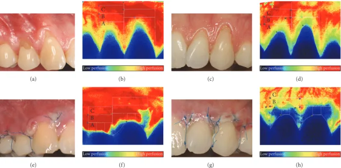
Figure 1: Representative photographs and LSCI images of a male (a, b, e, f) and a female (c, d, g, h) subjects. Combination of the modified coronally advanced tunnel and Geistlich Mucograft in both cases. (a, b, c, d) Images representing the preoperative perfusion. (e, f, g, h) Images showing the wound healing and perfusion 3 days postoperatively. Capital letters (A, B, and C) indicate the regions of interest for the blood flow evaluation.
surface to the LSCI instrument’s objective was set to 10 cm.
The LSCI instrument was connected to a computer and the measured values were displayed and recorded with a software application (PimSoft, Perimed AB, Stockholm, Sweden). The instrument was set to take snapshots of each area. Each snapshot was constructed by averaging 20 images in 2 secs in order to average out pulsatile variation in the blood flow.
Three regions of interest (ROI) were defined at each tooth as shown in Figure 1 (zone A, zone B, and zone C, moving away from the crown). The selection of regions and further steps of the data process were accomplished by blind analysis.
The blood flow value of ROI was defined as the average of all the pixel perfusion values in the ROI. The approximate pixel number was 7000 for zone A and 3500 for zones B and C. According to the point density it spanned 20 mm2 and 14.5 mm2, respectively. Blood flow was expressed in an arbitrary value called Laser Speckle Perfusion Unit (LSPU).
2.7. Wound Fluid Measurement. After the blood flow mea- surements, the relative volume of the wound fluid (WF) was assessed by Periotron 8000 (OraFlow Inc., NY, USA) with a filter paper (Periopaper, OraFlow Inc., NY, USA). The surface of the teeth was gently dried and Periopaper was placed for 10 seconds close to the orifice of the sulcus in the midbuccal area of treated and nontreated teeth. The values are shown in Periotron Scores (PS).
2.8. Statistical Analysis. Data in the text and the figures are presented as mean±standard error of mean (SE). The blood flow changes were analyzed by a mixed-model approach.
For pairwise comparison thepvalues were adjusted by the Benjamini and Hochberg method in order to control the false
discovery rate in multiple testing. To determine the associa- tion between the clinical outcome and baseline clinical parameters and between the blood flow and WF data, Spear- man’s correlation coefficients were calculated. The differences in clinical parameters between grafts and between genders were tested using the nonparametric Mann–WhitneyUtest.
Statistical evaluation was carried out by IBM SPSS Statistics for Windows (Armonk, NY: IBM Corp., USA).
3. Results
3.1. Systemic Blood Pressure and Blood Flow at the Reference Sites. No significant change was found in MAP throughout the observation period (90 ± 2.6mm Hg at bsl. versus85 ± 2.9mm Hg on day 180). There was no correlation between MAP and blood flow in either zone (zone A: 𝑟 = −0.115, 𝑝 = 0.140; zone B: 𝑟 = −0.130, 𝑝 = 0.096; zone C:𝑟 =
−0.057,𝑝 = 0.471). Therefore, the blood flow values were used instead of vascular resistance in the following analysis.
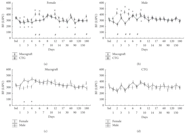
3.2. Blood Flow at the Treated Sites on the Days following the Surgery in Zone A. The statistical analysis showed that not only the graft (graft × time:𝑝 < 0.001) but also gender has a strong influence (gender×time:𝑝 < 0.001) on the blood flow of the healing mucosa. Furthermore, a significant interaction was observed between gender, graft type, and time (𝑝 < 0.001). The data were therefore split into two subgroups based on gender in addition to the two graft types (Figure 2).
In females, blood flow at the treated teeth dropped signifi- cantly, approximately to half of the baseline values in the case of both Geistlich Mucograft and CTG on the first day after the surgery (Figure 2(a)). After day 2, blood flow increased
Female
1 3 5 7 10 14 30 90 150
bsl 2 4 6 8 12 17 60 120 180
Days + + + + + + +
+ + + +
+ + + +
# # # # #
#
Mucograft CTG 0
100 200 300 400 500 600
BF (LSPU)
× × × × × × × × × ×
× ×
(a)
Male
1 3 5 7 10 14 30 90 150
bsl 2 4 6 8 12 17 60 120 180
Days + +
+ +
+
# # # #
Mucograft CTG 0
100 200 300 400 500 600
BF (LSPU)
×
× × × ×
(b) Mucograft
1 3 5 7 10 14 30 90 150
bsl 2 4 6 8 12 17 60 120 180
Days
∗ ∗ ∗ ∗ ∗ ∗ ∗ ∗ ∗
Female Male 0
100 200 300 400 500 600
BF (LSPU)
(c)
CTG
1 3 5 7 10 14 30 90 150
bsl 2 4 6 8 12 17 60 120 180
Days
∗ ∗ ∗ ∗
Female Male 0
100 200 300 400 500 600
BF (LSPU)
(d)
Figure 2: Time-course of the changes of gingival blood flow (BF) in zone A, expressed in Laser Speckle Perfusion Unit (LSPU). Time points include preoperative data (bsl.) and postoperative days (1 to 180). Data are presented as means±SE. In (a, b), statistically significant differences in the postoperative values versus bsl. are indicated by×in Geistlich Mucograft (𝑛 = 14) and by + in CTG (𝑛 = 13). The differences between grafts at the respective time points are indicated by #. In (c, d), the same data are shown in a different grouping as gender differences are depicted separately for Geistlich Mucograft and for CTG.∗indicates significantly different time points between the genders.×, +, #, and∗ mark significance levels of𝑝 < 0.05after being adjusted by the Benjamini and Hochberg method.
towards the baseline but remained below it until day 12 in Geistlich Mucograft patients and until day 7 in CTG patients.
Over the six-month period, there was only a slight difference in flap circulation between the two graft groups.
In males, contrary to females, there were marked dif- ferences in blood flow between the two grafted sites (Fig- ure 2(b)). Blood flow at Geistlich Mucograft-treated sites returned to baseline on day 2 and a hyperemic response occurred from day 4 to day 8. At CTG-treated sites, blood flow returned to baseline on day 3, and a reduced and shorter hyperemic response developed between day 5 and day 7.
Perfusion at Geistlich Mucograft-treated sites significantly exceeded the corresponding values for CTG on days 1, 2, 4, and 8 in males.
On Figures 2(c) and 2(d), the same data as on the upper panels were reconstructed for comparison between the genders at each time point, separately for each graft.
The blood flow values of males significantly exceed those of females between days 1 and 10 in the case of Geistlich Muco- graft and from day 3 to day 6 in the case of CTG.
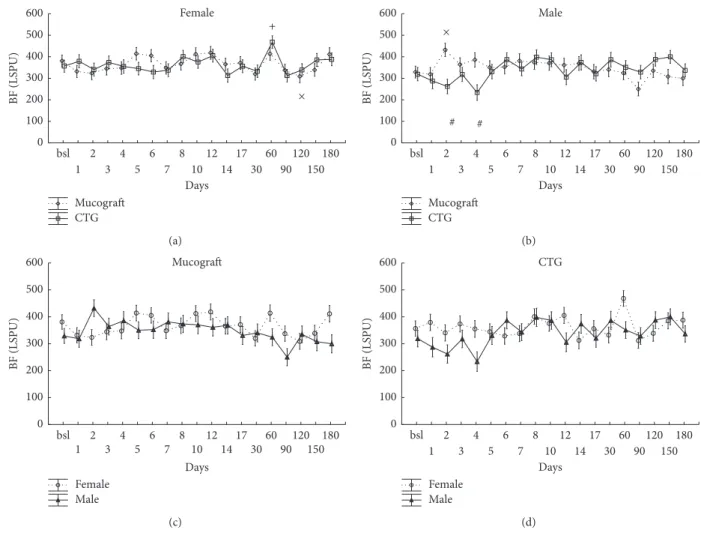
3.3. Blood Flow at the Treated Sites on the Days following the Surgery in Zone B. Similarly to zone A, the analysis was done on the level of gender×graft×time (𝑝 < 0.001) interaction (Figures 3(a) and 3(b)). However, contrary to zone A, the effect of the graft as a main factor was strong and significant (𝑝 < 0.001) while gender×graft was not due to the fact that blood flow at Geistlich Mucograft-treated sites was always above CTG values regardless of gender.
The graphs in Figures 3(c) and 3(d) demonstrate that, only in the case of Geistlich Mucograft, blood flow values were significantly higher in males than in females on days 2 and 4 while no difference was detected at CTG-treated sites.
3.4. Blood Flow at the Treated Sites on the Days following the Surgery in Zone C. Blood flow in zone C was less affected by the surgery, but still the effect of time was significant (𝑝 <
0.001). Neither the graft nor the graft-gender interaction had an overall effect on blood flow (𝑝 = 0.84; 𝑝 = 0.89). As in the case of zones A and B, the analysis showed that all interactions with the time factor (graft×time:𝑝 < 0.001;
Female
1 3 5 7 10 14 30 90 150
bsl 2 4 6 8 12 17 60 120 180
Days
+ + + + + + +
# # # # #
Mucograft CTG 0
100 200 300 400 500 600
BF (LSPU) × ×
× × ×
(a)
Male
1 3 5 7 10 14 30 90 150
bsl 2 4 6 8 12 17 60 120 180
Days
× × ×
× + ×
+
# # # #
Mucograft CTG 0
100 200 300 400 500 600
BF (LSPU)
(b) Mucograft
1 3 5 7 10 14 30 90 150
bsl 2 4 6 8 12 17 60 120 180
Days Female
Male
∗ ∗
0 100 200 300 400 500 600
BF (LSPU)
(c)
CTG
1 3 5 7 10 14 30 90 150
bsl 2 4 6 8 12 17 60 120 180
Days Female
Male 0
100 200 300 400 500 600
BF (LSPU)
(d)
Figure 3: Time-course of the changes of gingival blood flow (BF) in zone B, expressed in Laser Speckle Perfusion Unit (LSPU). Time points include preoperative data (bsl.) and postoperative days (1 to 180). Data are presented as means±SE. In (a, b), statistically significant differences of the postoperative values versus bsl. are indicated by×in Geistlich Mucograft (𝑛 = 14) and by + in CTG (𝑛 = 13). The differences between grafts at the respective time points are indicated by #. In (c, d), the same data are shown in a different grouping as gender differences are depicted separately for Geistlich Mucograft and for CTG.∗indicates significantly different time points between the genders.×, +, #, and∗ mark significance levels of𝑝 < 0.05after being adjusted by the Benjamini and Hochberg method.
gender×time:𝑝 < 0.001; gender×graft×time:𝑝 < 0.001) were significant, indicating some variations in blood flow by time over the observation period (Figure 4). The graphs on the lower panels of Figure 4 demonstrate that there were no differences observed in this zone between the genders.
3.5. Wound Fluid Measurements. The two main factors, graft type (𝑝 = 0.85) and gender (𝑝 = 0.13), were not significant but time (𝑝 < 0.001) was. Interactions between graft×time (𝑝 = 0.70) and graft × gender × time (𝑝 = 0.46) were not significant either, but the graft×gender interaction was significant (𝑝 < 0.001). This means that, overall, the WF production of Geistlich Mucograft-treated sites (13.8 [+2.6,
−2.2] PS) exceeded that of CTG-treated sites (10.7 [+2.1,−1.8]
PS) in females (Figure 5(a)). On the other hand, in males, the opposite was found: Geistlich Mucograft-treated sites had less WF (6.9 [+1.5,−1.2] PS) than CTG-treated sites (10 [+2.4,
−1.9] PS) (Figure 5(b)).
As time interacted with gender (𝑝 < 0.001), pairwise comparisons were made at each time point (Figure 5(c)). PSs
increased dramatically in both genders on the first day after surgery. They remained significantly higher than the baseline until day 10 in females and until day 5 in males. On the first two days, WF looked similar in both genders but from day 3 the values in males dropped steeper than in females. One month after the surgery, WF tended to be lower than the respective baseline values in both genders.
3.6. The Correlation between WF and Blood Flow. During the early healing period, blood flow in zone A showed a moderate inverse correlation with WF production on day 4 (𝑟 = −0.55, 𝑝 < 0.05), day 5 (𝑟 = −0.49,𝑝 < 0.05), day 6 (𝑟 = −0.51, 𝑝 < 0.05), and day 7 (𝑟 = −0.61,𝑝 < 0.01).
3.7. Clinical Parameters. Baseline GRD0 and GRW0 were very similar in the Geistlich Mucograft- and the CTG-treated groups (Table 1); however, the initial KT0 was significantly less in the CTG-treated group. Gains in the depth (REC) and width (RW) of the recessions were similar in the two groups. The increase in KT at Geistlich Mucograft-treated
Female
1 3 5 7 10 14 30 90 150
bsl 2 4 6 8 12 17 60 120 180
Days
× +
Mucograft CTG 0
100 200 300 400 500 600
BF (LSPU)
(a)
Male
1 3 5 7 10 14 30 90 150
bsl 2 4 6 8 12 17 60 120 180
Days
×
# #
Mucograft CTG 0
100 200 300 400 500 600
BF (LSPU)
(b) Mucograft
1 3 5 7 10 14 30 90 150
bsl 2 4 6 8 12 17 60 120 180
Days Female
Male 0
100 200 300 400 500 600
BF (LSPU)
(c)
CTG
1 3 5 7 10 14 30 90 150
bsl 2 4 6 8 12 17 60 120 180
Days Female
Male 0
100 200 300 400 500 600
BF (LSPU)
(d)
Figure 4: Time-course of the changes of gingival blood flow (BF) in zone C, expressed in Laser Speckle Perfusion Unit (LSPU). Time points include preoperative data (bsl.) and postoperative days (1 to 180). Data are presented as means±SE. In (a, b), statistically significant differences of the postoperative values versus bsl. are indicated by×in Geistlich Mucograft (𝑛 = 14) and by + in CTG (𝑛 = 13). The differences between grafts at the respective time points are indicated by #. In (c, d), the same data are shown in a different grouping as gender differences are depicted separately for Geistlich Mucograft and for CTG. There were no significant differences observed between the genders.×, +, and # indicate significance levels of𝑝 < 0.05after being adjusted by the Benjamini and Hochberg method.
Table 1: Gingival recession characteristics obtained at baseline and six months after the surgery.
Geistlich Mucograft (𝑛 = 14sites) CTG (𝑛 = 13sites)
mean±SE in mm mean±SE in mm
GRD0 2.4±0.23 2.8±0.27
GRW0 3.2±0.28 3.2±0.26
KT0 3 ± 0.41# 1.4±0.35
REC 1.9±0.29 2.6±0.27
RW 2.1±0.47 2.7±0.40
KT −0.7 ± 0.29# 0.7±0.41
GRD0, gingival recession depth at baseline; GRW0, gingival recession width at baseline; KT0, width of the keratinized tissue at baseline; REC, recession depth reduction; RW, recession width reduction; KT, increase in the width of the keratinized tissue. # represents the statistically significant differences between graft types;𝑝 < 0.05.
sites was significantly less than that at CTG-treated sites (Table 1).
No statistically significant differences were observed between females and males either in the baseline values or in REC, RW, and KT (data not shown).
REC and RW were positively correlated with the baseline values (𝑟 = 0.92,𝑝 < 0.001and𝑟 = 0.64,𝑝 < 0.001). In contrast, KT was negatively correlated with KT0 (𝑟 = −0.79, 𝑝 < 0.001).
4. Discussion
Our primary aim was to introduce the LSCI method to mon- itor the microcirculation of the oral mucosa after periodontal plastic surgery interventions. Imaging technique simultane- ously displays several areas of the flap contrary to the single- point laser Doppler flowmetry. Another unique property of LSCI is the rapid imaging which reduces movement artefact and decreases time of each measurement session especially when multiple images within a mouth has to be captured. These features facilitate patient compliance during many visits. This new method has not been tested before
bsl 2 4 6 8 12 17 60 120 180 (Days)
Female
Mucograft CTGp < 0.05 0
20 40 60 80 100 120
WF (PS)
(a)
Male
Mucograft CTGp < 0.01
2 4 6 8 12 17 60 120 180
bsl
(Days) 0
20 40 60 80 100 120
WF (PS)
(b)
× ×
× × × × × ×
× × × × + +
+ + + +
+
Female Male
∗ ∗ ∗
2 4 6 8 12 17 60 120 180
bsl
(Days) 0
20 40 60 80 100 120
WF (PS)
(c)
Figure 5: The effect of time, graft, and gender on wound fluid (WF) production. The two upper graphs (a and b) show the interaction between graft and gender in wound fluid (WF) production during the whole period, expressed in Periotron Scores (PS). The lower plot (c) shows the changes of WF production over time when the graft data were grouped. Time points include preoperative data (bsl.) and postoperative days (1 to 180). Data are presented as means±SE. Statistically significant differences of the postoperative values versus bsl. are indicated by× in females and by + in males. The differences between the genders are indicated by∗(𝑝 < 0.05, adjusted by the Benjamini and Hochberg method).
on postoperative mucosal flap; therefore we had no data available about the intraday and interday variability. In order to get the best estimation of the time-course we performed multiply repeats on each day and we made measurements on numerous days during the wound healing period. Impor- tantly due to the great number of measurements approxi- mately 8000 ROIs (∼1000 per patient,∼150 per tooth-site) were drawn manually and resulted in hard work of data processing. In this exploratory study we managed to define the most characteristic days (1, 3, 7, and 10) for the flap circulation which could decrease the necessary session of the measurements in further high scale studies. The split-mouth design is also a good way to decrease the number of the patients involved as it decreases the error rate due to the low relative standard deviation (<7%) of the gingival sites within a patient. This data also suggests that the effect of surgery intervention on the variability between parallel tooth sites within a surgical area managed to be kept fairly standardized probably due to the single experienced operator. Involving some reference sites to normalize the values at the test sites has only slightly decreased the error rate; thus it could be dropped. Contrary to the LSCI method the single-point laser Doppler technique suffers from the difficulty in repositioning of the probe during day by day follow-up and it is very
sensitive to the measurement distance and angulation [21, 24–
26]. In our study the spatial variability was decreased by using thousands of pixels for each ROI spanning 10–20 mm2 and the instrument was set to fixed focal distance. Overall these carefully settings in our design resulted in low variability among patients (<11%), which promoted the better power for evaluation of the between-group effect such as the gender.
The LSCI method was able to capture not just the massive effect of surgical intervention but also small differences in the surgical technique used. We assume that this method may help to understand physiological and pathophysiological changes during mucosal healing. Understanding the mecha- nism would help us to readjust postoperative care and select the best available surgical techniques, such as incision and flap design, suture, and graft size and type.
According to our results, the most apical area (zone C) was the least influenced by the surgery, as this was repositioned directly to vital tissues without an intermediate graft. Moreover, it is a more distensible mucosal area with the best collateral circulation. On the other hand, the most severe ischemia was observed in the marginal area (zone A) due to the intrasulcular incisions which cut off the main collateral circulation (with the periodontal plexus) of this area. Furthermore, the suspended sutures and the underlying
grafts cause probably the highest tension in this area. Blood flow in this area returned to the baseline level within 14 days in all cases and in some cases much earlier. In addition, in males, a hyperemic response was also observed after the hypoperfusion between day 4 and day 8 postoperative- ly.
Only a few studies investigated the blood flow of the flap in the oral mucosa. A study of four dogs [27] showed that the simple elevation of mucoperiosteal flaps causes a postoperative ischemia for seven days before blood flow would return to baseline. In a human study [28] using laser Doppler flowmetry, a postoperative hyperemic response was observed one day after the periodontal flap surgery. In this study the vascular response represented the mean of the two sex groups and neither of these studies applied graft material for the surgery. Histological observations suggested that tissue revascularization begins in mucosal flaps as early as two or three days postoperatively [29, 30]. Vascularity regained its normal level in 10 days after surgery if there was no significant interface (i.e., any grafting material) between the bone and the repositioned flap [31, 32]. However, if bone grafts and membranes separated the flap from the bone it took three weeks for vascularity to be normalized [33], which highlights the role of reuniting the alveolar and periodontal plexus to the mucosal one.
Furthermore, xenogeneic matrices were not bearing a vasculature contrary to autologous grafts, where revascu- larization can occur earlier by inosculation [16, 34]. The vascularization of the xenogeneic graft area only begins after the graft is almost disrupted which takes months [35, 36].
In animal study [12] the vascularization begins very slowly and sparsely after application of Geistlich Mucograft collagen matrix. The quick restoration of the blood flow long before the expected revascularization confirmed that MCAT only minimally compromised the mucosal vascular architecture and the full graft vascularization is not necessary for mucosal regeneration. The early recovery of flap circulation helps to cover and protect the grafted area and to promote tissue inte- gration. Similarly, it was observed previously [37] that a care- ful and less invasive surgical approach—for example, employ- ing microsurgical rather than macrosurgical techniques—
may better maintain circulation and speeds up revasculariza- tion. This also resulted in better clinical performance [5, 38].
Favorable flap circulation and the relatively intact periosteal plexus could provide a good double-layered recipient bed for the grafts. This serves as a good nutritive supply for both autogeneic and xenogeneic grafts by imbibition until new vessels develop within the grafts.
In spite of the fact that flap circulation slightly favored the xenogeneic matrix, the CTG resulted in similar root coverage compared to Geistlich Mucograft, with comparable mean baseline recession depth in both groups. This is in accordance with the findings of randomized clinical trials (RCT) [13, 39] where the percentage of root coverage by Geistlich Mucograft remained only slightly below that of the CAF + CTG. Although the gain in keratinized tissue width was slightly less in the Geistlich Mucograft than in the CTG group, which is also confirmed by an RCT [13], another RCT found no differences [10, 39]. Mean KT at baseline was slightly
different across the different groups which may have some effect on the clinical outcome.
Interestingly, unlike the moderate effects of the graft types, gender made a considerable impact on flap circulation.
This was an unexpected result compelling us to split our data into two subgroups. Ignoring the gender factor would have resulted in a failure to correctly assess the recovery time of blood flow as the two curves would have cancelled each other out. Furthermore, the effect of the different grafts on micro- circulation in both zones A and B would not have been assessed either. Males had an ischemic phase lasting a few days, followed by a hyperemic response in the marginal gingival zone whereas females were characterized by a slower recovery of the blood flow. All baseline clinical parameters of the gingiva including crevicular fluid and tissue morphology (e.g., recession depth, recession width, width of the kera- tinized mucosa, and thickness of the keratinized mucosa) were similar in the two gender groups. These suggest that neither the initial subclinical inflammation nor the morphol- ogy of the gingival recession can explain the gender-specific postoperative alteration in blood flow. The differences in blood flow between the genders were less pronounced in zone B and disappeared in zone C, implying that the gender effect is more important in the marginal zone where the circulation is most severed. To the best of our knowledge, there are no data available on the effect of gender on the flap microcircu- lation on the oral mucosa. Clinical observations [40–43] and findings in an experimental excisional palatal wound model [44] suggest that mucosal wound healing is faster in males than in females; however blood flow was not measured in these studies. It can only be supposed that better blood flow recovery may have facilitated wound healing in males.
The MCAT recovered earlier than the expected complete revascularization period suggesting that higher blood flow in males occurred due to the vasodilation of the remaining vessels. We can further suppose that there may be differences in terms of vascular reactivity between the genders. After surgery blood flow was reduced as the vascular supply of the flap was compromised which may result in low-flow mediated vasoconstriction. In the brachial artery an in vivo prolonged low-flow condition augments vasoconstriction during occlusion and attenuates hyperemic response [45] and low-flow mediated vasoconstriction seems to be more intense in women [46].
Arteriogenesis or the so called “collateralization” is another important mechanism to maintain perfusion in case of reduced vascularity before neovascularization can be com- pleted. This involves a proliferative increase in the diameter and length of the arterioles to compensate for the reduced perfusion of the flap [47]. In an ischemic limb mouse model blood flow recovered faster in males and more alpha smooth muscle cell positive vessels—arterioles—could be found [48] suggesting greater arteriogenesis. Furthermore, higher maximal vasodilation in response to acetylcholine and nitro- glycerin in male ischemic limbs was also recorded. Similarly, in patients with stable angina and chronic total occlusion of at least one major epicardial coronary artery [49] and in a rat myocardial infraction model the remodeling of arte- riolar vessels (arteriogenesis) was found to be reduced in
females [50]. There is evidence that the gingiva reacts by collateralization to pathophysiological stimuli such as perio- dontitis [51–53]. As a conclusion we hypothesized that in the gingival tissues of males there may be more native collaterals and/or increased collateralization after surgery and/or higher reactivity to vasodilation agents which might be attributable to gender differences.
Until the recovery of graft vascularization, a vascular leak maintains the nutritive supply of the graft via imbibition [14, 16]. However, not only the graft but also the distal part of the mucosal flap is supplied by nutrition via extravasation in the early ischemic period of healing [27]. In the present study, wound fluid production was measured in order to indirectly and noninvasively assess vascular permeability. Interestingly, blood flow recovery showed a correlation with recovery to normal tissue transudation. In males, this happened earlier—
within 2-3 days for blood flow and within six days for wound fluid—whereas in females, it was 8–14 days and 12 days, respectively. As in the case of blood flow in the flap (in zone A), differences in vascular leakage between the grafts showed some gender specificity. In females, Geistlich Mucograft- treated sites had higher fluid production which may be a compensatory mechanism of the lower blood flow while in males the tissue may require less diffusive nutrition due to the superior blood flow compared to CTG-treated sites. Similarly, it was observed in male mice [15] that skin graft angiogenesis peaked at 10 days after grafting and this was coincident with maximum vascular leakage.
5. Conclusions
The LSCI method seems to be feasible way to characterize the postoperative flap circulation in oral mucosa. It can be concluded that the application of both grafts resulted in excellent recirculation patterns in the flap. Gender was the most substantial influencing factor. Males showed a more rapid reestablishment of mucosal blood flow. Further study is necessary to investigate the mechanism of the gender- specific blood flow regulation in the healing mucosal flap. We found that earlier blood flow recovery is strongly associated with earlier normalization of vascular permeability. It is conceivable that the opposite changes observed in blood flow versus vascular permeability are equally able to ensure the supply required for the tissues to heal. This, however, did not influence the apparent clinical outcome; favorable recession coverage could be achieved with both CTG and Geistlich Mucograft in males and females.
Competing Interests
All authors declare that they have no conflict of interests.
Acknowledgments
The authors wish to thank dental students Barbara Mikecs, J´ulia N´emeth, P´eter Cs´anyi, and Ad´el Ruszin for their assist- ance in collecting data. This study was funded by the Research Fund of the Faculty of Dentistry, Semmelweis University, Hungary (4030511599 and 5111141075), and the Hungarian
Scientific Research Fund (OTKA K112364 and K-112964). The Mucograft was kindly donated by the manufacturer (Geistlich Pharma AG, Switzerland).
References
[1] S.-H. Park and H.-L. Wang, “Clinical significance of incision location on guided bone regeneration: human study,”Journal of Periodontology, vol. 78, no. 1, pp. 47–51, 2007.
[2] J. Kleinheinz, A. B¨uchter, B. Kruse-L¨osler, D. Weingart, and U.
Joos, “Incision design in implant dentistry based on vascular- ization of the mucosa,”Clinical Oral Implants Research, vol. 16, no. 5, pp. 518–523, 2005.
[3] R. Burkhardt and N. P. Lang, “Fundamental principles in peri- odontal plastic surgery and mucosal augmentation—a narrative review,”Journal of Clinical Periodontology, vol. 41, no. 15, pp.
S98–S107, 2014.
[4] A. L. Allen, “Use of the supraperiosteal envelope in soft tissue grafting for root coverage. I. Rationale and technique,”The Inter- national Journal of Periodontics & Restorative Dentistry, vol. 14, no. 3, pp. 216–227, 1994.
[5] P. F. Andrade, M. F. M. Grisi, A. M. Marcaccini et al., “Compari- son between micro- and macrosurgical techniques for the treat- ment of localized gingival recessions using coronally positioned flaps and enamel matrix derivative,”Journal of Periodontology, vol. 81, no. 11, pp. 1572–1579, 2010.
[6] M. S. Tonetti and S. Jepsen, “Clinical efficacy of periodontal plastic surgery procedures: consensus Report of Group 2 of the 10th European Workshop on Periodontology,”Journal of Clini- cal Periodontology, vol. 41, pp. S36–S43, 2014.
[7] R. Burkhardt, C. H. F. H¨ammerle, and N. P. Lang, “Self-reported pain perception of patients after mucosal graft harvesting in the palatal area,”Journal of Clinical Periodontology, vol. 42, no. 3, pp. 281–287, 2015.
[8] K. M. Soileau and R. B. Brannon, “A histologic evaluation of various stages of palatal healing following subepithelial con- nective tissue grafting procedures: a comparison of eight cases,”
Journal of Periodontology, vol. 77, no. 7, pp. 1267–1273, 2006.
[9] M. Camelo, M. Nevins, M. L. Nevins, P. Schupbach, and D. M.
Kim, “Treatment of gingival recession defects with xenogenic collagen matrix: a histologic report.,”The International journal of periodontics & restorative dentistry, vol. 32, no. 2, pp. 167–173, 2012.
[10] D. Cardaropoli, L. Tamagnone, A. Roffredo, and L. Gaveg- lio, “Treatment of gingival recession defects using coronally advanced flap with a porcine collagen matrix compared to coro- nally advanced flap with connective tissue graft: a randomized controlled clinical trial,”Journal of Periodontology, vol. 83, no. 3, pp. 321–328, 2012.
[11] K. Jepsen, S. Jepsen, G. Zucchelli et al., “Treatment of gingival recession defects with a coronally advanced flap and a xeno- geneic collagen matrix: a multicenter randomized clinical trial,”
Journal of Clinical Periodontology, vol. 40, no. 1, pp. 82–89, 2013.
[12] S. Ghanaati, M. Schlee, M. J. Webber et al., “Evaluation of the tissue reaction to a new bilayered collagen matrix in vivo and its translation to the clinic,”Biomedical Materials, vol. 6, no. 1, Article ID 015010, 2011.
[13] S. Aroca, B. Moln´ar, P. Windisch et al., “Treatment of multiple adjacent Miller class I and II gingival recessions with a Modified Coronally Advanced Tunnel (MCAT) technique and a collagen
matrix or palatal connective tissue graft: a randomized, con- trolled clinical trial,”Journal of Clinical Periodontology, vol. 40, no. 7, pp. 713–720, 2013.
[14] X. Wang, Y. Zhang, and C. Han, “Topical negative pressure improves autograft take by altering nutrient diffusion: a hypoth- esis,”Medical Science Monitor, vol. 20, pp. 61–63, 2014.
[15] A. Shaterian, A. Borboa, R. Sawada et al., “Real-time analysis of the kinetics of angiogenesis and vascular permeability in an animal model of wound healing,”Burns, vol. 35, no. 6, pp. 811–
817, 2009.
[16] R. C. Oliver, H. L¨oe, and T. Karring, “Microscopic evaluation of the healing and revascularization of free gingival grafts,”Journal of Periodontal Research, vol. 3, no. 2, pp. 84–95, 1968.
[17] J. D. Briers and S. Webster, “Laser speckle contrast analysis (LASCA): a nonscanning, full-field technique for monitoring capillary blood flow,”Journal of Biomedical Optics, vol. 1, no. 2, pp. 174–179, 1996.
[18] S. Eriksson, J. Nilsson, G. Lindell, and C. Sturesson, “Laser speckle contrast imaging for intraoperative assessment of liver microcirculation: a clinical pilot study,”Medical Devices: Evi- dence and Research, vol. 7, no. 1, pp. 257–261, 2014.
[19] N. Hecht, J. Woitzik, J. P. Dreier, and P. Vajkoczy, “Intraoperative monitoring of cerebral blood flow by laser speckle contrast analysis,”Neurosurgical Focus, vol. 27, no. 4, p. E11, 2009.
[20] L. Yuan, Y. Li, H. Li, H. Lu, and S. Tong, “Intraoperative laser speckle contrast imaging improves the stability of rodent mid- dle cerebral artery occlusion model,” Journal of Biomedical Optics, vol. 20, no. 9, Article ID 096012, 2015.
[21] F. Lindahl, E. Tesselaar, and F. Sj¨oberg, “Assessing paediatric scald injuries using laser speckle contrast imaging,”Burns, vol.
39, no. 4, pp. 662–666, 2013.
[22] D. M. J. Milstein, C. Ince, S. S. Gisbertz et al., “Laser speckle con- trast imaging identifies ischemic areas on gastric tube recon- structions following esophagectomy,”Medicine, vol. 95, no. 25, Article ID e3875, 2016.
[23] B. Moln´ar, S. Aroca, T. Keglevich et al., “Treatment of multiple adjacent Miller Class I and II gingival recessions with collagen matrix and the modified coronally advanced tunnel technique,”
Quintessence international (Berlin, Germany : 1985), vol. 44, no.
1, pp. 17–24, 2013.
[24] G. A. Tew, M. Klonizakis, H. Crank, J. D. Briers, and G. J.
Hodges, “Comparison of laser speckle contrast imaging with laser Doppler for assessing microvascular function,”Microvas- cular Research, vol. 82, no. 3, pp. 326–332, 2011.
[25] P. Rousseau, G. Mah´e, F. Haj-Yassin et al., “Increasing the
“region of interest” and “time of interest”, both reduce the variability of blood flow measurements using laser speckle con- trast imaging,”Microvascular Research, vol. 82, no. 1, pp. 88–91, 2011.
[26] G. Mah´e, F. Haj-Yassin, P. Rousseau et al., “Distance between laser head and skin does not influence skin blood flow values recorded by laser speckle imaging,”Microvascular Research, vol.
82, no. 3, pp. 439–442, 2011.
[27] T. N. McLean, B. A. Smith, E. C. Morrison, C. E. Nasjleti, and R. G. Caffesse, “Vascular changes following mucoperiosteal flap surgery: a fluorescein angiography study in dogs,”Journal of Periodontology, vol. 66, no. 3, pp. 205–210, 1995.
[28] M. Retzepi, M. Tonetti, and N. Donos, “Comparison of gingival blood flow during healing of simplified papilla preservation and modified Widman flap surgery: a clinical trial using laser Doppler flowmetry,”Journal of Clinical Periodontology, vol. 34, no. 10, pp. 903–911, 2007.
[29] D. E. Cutright, “The proliferation of blood vessels in gingival wounds,”Journal of Periodontology, vol. 40, no. 3, pp. 137–141, 1969.
[30] R. G. Caffesse, W. A. Castelli, and C. E. Nasjleti, “Vascular res- ponse to modified Widman flap surgery in monkeys,”Journal of Periodontology, vol. 52, no. 1, pp. 1–7, 1981.
[31] J. A. H. Lindeboom, K. R. Mathura, I. H. A. Aartman, F. H. M.
Kroon, D. M. J. Milstein, and C. Ince, “Influence of the appli- cation of platelet-enriched plasma in oral mucosal wound heal- ing,”Clinical Oral Implants Research, vol. 18, no. 1, pp. 133–139, 2007.
[32] D. M. J. Milstein, J. A. H. Lindeboom, and C. Ince, “Intravital sidestream dark-field (SDF) imaging is used in a rabbit model for continuous noninvasive monitoring and quantification of mucosal capillary regeneration during wound healing in the oral cavity: a pilot study,”Archives of Oral Biology, vol. 55, no.
5, pp. 343–349, 2010.
[33] D. M. J. Milstein, K. R. Mathura, J. A. H. Lindeboom, D.
Ramsoekh, R. Lindeboom, and C. Ince, “The temporal course of mucoperiosteal flap revascularization at guided bone regenera- tion-treated implant sites: a pilot study,” Journal of Clinical Periodontology, vol. 36, no. 10, pp. 892–897, 2009.
[34] M. W. Laschke and M. D. Menger, “Vascularization in tissue engineering: angiogenesis versus inosculation,”European Sur- gical Research, vol. 48, no. 2, pp. 85–92, 2012.
[35] F. Schwarz, D. Rothamel, M. Herten, M. Sager, and J. Becker,
“Angiogenesis pattern of native and cross-linked collagen mem- branes: an immunohistochemical study in the rat,”Clinical Oral Implants Research, vol. 17, no. 4, pp. 403–409, 2006.
[36] J. A. Vergara, C. R. Qui˜nones, C. E. Nasjleti, and R. G. Caffesse,
“Vascular response to guided tissue regeneration procedures using nonresorbable and bioabsorbable membranes in dogs,”
Journal of Periodontology, vol. 68, no. 3, pp. 217–224, 1997.
[37] R. Burkhardt and N. P. Lang, “Coverage of localized gingival recessions: comparison of micro- and macrosurgical tech- niques,”Journal of Clinical Periodontology, vol. 32, no. 3, pp. 287–
293, 2005.
[38] N. Nizam, O. Bengisu, and S¸. S¨onmez, “Micro- and macro- surgical techniques in the coverage of gingival recession using connective tissue graft: 2 years follow-up,”Journal of Esthetic and Restorative Dentistry, vol. 27, no. 2, pp. 71–83, 2015.
[39] M. K. McGuire and E. T. Scheyer, “Xenogeneic collagen matrix with coronally advanced flap compared to connective tissue with coronally advanced flap for the treatment of dehiscence- type recession defects,”Journal of Periodontology, vol. 81, no. 8, pp. 1108–1117, 2010.
[40] S. M. Conrad, G. H. Blakey, D. A. Shugars, R. D. Marciani, C.
Phillips, and R. P. White Jr., “Patients’ perception of recovery after third molar surgery,”Journal of Oral and Maxillofacial Surgery, vol. 57, no. 11, pp. 1288–1294, 1999.
[41] C. Phillips, R. P. White Jr., D. A. Shugars, and X. Zhou, “Risk fac- tors associated with prolonged recovery and delayed healing after third molar surgery,”Journal of Oral and Maxillofacial Surgery, vol. 61, no. 12, pp. 1436–1448, 2003.
[42] I. S. Benediktsd´ottir, A. Wenzel, J. K. Petersen, and H. Hintze,
“Mandibular third molar removal: risk indicators for extended operation time, postoperative pain, and complications,”Oral Surgery, Oral Medicine, Oral Pathology, Oral Radiology, and Endodontics, vol. 97, no. 4, pp. 438–446, 2004.
[43] W. L. Adeyemo, A. L. Ladeinde, and M. O. Ogunlewe, “Clinical evaluation of post-extraction site wound healing,”Journal of Contemporary Dental Practice, vol. 7, no. 3, pp. 40–49, 2006.
[44] C. G. Engeland, J. A. Bosch, J. T. Cacioppo, and P. T. Marucha,
“Mucosal wound healing: the roles of age and sex,”Archives of Surgery, vol. 141, no. 12, pp. 1193–1198, 2006.
[45] M. Rakobowchuk, E. R. Parsloe, S. E. Gibbins, E. Harris, and K.
M. Birch, “Prolonged low flow reduces reactive hyperemia and augments low flow mediated constriction in the brachial artery independent of the menstrual cycle,”PLoS ONE, vol. 8, no. 2, Article ID e55385, 2013.
[46] J. Levenson, F. Pessana, J. Gariepy, R. Armentano, and A. Simon,
“Gender differences in wall shear–mediated brachial artery vasoconstriction and vasodilation,” Journal of the American College of Cardiology, vol. 38, no. 6, pp. 1668–1674, 2001.
[47] K. Merz, R. Schweizer, S. Schlosser, P. Giovanoli, D. Erni, and J.
A. Plock, “Distinct microhemodynamic efficacy of arteriogen- esis and angiogenesis in critically ischemic skin flaps,”Microvas- cular Research, vol. 83, no. 2, pp. 249–256, 2012.
[48] X. Peng, J. Wang, R. M. Lassance-Soares et al., “Gender differ- ences affect blood flow recovery in a mouse model of hindlimb ischemia,”American Journal of Physiology—Heart and Circula- tory Physiology, vol. 300, no. 6, pp. H2027–H2034, 2011.
[49] Y. Shen, F. H. Ding, R. Y. Zhang, Q. Zhang, L. Lu, and W. F. Shen,
“Serum cystatin c reflects angiographic coronary collateraliza- tion in stable coronary artery disease patients with chronic total occlusion,”PLoS ONE, vol. 10, no. 9, Article ID e0137253, 2015.
[50] E. I. Dedkov, K. Oak, L. P. Christensen, and R. J. Tomanek,
“Coronary vessels and cardiac myocytes of middle-aged rats demonstrate regional sex-specific adaptation in response to postmyocardial infarction remodeling,”Biology of Sex Differ- ences, vol. 5, no. 1, article 1, 2014.
[51] J. E. Kennedy, “Effect of inflammation on collateral circulation of the gingiva,”Journal of Periodontal Research, vol. 9, no. 3, pp.
147–152, 1974.
[52] G. Soderholm and J. Egelberg, “Morphological changes in gingi- val blood vessels during developing gingivitis in dogs,”Journal of Periodontal Research, vol. 8, no. 1, pp. 16–20, 1973.
[53] H. Zoellner, C. C. Chapple, and N. Hunter, “Microvasculature in gingivitis and chronic periodontitis: disruption of vascular net- works with protracted inflammation,”Microscopy Research and Technique, vol. 56, no. 1, pp. 15–31, 2002.
Submit your manuscripts at https://www.hindawi.com
Stem Cells International
Hindawi Publishing Corporation
http://www.hindawi.com Volume 2014
Hindawi Publishing Corporation
http://www.hindawi.com Volume 2014
INFLAMMATION
Hindawi Publishing Corporation
http://www.hindawi.com Volume 2014
Behavioural Neurology
Endocrinology
International Journal ofHindawi Publishing Corporation
http://www.hindawi.com Volume 2014
Hindawi Publishing Corporation
http://www.hindawi.com Volume 2014
Disease Markers
Hindawi Publishing Corporation
http://www.hindawi.com Volume 2014
BioMed
Research International
Oncology
Journal ofHindawi Publishing Corporation
http://www.hindawi.com Volume 2014
Hindawi Publishing Corporation
http://www.hindawi.com Volume 2014
Oxidative Medicine and Cellular Longevity
Hindawi Publishing Corporation
http://www.hindawi.com Volume 2014
PPAR Research The Scientific World Journal
Hindawi Publishing Corporation
http://www.hindawi.com Volume 2014
Immunology Research
Hindawi Publishing Corporation
http://www.hindawi.com Volume 2014
Journal of
Obesity
Journal ofHindawi Publishing Corporation
http://www.hindawi.com Volume 2014
Hindawi Publishing Corporation
http://www.hindawi.com Volume 2014
Computational and Mathematical Methods in Medicine
Ophthalmology
Journal ofHindawi Publishing Corporation
http://www.hindawi.com Volume 2014
Diabetes Research
Journal ofHindawi Publishing Corporation
http://www.hindawi.com Volume 2014
Hindawi Publishing Corporation
http://www.hindawi.com Volume 2014
Research and Treatment
AIDS
Hindawi Publishing Corporation
http://www.hindawi.com Volume 2014
Gastroenterology Research and Practice
Hindawi Publishing Corporation
http://www.hindawi.com Volume 2014
Parkinson’s Disease
Evidence-Based Complementary and Alternative Medicine
Volume 2014 Hindawi Publishing Corporation
http://www.hindawi.com