Article
Connective tissue growth factor is a new ligand of epidermal growth factor receptor
Sandra Rayego-Mateos
1, Raquel Rodrigues-Dı´ez
1, Jose Luis Morgado-Pascual
1, Raul R. Rodrigues Dı´ez
1, Sebastian Mas
3, Carolina Lavoz
1, Matilde Alique
1, Janos Pato
2, Gyorgy Keri
2,3, Alberto Ortiz
4, Jesus Egido
4, and Marta Ruiz-Ortega
1,*
1 Cellular Biology in Renal Diseases Laboratory, Universidad Auto´noma de Madrid,28040Madrid, Spain
2 Pathobiochemistry Research Group of Hungarian Academy of Sciences, Semmelweis University, H-1094Budapest, Hungary
3 Vichem Chemie Research Ltd., H-1022Budapest, Hungary
4Renal Research Laboratory, IIS-Fundacio´n Jime´nez Dı´az, Universidad Auto´noma de Madrid,28040Madrid, Spain
* Correspondence to: Marta Ruiz-Ortega, E-mail:mruizo@fjd.es
Chronic kidney disease is reaching epidemic proportions worldwide and there is no effective treatment. Connective tissue growth factor (CCN2) has been suggested as a risk biomarker and a potential therapeutic target for renal diseases, but its specific receptor has not been identified. Epidermal growth factor receptor (EGFR) participates in kidney damage, but whether CCN2activates the EGFR pathway is unknown. Here, we show that CCN2is a novel EGFR ligand. CCN2binding to EGFR extracellular domain was demonstrated by surface plasmon resonance. CCN2contains four distinct structural modules. The carboxyl-terminal module (CCN2(IV)) showed a clear inter- action with soluble EGFR, suggesting that EGFR-binding site is located in this module. Injection of CCN2(IV) in mice increased EGFR phosphorylation in the kidney, mainly in tubular epithelial cells. EGFR kinase inhibition decreased CCN2(IV)-induced renal changes (ERK activation and inflammation). Studies in cultured tubular epithelial cells showed that CCN2(IV) binds to EGFR leading to ERK ac- tivation and proinflammatory factors overexpression. CCN2interacts with the neurotrophin receptor TrkA, and EGFR/TrkA receptor crosstalk was found in response to CCN2(IV) stimulation. Moreover, endogenous CCN2blockade inhibited TGF-b-induced EGFR acti- vation. These findings indicate that CCN2is a novel EGFR ligand that contributes to renal damage through EGFR signalling.
Keywords:CCN2, receptors, EGFR, TrkA, renal, inflammation
Introduction
Chronic kidney disease is a major health problem that has reached epidemic proportions and it may lead to end-stage renal disease or early cardiovascular death. Moreover, available clinical treatments only retard renal disease progression. Connective tissue growth factor (CCN2/CTGF), a member of the CCN (Cyr61/ CCN2/Nov) family, is over-expressed in many human renal patholo- gies (Perbal,2004;De Winter et al.,2008). Experimental studies have shown that CCN2inhibition slows disease progression in dia- betic nephropathy, unilateral ureteral obstruction, and nephrecto- mized TGF-b1transgenic mice (Yokoi et al.,2004;Okada et al., 2005;Guha et al.,2007;Phanish et al.,2010) suggesting that thera- peutic approaches that selectively block CCN2activity could be beneficial for renal disease treatment.
CCN2has to be considered a matricellular protein rather than a conventional growth factor. This protein, as other CNN members, contains four distinct structural modules that can be cleaved by pro- teases: an amino-terminal insulin-like growth-factor-binding
domain, a cysteine-rich domain, a thrombospondin type1repeat, and a carboxyl-terminal cystine-knot domain (Rachfal and Brigstock,2005;Leask and Abraham,2006;De Winter et al.,2008; Chen and Lau,2010). CCN2and its degradation fragments have been detected in biological fluids and have been proposed as risk biomarkers in several nephropathies (Riser et al.,2003;Tam et al., 2009;Slagman et al.,2011). Among these degradation fragments, the11kDa carboxyl-terminal module (namely here CCN2(IV)) has received special interest. In cultured cells, this fragment regulates cell migration and proliferation, increases chemokines and extracel- lular matrix production, and has been involved in renal inflammation (Liu et al.,2006;De Winter et al.,2008;Sanchez-Lopez et al.,2009; Markiewicz et al.,2011). Although several studies have investigated the intracellular mechanisms activated by CCN2and its fragments, the identification of a specific receptor for CCN2remains elusive.
The epidermal growth factor receptor (EGFR) is the founding member of the ErbB receptor tyrosine kinase family. EGFR signalling controls key cellular programmes, including survival, proliferation, differentiation, and locomotion, both during development and post- natally. The EGFR is over-expressed, dysregulated, or mutated in many epithelial malignancies, participating in human cancer includ- ing lung, colon, breast, ovary, and gliomas (Sibilia et al.,2007;Bronte
Received October2,2012. Revised May13,2013. Accepted June3,2013.
#The Author (2013). Published by Oxford University Press on behalf ofJournal of Molecular Cell Biology, IBCB, SIBS, CAS. All rights reserved.
at ELTE on April 21, 2015http://jmcb.oxfordjournals.org/Downloaded from
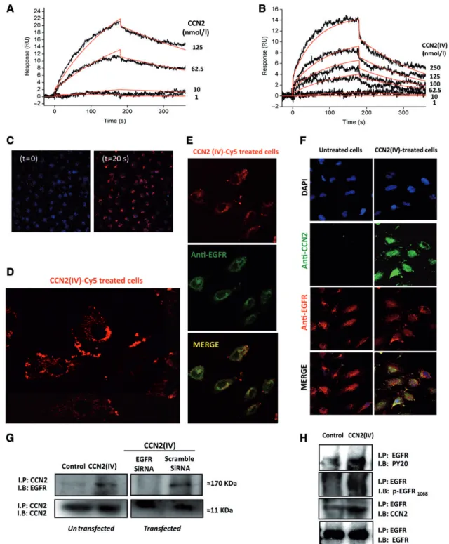
Figure1CCN2binds to EGFR. Surface plasmon resonance interaction analysis of full-length CCN2(A) and the carboxyl-terminal CCN2fragment (CCN2(IV)) (B) with immobilized EGFR extracellular domain (sEGFR) was performed using Biacore3000. Increasing concentrations of ligands (1– 250nmol/L) were injected over a surface with a density of500response units (RU) of immobilized sEGFR. The response in RU was recorded as a function of time. An overlay plot is shown of all sensorgrams after subtraction of their respective control sensorgrams. Binding parameters, cal- culated by applying the nonlinear curve-fitting software package BIAevaluation3.2(BIAcore, Inc.) to all sensorgrams simultaneously using a single-site model with drifting baseline, indicate that CCN2(IV)/EGFR interaction occurs with Kd¼126+2nmol/L. (CandD) CCN2(IV) interacts with EGFR in human tubular epithelial cells. Fluorescent labelled CCN2(IV)-Cy5(100ng/ml) was added to HK2cells and live confocal microscopy images were taken once every1.3sec for a period of2min (time0and20sec,C; time2min,D). Nuclei were stained with DAPI (blue). CCN2– EGFR interaction was evaluated by immunocytochemistry. Serum-starved HK2cells were stimulated or not with100ng/ml CCN2(IV)-Cy5(E) or CCN2(IV) (F) for10min, and fixed by cross-linking. (E) CCN2(IV)-Cy5-treated cells presented a red membrane immunostaining, while EGFR was immunode- tected by an secondary AlexaFluorw488labelled antibody (green). EGFR/CCN2(IV) colocalization was found (yellow staining, merge). (F) EGFR and CCN2were detected using specific primary antibodies followed by their corresponding secondary AlexaFluorw633/488antibodies (red/green, respectively). EGFR/CCN2colocalization was found only in CCN2(IV)-treated cells (yellow staining), but not in control ones. Figures show a
324 | Journal of Molecular Cell Biology Rayego-Mateos et al.
at ELTE on April 21, 2015http://jmcb.oxfordjournals.org/Downloaded from
et al.,2011). Besides tumour biology, EGFR family members are implicated in the development of end organ damage in hypertension (Hao et al.,2004) and atherosclerosis (Dreux et al.,2006). In the kidney, EGFR signalling is critically involved in renal electrolyte homeostasis (Melenhorst et al., 2008), and EGFR blockade, by genetic or pharmacological approaches, ameliorates experimental renal disease progression (Terzi et al.,2000;Lautrette et al.,2005; Flamant et al.,2012;Liu et al.,2012). Our aim was to investigate the capacity of CCN2and its carboxyl-terminal fragment to interact with and activate EGFR, and whether the activation of EGFR signalling is involved in CCN2-induced responses in the kidney.
Results
CCN2binds to EGFR via the carboxyl-terminal module
Surface plasmon resonance analysis was used to assess the ability of CCN2 to bind the extracellular domain of EGFR. CCN2 bound to immobilized EGFR on a BIAcore sensor chip (Figure1A).
The full-length CCN2 protein contains four distinct structural modules (De Winter et al.,2008). Binding experiments using the carboxyl-terminal degradation fragment CCN2(IV) as a ligand showed a clear interaction with EGFR (Figure1B), suggesting that the EGFR-binding site is present in the carboxyl-terminal module.
To investigate whether CCN2directly interacts with EGFR in cells, we performed studies in cultured human tubular epithelial cells (HK2cell line). First, live-cell imaging by confocal time-lapse microscopy was performed to visualize CCN2(IV) binding to the cell. After adding la- belled CCN2(IV)-Cy5 to cells, the immunofluorescent signal was rapidly located at the cell membrane, indicating CCN2(IV) cellular binding (Figure1C and D). The potential CCN2–EGFR interaction was furtherdemonstratedbyimmunocytochemistryandimmunoprecipita- tion (IP) experiments, using a cross-linking procedure to fix the pro- teins anchored to the cell surface. EGFR is expressed in untreated HK2cells. Moreover, CCN2(IV)-treated cells showed a clear cellular binding that colocalized with EGFR immunostaining (Figure1E and F). IP studies showed that in CCN2(IV)-treated cells, but not in untreat- ed ones, CCN2–EGFR complexes were formed (Figure1G and H). One of the earliest steps of EGFR activation is its auto-phosphorylation on tyrosine (Y) residues (Sweeney and Carraway,2000). In CCN2(IV)- treatedcells,complexescontainingtyrosine-phosphorylatedproteins, including Y1068on EGFR, were found (Figure1H). Transfection with a small interfering RNA molecule (siRNA) targeting EGFR, but not with a nonspecific scramble siRNA, abolished the CCN2–EGFR complex for- mation, showing the specificity of this interaction (Figure1G). These data demonstrate that in cultured tubular epithelial cells, stimulation with CCN2(IV) led to CCN2–EGFR complex formation.
CCN2induces EGFR phosphorylation in cultured tubular epithelial cells
In cultured human tubular epithelial cells, CCN2(IV) increased EGFR phosphorylation on Y1068and Y1173(Figure2A). In murine
tubular epithelial cells, CCN2 (IV)-induced EGFR activation was dose- and time-dependent, starting as early as5min and peaking after15min with a maximal response at50ng/ml (Figure2B and C). EGFR specific activation was demonstrated by pharmacological inhibition using two different EGFR kinase inhibitors, erlotinib and AG1478(Figure2D), and EGFR gene silencing (Figure2E). CCN2(IV) also increased EGFR phosphorylation in other cell types, including murine fibroblasts and human mesangial cells (Figure2F and G).
Moreover, the full-length CCN2protein also induced EGFR phosphor- ylation (Figure2H), showing a similar response as obtained with the carboxyl-terminal fragment CCN2(IV). For this reason, only CCN2(IV) was used in the following experiments. Our data demonstrate that both CCN2 and its carboxyl-terminal fragment bind and activate EGFR signalling in cells.
CCN2(IV) induces EGFR phosphorylation in the kidney
Next, we investigated whether CCN2could activate EGFR signal- ling in the kidney. Renal levels of phosphorylated EGFR protein were elevated in CCN2(IV)-injected mice compared with control mice (Figure 3A). In the kidney, EGFR is mainly expressed in tubular cells (Melenhorst et al., 2008). Immunohistochemistry and immunofluorescence using antibodies that recognized phos- phorylated EGFR on Y1173and Y1068, respectively, revealed that CCN2(IV) activated EGFR in tubular cellsin vivo(Figure3B, C, and E). Treating the CCN2(IV)-injected mice with erlotinib, a small mol- ecule tyrosine kinase inhibitor that targets the receptor catalytic domain of EGFR, diminished renal phosphorylated EGFR levels to control levels (Figure3B, C, and E).
EGFR activation by CCN2(IV) is linked to ERK signalling
Several auto-phosphorylation sites have been identified in the carboxyl-terminal region of EGFR that varies dependent on the ligand and are linked to different downstream signalling systems (Sweeney and Carraway,2000). EGFR phosphorylation on Y1068 and Y1173 is involved in ERK signalling (Rojas et al., 1996; Pourazar et al., 2008). Both tyrosines were phosphorylated in kidneys of CCN2(IV)-injected mice (Figure 3) and in cultured tubular epithelial cells exposed to CCN2(IV) (Figure2A). CCN2(IV)- injected mice also presented elevated renal levels of phosphorylated ERK1/2compared with controls, which were inhibited by erlotinib (Figure 3D). EGFR activation by CCN2 was also linked to ERK signalling in cultured tubular epithelial cells. Blockade of EGFR, by kinase inhibition or gene silencing, diminished ERK phosphorylation levels in CCN2(IV)-treated cells to levels similar to their correspond- ing controls (Figure2D and E).
CCN2(IV) via EGFR activation regulates renal inflammatory response in vivo and in vitro
We further investigated thein vivoeffect of EGFR blockade on CCN2-induced renal damage. Treatment of CCN2(IV)-injected mice with erlotinib diminished the presence of infiltrating mono- cytes/macrophages (F4/801 cells) and T lymphocytes (CD31
representative experiment of four with similar results. (G) CCN2– EGFR complexes were found by coprecipitation experiments. Cell lysates were immunoprecipitated with anti-CCN2, followed by SDS – PAGE and western blotting (IB) using an anti-EGFR antibody. In some points, cells were transfected with an EGFR siRNA or its corresponding scramble siRNA. In CCN2(IV)-treated cells, the170kDa band corresponding to EGFR molecular weight was found, while it disappeared in EGFR silenced cells, showing the formation of CCN2– EGFR complexes. The IB with anti-CCN2antibody was used as loading control. (H) IP with anti-EGFR antibody followed by IB with several antibodies: anti-phosphorylated tyrosine (PY20), anti- phosphorylated EGFR (p-EGFR1068), anti-CCN2, and anti-EGFR (used as loading control). In CCN2(IV)-treated cells, bands for PY20, p-EGFR1068
and CCN2were detected, confirming CCN2– EGFR complex formation. Figures show a representative IP experiment of five with similar results.
at ELTE on April 21, 2015http://jmcb.oxfordjournals.org/Downloaded from
Figure2CCN2activates EGFR signalling in renal cells. EGFR activation was evaluated using antibodies against phosphorylated EGFR on Y1068 (p-EGFR1068) or Y1173(p-EGFR1173), both implicated in ERK activation. CCN2(IV) increased EGFR phosphorylation in human (A) and murine (B–
D) tubular cells. (A) Human tubular epithelial cells (HK2cells) were treated with50ng/ml CCN2(IV) for15min. Some cells were pre-incubated with erlotinib (10mmol/L). Murine tubular epithelial cells were treated with10ng/ml CCN2(IV) for increasing time periods (B) or with several concentrations of CCN2(IV) (range50–0.1ng/ml) for15min (C). (D) Cells were pre-incubated for1h with erlotinib (10mmol/L) or AG1478 (100nmol/L) before the stimulation with10ng/ml CCN2(IV) for15min. (E) CCN2(IV) induces EGFR phosphorylation linked to ERK activation in tubular epithelial cells. HK2cells were incubated with transfection reagent alone (untransfected) or transfected with EGFR siRNA or scramble siRNA, and then treated or not with CCN2(IV). ERK activity was determined by levels of phosphorylated ERK1/2. Total EGFR, ERK, and GAPDH levels were used as loading/silencing controls. Data of phosphorylated protein vs. total protein levels are expressed as mean+SEM of8inde- pendent western blot experiments. *P,0.05vs. control-untransfected.}P,0.05vs. untreated scramble siRNA-transfected cells.†P,0.05vs.
CCN2(IV)-treated scramble siRNA-transfected cells.&P,0.05vs. CCN2(IV)-treated untransfected cells. Renal fibroblasts (F) and human mesangial cells (G) were treated with10ng/ml CCN2(IV) for15min and50ng/ml CCN2(IV) for increasing time periods (5,10, and15min), respectively. (H) Full-length CCN2activates the EGFR pathway. Murine tubular epithelial cells were stimulated with34ng/ml CCN2(full-length recombinant protein) for increasing time periods. *P,0.05vs. control.#P,0.05vs. CCN2(IV) alone. Figures (exceptE) show a representative western blot experiment and data are expressed as mean+SEM of5–8independent experiments.
326 | Journal of Molecular Cell Biology Rayego-Mateos et al.
at ELTE on April 21, 2015http://jmcb.oxfordjournals.org/Downloaded from
cells) in the kidney to levels similar to control mice (Figure4A and B). EGFR inhibition also down-regulated renal gene expression and protein levels of several proinflammatory factors (CCL-2and IL-6) to control levels (Figure4C and D).
In cultured murine tubular epithelial cells, CCN2(IV) regulates some proinflammatory factors (Sanchez-Lopez et al.,2009). The blockade of EGFR by erlotinib or AG1478diminished CCN2(IV)- induced gene overexpression and protein release of CCL-2 and IL-6to control levels (Figure4E and F). Similar inhibitory effect was found by EGFR gene silencing (Figure4G). These data link EGFR activation by CCN2(IV) with the up-regulation of proinflamma- tory factors in tubular epithelial cells and the inflammatory re- sponse observed in the kidney.
ADAMs are not involved in CCN2-mediated EGFR-signalling activation
Besides direct activation of EGFR by ligand binding, several factors can indirectly activate EGFR by a process termed ‘transactivation’.
EGFR transactivation is regulated by ADAMs, disintegrins, and matrix metalloproteases (MMPs) that mediate EGFR ligand shedding
(Ohtsu et al.,2006). In renal cells, ADAM-17regulates EGFR transac- tivation (Lautrette et al.,2005;Wolf,2005). We have observed that a pan-specific inhibitor of MMPs, GM6001, did not modify CCN2(IV)-induced EGFR phosphorylation (Figure 5A). Moreover, the pharmacological inhibition of ADAM-17 using TAPI-2 or ADAM-17 gene silencing did not modify CCN2(IV)-induced EGFR phosphorylation (Figure5A and B). These data clearly demonstrate that ADAMs are not involved in CCN2-mediated EGFR-signalling acti- vation, and support our findings that CCN2directly interacts with EGFR.
Role of integrins in CCN2-induced EGFR activation
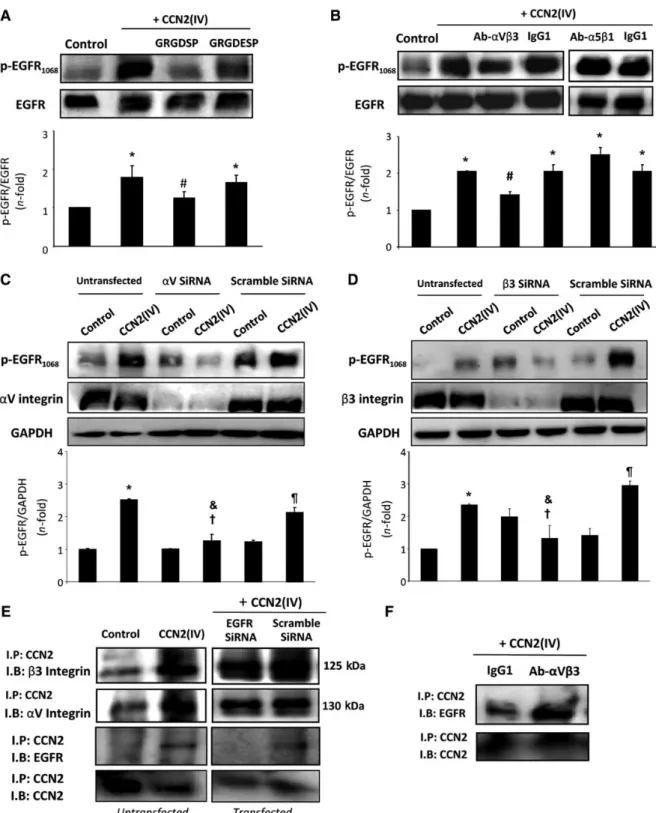
Integrins are heterodimeric receptors for cell-surface adhesion molecules and extracellular matrix proteins, which are composed of two subunits,aandb. Eachabcombination has specific signalling properties (Juliano,2002). To date, eighteenaand eightbsubunits have been identified, which form at least24differentabintegrins (Humphries et al.,2006). Integrin-binding sites are present in CCN2 and mediate several effects (Chen et al.,2004;Gao and Brigstock, 2005). We first tested the involvement of integrins in EGFR activation Figure3CCN2induces EGFR phosphorylation in the kidney. (A) C57BL/6mice were i.p. injected with2.5ng/g of body weight of recombinant CCN2(IV) or vehicle (saline) and sacrificed24h later. Some animals were treated with erlotinib (40mg/kg per day) or its vehicle (control group), starting24h before CCN2(IV) injection. EGFR activation was determined in total renal extracts by western blot analysis. Figure shows two representative mice from each group and data are expressed as mean+SEM of8–10mice per group. The localization of activated EGFR was determined by immunohistochemistry using antibody against p-EGFR1173 (B) and by immunofluorescence using antibody against p-EGFR1068(E), which showed increased p-EGFR immunostaining mainly in tubular epithelial cells. (C) The pEGFR1173immunostaining inBwas quantified and expressed as mean+SEM of8–10animals per group. (D) ERK activation is a downstream mechanism of CCN2/EGFR signalling in the kidney. Figure shows a representative experiment and data of p-ERK levels are expressed as mean+SEM of8–10mice per group. *P, 0.05vs. control.#P,0.05vs. CCN2(IV).
at ELTE on April 21, 2015http://jmcb.oxfordjournals.org/Downloaded from
by CCN2(IV) using arginine-glycine-aspartic acid (RGD) peptides.
RGD was originally identified as the sequence in fibronectin that is a recognition site fora5b1integrin, but it also recognizesa3b1, a8b1,aVb1,aVb3,aVb5,aVb6, anda2bb3integrins (Plow et al., 2000). Pre-incubation of HK2 cells with cyclic RGD peptide (GRGDSP), but not with control peptide (GRGDESP), reduced CCN2(IV)-induced EGFR activation (Figure6A), suggesting that integ- rins with RGD recognition specificity may be involved in CCN2 responses.
The specific integrins involved in CCN2 actions are cell- dependent. In hepatic stellate cells,aVb3integrin interacts with CCN2(IV) (Gao and Brigstock,2004), while in pancreatic stellate cells it isa5b1(Gao and Brigstock,2005). Therefore, we tested the involvement of these two integrins in tubular epithelial cells.
Pre-incubation of HK2cells with a neutralizing antibody against aVb3integrin inhibited CCN2(IV)-induced EGFR phosphorylation, while a control IgG or aa5b1-neutralizing antibody had no effect (Figure 6B). Using siRNA against b3 or aV integrins showed Figure4EGFR kinase inhibition decreases CCN2(IV)-induced renal inflammatory cell infiltration. In paraffin-embedded kidney sections, immuno- histochemistry using anti-F4/80and anti-CD3was performed to characterize monocyte/macrophages and T lymphocytes, respectively (represen- tative sections from each group,A; staining quantification,B). Magnification200×. Erlotinib inhibits CCN2(IV)-induced up-regulation of renal inflammatory molecules. CCL-2and IL-6gene expression levels were determined by real-time PCR (C) and protein levels by ELISA (D) in total renal extracts from different animal groups. Data are expressed as mean+SEM of8–10animals per group. *P,0.05vs. control.#P,0.05 vs. CCN2(IV). (E–G) CCN2(IV) increases proinflammatory factors via EGFR activation in murine tubular epithelial cells. Cells were pre-incubated for1h with erlotinib (10mmol/L) or AG1478(100nmol/L) before the stimulation with10ng/ml CCN2(IV) for6h (gene studies;E) or24h (protein studies, cell-conditioned medium;F). (G) EGFR gene silencing inhibits upregulation of proinflammatory molecules caused by CCN2(IV) in human tubular epithelial cells. HK2cells were transfected or not with an EGFR or scramble siRNA before the stimulation with50ng/ml CCN2(IV) for 6h (gene studies). Data are expressed as mean+SEM of6 (E),5 (F), and 4(G) independent experiments. *P,0.05vs.
control-untransfected. #P,0.05 vs. CCN2(IV). †P,0.05 vs. CCN2(IV)-treated scramble siRNA-transfected cells. &P,0.05 vs.
CCN2(IV)-treated untransfected cells.
328 | Journal of Molecular Cell Biology Rayego-Mateos et al.
at ELTE on April 21, 2015http://jmcb.oxfordjournals.org/Downloaded from
similar inhibitory effects, while a scrambled siRNA had no effect (Figure6C and D). These data clearly show thataVb3integrin med- iates CCN2(IV)-induced EGFR activation.
Next, we evaluated whether CCN2(IV) could directly bind toaVb3 integrin by IP experiments. In CCN2(IV)-treated HK2cells, the for- mation of CCN2-b3 and CCN2-aV complexes was found (Figure 6E). Moreover, EGFR gene silencing did not modify the CCN2(IV) binding toaV orb3integrin subunits (Figure6E), demon- strating the direct binding of CCN2(IV) toaVb3integrin, even in the absence of EGFR. Interestingly, pre-incubation of cells with the neu- tralizing antibody againstaVb3integrin did not modify the CCN2– EGFR complex formation, assessed by EGFR co-IP upon CCN2(IV) treatment (Figure6F). These data showed thataVb3integrin direct- ly binds to CCN2(IV), but is not necessary for the binding of CCN2(IV) to EGFR and the subsequent complex formation.
Potential crosstalk between EGFR and TrkA in response to CCN2(IV) stimulation in cultured tubular epithelial cells
In mesangial cells, CCN2stimulated tyrosine phosphorylation of proteins at75–80and140–180kDa within10min, and previous studies have identified the neurotrophin receptor TrkA (molecular weight 140kDa) as a potential CCN2 receptor (Wahab et al., 2005), also in other cell types, such as cardiomyocytes (Wang et al.,2010). Therefore, the role of TrkA in CCN2-induced responses in tubular epithelial cells was evaluated. Western blot was performed using an antibody that recognizes TrkA phosphorylated on Y490, pre- viously related to CCN2responses in mesangial cells (Wahab et al., 2005). We found that CCN2(IV) increased TrkA phosphorylation levels in HK2 cells, which was abolished in TrkA-silenced cells (Figure7A), showing the specificity of this CCN2(IV) response.
Next, we further evaluated the potential interrelation between EGFR and TrkA in HK2 cells. Gene silencing of TrkA diminished CCN2(IV)-induced EGFR phosphorylation (Figure7A). Moreover, pharmacological inhibition of TrkA using K252a also blocked CCN2(IV)-mediated EGFR activation (Figure 7B). On the other hand, EGFR gene silencing inhibited TrkA phosphorylation induced by CCN2(IV) (Figure7C). These data indicate an EGFR/
TrkA receptor crosstalk.
CCN2is a downstream mediator of TGF-b-induced EGFR activation CCN2is a downstream mediator of TGF-b-induced profibrotic responses (Ruiz-Ortega et al.,2007). In HK2cells, blockade of en- dogenous CCN2production by specific CCN2gene silencing mark- edly diminished TGF-b-induced EGFR phosphorylation after24h of TGF-bincubation, compared with scramble siRNA controls (Figure8).
Our results confirm and extend previous data, showing that CCN2is a downstream mediator of TGF-b-induced responses, including EGFR signalling.
Discussion
By surface plasmon resonance, we have detected direct binding of CCN2 to the immobilized extracellular fraction of EGFR.
Interestingly, both full-length CCN2and CCN2(IV) bound to EGFR and increased EGFR phosphorylation in cultured renal cells, sug- gesting that the EGFR-binding site is present in the carboxyl- terminal module. Ourin vivostudies show that CCN2(IV) activates EGFR/ERK pathway in the kidney, mainly in tubular epithelial cells.
Ourin vitrostudies in these cells demonstrate that CCN2(IV) rapidly binds to the cellular membrane and leads to CCN2– EGFR complex formation, increases EGFR phosphorylation, and activates down- stream signalling mechanisms. Seven ligands for EGFR have been identified so far: EGF, TGF-a, heparin binding EGF-like growth factor, amphiregulin, betacellulin, epigen, and epiregulin (Dreux et al., 2006). Data presenting here extend this list, suggesting that CCN2is another ligand for EGFR.
Ligand binding to EGFR induces a conformational change leading to the formation of receptor homo- or heterodimers and subsequent activation of the intrinsic tyrosine kinase domain by phosphorylation of specific tyrosine residues within the cytoplasmic tail of the recep- tor (Sweeney and Carraway,2000). Phosphorylation of different tyro- sine residues occurs upon binding of different ligands to the same EGFR, leading to a variety of downstream signal transduction path- ways that can be selectively activated (Sweeney and Carraway, 2000). Ourin vivodata clearly demonstrated that CCN2(IV) adminis- tration activated renal EGFR, as shown by increased EGFR phosphor- ylation, mainly in tubular epithelial cells. In these cellsin vitroandin Figure5Pharmacological inhibition of MMPs or ADAM-17and gene silencing of ADAM-17do not modify CCN2(IV)-induced EGFR phosphorylation in human tubular epithelial cells. (A) HK2cells were pre-incubated for1h with the pan-specific MMPs inhibitor GM6001(1mmol/L) or the ADAM-17-specific inhibitor TAPI-2(20mmol/L) before the stimulation with50ng/ml CCN2(IV) for15min. Values are mean+SEM from at least6independent experiments. *P,0.05vs. control. (B) HK2cells were transfected with ADAM-17siRNA or scramble siRNA, and then treated with50ng/ml CCN2(IV) for15min. Values are mean+SEM from at least4independent experiments. *P,0.05vs. control-untransfected.
at ELTE on April 21, 2015http://jmcb.oxfordjournals.org/Downloaded from
Figure6Role of integrins in CCN2(IV)-induced EGFR activation in cultured human tubular epithelial cells. HK2cells were pre-incubated with 0.2mmol/L RGDSP or its control peptide RGDESP (A) or with5mg/ml neutralizing antibodies againstaVb3integrin,a5b1integrin, or their cor- responding IgG1control (B). *P,0.05vs. control.#P,0.05vs. CCN2(IV). HK2cells were non-transfected or transfected withaV (C),b3(D), or scramble siRNA, and then treated or not with50ng/ml CCN2(IV) for15min. Figures show a representative western blot and data are expressed as mean+SEM of8independent experiments.aV orb3integrin was used as silencing control and GAPDH as loading control. *P,0.05vs.
control-untransfected.}P,0.05vs. untreated scramble siRNA-transfected cells.†P,0.05vs. CCN2(IV)-treated scramble siRNA-transfected cells.&P,0.05vs. CCN2(IV)-treated untransfected cells. (E) EGFR gene-silenced cells were stimulated with CCN2(IV) and cross-linked, and cell lysates were immunoprecipitated with anti-CCN2antibody (IP) and analysed by western blot (IB) with antibodies against EGFR, CCN2,aV, orb3integrins. The formation ofaV/b3-CCN2(IV) complexes in the presence or absence of EGFR (blocked by gene silencing) was shown. (F) Cells were pre-incubated with a neutralizingaVb3integrin antibody or IgG control before CCN2(IV) stimulation. TheaVb3integrin neutralization did not modify CCN2(IV)-EGFR complex formation. One representative experiment out of three with similar results was shown inEandF.
330 | Journal of Molecular Cell Biology Rayego-Mateos et al.
at ELTE on April 21, 2015http://jmcb.oxfordjournals.org/Downloaded from
vivo, CCN2(IV) induced EGFR phosphorylation on Y1068and Y1173, which have been previously associated to MAPK cascade (Rojas et al.,1996;Sweeney and Carraway,2000). In cultured tubular epi- thelial cells, EGFR blockade by gene silencing or kinase inhibition inhibited CCN2(IV)-induced ERK activation and upregulation of proinflammatory genes.In vivotreatment with erlotinib markedly diminished the number of inflammatory cells, the up-regulation of proinflammatory markers, and ERK activation in the kidneys of CCN2(IV)-injected mice. Our results suggest that CCN2(IV) directly binds to EGFR and activates its signalling pathway leading to the modulation of downstream mechanisms, such as ERK activation, and cellular responses, including renal inflammatory cell infiltration.
EGF ligands exist as inactive transmembrane precursors, requir- ing ADAM-mediated proteolytic cleavage of their ectodomain to be released as mature soluble ligands, whereby ADAMs regulate EGFR ligands availability (Melenhorst et al., 2008). Regarding CCN2, the full-length protein can be digested by proteases in- cluding MMPs. In particular, MMP-2leads to the generation of a 11kDa carboxyl-terminal fragment, which corresponds to CCN2(IV) (Hashimoto et al.,2002;De Winter et al.,2008;Tam et al.,2009).
In the urine of patients with diabetic nephropathy, full-length CCN2 and CCN2(IV) were both found (Riser et al., 2003).
However, the mechanisms involved in the regulation of CCN2deg- radation in renal diseases are unknown and future studies are needed.
EGFR transactivation is mediated by ADAM-dependent EGFR ligand shedding by factors that bind G protein-coupled receptors (Ohtsu et al., 2006). Depending on the tissue, different ADAMs may be involved in EGFR ligand shedding. In this sense, Angiotensin II-induced EGFR transactivation in the kidney is regu- lated by ADAM-17 (Lautrette et al., 2005; Wolf,2005; Flamant et al., 2012), while ADAM-12mediates this process in the heart (Asakura et al.,2002). We have observed that the pharmacological inhibition of MMPs or ADAM-17and gene silencing of ADAM-17 did not modify CCN2(IV)-induced EGFR phosphorylation in renal cells, demonstrating that MMPs are not involved in CCN2(IV)- mediated EGFR-signalling activation and confirming the direct interaction of CCN2(IV) with EGFR.
Severalin vitrostudies have shown that CCN2, through its binding to different domains, regulates different processes. The aminus ter- minal portion binds IGF-I and synergizes in the production of matrix proteins in renal cells (Kim et al.,1997;Wang et al.,2001;Lam et al., 2003). InXenopuscells, CCN2, through the cysteine-rich domain, dir- ectly binds to TGF-band acts as a cofactor that enhances TGF-b binding to its receptors and Smad-responsive promoter activation (Abreu et al.,2002). TSP-1domains have been implicated in the binding to extracellular matrix proteins, integrins, heparan sulphate proteoglycans, low-density lipoprotein receptor-related protein, and vascular endothelial growth factor (Adams and Tucker, 2000; Segarini et al.,2001;Inoki et al.,2002;Leask and Abraham,2006; Figure7Blockade of TrkA by gene silencing or a specific TrkA receptor inhibitor inhibits CCN2(IV)-induced EGFR activation and EGFR gene silencing inhibits CCN2(IV)-induced TrkA activation. (AandC) HK2cells were transfected with siRNA against TrkA (A) or EGFR (C) or its corresponding scram- ble. (B) HK2cells were pre-incubated with1×1024mmol/L K252a for1h. In all experiments, cells were treated or not with50ng/ml CCN2(IV) for 15min. Activation of EGFR or TrkA was evaluated using specific p-EGFR1068or p-TrkA490antibodies. EGFR and pTrkA were used as silencing controls and GAPDH as loading control. Figures show a representative western blot experiment and data are expressed as mean+SEM of4(A),3(B), and4 (C) independent experiments. *P,0.05vs. control-untransfected.}P,0.05vs. untreated scramble siRNA-transfected cells.†P,0.05vs.
CCN2(IV)-treated scramble siRNA-transfected cells.&P,0.05vs. CCN2(IV)-treated untransfected cells. at ELTE on April 21, 2015http://jmcb.oxfordjournals.org/Downloaded from
Chen and Lau, 2010). The carboxyl-terminal CCN2 cystine-knot module binds to integrins and exerts additional signalling capabil- ities, including regulation of fibrosis and inflammation (Leask and Abraham,2006;Liu et al.,2006;De Winter et al.,2008). Our in vitrodata show that this carboxyl-terminal fragment binds to EGFR leading to the regulation of proinflammatory factors. Our findings implicate integrins as key mediators of CCN2(IV)-induced EGFR acti- vation using RGD peptides and provide evidence that integrinaVb3 is involved in CCN2(IV)-induced EGFR activation based on results of the in vitro experiments utilizing neutralizing anti-integrin anti- bodies and siRNA. The ability of integrins to cooperate with receptor tyrosine kinases, including EGFR, to transduce proliferative signals and regulate cell survival and migration has been discussed previ- ously (Miranti and Brugge,2002;Schwartz and Ginsberg,2002).
Integrins are able to form physical complexes with EGFR at the cell membrane and trigger ligand-independent phosphorylation of Y845, Y1068, Y1086, and Y1173 residues in the EGFR molecule (Moro et al., 1998). This integrin-dependent EGFR activation appears necessary for full EGFR-dependent transcriptional responses (Cabodi et al.,2004). Our data show thataVb3integrin binds to CCN2(IV) and is involved in EGFR-signalling transduction, but is not necessary for its binding to the EGFR and the formation of EGFR-CCN2(IV) complex.
TrkA is a member of the Trk family of cell membrane receptors (TrkA, TrkB, and TrkC). These receptors interact with neurotrophins and form homodimers or heterodimers with the low-affinity pan neurotrophin receptor, p75NTR. Neurotrophins, such as nerve growth factor, brain-derived neurotrophic factor, neurotrophin3
and4, and their receptors, are important for the development, sur- vival, and function of neurons (Allen and Dawbarn,2006). The neu- rotrophin receptor TrkA has been proposed as a CCN2receptor in mesangial cells (Wahab et al., 2005) and involved in diabetic nephropathy (Fragiadaki et al.,2012). In murine cardiomyocytes, CCN2 via TrkA induced profibrotic and proinflammatory effects (Wang et al.,2010). Interestingly, cardiomyocytes express add- itional CCN2 receptors that mediate proinflammatory actions, since CCN2-induced TNF-aand IL-6mRNA upregulation occurred in the absence of TrkA (Wang et al.,2010). In tubular epithelial cells, we have found that CCN2(IV) activates TrkA signalling.
Gene silencing and pharmacological inhibition of TrkA diminished EGFR phosphorylation, and EGFR gene silencing inhibited TrkA phosphorylation induced by CCN2(IV), demonstrating EGFR/TrkA crosstalk in response to CCN2(IV) stimulation. The similarity of phos- phoproteomic profiles between TrkA and EGFR indicates a consider- able overlap in downstream signallings originated in these tyrosine kinase receptors (Bradshaw et al.,2013). In monocytes, EGFR/TrkA crosstalk has been described in response to G protein-coupled receptors and linked to modulation of proinflammatory mediators (El Zein et al.,2010). The complexity of tyrosine kinase receptor sig- nalling and interactions will require future studies.
The incidence of chronic kidney disease is increasing and current treatments only retard disease progression. Many studies using different strategies for blocking CCN2activity have proven benefi- cial effects on experimental pathologies, including renal diseases (Leask and Abraham, 2006) However, these are far from being used in humans. CCN2overexpression has been described in a wide variety of progressive human renal diseases and has been proposed as a risk biomarker (Riser et al.,2003;Tam et al.,2009; Slagman et al.,2011). We describe here that CCN2(both full-length and the carboxyl-terminal fragment) interacts with and activates EGFR, leading to ERK activation and regulation of renal inflamma- tion. CCN2has been described as a downstream mediator of profi- brotic factors (Hashimoto et al.,2002;Ruperez et al.,2003;Leask and Abraham,2006). Our results showing that CCN2gene silencing inhibited EGFR pathway activation in response to TGF-bsupport these findings and extend the importance of EGFR signalling in the fibrotic process. Experimental evidences suggest that EGFR in- hibition may have therapeutic potential for kidney diseases (Lautrette et al.,2005;Flamant et al.,2012;Liu et al.,2012). Our findings indicate that CCN2is a new ligand of the EGFR and identify this receptor as an important therapeutic target for renal diseases.
Materials and methods Cell cultures
Human renal proximal tubular epithelial cells (HK2cell line, ATCC CRL-2190) were grown in RPMI1640medium with10% fetal bovine serum (FBS),1% glutamine,100U/ml penicillin,100mg/ml strepto- mycin, 5mg/ml insulin transferrin selenium (ITS), and 36ng/ml hydrocortisone in5% CO2at378C. Murine renal cortical fibroblasts (TFB cell line) and murine proximal tubular epithelial cells [murine tubular-epithelial (MCT) cell line] originally obtained from Dr Eric Neilson (Vanderbilt University) were grown in RPMI with10% FBS, 2mmol glutamine, 100U/ml penicillin, and 100mg/ml strepto- mycin in 5% CO2at378C. At60%–70% of confluence, HK2and TFB cells were growth-arrested in serum-free medium for 24h Figure 8 Blockade of endogenous CCN2 production diminishes
TGF-b-induced EGFR activation. CCN2 gene silenced HK2 cells and control cells were stimulated with 1ng/ml TGF-b for 24h. Figure shows a representative experiment and data are expressed as mean+ SEM of3independent western blot. *P,0.05vs. control-untransfected.
}P,0.05vs. untreated scramble siRNA-transfected cells.†P,0.05vs.
TGF-b-treated scramble siRNA-transfected cells. &P,0.05 vs.
TGF-b-treated untransfected cells.
332 | Journal of Molecular Cell Biology Rayego-Mateos et al.
at ELTE on April 21, 2015http://jmcb.oxfordjournals.org/Downloaded from
before the experiments, while MCT cells were maintained in1% FBS.
Reagents
The full-length molecule of CCN2(CCN2; Biovendor), TGF-band CCN2(IV) (Preprotech), erlotinib (Vichem), tyrphostin AG1478 (Alomone Labs), K-252a and GM6001(Calbiochem), TAPI-2(Enzo Life Sciences), RGDs peptides (Bachem), and neutralizing.
Neutralizing antibodies against integrin aVb3, integrin a5b1, and IgG1(Millipore) were used. DMSO, used as solvent of some reagents, had no effect on cell viability or gene expression.
Ligand – receptor interaction assays
Surface plasmon resonance interaction analysis was performed using Biacore3000(GE Healthcare). Data were collected using the highest collection rate. All experiments were carried out at258C using HBS-EP (10mmol/L HEPES, 150mmol/L NaCl, 3mmol/L EDTA,0.005% P20, pH7.4) as running buffer. CM5sensor chip, N-hydroxysuccinimide (NHS),N-ethyl-N′-(3-dimethylaminopropyl) carbodiimide (EDC), and ethanolamine HCl were obtained from GE Healthcare. EGFR extracellular domain (Genway) was immobi- lized on the surface of a CM5sensor chip by the standard amino coupling procedure at a flow rate of5ml/min. The surface was acti- vated for7min using a0.05mol/L NHS/0.2mol/L EDC mixture.
Then,5mg/ml EGFR in10mmol/L sodium acetate (pH5.0) was injected for 13min. Finally, residual activated groups on the surface were deactivated by a7min injection of1mol/L ethanola- mine (pH8.5). Immobilization density reached500RU. An add- itional flow cell was activated and deactivated and then used as a reference surface. For kinetic analysis, CCN2and CCN2(IV) were diluted in HBS-EP (range 1–250mmol/L). Concentration series and blank samples were injected for3min using a flow rate of 50ml/min and the dissociation was monitored for3min. Data pro- cessing and kinetic analysis were performed using BiaEvaluation 4.1.1. (GE Healthcare). Data were double referenced using refer- ence surface subtraction and blank correction. Processed data were globally fit to a simple1:1interaction model.
Live cell confocal microscopy
Cells were imaged using a Leica TCS SP5confocal microscope.
Fluorophore Cy-5-emitted fluorescence was monitored with a 550+2nm band pass or a670nm long pass filter and DAPI was excited using a DIODE laser. For video rate confocal, the images were captured (1frame every1.33sec) at400Hz for a period of 2min and digitalized using the LIF/LEICA program (LEICA microsys- tems). CCN2fluorescence by Cy5labelling was detected using Cy-5 fluorophore (1nmol/ml; Amersham) following the manufacturer instructions.
Chemical cross-linking
Chemical cross-linking was carried out as previously described, using DTSSP (Pierce) (Ardura et al.,2010), before immunocyto- chemistry and co-IP experiments.
Fluorescence immunocytochemistry
Growth-arrested HK2cells growing on glass coverslips were sti- mulated with CCN2(IV) or CCN2(IV)-Cy5. After chemical cross- linking, cells were fixed in4% paraformaldehyde, washed, and blocked (PBS/10% BSA/4% serum,1h). Then, cells were incu- bated overnight with anti-EGFR (1:200 dilution; Santa Cruz Biotechnology) or anti-CCN2(1:200dilution; Sigma) in PBS with 1% BSA, followed by AlexaFluorw633-conjugated goat anti-mouse (red) or AlexaFluorw488-conjugated goat anti-rabbit (green)
antibodies (1:300dilution; Invitrogen), respectively. Nuclei were stained with1mg/ml DAPI as control for equal cell density. The absence of primary antibody was used as negative control.
Samples were mounted in Mowiol40–88(Sigma) and examined by using a Leica DM-IRB confocal microscope.
Co-IP assays
After chemical cross-linking, cells were lysed in 300–500ml Triton– NP-40lysis buffer [50mmol/L Tris– HCl pH8,150mmol/L NaCl, 1mmol/L phenylmethylsulphonylfluoride, 1% NP-40/ IGEPAL, and a phosphatase-inhibitor cocktail (Set II, Calbiochem)], scraped off the dish, and incubated for1h at48C with shaking.
Cell lysates were pre-cleared by incubating with10ml of protein A-agarose bead slurries (0.5ml agarose/2ml PBS) for30min at 48C, and then centrifuged to wash away supernatants. Pre-cleared lysates were incubated with2.5–5mg antibody overnight at48C for IP experiment. The immune complexes were captured by the add- ition of protein A/G PLUS-agarose (20ml) bead slurries for1h at48C.
The agarose beads were collected by centrifugation, washed and subjected to SDS –PAGE, followed by western blot as described below (Ardura et al.,2010).
Western blot
Proteins were obtained from treated cells or mouse kidneys using lysis buffer (50mmol/L Tris–HCl,150mol/L NaCl,2mmol/L EDTA,2mmol/L EGTA,0.2% Triton X-100,0.3% IGEPAL,10ml/ml proteinase inhibitors cocktail,10ml/ml PMSF, and10ml/ml ortho- vanadate). To determine protein content the BCA method was used. Cell (25mg/lane) and kidney (100–150mg/lane) protein extracts were separated on 6% –12% polyacrylamide-SDS gels under reducing conditions.
Samples were then transferred onto nitrocellulose membranes (BioRad), blocked with TBS/5% defatted milk/0.05% Tween-20, and incubated overnight at 48C with the following antibodies (dilution): p-EGFR1068(1:250; Calbiochem), p-EGFR1173(1:250; Cell Signalling), p-TrkA490(1:1000; Cell Signalling), ADAM-17(1:1000; Abcam), CCN2(1:1000; Sigma), EGFR (1:250), ERK1/2(1:200), in- tegrinaV (1:200), integrinb3(1:200), pERK1/2(1:200), and anti- phosphotyrosine PY20 (1:250; Santa Cruz). Membranes were subsequently incubated with peroxidase-conjugated IgG secondary antibody and developed using an ECL chemiluminiscence kit (Amersham). Loading controls were done using an anti-GAPDH anti- body (1:10000; Chemicon) or total protein levels in phosphorylation studies. Autoradiographs were scanned using the Gel DocTM EZ imager and analysed with the Image Lab3.0software (BioRad).
Gene silencing
Gene silencing in cultured cells was performed using either a pre-designed siRNA corresponding to EGFR, ADAM-17, TrkA, CCN2 (Ambion), integrinaV, or integrin b3(Santa Cruz Biotechnology) or their corresponding scramble siRNAs. Subconfluent cells were transfected for24h with 25nmol/L siRNA using50nmol/L Lipofectamine RNAiMAX (Invitrogen) or treated only with lipofecta- mine—vehicle, according to the manufacturer’s instructions. Then, cells were incubated with10% heat-inactivated FBS for24h, followed by24h in serum-free medium and then treated or not with CCN2(IV).
Animal model
Studies were performed in adult male C57BL/6 mice (9– 12weeks old,20g; Harlan Interfauna Ibe´rica) and maintained at local animal facilities. All the procedures on animals were
at ELTE on April 21, 2015http://jmcb.oxfordjournals.org/Downloaded from
performed according to the European Community and Instituto de Investigacio´n Sanitaria Fundacio´n Jime´nez Dı´az Animal Research Ethical Committee guidelines. C57BL/6 mice received a single intraperitoneal injection of recombinant CCN2(IV) (endotoxin levels,0.01units; Preprotech) dissolved in saline at the dose of 2.5ng/g of body weight as described (Sanchez-Lopez et al., 2009), and studied 24h later (n¼8–10 mice per group). To block EGFR activation, animals were treated with erlotinib (40mg/kg/day) or its vehicle (10% Ethanol) at 24h before CCN2injection (n¼8–10mice per group). Mice were sacrificed under anaesthesia (Ketamine-HCl/Xylazine-HCl) and then kidneys were perfusedin situwith cold saline before removal.
We have previously demonstrated that CCN2(IV) administration did not cause tubular damage or fibrosis (Sanchez-Lopez et al., 2009).
Renal histology and immunohistochemistry
Immunohistochemistry was carried out on 3mm paraffin- embedded kidney sections. Sections were deparafinized and exposed to the PT Link (Dako) with Sodium Citrate Buffer (10mmol/L, pH6 or9 depending on the immunohistochemical marker) for antigen retrieval. After endogenous peroxidase was blocked, sections were incubated with4% BSA/8% serum in1× wash buffer ‘en vision’ (Dako) to eliminate nonspecific protein binding, followed by primary antibodies (dilution) F4/80 (1:50), CD3(1:300; Serotec), and p-EGFR1173(1:200; Cell Signalling) over- night at 48C. After washing, they were incubated with anti-IgG secondary biotinylated-conjugated antibodies (Amersham) fol- lowed by the avidin-biotin-peroxidase complex (Dako) and3,3′-dia- minobenzidine as chromogen. Sections were counterstained with Carazzi’s haematoxylin.
Immunofluorescence was performed by incubating sections with 4% BSA/8% serum in PBS (for blockade), then anti-p-EGFR anti- body (1:200; Dako), followed by AlexaFluorw633-conjugated anti- body (1:200). The total number of positive stained cells was quantified in five randomly chosen fields (20×) using the Image-Pro Plus software. Data are expressed as positive stained area vs. total analysed area. Triplicate samples from each animal were examined in a blind manner.
ELISA for proinflammatory factors
CCL-2 and IL-6 protein levels were assayed by an ELISA kit (eBioscience), and quantified by comparison with a standard curve. Data are expressed as n-fold increase over the mean of control levels.
Gene expression studies
Total RNA was isolated from cells and mouse kidney samples with Trizol (Invitrogen). The cDNA was synthesized using the High- Capacity cDNA Archive Kit (Applied Biosystems) using2mg of total RNA primed with random hexamer primers. Multiplex real-time PCR was performed using Applied Biosystems expression assays mouse CCL-2 Mm00441242_m1 and mouse IL-6 Mm00446 190_m1. Data were normalized to18S eukaryotic ribosomal RNA 4210893E (VIC). The mRNA copy numbers were calculated for each sample by the instrument software using Ct value. Results are expressed in copy numbers, calculated relative to unstimulated cells or control mice, after normalization against18S.
Statistical analysis
All results are expressed as mean+SEM. Differences between agonist-treated groups and controls were assessed by Mann – Whitney test.P,0.05was considered significant. Statistical ana- lysis was conducted using the SPSS statistical software (version 11.0).
Acknowledgments
We thank MaMar Gonzalez Garcia-Parren˜o and Susana Carrasco at the IIS-Fundacio´n Jime´nez Dı´az for their technical help with immunohistochemistry.
Funding
This work was supported by grants from the Instituto de Salud Carlos III (ISCIII) (REDINREN RD06/0016; RD12/0021, PI081564, PI11/01854, PI12/02587, PI12/00204, and PI10/00072), Comunidad de Madrid (S2010/BMD-2321, S2010/BMD-2378), Sociedad Espan˜ola de Nefrologı´a, DIALOK Eurpean project LSHB-CT-2007-036644, and Fundacion Lilly and Research Institute Queen Sophia (FRIAT). ISCIII fellowships to R.R.D., C.L., and M.A. and programa de intensificacion (ISCIII/Lain Entralgo) to A.O.
Conflict of interest:none declared.
References
Abreu, J.G., Ketpura, N.I., Reversade, B., et al. (2002). Connective-tissue growth factor (CTGF) modulates cell signalling by BMP and TGF-beta. Nat. Cell Biol.4, 599–604.
Adams, J.C., and Tucker, R.P. (2000). The thrombospondin type1repeat super- family: diverse proteins with related roles in neuronal development. Dev. Dyn.
218,280–299.
Allen, S.J., and Dawbarn, D. (2006). Clinical relevance of the neurotrophins and their receptors. Clin. Sci. (Lond)110,175–191.
Ardura, J.A., Rayego-Mateos, S., Ra´mila, D., et al. (2010). Parathyroid hormone-related protein promotes epithelial-mesenchymal transition.
J. Am. Soc. Nephrol.21,237–248.
Asakura, M., Kitakaze, M., Takashima, S., et al. (2002). Cardiac hypertrophy is inhibited by antagonism of ADAM12processing of HB-EGF: metalloproteinase inhibitors as a new therapy. Nat. Med.8,3–5.
Bradshaw, R.A., Chalkley, R.J., Biarc, J., et al. (2013). Receptor tyrosine kinase signaling mechanisms: devolving TrkA responses with phosphoproteomics.
Adv. Biol. Regul.53,87–96.
Bronte, G., Terrasi, M., Rizzo, S., et al. (2011). EGFR Genomic alterations in cancer: prognostic and predictive values. Front. Biosci.1,879–887.
Cabodi, S., Moro, L., Bergatto, E., et al. (2004). Integrin regulation of epidermal growth factor (EGF) receptor and of EGF dependent responses. Biochem. Soc.
Trans.32,438–442.
Chen, C.C., and Lau, L.F. (2010). Deadly liaisons: fatal attraction between CCN matricellular proteins and the tumor necrosis factor family of cytokines.
J. Cell Commun. Signal.4,63–69.
Chen, Y., Abraham, D.J., Shi-Wen, X., et al. (2004). CCN2Promotes fibroblast ad- hesion to fibronectin. Mol. Biol. Cell15,5635–5646.
De Winter, P., Leoni, P., and Abraham, D. (2008). Connective tissue growth factor:
structure-function relationships of a mosaic, multifunctional protein. Growth Factors26,80–91.
Dreux, A.C., Lamb, D.J., Modjtahedi, H., et al. (2006). The epidermal growth factor receptors and their family of ligands: their putative role in atherogenesis.
Atherosclerosis186,38–53.
El Zein, N., D’Hondt, S., and Sariban, E. (2010). Crosstalks between the receptors tyrosine kinase EGFR and TrkA and the GPCR, FPR, in human monocytes are
334 | Journal of Molecular Cell Biology Rayego-Mateos et al.
at ELTE on April 21, 2015http://jmcb.oxfordjournals.org/Downloaded from
essential for receptors-mediated cell activation. Cell. Signal.22,1437–1447.
Flamant, M., Bolle´e, G., He´nique, C., et al. (2012). Epidermal growth factor: a new therapeutic target in glomerular disease. Nephrol. Dial. Transplant. 27, 1297–1304.
Fragiadaki, M., Hill, N., Hewitt, R., et al. (2012). Hyperglycemia causes renal cell damage via CCN2-induced activation of the TrkA receptor: implications for dia- betic nephropathy. Diabetes61,2280–2288.
Gao, R., and Brigstock, D.R. (2004). Connective tissue growth factor induces ad- hesion of rat activated hepatic stellate cells by binding of its C-terminal domain to integrin alpha(v)beta(3) and heparan sulfate proteoglycan.
J. Biol. Chem.279,8848–8855.
Gao, R., and Brigstock, D.R. (2005). Connective tissue growth factor in rat pan- creatic stellate cell function: integrin a5b1 as a novel CCN2 receptor.
Gastroenterology129,1019.
Guha, M., Xu, Z.G., Tung, D., et al. (2007). Specific down-regulation of connective tissue growth factor attenuates progression of nephropathy in mouse models of type1and type2diabetes. FASEB J.21,3355–3368.
Hao, L., Du, M., Lopez-Campistrous, A., et al. (2004). Agonist induced activation of matrix metalloproteinase-7promotes vasoconstriction through the epider- mal growth factor-receptor pathway. Circ. Res.94,68–76.
Hashimoto, G., Inoki, I., Fujii, Y., et al. (2002). Matrix metalloproteinases cleave connective tissue growth factor and reactivate angiogenic activity of vascular endothelial growth factor165. J. Biol. Chem.277,36288–36295.
Humphries, J.D., Byron, A., and Humphries, M.J. (2006). Integrin ligands at a glance. J. Cell Sci.119,3901–3903.
Inoki, I., Shiomi, T., Hashimoto, G., et al. (2002). Connective tissue growth factor binds vascular endothelial growth factor (VEGF) and inhibits VEGF-induced angiogenesis. FASEB J.16,219–221.
Juliano, R.L. (2002). Signal transduction by cell adhesion receptors and the cyto- skeleton: functions of integrins, cadherins, selectins, and immunoglobulin- superfamily members. Annu. Rev. Pharmacol. Toxicol.42,283–323. Kim, H.S., Nagalla, S.R., Oh, Y., et al. (1997). Identification of a family of low-
affinity insulin-like growth factor binding proteins (IGFBPs): characterization of connective tissue growth factor as a member of the IGFBP superfamily.
Proc. Natl Acad. Sci. USA94,12981–12986.
Lam, S., van der Geest, R.N., Verhagen, N.A., et al. (2003). Connective tissue growth factor and igf-I are produced by human renal fibroblasts and cooperate in the induction of collagen production by high glucose. Diabetes 52, 2975–2983.
Lautrette, A., Li, S., Alili, R., et al. (2005). Angiotensin II and EGF receptor cross- talk in chronic kidney diseases: a new therapeutic approach. Nat. Med.11, 867–874.
Leask, A., and Abraham, D.J. (2006). All in the CCN family: essential matricellular signaling modulators emerge from the bunker. J. Cell Sci.119, 4803–4810.
Liu, B.C., Zhang, J.D., Zhang, X.L., et al. (2006). Role of connective tissue growth factor (CCN2) module4in regulating epithelial mesenchymal transition in HK-2cells. Clin. Chim. Acta373,144–150.
Liu, N., Guo, J.K., Pang, M., et al. (2012). Genetic or pharmacologic blockade of EGFR inhibits renal fibrosis. J. Am. Soc. Nephrol.23,854–867.
Markiewicz, M., Nakerakanti, S.S., Kapanadze, B., et al. (2011). Connective tissue growth factor (CCN2/CCN2) mediates angiogenic effect of S1P in human dermal microvascular endothelial cells. Microcirculation18,1–11. Melenhorst, W.B., Mulder, G.M., Xi, Q., et al. (2008). Epidermal growth factor re-
ceptor signaling in the kidney: key roles in physiology and disease.
Hypertension52,987–993.
Miranti, C.K., and Brugge, J.S. (2002). Sensing the environment: a historical per- spective on integrin signal transduction. Nat. Cell Biol.4, E83– E90. Moro, L., Venturino, M., Bozzo, C., et al. (1998). Integrins induce activation of EGF
receptor: role in MAP kinase induction and adhesion-dependent cell survival.
EMBO J.17,6622–6632.
Ohtsu, H., Dempsey, P.J., and Eguchi, S. (2006). ADAMs as mediators of EGF re- ceptor transactivation by G protein-coupled receptors. Am. J. Physiol. Cell Physiol.291, C1– C10.
Okada, H., Kikuta, T., Kobayashi, T., et al. (2005). Connective tissue growth factor expressed in tubular epithelium plays a pivotal role in renal fibrogenesis.
J. Am. Soc. Nephrol.16,133–143.
Perbal, B. (2004). CCN Proteins: multifunctional signalling regulators. Lancet 363,62–64.
Phanish, M.K., Winn, S.K., and Dockrell, M.E. (2010). Connective tissue growth factor-(CTGF, CCN2) a marker, mediator and therapeutic target for renal fibro- sis. Nephron Exp. Nephrol.114, e83– e92.
Plow, E.D., Haas, T.A., Zhang, L., et al. (2000). Ligand binding to integrins. J. Biol.
Chem.275,21785–21788.
Pourazar, J., Blomberg, A., Kelly, F.J., et al. (2008). Diesel exhaust increases EGFR and phosphorylated C-terminal Tyr1173in the bronchial epithelium. Part.
Fibre Toxicol.6,5–8.
Rachfal, A.W., and Brigstock, D.R. (2005). Structural and functional properties of CCN proteins. Vitam. Horm.70,69–103.
Riser, B.L., Cortes, P., De Nichilo, M., et al. (2003). Urinary CCN2(CTGF) as a pos- sible predictor of diabetic nephropathy: preliminary report. Kidney Int.64, 451–458.
Rojas, M., Yao, S., and Lin, Y.Z. (1996). Controlling epidermal growth factor (EGF)-stimulated Ras activation in intact cells by a cell-permeable peptide mimicking phosphorylated EGF receptor. J. Biol. Chem.271,27456–27461.
Ruiz-Ortega, M., Rodrı´guez-Vita, J., Sanchez-Lopez, E., et al. (2007). TGF-beta signaling in vascular fibrosis. Cardiovasc. Res.74,196–206.
Rupe´rez, M., Lorenzo, O., Blanco-Colio, L.M., et al. (2003). Connective tissue growth factor is a mediator of angiotensin II-induced fibrosis. Circulation 108,1499–1505.
Sa´nchez-Lo´pez, E., Rayego, S., Rodrigues-Dı´ez, R., et al. (2009). CTGF promotes inflammatory cell infiltration of the renal interstitium by activating NF-kappa B. J. Am. Soc. Nephrol.20,1513–1526.
Schwartz, M.A., and Ginsberg, M.H. (2002). Networks and crosstalk: integrin sig- nalling spreads. Nat. Cell Biol.4, E65– E68.
Segarini, P.R., Nesbitt, J.E., Li, D., et al. (2001). The low density lipoprotein receptor-related protein/alpha2-macroglobulin receptor is a receptor for con- nective tissue growth factor. J. Biol. Chem.276,40659–40667.
Sibilia, M., Kroismayr, R., Lichtenberger, B.M., et al. (2007). The epidermal growth factor receptor: from development to tumorigenesis. Differentiation 75,770–787.
Slagman, M.C., Nguyen, T.Q., Waanders, F., et al. (2011). Effects of antiproteinu- ric intervention on elevated connective tissue growth factor (CCN2/CCN2) plasma and urine levels in nondiabetic nephropathy. Clin. J. Am. Soc.
Nephrol.6,1845–1850.
Sweeney, C., and Carraway, K.L. (2000). Ligand discrimination by ErbB receptors:
differential signaling through differential phosphorylation site usage.
Oncogene19,5568–5573.
Tam, F.W., Riser, B.L., Meeran, K., et al. (2009). Urinary monocyte chemoattract- ant protein-1(MCP-1) and connective tissue growth factor (CCN2) as prognos- tic markers for progression of diabetic nephropathy. Cytokine47,37–42.
Terzi, F., Burtin, M., Hekmati, M., et al. (2000). Targeted expression of a dominant-negative EGF-R in the kidney reduces tubulo-interstitial lesions after renal injury. J. Clin. Invest.106,225–234.
Wahab, N.A., Weston, B.S., and Mason, R.M. (2005). Connective tissue growth factor CCN2interacts with and activates the tyrosine kinase receptor TrkA.
J. Am. Soc. Nephrol.16,340–351.
Wang, S., Denichilo, M., Brubaker, C., et al. (2001). Connective tissue growth factor in tubulointerstitial injury of diabetic nephropathy. Kidney Int.60, 96–105.
Wang, X., McLennan, S.V., Allen, T.J., et al. (2010). Regulation of pro-inflammatory and pro-fibrotic factors by CCN2/CTGF in H9c2cardiomyo- cytes. J. Cell Commun. Signal.4,15–23.
Wolf, G. (2005). ‘As time goes by’: angiotensin II-mediated transactivation of the EGF receptor comes of age. Nephrol. Dial. Transplant.20,2050–2053. Yokoi, H., Mukoyama, M., Nagae, T., et al. (2004). Reduction in connective tissue
growth factor by antisense treatment ameliorates renal tubulointerstitial fi- brosis. J. Am. Soc. Nephrol.15,1430–1440.
at ELTE on April 21, 2015http://jmcb.oxfordjournals.org/Downloaded from