COMPUTED TOMOGRAPHY
The effect of four-phasic versus three-phasic contrast media injection protocols on extravasation rate in coronary CT angiography: a randomized controlled trial
Júlia Karády1&Alexisz Panajotu1&Márton Kolossváry1&Bálint Szilveszter1&
Ádám L. Jermendy1&Andrea Bartykowszki1&Mihály Károlyi1&Csilla Celeng1&
Béla Merkely1&Pál Maurovich-Horvat1
Received: 19 October 2016 / Revised: 20 March 2017 / Accepted: 3 April 2017 / Published online: 24 May 2017
#The Author(s) 2017. This article is an open access publication
Abstract
Objectives Contrast media (CM) extravasation is a well- known complication of CT angiography (CTA). Our prospec- tive randomized control study aimed to assess whether a four- phasic CM administration protocol reduces the risk of extrav- asation compared to the routinely used three-phasic protocol in coronary CTA.
Methods Patients referred to coronary CTA due to suspected coronary artery disease were included in the study. All patients received 400 mg/ml iomeprol CM injected with dual-syringe automated injector. Patients were randomized into a three- phasic injection-protocol group, with a CM bolus of 85 ml followed by 40 ml of 75%:25% saline/CM mixture and 30 ml saline chaser bolus; and a four-phasic injection-protocol group, with a saline pacer bolus of 10 ml injected at a lower flow rate before the three-phasic protocol.
Results2,445 consecutive patients were enrolled (mean age 60.6 ± 12.1 years; females 43.6%). Overall rate of extravasa- tion was 0.9% (23/2,445): 1.4% (17/1,229) in the three-phasic group and 0.5% (6/1,216) in the four-phasic group (p = 0.034).
Conclusions Four-phasic CM administration protocol is easy to implement in the clinical routine at no extra cost. The ex- travasation rate is reduced by 65% with the application of the
four-phasic protocol compared to the three-phasic protocol in coronary CTA.
Key Points
• Four-phasic CM injection-protocol reduces extravasation rate by 65% compared to three-phasic.
•The saline pacer bolus substantially reduces the risk of CM extravasation.
•The implementation of four-phasic injection-protocol is at no cost.
Keywords Computed tomography angiography . Contrast media . Contrast media extravasation . Multidetector-row computed tomography . Coronary heart disease
Abbreviations
AMI Acute myocardial infarction CM Contrast media
CTA Computer tomography angiography DLP Dose-length product
PAD Peripheral artery disease TIA Transient ischaemic attack
Introduction
To achieve robust diagnostic performance in coronary com- puted tomography angiography (CTA) adequate intraluminal iodinated contrast media (CM) concentration is required.
Therefore, high flow rate injection, high concentration and relatively large volume of CM is used in daily practice.
However, the highly viscous iodinated CM and the high in- jection flow rate increase the risk of vessel wall injury resulting in CM extravasation.
Drs. Júlia Karády, Alexisz Panajotu, Béla Merkely and Pál Maurovich- Horvat contributed equally to this work.
* Pál Maurovich-Horvat
p.maurovich.horvat@mail.harvard.edu
1 MTA-SE Cardiovascular Imaging Research Group, Heart and Vascular Center, Semmelweis University, 68 Varosmajor Street, 1122 Budapest, Hungary
Contrast media extravasation is a well-known complication of CTA, with an incidence rate of 0.3–1.3% [1–6]. In case of CM extravasation, image quality is deteriorated due to insuf- ficient intraluminal attenuation [7,8], leading to an increased number of repeated CTA examinations, which results in extra radiation doses, additional CM load and increased costs.
Extravasation usually resolves without any serious complica- tions; however, in some instances it can lead to severe injuries [9]. CM has toxic effects on perivascular tissues that may trigger acute and chronic local inflammatory response, tissue necrosis or compartment syndrome [4,5,10,11]. It has been shown that female gender, elderly age, history of chemo- or radiotherapy, low muscle volume and peripheral locations oth- er than the cubital region as injection site increase the risk of CM extravasation [2,3,12].
A three-phasic CM injection-protocol is widely used to achieve optimal attenuation during coronary CTA, which re- sults in high contrast enhancement in the left side of the heart and in a lower enhancement in the right [13,14]. The tradi- tional three-phasic injection-protocol starts with a high flow rate CM injection (>5 ml/s), continues with a mixture of CM and saline, and finishes with a saline chaser bolus. We hypoth- esized that the relatively large quantity of high viscosity CM could place an increased strain on the vein’s wall, which in- creases the risk of extravasation. Extending the three-phasic injection-protocol with an initial slower saline flux of pacer bolus right before CM administration may open the possibly collapsed vein lumen with less stress on the vessel wall, thus when the contrast material enters the lumen with a higher flow rate, the already pre-dilated lumen is less likely to rupture.
Therefore, we sought to evaluate the effect of a four-phasic contrast injection-protocol on the CM extravasation rate in coronary CTA in a randomized controlled clinical trial.
Materials and methods Study design
In this prospective, single-centre, single-blinded, randomized, controlled clinical trial we compared two contrast media injection-protocols in patients who were referred for coronary CTA examination. We randomized patients into two groups:
(1) a three-phasic injection-protocol group and (2) a four- phasic injection-protocol group. To achieve randomization, we alternated the use of three-phasic and four-phasic CM ad- ministration protocols on a weekly basis: on even weeks, we applied the three-phasic protocol, and on odd weeks we used the four-phasic protocol. The primary endpoint was the occur- rence of CM extravasation. CM extravasation was defined as (1) presence of pain and local swelling close to the cannula insertion site occurring after the initiation of CM injection, and (2) absence of CM or minimal CM attenuation in cardiac
chambers on the CTA images. Our institutional review board approved the study and informed consent was waived.
Study population and CTA protocol
We included consecutive patients who were referred for cor- onary CTA from January 2014 to August 2015. Exclusion criteria were contraindications to iodinated CM, age under 18 years and the presence of a cannula that was not inserted by our radiographers.
We performed all coronary CTA examinations with a 256- slice multi-detector row CT scanner (Brilliance iCT 256;
Philips Healthcare, Best, The Netherlands). Contrast- enhanced image acquisition was performed in inspiration dur- ing a single breath-hold in a cranio-caudal direction. The fol- lowing imaging parameters were used: slice collimation of 128 mm × 0.625 mm, rotation time of 270 ms, tube voltage 100–120 kV and tube current 150–300 mAs depending on the patient’s weight. The majority of scans (99.8%) were acquired by using prospective ECG triggering at 78% phase of the cardiac cycle with 3% padding. Bolus tracking was used with a region of interest (ROI) placed in the left atrium. Images were reconstructed with a slice thickness of 0.8 mm and 0.4 mm increment.
Contrast media injection-protocol
All patients received the same type of cannula (B. Braun Medical Inc., Melsungen, Germany). All cannulas were inserted by certified radiographers. The preferred location of vein puncture was the right antecubital region. Other distal venous access locations were used if no suitable vein was found in the antecubital region. We registered venipuncture characteristics, such as the side and location of venous access, the size of the inserted cannula and the number of insertion attempts. In all patients the injection site was tested with a 20- ml saline bolus. All patients received a 400 mg/ml concentra- tion iomeprol (Iomeron 400, Bracco Spa, Milan, Italy) CM injected with dual-syringe automated mechanical injector. CM was pre-heated to 37 °C. In patients having more than 80 kg of bodyweight we used a 5.5 ml/s injection rate and 95 ml CM and 120 kV tube voltage. In patients less than 80 kg in bodyweight we used an injection rate of 4.5 ml/s, 80 ml CM and 100 kV tube voltage.
In the three-phasic protocol group the injection started with the CM bolus, followed by 40 ml of 75%:25% saline-CM mixture, and finished with 30 ml of chaser saline bolus.
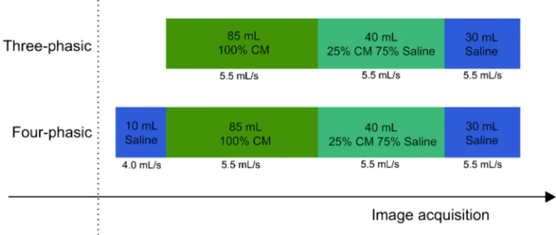
With the four-phasic protocol the injection started with the saline pacer bolus of 10 ml, administered with 1.5 ml/s lower flow rate than the CM bolus; specifically, a saline pacer bolus flow rate of 4.0 ml/s if the injection rate of CM was 5.5 ml/s and 3 ml/s if the CM flow rate was 4.5 ml/s and continued with the steps of the three-phasic protocol (Fig.1).
Statistical analysis
Continuous variables are reported as mean ± standard devia- tion. Based on our relatively large sample size the central limit theorem allows the use of parametric tests, therefore we com- pared the continuous variables using Student’s t-test. To eval- uate the differences between categorical variables we used Fisher’s exact test. Two-sided P values below 0.05 were con- sidered statistically significant. For risk estimation, we calcu- lated the odds ratio (OR) with 95% confidence intervals (CIs).
All statistical data analysis was performed with IBM SPSS (IBM Corp: version 23, Armonk, NY, USA).
Results
In total, 2,445 consecutive patients with suspected coronary artery disease were enrolled between 2014 January and 2015 August. The mean age was 60.6 ± 12.1 years and there were less female patients than males (females 43.6%). The clinical characteristics of included patients are summarized in Table1.
Out of the 2,445 patients, 1,229 (50.3%) received a three- phasic and 1,216 (49.7%) a four-phasic CM injection-protocol (Table2). The overall number of CM extravasation was 23 out of 2,445 patients (0.9%). The CM extravasation rate in the three-phasic group was 1.4% (17/1,229), whereas in the four-phasic group the extravasation rate was 0.5% (6/1,216), p = 0.034 (Fig.2). The four-phasic CM injection-protocol re- sulted in 65% reduction in extravasation rate as compared to the three-phasic CM injection-protocol in coronary CTA (odds ratio (OR): 0.354; CI: 0.139–0.900; p = 0.029). The majority of the patients received an 18 G cannula for CM injection (97.2% of all patients). The use of a 20 G cannula did not differ between the two groups (three-phasic protocol
group 34 (3.1%), four-phasic protocol group 38 (3.1%), re- spectively, p = 0.63).
Besides the CM injection-protocol none of the clinical and vein quality characteristics of patients who had extravasation versus patients with no extravasation showed any differences (Table 2). In the four-phasic group a CM injection rate of 5.5 ml/s was administered in 88.4% (1,075/1,216) of the pa- tients, which was lower than the three-phasic protocol group (91.2%, 1,121/1,229), p = 0.02. Even though we found a sig- nificant difference between the two groups, this did not influ- ence extravasation rates, since there was no difference in in- jection rates among patients with extravasation (5.5 ml/s flow Fig. 1 Schematic representation of the three-phasic and the four-phasic
contrast media (CM) injection-protocols. The three-phasic protocol starts with an undiluted CM bolus, followed by a 75%:25% saline and CM mixture and ends with a 30-ml chaser saline bolus. The four-phasic
protocol starts with a 10-ml saline pacer bolus, administered at a 1.5 ml/s slower flow rate than the CM bolus, and continues with the three-phasic protocol. The injection rate settings are dependent on the body weight of the patient and on the tube voltage settings
Table 1 Clinical characteristics of the patients
n = 2445
Age (y) 60.6 ± 12.1
Female (%) 1,065 (43.6)
Height (cm) 171.2 ± 10.1
Weight (kg) 84.0 ± 17.2
BMI (kg/m2) 28.5 ± 4.8
Hypertension (%) 1,605 (65.6)
Diabetes (%) 393 (16.1)
Dyslipidaemia (%) 1,142 (46.7)
AMI (%) 185 (7.6)
PAD (%) 236 (9.7)
Stroke/TIA (%) 135 (5.5) Current smoking (%) 990 (40.5) Total DLP (mGy*cm) 356.8 ± 142.0 Effective dose (mSv) 5.0 ± 2.0 Contrast material (ml) 91.2 ± 7.3 AMIacute myocardial infarction, BMI body mass index,PADperipheral artery disease,TIAtransient ischaemic attack, DLPdose-length product
rate: 95.7% (22/23)) versus patients who had no extravasation (5.5 ml/s flow rate: 89.8% (2,174/2,422)), p = 0.72.
We assessed the effect of the three- and four-phasic CM injection protocols in subgroups considered prone to develop- ing extravasation. Among females, less extravasation events occurred in the four-phasic group compared to the three- phasic group (5.6% (3/533) vs. 23.2% (12/517), respectively p = 0.02). Similarly, we could detect significantly less extrav- asation when the four-phasic protocol was administered to patients older than 60 years compared to the three-phasic group (4.0% (3/732) vs. 19.4% (14/720), respectively p = 0.007). Furthermore, we did not experience any extravasation in patients who received a 20 G cannula.
Discussion
In the present study, we reported a new contrast injection- protocol, which resulted in a 65% reduction of the extravasa- tion rate as compared to the conventionally used three-phasic CM injection-protocol in coronary CTA. The addition of a Table 2 Comparison of the
extravasation rate, and clinical, vein quality and image acquisition characteristics between the three-phasic contrast media (CM) injection and four- phasic CM injection-protocol groups
Three-phasic group (n = 1,229)
Four-phasic group (n = 1,216)
p
Clinical characteristic
Age (y) 60.4 ± 12.1 60.8 ± 12.0 0.44
Female, n (%) 529 (44.1) 536 (43.0) 0.63
Height (cm) 171.2 ± 10.0 171.3 ± 10.2 0.88
Weight (kg) 83.8 ± 17.2 84.1 ± 17.2 0.62
BMI (kg/m2) 28.5 ± 4.9 28.6 ± 4.7 0.76
Hypertension, n (%) 816 (66.4) 789 (64.9) 0.44
Diabetes, n (%) 202 (16.4) 191 (15.7) 0.66
Dyslipidaemia, n (%) 582 (47.4) 560 (46.1) 0.54
AMI, n (%) 99 (8.1) 86 (7.1) 0.36
PAD, n (%) 120 (10.6) 116 (10.1) 0.73
Stroke or TIA (%) 62 (5.0) 73 (6.0) 0.33
Smoking (%) 497 (40.5) 493 (40.4) 0.97
Vein quality characteristics
Venous access side (right %) 1,103 (89.7) 1,066 (87.7) 0.11
Cubital cannula (%) 1,163 (94.6) 1,130 (92.9) 0.09
Cannula size
18 G, n (%) 1,195 (97.2) 1,178 (96.9) 0.63
20 G, n (%) 34 (3.1) 38 (3.1) 0.63
Cannula size (18 G%) 1,195 (97.2) 1,178 (96.9) 0.63
Successful cannula insertion at first attempt (%)
1,124 (91.5) 1,122 (92.7) 0.77
Contrast material characteristics
Contrast material volume 91.3 ± 7.3 91.0 ± 7.4 0.23
Injection flow rate
5.5 ml/s, n (%) 1,121 (91.2) 1,075 (88.4) 0.02
AMIacute myocardial infarction,BMIbody mass index,PADperipheral artery disease,TIAtransient ischaemic attack,DLPdose-length product
Fig. 2 The contrast media extravasation rate in the three-phasic group was 1.4% (17/1,229), whereas in the four-phasic contrast media injection- protocol group the extravasation rate was 0.5% (6/1,216), p = 0.034
saline pacer bolus to the three-phasic CM injection-protocol is easy to implement at no additional cost.
We found an overall extravasation rate of 0.9%, which is similar to that in the published literature (0.3–1.3%) [1–5].
Interestingly, from the comprehensive review of Cohan et al.
published in 1996 [2] through recent publications, the same range of extravasation rate is reported, which suggests that no effective strategy is available to reduce the risk of CM extrav- asation [1–5]. To the best of our knowledge our study is the first to describe the four-phasic CM injection-protocol, in which a saline pacer bolus is added to the conventional three-phasic CM protocol to reduce the risk of extravasation.
We detected a statistically significant difference between the three- and four-phasic group regarding contrast injection rates (5.5 ml/s: 91.2% (1,121/1,229) vs. 88.4% (1,075/1,216), respectively, p = 0.02); however, we did not find any differ- ence in injection flow rates among patients with versus with- out extravasation (5.5 ml/s flow rate: 95.7% (22/23) vs. 89.8%
(2,174/2,422), respectively, p = 0.72). In a study by Federle et al., the effect of contrast bolus flow rate was evaluated in 5,106 patients who received CM for CTA examination, and they detected no correlation between extravasation and injec- tion flow rate [3]. The mean CM injection flow rate was 2.8 ml/s (range 1–5 ml/s) and they observed an overall extrav- asation in 0.9% of the scans. Although the authors used low flow rates, they still experienced the same percentage of ex- travasation independent of the injection speed as we did in our study. This suggests that instead of the flow rate other charac- teristics, such as CM viscosity and collapsed vein wall, might play a role in extravasation. We adjusted the CM injection rates according the tube voltage setting. In case of 120 kV we used a higher injection rate (5.5 ml/s) in order to achieve higher intracoronary attenuation. In case of 100 kV we used lower injection rates (4.5 ml/s), due to the increased iodine x- ray absorption at lower tube voltages.
Davenport et al. assessed whether extrinsic warming of low- and high-osmolality CM affects the extravasation rate [15]. They could not detect any beneficial effect of preheating on low-osmolality CM extravasation rates (preheated: 0.30%
(32/10,831), non-heated: 0.23% (23/10,064); p = 0.64); how- ever, pre-heating of high-osmolality CM decreased extravasa- tion rate as compared to non-heated (0.27% (5/1,851) vs.
0.87% (18/2,074), respectively; p = 0.05. Similar to these findings, in a prospective study of 4,457 patients iodine con- centration and flow rate did not show any association with CM extravasation [4]. In these studies, besides the injection flow rate, the CM injection-protocol was not described in detail, rendering direct comparisons with our study difficult.
Other studies have identified several risk factors of CM extravasation that are unrelated to CM administration proto- cols and are not modifiable. These risk factors are mainly associated with the fragility of the patients’vasculature, such as atherosclerosis, diabetes, chemo- or radiotherapy, and
autoimmune diseases [2,12]. Female gender and elderly age (>60 years) were predictors of CM extravasation in a study by Shaquan et al. [11]. Our results suggest that four-phasic pro- tocol reduces extravasation rate independently of these risk factors.
CM extravasation may cause severe complications due to the toxic effects of iodinated CM on the perivascular tissues [2,5]. Furthermore, it may lead to repeated CTA exams with a consequently higher radiation dose, increased CM load and higher costs. Therefore, the reduction of CM extravasation is of importance. It seems that the beneficial effect of a four- phasic CM injection-protocol is due to the saline pacer bolus, which opens the vein before the high flow-rate CM injection and reduces the risk of vessel wall injury and extravasation, and reduces the risk of vessel wall injury and extravasation.
Some state-of-the art power injectors offer‘keep vein open’
functionality with an intravenous saline drip that is flowing just enough (e.g. 0.25 ml every 30 s) to keep the vein open for a longer time period and prevent coagulation or clot formation at the injection site. Intuitively, this technique might also re- duce the risk of extravasation to some extent. However, it is unlikely that the slow drip of saline prevents extravasation as effectively as the saline pacer bolus described in our study, although this needs further investigation. Furthermore, it is important to note that the four-phasic CM injection-protocol is vendor independent and can be programmed with all power injectors.
It is important to note that with the introduction of novel CT technologies, the amount of CM needed to achieve diag- nostic quality has markedly decreased. In a recently published study by Kim et al., CM volume usage in coronary CTA performed with 320-row CT could be decreased from 60 to 40 ml with preserved image quality and diagnostic accuracy [16]. In addition, Felmly et al. demonstrated that with the latest generation dual source CT a comprehensive transcathe- ter aortic valve replacement planning was feasible with re- duced CM volumes [17]. In line with these findings, Mangold et al. demonstrated that the use of automated tube voltage selection and CM volume adjustment reduces CM volumes and provides excellent image quality and optimal intravascular attenuation [18]. The effect of novel CT technol- ogies and reduced CM volumes on extravasation rate warrants further investigation.
Our study has some limitations, which should be acknowl- edged. First, this was a single-centre study, which might limit the generalizability of our results. We used a deterministic method for randomization, which involves open allocation based on odd and even weeks. This might potentially influ- ence recruitment. However, in our study we enrolled all eligi- ble patients, therefore the risk of selection bias is minimized.
In addition, we did not perform a power calculation. However, during the 20-month study period we enrolled the maximum number of patients. Furthermore, we defined extravasation
based on local symptoms and the inadequate CM enhance- ment in CTA images. To further objectivize extravasation events a dedicated extravasation monitor system or pressure monitoring would have been beneficial; however, at the time of the study this was not available at our site.
In conclusion, the implementation of four-phasic CM injection-protocol in routine coronary CTA practice is easy and reduces the risk of extravasation at no extra cost. The addition of a saline pacer bolus might be beneficial for all CM injection-protocols in general CT angiography as well;
however, this warrants further investigations.
Acknowledgements This study was supported by National Research, Development and Innovation Office of Hungary (NKFIA; NVKP-16-1- 2016-0017).
Compliance with ethical standards
Guarantor The scientific guarantor of this publication is Pál Maurovich-Horvat MD PhD MPH.
Conflict of interest The authors of this manuscript declare no relation- ships with any companies, whose products or services may be related to the subject matter of the article.
Funding The authors state that this work has not received any funding.
Statistics and biometry One of the authors has significant statistical expertise.
Ethical approval Institutional Review Board approval was obtained.
Informed consent Written informed consent was waived by the Institutional Review Board.
Methodology
•prospective
•randomised controlled trial
•performed at one institution
Open AccessThis article is distributed under the terms of the Creative C o m m o n s A t t r i b u t i o n 4 . 0 I n t e r n a t i o n a l L i c e n s e ( h t t p : / / creativecommons.org/licenses/by/4.0/), which permits unrestricted use, distribution, and reproduction in any medium, provided you give appro- priate credit to the original author(s) and the source, provide a link to the Creative Commons license, and indicate if changes were made.
References
1. Moreno CC, Pinho D, Nelson RC et al (2013) Lessons learned from 118,970 multidetector computed tomographic intravenous contrast material administrations: impact of catheter dwell time and gauge, catheter location, rate of contrast material administration, and pa- tient age and sex on volume of extravasate. J Comput Assist Tomogr 37:286–288
2. Cohan RH, Ellis JH, Garner WL (1996) Extravasation of radio- graphic contrast material: recognition, prevention, and treatment.
Radiology 200:593–604
3. Federle MP, Chang PJ, Confer S, Ozgun B (1998) Frequency and effects of extravasation of ionic and nonionic CT contrast media during rapid bolus injection. Radiology 206:637–640
4. Wienbeck S, Fischbach R, Kloska SP et al (2010) Prospective study of access site complications of automated contrast injection with peripheral venous access in MDCT. AJR Am J Roentgenol 195:
825–829
5. Wang CL, Cohan RH, Ellis JH, Adusumilli S, Dunnick NR (2007) Frequency, management, and outcome of extravasation of nonionic iodinated contrast medium in 69,657 intravenous injections.
Radiology 243:80–87
6. Shuman WP, Adam JL, Schoenecker SA, Tazioli PR, Moss AA (1986) Use of a power injector during dynamic computed tomog- raphy. J Comput Assist Tomogr 10:1000–1002
7. Behrendt FF, Bruners P, Keil S et al (2009) Impact of different vein catheter sizes for mechanical power injection in CT: in vitro evalu- ation with use of a circulation phantom. Cardiovasc Intervent Radiol 32:25–31
8. Cademartiri F, de Monye C, Pugliese F et al (2006) High iodine concentration contrast material for noninvasive multislice comput- ed tomography coronary angiography: iopromide 370 versus iomeprol 400. Invest Radiol 41:349–353
9. Dykes TM, Bhargavan-Chatfield M, Dyer RB (2015) Intravenous contrast extravasation during CT: a national data registry and prac- tice quality improvement initiative. J Am Coll Radiol 12:183–191 10. Wilson BG (2011) Contrast media-induced compartment syn-
drome. Radiol Technol 83:63–77
11. Shaqdan K, Aran S, Thrall J, Abujudeh H (2014) Incidence of contrast medium extravasation for CT and MRI in a large academic medical centre: a report on 502,391 injections. Clin Radiol 69:
1264–1272
12. Bellin MF, Jakobsen JA, Tomassin I et al (2002) Contrast medium extravasation injury: guidelines for prevention and management.
Eur Radiol 12:2807–2812
13. Litmanovich D, Zamboni GA, Hauser TH, Lin PJ, Clouse ME, Raptopoulos V (2008) ECG-gated chest CT angiography with 64-MDCT and tri-phasic IV contrast administration regimen in patients with acute non-specific chest pain. Eur Radiol 18:
308–317
14. Lu JG, Lv B, Chen XB, Tang X, Jiang SL, Dai RP (2010) What is the best contrast injection protocol for 64-row multi-detector cardi- ac computed tomography? Eur J Radiol 75:159–165
15. Davenport MS, Wang CL, Bashir MR, Neville AM, Paulson EK (2012) Rate of contrast material extravasations and allergic-like reactions: effect of extrinsic warming of low-osmolality iodinated CT contrast material to 37 degrees C. Radiology 262:475–484 16. Kim R, Park EA, Lee W, Chung JW (2016) Feasibility of 320-row
area detector CT coronary angiography using 40 ml of contrast material: assessment of image quality and diagnostic accuracy.
Eur Radiol 26:3802–3810
17. Felmly LM, De Cecco CN, Schoepf UJ et al (2016) Low contrast medium-volume third-generation dual-source computed tomogra- phy angiography for transcatheter aortic valve replacement plan- ning. Eur Radiol. doi:10.1007/s00330-016-4537-6
18. Mangold S, Wichmann JL, Schoepf UJ et al (2016) Coronary CT angiography in obese patients using 3(rd) generation dual-source CT: effect of body mass index on image quality. Eur Radiol 26:
2937–2946