Adaptive Coding of Face Identity in the Human Visual Cortex
Theses of the Ph.D. dissertation
Petra Hermann
Scientific advisor:
Prof. Zoltán Vidnyánszky, Ph. D., D.Sc.
Roska Tamás Doctoral School of Sciences and Technology Faculty of Information Technology and Bionics
Pázmány Péter Catholic University
Budapest, 2015
“Because I am always interested in faces. I just want you to sit down and look at the human face. But if there is too much going on in the background, if the face moves too much, if you can’t see the eyes, if the lighting is too artistic, the face is lost.”
(Ingmar Bergman) [1]; [2];
[3]; [4];
;[5]; [6]; [7];
[8]; [9]; [10]; [11]; [12]; [13]; [14]; [15]
1. Introduction
Face perception is one of the most important functions of the human visual system. Faces convey the majority of socially relevant information, therefore the ability to process faces is essential for normal social functioning. Extensive experimental and modelling research has made significant progress in identifying the neural basis of the remarkably efficient and seemingly effortless face perception in humans. However, the majority of these results might have limited interpretability since they are based on research involving faces that were clear and isolated. On the contrary, in the natural environment, faces occur often under low visibility conditions and/or in rapid succession, thus well-functioning, optimized processing system is needed to enable successful face perception. Uncovering the neural mechanisms underlying face perception in a more realistic context is not only invaluable for a better insight into how the visual system works but also could facilitate the development of more efficient training programs on face perception. Furthermore, it could form the basis of more reliable machine-based face recognition algorithms which is a key issue in computer vision.
The very rich information that is crucial for intact social interaction such as a person’s identity, age, gender, expression is conveyed by the face rendering it as a stimulus of exquisite importance. Converging behavioral, neuropsychological, and neuroimaging evidence suggests that faces constitute a special class of visual stimuli with dedicated processing mechanisms that differ from that of other non-face objects (for reviews see [16, 17]). Face- selective areas that were found in the human extrastriate cortex (for reviews, see [16, 18]) might provide the neural substrate for such processes. Neuroimaging studies demonstrated that faces elicit robust and selective responses in regions of the human occipital and temporal cortex [19–26] with considerably high reproducibility and reliability in the fusiform gyrus [27]. The region in the mid-fusiform gyrus that consistently shows significantly greater response to faces
than to non-face objects has become known as fusiform face area (FFA) [20]. The FFA is thought to subserve face perception, since its activity measured with BOLD fMRI was found to be strongly correlated with detection and identification of face images [28–30].
However, in these studies face perception was investigated using intact face images, presented without any contextual information. On the contrary, faces that we encounter in real life are often poorly visible due to suboptimal viewing conditions such as insufficient illumination, odd poses etc., and thus their recognition becomes more effortful. In addition, in the majority of social interactions more than two people are engaged and thus it can dynamically change whose face is in the focus of our attention. To provide efficient communication flow through reacting rapidly and accurately, the visual system must optimize its processing mechanisms under these challenging conditions.
It has been suggested [31, 32] that under low visibility conditions the visual system must recruit additional resources to handle the noisy and deteriorated visual image via re-entrant processing mechanisms involving the shape-sensitive lateral occipital cortex (LOC, [33]). Furthermore, when the visual system is put into a continuously changing environment where faces occur in a temporal context, based on short-term prior experience, iterative recurrent mechanisms might help re-estimate and update predictions about sensory input (the same or a different face will be seen), maximizing the efficiency of neural processing, which is supported by the predictive coding model of perception [34–37]. Such processes were suggested to be involved within the core face- processing network composed of the FFA and the occipital face area (OFA, [38]) of the inferior occipital cortex in a study by Ewbank et al. [39]. In sum, the visual system is able to adapt to the challenging conditions of the current environment and provide an accurate perception by optimizing its function, presumably engaging a re- entrant processing loop between higher- and lower-level visual
cortical areas. However, the exact neural mechanisms and their relationship to behavior are not yet understood.
The dissertation focuses on how visual cortical processing of faces is affected by the deterioration of image quality and prior perceptual experience. In particular, the research was aimed at:
uncovering the re-entrant neural processes that enable the extraction of identity information under challenging conditions when face images are deteriorated and noisy.
revealing the contribution of short-term face adaptation processes mediating the effect of prior experience to face perception.
2. Methods
Throughout the course of my work, I have used the following experimental methods applicable in cognitive neuroscience research:
psychophysics, traditional task-based, and resting-state functional connectivity fMRI methods. For writing experimental presentation scripts and codes for data analysis, I used MATLAB R2010a and R2013b (The MathWorks Inc., Natick, MA, USA) with various toolboxes. For stimulus presentation, I applied Psychophysics Toolbox Version 3 (PTB-3) [40, 41] (http://psychtoolbox.org/), for fMRI preprocessing and statistical analysis, SPM8 and SPM12 (Wellcome Trust Centre for Neuroimaging, London, UK), and for correlation analysis, Robust Correlation Toolbox [42]. Further statistical analyses were performed in STATISTICA 12 (StatSoft, Tulsa, OK, USA). We have implemented the seed-based intrinsic functional connectivity fMRI analysis in MATLAB based on directions in the literature [43–46]. For data visualization, I adopted the BrainNet Viewer [47] (http://www.nitrc.org/projects/bnv/). The fMRI experiments were conducted at the MR Research Center of Szentágothai Knowledge Center (Semmelweis University, Budapest, Hungary) on a 3 T Philips Achieva scanner (Philips Healthcare, Best,
The Netherlands) equipped with an 8-channel SENSE head coil and at the Friedrich-Schiller-University Jena (Jena, Germany) on a 3 T Siemens Magnetom Trio scanner (Siemens Healthineers, Erlangen, Germany) equipped with 20-channel head coil.
3. New scientific results
1. Thesis: I have shown that perception of facial identity in the case of noisy face images is subserved by neural computations within the right FFA as well as a re-entrant processing loop involving bilateral FFA and LOC.
Published in [1], [3].
Previous research has made significant progress in identifying the neural basis of the remarkably efficient and seemingly effortless face perception in humans. However, the neural processes that enable the extraction of facial information under challenging conditions when face images are noisy and deteriorated remains poorly understood. Here we investigated the neural processes underlying the extraction of identity information from noisy face images using fMRI. For each participant, we measured (1) face identity discrimination performance outside the scanner, (2) visual cortical fMRI responses for intact and phase-randomized face stimuli, and (3) intrinsic functional connectivity using resting-state fMRI.
1.1. I have shown that noisy face discrimination is also based on face-specific processes as opposed to discrimination based on low-level stimulus features.
Combined behavioral and neuroimaging results provided strong evidence for specialized face-processing (for reviews see [16, 17]) linked to FFA mechanisms [28, 30, 48]. Yovel and Kanwisher [49]
has revealed that the most reliable marker of face-specific processing, namely the behavioral face-inversion effect (FIE, [50])—
i.e. the significant drop in discrimination of upside-down (inverted) relative to upright faces—is closely associated with the fMRI response in the FFA. Therefore, we reasoned that if FFA is the primary neural substrate also for noisy face perception, face inversion would impair behavioral responses in the case of noisy face stimuli as well. We found robust face inversion effects (i.e.
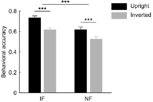
decreased accuracy for inverted faces) in the case of both intact and noisy face conditions, which did not differ significantly in magnitude (Fig. 1). These behavioral findings suggest that the neural mechanisms involved in the processing of noisy faces might be similar to those of faces without noise, presumably mediated by the FFA.
Figure 1. Behavioral results. Identity discrimination performance was significantly higher for intact as compared to noisy faces, however the face inversion equally impaired accuracy in both cases. Provided data are mean correct response ratio ± SEM across participants (N = 26). Black bars represent data for upright faces; gray bars represent data for inverted faces.
IF, intact faces; NF, noisy faces (***p < 0.001).
1.2. Based on whole-brain analysis, I found that the presence of noise led to reduced and increased fMRI responses in the mid-fusiform gyrus and the lateral occipital cortex, respectively. Furthermore, the noise-induced modulation of the fMRI responses in the right face-selective fusiform face area (FFA) was closely associated with individual differences in the identity discrimination performance of noisy faces: smaller decrease of the fMRI responses was accompanied by better identity discrimination.
It has been suggested [31, 32] that in the case of phase- randomized face images the increased processing demand due to the distorted spatial localization of the facial features might lead to the engagement of a re-entrant processing loop involving the FFA and a region of the lateral occipital cortex (LOC), which represents shape information within a spatial coordinate system [33, 51] and shows increased fMRI responses to noisy face images [31]. However, an important question that remains to be explored is whether it is the FFA or the LOC on whose neural representations the perception of deteriorated and noisy face images is based. Even though combined behavioral and neuroimaging results provided strong evidence for a close link between face perception and the neural processes in the FFA in the case of intact face images [28, 30, 48, 49], it has not been investigated whether this holds true also for faces that are noisy and poorly visible.
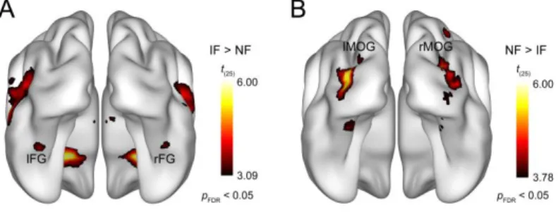
We have found that adding phase noise to face images leads to reduced and increased fMRI responses to faces in bilateral mid- fusiform gyrus (Fig. 2A) and bilateral LOC (Fig. 2B), respectively, which is in agreement with previous results [31, 52].
Figure 2. Results of the whole-brain random-effects analysis. Bilateral areas of the fusiform gyrus showed significantly lower activation for noisy relative to intact faces (A), while larger responses to noisy than intact faces were found bilaterally in the middle occipital gyrus (B). Statistical maps are displayed with pFDR < 0.05 on the smoothed ICBM152 brain [53–55]. IF, intact faces; NF, noisy faces; lFG, left fusiform gyrus; rFG, right fusiform gyrus; lMOG, left middle occipital gyrus; rMOG, right middle occipital gyrus.
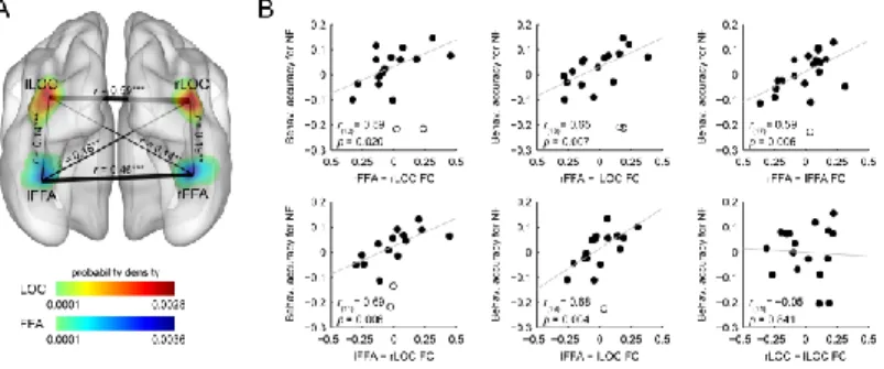
Importantly, our results provide the first evidence that only in the right face-selective FFA did noise-induced modulation of the fMRI responses show a close association with the individual differences in face identity discrimination performance of noisy faces (Fig. 3B): smaller decrease of the fMRI responses was associated with better identity discrimination. This relationship was not driven by the overall face perception ability of the participants, because performance for intact faces was regressed out from that for noisy faces. Our results imply that the perception of noisy face images is based on the neural representations extracted from the right FFA.
Figure 3. Results of the ROI-based correlation analysis. A, Probability density map illustrating the spatial distribution of the highest noise-effect voxels across participants in bilateral FFA and LOC. Color scales reflect probability density estimates (cool colors, FFA; warm colors, LOC). B, Relationship between the noise-induced modulation of the fMRI responses and the behavioral accuracy in discriminating noisy faces: smaller decrease
of the fMRI responses in the right FFA indicated better identity discrimination. Due to the semipartial correlation procedure, correlation scatter plots depict residual values on the y-axis. The y-axis values denote behavioral accuracy for noisy faces indexed by the residual correct response ratio. The x-axis values denote noise effect on the fMRI responses indexed by the beta difference in the IF versus NF contrast. Circles represent individual participants and bivariate outliers are marked with open circles.
Diagonal line indicates linear least-squares fit. IF, intact faces; NF, noisy faces.
1.3. I found that the strength of the intrinsic functional connectivity within the visual cortical network composed of bilateral FFA and bilateral object-selective lateral occipital cortex (LOC) predicted the participants’ ability to discriminate the identity of noisy face images.
Based on the suggested role of the re-entrant neural mechanisms in the processing of noisy faces, we predicted that the individual ability to handle stimulus noise might depend on the strength of functional interactions between FFA and LOC. To test this prediction, we estimated the strength of intrinsic functional connectivity between bilateral FFA and LOC (Fig. 4A) using resting- state fMRI [43] (for review see [56]) and computed correlations between these measures and the face identity discrimination performance for noisy faces. In the correlation analysis the intact face performance was used as a covariate to control for the confounding effect of the overall face perception ability of the participants. Our correlation analysis revealed that the functional connectivity strength between bilateral FFA and bilateral LOC correlated positively with the behavioral accuracy for noisy faces (Fig. 4B): the stronger the functional connectivity between these regions during rest, the better the face identity discrimination performance in the noisy condition. These results suggest that face identity perception in the case of noisy faces is based on functional interactions between bilateral FFA and LOC.
Figure 4. Results of the intrinsic functional connectivity analysis. A, Connections between the pairs of ROIs displayed as edges and overlaid on the probability density map from Figure 3. The thickness of an edge represents the strength of the connection (correlation coefficients (r) averaged across subjects); significant correlations were found between all ROI pairs investigated. B, Scatter plots indicating the relationship between the intrinsic functional connectivity and the behavioral accuracy for noisy faces. The strength of the functional connectivity between bilateral FFA and LOC, as well as between the right and left FFA, correlated positively with the identity-discrimination performance in the case of noisy faces. Due to the partial correlation procedure, correlation scatter plots depict residual values on both axes. The y-axis values denote the behavioral accuracy for noisy faces indexed by the residual correct response ratio. The x-axis values denote the connection strength between a ROI pair indexed by the residual correlation coefficient. Circles represent individual participants and bivariate outliers are marked with open circles. Diagonal line indicates linear least- squares fit. NF, noisy faces; FC, functional connectivity (**p < 0.01, ***p <
0.001).
2. Thesis: I have shown that there is a face-selective repetition- induced fMRIa within the core face-processing network composed of the FFA and OFA which reflects functionally relevant adaptation processes involved in face identity perception.
Published in [2], [4].
It has been shown that sensory information processing is highly affected by the short-term prior perceptual experience. When a sensory stimulus is repeated, the evoked neural signal is invariably smaller than the one observed for its first presentation, an effect termed as repetition suppression (RS) [57]. Similarly, in functional magnetic resonance imaging (fMRI) experiments stimulus repetitions elicit the reduction of the blood oxygenation level- dependent (BOLD) signal when compared to non-repeating stimuli (for review see [58]), a phenomenon called fMRI adaptation (fMRIa). It has been shown that repetition of identical face stimuli leads to fMRIa in the core face-selective occipitotemporal visual cortical network, involving the bilateral fusiform face area (FFA) and the occipital face area (OFA) [39, 59, 60]. Extensive previous experimental and modeling research has made significant progress in revealing the neural processes involved in RS (for reviews see [61, 62]). However, surprisingly little is known about its behavioral relevance. Therefore, here we aimed at investigating the relationship between fMRIa and face perception ability by measuring in the same human participants both the repetition-induced reduction of fMRI responses in these regions and face identity discrimination performance outside the scanner for upright and inverted face stimuli.
2.1. I found a significant fMRIa, i.e. reduced BOLD signal for repeated as compared to alternating faces in the fusiform face area (FFA) and a moderate fMRIa in the occipital face area (OFA). Furthermore, the magnitude of the face- selective fMRIa measured in these face-processing areas was closely associated.
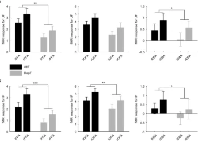
In agreement with previous results [39, 59, 60], the repetition of identical face stimuli led to significant fMRIa, i.e. reduced BOLD signal in the FFA, and a moderate fMRIa in the OFA, and we also found fMRIa in the extrastriate body area (EBA) for both upright (Fig. 5A) and inverted (Fig. 5B) faces.
Figure 5. Average activation (± SEM) profiles for the left and right FFA (left), OFA (middle), and EBA (right) when faces were presented upright (A) and inverted (B). We found fMRIa, i.e. reduced fMRI responses for repeated (RepT) as compared to alternating faces (AltT) for all ROIs investigated in the case of both upright and inverted conditions. Black bars represent AltT; gray bars represent RepT. UF, upright faces; IF, inverted faces (*p < 0.05, **p < 0.01, ***p < 0.001).
However, it is not known whether fMRIa reflects common or different underlying mechanisms in the tested visual cortical areas.
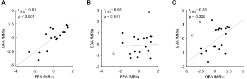
To test this, we calculated pairwise correlations of fMRIa magnitudes among the three regions. In the correlation analysis, the fMRIa for the inverted faces was used as a covariate to control for the individual differences in low-level visual feature adaptation processes. We found a strong correlation of the face-selective fMRIa between OFA and FFA (Fig. 6A) and also between OFA and EBA (Fig. 6C), but not between FFA and EBA (Fig. 6B). These findings imply that fMRIa might involve different components: one is mediated by neural mechanisms that are specific to the core face-
processing network and another which affects the fMRI responses in the OFA and EBA, but not in FFA.
Figure 6. Correlation between fMRIa observed in the FFA and OFA (A), in the FFA and EBA (B) and in the OFA and EBA (C). Significant correlation was found between the magnitude of fMRIa measured in the FFA and OFA, as well as in the OFA and EBA, but not in the FFA and EBA. Due to the regression-based approach, correlation scatter plots depict residual values on both axes. y- and x-axis values denote the fMRIa indexed by the residual beta difference in the AltT vs. RepT contrast. Circles represent individual participants and bivariate outliers are marked with open circles. Diagonal line indicates linear least squares fit.
2.2. I have shown that the face-selective fMRIa in the two regions of the core face-processing network, namely in the fusiform face area (FFA) and occipital face area (OFA) predicts individual differences in face-selective perceptual ability.
The visual system as an inference machine actively generates and optimizes predictions about the incoming sensory input to make the information processing more efficient as suggested by the predictive coding model of perception [34–37]. From this perspective, RS is a manifestation of minimizing prediction error through adaptive changes in predictions. At the neuronal level, RS is generally believed to reflect short-term plastic processes of the neurons, as they adapt to the temporal context of the current environment, presumably as a consequence of dynamic synaptic
change within recurrent neural networks [58, 63–65]). Thereby, RS reflects the flexibility of the neural system and its ability to adjust to continuously changing requirements, optimizing the performance of the individual. We reasoned that if RS (and the consequent fMRIa) indeed reflects the better predictive ability of the neural system then this should manifest on the perceptual level as well: a good generative model of faces can produce better predictions of subsequent stimulation, which leads to better performance and reduced concomitant prediction error unit activity, i.e. fMRIa. To test this prediction, we correlated the individual fMRIa magnitudes measured in the core face-processing areas, namely the FFA and OFA, as well as in the body-selective EBA with the participants’
face identity discrimination performance. In the correlation analysis the behavioral and fMRI results for the inverted faces were used as covariates to control for the individual differences in overall object perception ability and low-level visual feature adaptation processes, respectively.
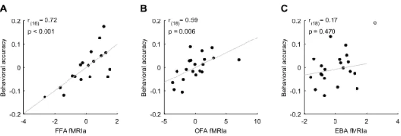
Figure 7. Correlation between behavioral accuracy and fMRIa for the FFA (A), OFA (B), and EBA (C). Significant correlation was found in the case of the FFA and OFA, but not for the EBA. Due to the regression-based approach correlation scatter plots depict residual values on both axes. y-axis values denote behavioral accuracy in the face identity discrimination task indexed by the residual correct response ratio. x-axis values denote the fMRIa indexed by the residual beta difference in the AltT vs. RepT contrast.
Circles represent individual participants and bivariate outliers are marked with open circles. Diagonal line indicates linear least squares fit.
Our correlation analysis revealed that the magnitude of the fMRIa measured in the FFA (Fig. 7A) and OFA (Fig. 7B), but not in the EBA (Fig. 7C) correlated positively with the behavioral accuracy: the higher the magnitude of the fMRIa for repeated faces, the better the face identity discrimination performance. These results suggest that RS in the core face-processing areas predicts face- selective perceptual ability and thus reflects functionally relevant adaptation processes involved in face identity perception.
4. Conclusions and possible applications
Our results provide important new insights into the adaptive information coding processes within the extensive visual cortical face-processing network, especially regarding the recurrent neural mechanisms that enable efficient and robust human face perception even under suboptimal viewing conditions.
Understanding the strategies that the visual system employs in natural unconstrained settings could be the first step translating them into machine-based face recognition algorithms which have a huge impact in computer vision (see [66] for a review).
Furthermore, advancing the knowledge of neural mechanisms underlying face perception at both regional and network level is a key issue to develop training programs including fMRI-based neurofeedback techniques (see [67, 68] for reviews) which could help to improve the efficacy of visual cortical processing of facial information, especially in prosopagnosia.
5. Acknowledgements
I would like to express my gratitude for those who accompanied me during my exciting journey. First of all, I would like to say thanks to my supervisor, Prof. Zoltán Vidnyánszky, for his support and careful guidance throughout my study and research. Zoli, your attitude to scientific thinking and writing makes you a role model for me. I am especially grateful for your trust in me, your attention, patience and impatience as well. I gladly remember our personal discourses about Hantai (“Hantás”). Thank you.
Hereby I also acknowledge the collaborative work and help of Prof. Gyula Kovács. Gyula, I am particularly thankful to you for the time that I could spend in your lab of Jena.
I am also very grateful to the Doctoral School, especially to Prof. Tamás Roska and Prof. Péter Szolgay for providing the opportunity to spend my PhD years in a multi-disciplinary environment. I am indebted to Prof. József Hámori for his attention and for sharing his educational experiences and abundant knowledge of science and art with me.
I am tremendously thankful to my close tutor colleagues, Éva Bankó, Viktor Gál, István Kóbor, and Béla Weiss, for their help, inspiring discussions, and useful advice. Éva, I cannot express enough my gratitude for your invaluable scientific and friendly support that I received from you from my first time in the lab. I am very glad that I found a tutor and friend in you. Viktor, your grandiose knowledge in MRI methods impressed me and I learned from you that humor is essential in research. Thank you for letting me be a part of the “Viktor milieu”, and thank you for your friendship.
I am indebted to Anita Deák and Beatrix Lábadi from the Institute of Psychology of the University of Pécs for encouraging me to take the first steps. Anita, I feel enormously grateful to you that I
could be part of the fMRI Research Group in Pécs. Thank you for tutoring me and still being my genuine friend up to this day.
Very special thanks to all my fellow PhD students and colleauges in our lab, especially to Balázs Knakker, Vanda Nemes, Pál Vakli, Petra Kovács, Gergely Drótos, Regina Meszlényi, Ádám Kettinger, Máté Kiss, Emese Maróti, Krisztián Buza, and Annamária Manga for all their help, productive discussions and for the everyday lunch. Balázs, we took our first steps in this journey together, sometimes we felt lost, but we could lean on each other and continue our ride. I am very grateful for your continuous scientific and emotional support, and also for our deep conversations. Thank you for being by my side in trouble too. I am privileged to have such a companion and friend.
I would like to thank my two opponents, István Hernádi and Lajos Kozák, for evaluating my dissertation and their relevant questions and valuable suggestions contributing to the finalization of this work.
I would also like to thank Gábor Rudas, the director of the MR Research Center of the Semmelweis University, for the opportunity to conduct the measurements and the wonderful atmosphere.
I owe a lot to Katinka Vida Tivadarné for her help in all the administrative issues. Katinka néni, I am very grateful for your genuine humanity and limitless patience. I am very pleased to know you. I am also very thankful to Viktória Sifter from the Library.
I would like to say thanks to all of my friends for accepting me and being so patient and supportive and giving me new energy to move on during my busy days. Thank you all for your friendship.
I say thanks to Prof. Zsuzsa Hetényi for encouraging and guiding me along the paths of Nabokov. I am especially thankful to Ágnes Juhász for guiding me along my own paths.
I am also grateful to Dvořak, Brahms, and the experimental minimal techno artists, especially to S Olbricht for inspiring me with their music. I owe special thanks to Zoltán Nagy from Zürich for involving me in the AntiNode shows.
Last but not least, I am most grateful to my Mother and Father who have helped me tremendously in every possible way so that I could focus on research. Anya, thank you for being the source of continuous, limitless, unconditional support for me. Thank you for believing in me all the time, especially when I could not.
6. References
The author's journal publications
[1] P. Hermann, É. M. Bankó, V. Gál, and Z. Vidnyánszky,
“Neural basis of identity information extraction from noisy face images,” J. Neurosci., vol. 35, no. 18, pp. 7165–7173, May 2015.
[2] P. Hermann, M. Grotheer, G. Kovács, and Z. Vidnyánszky,
“The relationship between repetition suppression and face perception,” Brain Imaging Behav., pp. 1–11, Jul. 2016.
The author's conference publications
[3] P. Hermann, É. M. Bankó, V. Gál, and Z. Vidnyánszky, “The human face network: insights from intrinsic functional connectivity analysis,” presented at the 4th Neuroimaging Workshop, Debrecen, Hungary, 2014, p. 22.
[4] P. Hermann, M. Grotheer, G. Kovács, and Z. Vidnyánszky,
“Repetition suppression of fMRI responses in fusiform and occipital face areas predicts individual differences in face perception ability,” presented at the 5th Neuroimaging Workshop, Szeged, Hungary, 2015, p. 7.
The author's other journal and conference publications [5] C. Amado, P. Hermann, P. Kovács, M. Grotheer, Z.
Vidnyánszky, and G. Kovács, “The contribution of surprise to the prediction based modulation of fMRI responses,”
Neuropsychologia, vol. 84, pp. 105–112, Apr. 2016.
[6] M. Grotheer, P. Hermann, Z. Vidnyánszky, and G. Kovács,
“Repetition probability effects for inverted faces,”
NeuroImage, vol. 102, Part 2, pp. 416–423, Nov. 2014.
[7] J. Körtvélyes, E. M. Bankó, A. Andics, G. Rudas, J. Németh, P. Hermann, and Z. Vidnyánszky, “Visual cortical responses to the input from the amblyopic eye are suppressed during binocular viewing,” Acta Biol. Hung., vol. 63 Suppl 1, pp. 65–
79, Mar. 2012.
[8] P. Hermann, Á. Kettinger, R. Meszlényi, and Z.
Vidnyánszky, “Comparison of the suitability of 32-channel and 64-channel head coils for application in fMRI research,”
presented at the IBRO Workshop 2016, Budapest, Hungary, 2016.
[9] A. Catarina, P. Hermann, P. Kovács, M. Grotheer, Z.
Vidnyánszky, and G. Kovács, “The role of surprise enhancement in predictions,” presented at the IBRO Workshop 2016, Budapest, Hungary, 2016.
[10] P. Kovács, P. Hermann, B. Knakker, G. Kovács, and Z.
Vidnyánszky, “Uncovering the configural coding of facial information using the face inversion effect,” presented at the IBRO Workshop 2016, Budapest, Hungary, 2016.
[11] P. Hermann, M. Grotheer, G. Kovács, and Z. Vidnyánszky,
“Resting-state functional connectivity predicts the repetition suppression of fMRI responses in the fusiform gyrus,”
presented at the 15th Biannual Conference of The Hungarian Neuroscience Society (MITT), Budapest, Hungary, 2015.
[12] M. Grotheer, Z. Vidnyánszky, P. Hermann, and G. Kovács,
“Repetition probability effects for inverted faces in the fusiform face area,” presented at the 9th PPRU Workshop:
Person Perception – From cortical areas to social functions, Jena, Germany, 2014, p. 14.
[13] P. Hermann, V. Gál, É. M. Bankó, and Z. Vidnyánszky,
“Resting-state functional connectivity predicts the face selectivity of fMRI responses in the fusiform gyrus,” presented at the XIV. Conference of The Hungarian Neuroscience Society (MITT), Budapest, Hungary, 2013, pp. 217–218.
[14] P. Hermann, É. M. Bankó, and Z. Vidnyánszky,
“Representation of facial identity information in the medial and anterior temporal lobe,” Clin. Neurosci., vol. 65, no. S1, p.
27, 2012.
[15] P. Hermann, “Electrophysiological correlates of object- specific processing deficits in amblyopia,” Pázmány Péter Cathol. Univ. PhD Proc., pp. 153–158, 2011.
CUMMULATIVE IMPACT FACTOR: 19.441;
NUMBER OF INDEPENDENT CITATIONS: 11.
Selected publications cited in the dissertation
[16] B. Duchaine and G. Yovel, “A revised neural framework for face processing,” Annu. Rev. Vis. Sci., vol. 1, no. 1, pp. 393–
416, Nov. 2015.
[17] G. Yovel, “Neural and cognitive face-selective markers: An integrative review,” Neuropsychologia, vol. 83, pp. 5–13, Mar.
2016.
[18] J. V. Haxby, E. A. Hoffman, and M. I. Gobbini, “The distributed human neural system for face perception,” Trends Cogn. Sci., vol. 4, no. 6, pp. 223–233, Jun. 2000.
[19] A. Puce, T. Allison, J. C. Gore, and G. McCarthy, “Face- sensitive regions in human extrastriate cortex studied by functional MRI,” J. Neurophysiol., vol. 74, no. 3, pp. 1192–
1199, Sep. 1995.
[20] N. Kanwisher, J. McDermott, and M. M. Chun, “The fusiform face area: a module in human extrastriate cortex specialized for face perception,” J. Neurosci., vol. 17, no. 11, pp. 4302–
4311, Jun. 1997.
[21] A. Ishai, C. F. Schmidt, and P. Boesiger, “Face perception is mediated by a distributed cortical network,” Brain Res. Bull., vol. 67, no. 1–2, pp. 87–93, Sep. 2005.
[22] D. Y. Tsao, S. Moeller, and W. A. Freiwald, “Comparing face patch systems in macaques and humans,” Proc. Natl. Acad.
Sci., vol. 105, no. 49, pp. 19514–19519, Dec. 2008.
[23] R. Rajimehr, J. C. Young, and R. B. H. Tootell, “An anterior temporal face patch in human cortex, predicted by macaque maps,” Proc. Natl. Acad. Sci., vol. 106, no. 6, pp. 1995–2000, Feb. 2009.
[24] C. J. Fox, G. Iaria, and J. J. S. Barton, “Defining the face processing network: optimization of the functional localizer in fMRI,” Hum. Brain Mapp., vol. 30, no. 5, pp. 1637–1651, May 2009.
[25] K. S. Weiner and K. Grill-Spector, “Sparsely-distributed organization of face and limb activations in human ventral temporal cortex,” NeuroImage, vol. 52, no. 4, pp. 1559–1573, Oct. 2010.
[26] B. Rossion, B. Hanseeuw, and L. Dricot, “Defining face perception areas in the human brain: A large-scale factorial fMRI face localizer analysis,” Brain Cogn., vol. 79, no. 2, pp.
138–157, Jul. 2012.
[27] R. W. McGugin and I. Gauthier, “The reliability of individual differences in face-selective responses in the fusiform gyrus and their relation to face recognition ability,” Brain Imaging Behav., pp. 1–12, Nov. 2015.
[28] K. Grill-Spector, N. Knouf, and N. Kanwisher, “The fusiform face area subserves face perception, not generic within- category identification,” Nat. Neurosci., vol. 7, no. 5, pp. 555–
562, May 2004.
[29] N. Furl, L. Garrido, R. J. Dolan, J. Driver, and B. Duchaine,
“Fusiform gyrus face selectivity relates to individual differences in facial recognition ability,” J. Cogn. Neurosci., vol. 23, no. 7, pp. 1723–1740, Jul. 2010.
[30] L. Huang, Y. Song, J. Li, Z. Zhen, Z. Yang, and J. Liu,
“Individual differences in cortical face selectivity predict behavioral performance in face recognition,” Front. Hum.
Neurosci., vol. 8, p. 483, Jul. 2014.
[31] É. M. Bankó, V. Gál, J. Körtvélyes, G. Kovács, and Z.
Vidnyánszky, “Dissociating the effect of noise on sensory processing and overall decision difficulty,” J. Neurosci., vol.
31, no. 7, pp. 2663–2674, Feb. 2011.
[32] É. M. Bankó, J. Körtvélyes, B. Weiss, and Z. Vidnyánszky,
“How the visual cortex handles stimulus noise: insights from amblyopia,” PLoS ONE, vol. 8, no. 6, p. e66583, Jun. 2013.
[33] J. Larsson and D. J. Heeger, “Two retinotopic visual areas in human lateral occipital cortex,” J. Neurosci., vol. 26, no. 51, pp. 13128–13142, Dec. 2006.
[34] D. Mumford, “On the computational architecture of the neocortex,” Biol. Cybern., vol. 66, no. 3, pp. 241–251, Jan.
1992.
[35] R. P. N. Rao and D. H. Ballard, “Predictive coding in the visual cortex: a functional interpretation of some extra- classical receptive-field effects,” Nat. Neurosci., vol. 2, no. 1, pp. 79–87, Jan. 1999.
[36] K. Friston, “A theory of cortical responses,” Philos. Trans. R.
Soc. Lond. B Biol. Sci., vol. 360, no. 1456, pp. 815–836, Apr.
2005.
[37] K. Friston, “The free-energy principle: a unified brain theory?,” Nat. Rev. Neurosci., vol. 11, no. 2, pp. 127–138, Feb. 2010.
[38] A. Puce, T. Allison, M. Asgari, J. C. Gore, and G. McCarthy,
“Differential sensitivity of human visual cortex to faces, letterstrings, and textures: a functional magnetic resonance imaging study,” J. Neurosci., vol. 16, no. 16, pp. 5205–5215, Aug. 1996.
[39] M. P. Ewbank, R. N. Henson, J. B. Rowe, R. S. Stoyanova, and A. J. Calder, “Different neural mechanisms within occipitotemporal cortex underlie repetition suppression across same and different-size faces,” Cereb. Cortex, vol. 23, no. 5, pp. 1073–1084, May 2013.
[40] D. H. Brainard, “The Psychophysics Toolbox,” Spat. Vis., vol.
10, no. 4, pp. 433–436, Jan. 1997.
[41] D. G. Pelli, “The VideoToolbox software for visual psychophysics: transforming numbers into movies,” Spat. Vis., vol. 10, no. 4, pp. 437–442, Jan. 1997.
[42] C. R. Pernet, R. Wilcox, and G. A. Rousselet, “Robust correlation analyses: false positive and power validation using a new open source matlab toolbox,” Front. Psychol., vol. 3, p.
606, Jan. 2013.
[43] B. Biswal, F. Zerrin Yetkin, V. M. Haughton, and J. S. Hyde,
“Functional connectivity in the motor cortex of resting human brain using echo-planar mri,” Magn. Reson. Med., vol. 34, no.
4, pp. 537–541, Oct. 1995.
[44] M. D. Fox, A. Z. Snyder, J. L. Vincent, M. Corbetta, D. C.
Van Essen, and M. E. Raichle, “The human brain is intrinsically organized into dynamic, anticorrelated functional networks,” Proc. Natl. Acad. Sci., vol. 102, no. 27, pp. 9673–
9678, Jul. 2005.
[45] A. Weissenbacher, C. Kasess, F. Gerstl, R. Lanzenberger, E.
Moser, and C. Windischberger, “Correlations and anticorrelations in resting-state functional connectivity MRI:
A quantitative comparison of preprocessing strategies,”
NeuroImage, vol. 47, no. 4, pp. 1408–1416, Oct. 2009.
[46] D. Zhang and M. E. Raichle, “Disease and the brain’s dark energy,” Nat. Rev. Neurol., vol. 6, no. 1, pp. 15–28, Jan. 2010.
[47] M. Xia, J. Wang, and Y. He, “BrainNet Viewer: a network visualization tool for human brain connectomics,” PLoS ONE, vol. 8, no. 7, p. e68910, Jul. 2013.
[48] N. Furl, L. Garrido, R. J. Dolan, J. Driver, and B. Duchaine,
“Fusiform gyrus face selectivity relates to individual differences in facial recognition ability,” J. Cogn. Neurosci., vol. 23, no. 7, pp. 1723–1740, Jul. 2010.
[49] G. Yovel and N. Kanwisher, “The neural basis of the behavioral face-inversion effect,” Curr. Biol., vol. 15, no. 24, pp. 2256–2262, Dec. 2005.
[50] R. K. Yin, “Looking at upside-down faces.,” J. Exp. Psychol., vol. 81, no. 1, pp. 141–145, Jul. 1969.
[51] E. H. Silson, D. J. McKeefry, J. Rodgers, A. D. Gouws, M.
Hymers, and A. B. Morland, “Specialized and independent
processing of orientation and shape in visual field maps LO1 and LO2,” Nat. Neurosci., vol. 16, no. 3, pp. 267–269, Mar.
2013.
[52] H. R. Heekeren, S. Marrett, P. A. Bandettini, and L. G.
Ungerleider, “A general mechanism for perceptual decision- making in the human brain,” Nature, vol. 431, no. 7010, pp.
859–862, Oct. 2004.
[53] J. Mazziotta, A. Toga, A. Evans, P. Fox, J. Lancaster, K.
Zilles, R. Woods, T. Paus, G. Simpson, B. Pike, C. Holmes, L.
Collins, P. Thompson, D. MacDonald, M. Iacoboni, T.
Schormann, K. Amunts, N. Palomero-Gallagher, S. Geyer, L.
Parsons, K. Narr, N. Kabani, G. Le Goualher, D. Boomsma, T.
Cannon, R. Kawashima, and B. Mazoyer, “A probabilistic atlas and reference system for the human brain: International Consortium for Brain Mapping (ICBM).,” Philos. Trans. R.
Soc. Lond. Ser. B, vol. 356, no. 1412, pp. 1293–1322, Aug.
2001.
[54] J. C. Mazziotta, A. W. Toga, A. Evans, P. Fox, and J.
Lancaster, “A probabilistic atlas of the human brain: theory and rationale for its development. The International Consortium for Brain Mapping (ICBM),” NeuroImage, vol. 2, no. 2, pp. 89–101, Jun. 1995.
[55] J. Mazziotta, A. Toga, A. Evans, P. Fox, J. Lancaster, K.
Zilles, R. Woods, T. Paus, G. Simpson, B. Pike, C. Holmes, L.
Collins, P. Thompson, D. MacDonald, M. Iacoboni, T.
Schormann, K. Amunts, N. Palomero-Gallagher, S. Geyer, L.
Parsons, K. Narr, N. Kabani, G. Le Goualher, J. Feidler, K.
Smith, D. Boomsma, H. H. Pol, T. Cannon, R. Kawashima, and B. Mazoyer, “A four-dimensional probabilistic atlas of the human brain,” J. Am. Med. Inform. Assoc. JAMIA, vol. 8, no.
5, pp. 401–430, Oct. 2001.
[56] M. D. Fox and M. E. Raichle, “Spontaneous fluctuations in brain activity observed with functional magnetic resonance imaging,” Nat. Rev. Neurosci., vol. 8, no. 9, pp. 700–711, Sep.
2007.
[57] R. Desimone, “Neural mechanisms for visual memory and their role in attention,” Proc. Natl. Acad. Sci., vol. 93, no. 24, pp. 13494–13499, Nov. 1996.
[58] R. N. A. Henson and M. D. Rugg, “Neural response suppression, haemodynamic repetition effects, and behavioural priming,” Neuropsychologia, vol. 41, no. 3, pp. 263–270, 2003.
[59] C. Summerfield, E. H. Trittschuh, J. M. Monti, M.-M.
Mesulam, and T. Egner, “Neural repetition suppression reflects fulfilled perceptual expectations,” Nat. Neurosci., vol.
11, no. 9, pp. 1004–1006, Sep. 2008.
[60] G. Kovács, L. Iffland, Z. Vidnyánszky, and M. W. Greenlee,
“Stimulus repetition probability effects on repetition suppression are position invariant for faces,” NeuroImage, vol.
60, no. 4, pp. 2128–2135, May 2012.
[61] K. Grill-Spector, R. Henson, and A. Martin, “Repetition and the brain: neural models of stimulus-specific effects,” Trends Cogn. Sci., vol. 10, no. 1, pp. 14–23, Jan. 2006.
[62] N. Bunzeck and C. Thiel, “Neurochemical modulation of repetition suppression and novelty signals in the human brain,”
Cortex, vol. 80, pp. 161–173, Jul. 2016.
[63] R. N. A. Henson, “Neuroimaging studies of priming,” Prog.
Neurobiol., vol. 70, no. 1, pp. 53–81, May 2003.
[64] R. N. Henson, “Repetition suppression to faces in the fusiform face area: A personal and dynamic journey,” Cortex, vol. 80, pp. 174–184, Jul. 2016.
[65] R. Auksztulewicz and K. Friston, “Repetition suppression and its contextual determinants in predictive coding,” Cortex, vol.
80, pp. 125–140, Jul. 2016.
[66] P. Sinha, B. Balas, Y. Ostrovsky, and R. Russell, “Face recognition by humans: Nineteen results all computer vision researchers should know about,” Proc. IEEE, vol. 94, no. 11, pp. 1948–1962, Nov. 2006.
[67] N. Weiskopf, “Real-time fMRI and its application to neurofeedback,” NeuroImage, vol. 62, no. 2, pp. 682–692, Aug. 2012.
[68] R. T. Thibault, M. Lifshitz, and A. Raz, “The self-regulating brain and neurofeedback: Experimental science and clinical promise,” Cortex, vol. 74, pp. 247–261, Jan. 2016.