Review Article
An epidemic of new-born photography poses: the potential dangers of passive end range
positioning during induced sleep in 0–14-day-old neonates: a scoping review
Edit Nagy, PT, PhD1), Regina Finta PT1)
1) Department of Physiotherapy, University of Szeged, Faculty of Health and Social Sciences:
Temesvári krt 31 Szeged Csongrad 6726, Hungary
Abstract. [Purpose] In the photography of new-borns, there is an epidemic trend in the posing of 0–14-day-old neonates that induces ethical and competence issues. The aim of this study is to map the key concepts underpin- ning the contraindications of this type of passive positioning of the new-borns. [Methods] During the search for literature, the following keywords were used in the PubMed database: neonates; new-born; neck position; hy- perextension; rotation; atlanto-occipital joint; sudden infant death; prone sleeping position; white noise; and pain perception. [Results] The white noise applied has been described as a pain perception modulator and an alternative pain reducing method in new-born care. There is evidence warning of the potential danger of passively produced cervical spine positions, considering the primary unstable atlanto-occipital joint during early infancy, the possible compression on vertebral arteries, and the intradural diameter decreasing the effect of extension. These factors may have an impact on healthy motor and cognitive development. [Conclusion] This perspective suggests that a wider debate should be called for concerning the role of medical professions to control this very dangerous practice, and that future cohort studies are necessary to monitor and follow up on the potential negative effects of this current trend.
Key words: Neonates, Passive position, Induced sleep
(This article was submitted Dec. 7, 2019, and was accepted Aug. 9, 2020)
INTRODUCTION
Passive movement is the link between the profession of physiotherapy and choreographed new-born photography. New- born photography is very popular and the heart-melting photographs create visceral emotional reactions, but during the last few years a dangerous and extreme trend has appeared in the photography sessions. The role of the photographer has changed dramatically, from that of a passive observer to that of an active choreographer, who plays white noise to modulate the behavioural responses of the new-borns being photographed. Photographers actively intervene by positioning the new-borns in different poses while they are in an induced sleep (by white noise). This intervention has a very limited time window, as it is possible to perform it until just two weeks after birth. This kind of active intervention evokes both ethical and competence issues, and, unfortunately, it appears rather uncontrolled. Many videos and tutorials on YouTube illustrate this problem, and they can be found with a search of such keywords as ‘new-born froggy pose’. For visual illustration of the existing trends, pictures of the positions were drawn to avoid any copyright issues. However, the ‘froggy pose’ (Fig. 1) is just the tip of the iceberg; a huge number of unnatural positions can be seen across social media, using passively produced and maintained, often end range cervical spine positions (Fig. 2). On social networks we can see frequently advertisements seeking 7–14-day-
J. Phys. Ther. Sci. 32: 788–794, 2020
*Corresponding author. Edit Nagy (nedit@etszk.u-szeged.hu)
©2020 The Society of Physical Therapy Science. Published by IPEC Inc.
This is an open-access article distributed under the terms of the Creative Commons Attribution Non-Commercial No Deriva- tives (by-nc-nd) License. (CC-BY-NC-ND 4.0: https://creativecommons.org/licenses/by-nc-nd/4.0/)
The Journal of Physical Therapy Science
The Journal of Physical Therapy Science
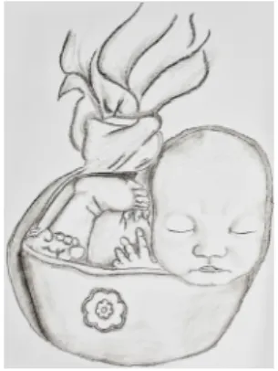
old new-born models for photography workshops (Fig. 3), offering copies of the images free of charge. From the perspective of physiotherapy, this practice appears very dangerous, especially the passive movement and the positioning of the upper cervical spine into unnatural, frequently end range positions for long periods of time, while under induced sleep in atonic states, merely for photographic purpose. In a video tutorial about the ‘froggy’ pose, the time spent in this hyperextended neck position was 30 minutes. In another public video, periodic breathing patterns could also be observed when the neonate was wrapped in tightly to create a ‘potato sack pose’ (Fig. 4).
The aim of this study is to map the key concepts underpinning the contraindications of passive, end range positioning of new-borns, to discover how it is possible to create such unnatural poses in early infancy (0–14 days), and what the potential hazards of this practice are in terms of healthy development of the babies, as well as the ethical issues concerning this practice. These questions are particularly important since this research area is complex and has not been reviewed comprehensively before from this perspective. This report suggests that a wider debate should be called for about the role of medical professions to control and regulate this very dangerous practice.
METHODS
Research question: What are the mechanisms underlying the positioning of the upper cervical joints into end range during induced deep sleep in early infancy (0–14 days), and what are the potential dangers of these extreme poses?
During the search for literature, the following keywords were used in the PubMed electronic database: neonates; new- born; neck position; hyperextension; rotation; atlanto-occipital joint; sudden infant death; prone sleeping position; white noise and pain perception. Because our aim was to identify the relevant literature regardless of study design, we included both post-mortem and in vivo studies dealing with the head positions and position-dependent cerebral blood flow in this early stage of life. The very first weeks of life is not really studied in the literature, therefore we included any studies dealing with infancies (generally age is between 0–12 months) that was relevant to answer our research question and had reflected one or two of our key words.
Fig. 1. The froggy pose.
Fig. 2. The womb position.
Fig. 3. Workshop positioning.
The results were summarized according to setting, aim, the sample size and age of the infants, and the main findings of the studies that are relevant to this scope of this review, that are one of the abovementioned cervical positions, brainstem blood flow, white noise, sleeping position. At the end of searching process 11 studies were selected as being eligible for the analysis, three of them assessing the white noise pain modulatory effects, but only one is focusing on the first week of life, and 8 studies dealing with the different head positions and their effects on brainstem blood flow. This work is complied with the current laws of our country, in line with the Helsinki declaration.
RESULTS
The nervous system of healthy, term neonates is not mature. The flexor muscle tone is higher in cases of normal postural tone in early infancy. The normal postural tone, the asymmetrical and symmetrical tonic neck reflexes, the tonic labyrinth reflexes and the righting reactions consist of the postural mechanisms elicited by neck movements and body positions in awake neonates with normal muscle tone. The receptors are proprioceptors located in the cervical muscles, in the vestibular system and the cutaneous somatosensory receptors. In case of the neck on body righting reaction, for example, if the head of the awake new-born is rotated passively, the whole body will follow in a block resulting in a log rolling1). Therefore, in an awake state it is impossible to passively position the cervical spine into end range, due to the appearance of reflexive movement patterns in early infancy in cases of term neonates.
Falling asleep generates physiological changes in muscle tone, which result in physiological hypotonia and atonia. This sleep atonia is especially prominent during rapid eye movement (REM) sleep, where the aforementioned natural, protective postural mechanisms are also absent2). Moreover, if muscle tone is decreased, the sensitivity threshold of proprioceptors is higher as well as reacting only to extreme stimuli.
At birth, one of the most developed functions of the nervous system is that of the auditory system, i.e. the perception of noises. This is in contrast to the immature somatosensation and body perception, as well as the postural control that will develop after birth as the characteristic indicators of movement development.
The fact that white noise induces deep sleep and pain modulatory effects is not new in medical literature. The analgesic property of white noise has been assessed in 100 healthy volunteer students. Pain perception threshold, pain threshold and pain tolerance were recorded. The measurements were performed without any acoustic stimulation and then with a 100 dB of white noise. Each measured parameter was significantly increased under white noise, indicating the modulatory effect of white noise on pain perception. However, the relatively lower average increase obtained for pain threshold and tolerance compared to the perception level, and the great variability of the analgesic efficiency of white noise among subjects, show that this analgesic process is not reliable enough to be useful in medical or dental practices3).
A randomised study has shown that, when exposed to white noise, the likelihood of a baby falling asleep is increased more than threefold (from 25% to 80%). Low frequency noise is known to be a more effective inhibitor of behaviour than high frequency sound. White noise likely acts by masking other external noises, thereby removing additional stimuli and calming the baby4).
The behavioural responses associated with pain perception of 120 new-borns were assessed comparing three groups un- dergoing a painful procedure. Infants in group 1 were held on the mothers’ laps, infants in group 2 were held on the mother’s laps and listened to white noise, and infants in group 3 lay in their cribs and listened to white noise during the procedure).
The Neonatal Infant Pain Scale was used to evaluate the behavioural responses to pain during a heel prick blood draw, and a new-born information sheet was developed by the researcher. Changes in cardiac and respiratory rates recorded during the invasive procedure were statistically significant among the three groups (p<0.05). Their results provided evidence about the pain modulatory effect of white noise in infants: the shortest crying period and the lowest behavioural reactions were among those infants lying in their cribs and listening to white noise. This group was then followed by the infants who listened to white noise while being held by their mothers. The highest behavioural reaction was reported by those infants who were held by their mothers but did not listen to white noise. According to their results, white noise is an effective nonpharmacological method to control pain: it reduces crying time and positively affects vital signs. Therefore, it was recommended that the use of white noise be practised on new-borns when they undergo painful procedures5).
In new-borns, REM sleep phases constitute about 50% of sleep, but these REM phases differ from their adult form. In general, muscle tone is lost during REM sleep because motor neurons are actively inhibited. In neonates, the atonia is very irregular, and REMs and muscle twitches occur on a background of low muscle tone. By some criteria, REM sleep might be considered lighter than non-REM sleep; for example, humans are more easily awakened from REM sleep than from non-REM. By other criteria, non-REM sleep might be considered lighter than REM sleep; muscle tone, spinal reflexes and the regulation of body temperature are maintained during non-REM sleep, but are reduced during REM sleep. In the non- REM phases of sleep, spindle waves occur in the sleep EEG, the rhythmic firing of thalamic and cortical cells occlude the transmission of sensory information through the thalamus and the cortex2).
It is a well-known fact that the atlanto-occipital joint is primarily unstable due to the proportions of the new-born’s body and the lack of head control in early infancy. Therefore, adventitious sliding and slipping movements between the vertebral column and skull are possible in cadavers. In ten of 17 infants, the posterior arch of the atlas inverted through the foramen magnum during extension of the head on the atlas, resulting in the anatomic potential of bilateral vertebral artery compres-
sion. These anatomic conditions may be the basis for a chain of events that contributes to death in neonates and infants with certain conventional diseases, and it may be one source of unanticipated death6).
The majority of the studies assessing the effect of extreme head movements (hyperextension of the upper cervical spine, rotation and combined rotation and hyperextension) on the brainstem blood flow are cadaver studies, since in medical studies the potential danger of extreme head positions of the neonates are out of question from an ethical point of view. Hyperexten- sion of the cervical spine can cause bilateral compression of vertebral arteries during early infancy6, 7). A central respiratory regulation disturbance—triggered by impaired oxygen supply to the brainstem—is being discussed as an aetiological factor in Sudden Infant Death Syndrome (SIDS). Alterations of position-induced pressure changes were simulated post-mortem, and according to the results the rotation and hyperextension movement combination related position-dependent changes occurred in the vertebral arteries of all cases, but only in three cases were bilateral8).
In vivo, the only frequently assessed area is the effect of normal sleeping positions (prone and supine) associated with passive cervical rotation on the vertebral artery blood flow. The cervical position-induced changes in the blood flow of vertebral arteries are associated with the disturbances in the blood supply of the brainstem, and several studies have assessed its role in SIDS9, 10). Based on their results, the authors suggest avoidance of risky rotated positions either supine or prone11), emphasising that the lateral position is urgently recommended as the sleeping position in infants12). This is because in some infants, the rotated head positions during sleep (especially rotation in prone) can cause position-dependent circulatory disturbance in the vertebrobasilar circuit.
The intradural sagittal diameter at the second cervical vertebra (SD/C2) of 62 SIDS cases was measured myelographically in a study. The SD/C2 was significantly decreased in extension compared to a neutral posture. With consideration of the primarily narrow spinal canal in infants, according to the measurements there is a potential hazard for infants in any further, significant shortening of the SD/C213).
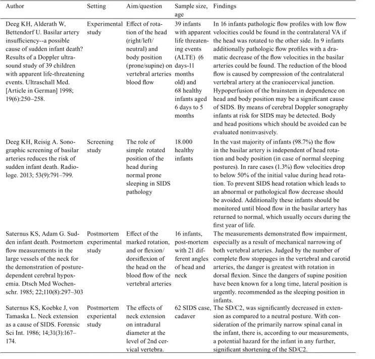
As a summary of our results we can state, that the inborn postural mechanisms in the new-borns are different in awake versus asleep states and the referred extreme positions such as the froggy pose can be performed easier while sleeping. The white noise applied during the photo sessions has a pain modulatory and sleep inducing effects especially in early infancy, therefore it is possible that white noise is masking the somatosensory stimuli while the newborns are positioned passively into end range positions. Moreover the findings of our scoping review indicate that the cervical end range positions while sleeping may cause disturbances in vertebrobasilar blood flow affecting brainstem circulation that may be hazardous in later motor development. In the medical literature, there is no evidence to show the effects of extreme cervical positions in vivo;
evidence only shows the effects post-mortem, due to the reasonable strict ethical concerns (Table 1).
DISCUSSION
Reviewing the relevant literature, only a limited number of studies focused on the first two weeks of life, there is a research gap about the complex effect of white noise induced sleep and passive end range positioning on cerebral blood flow in vivo in this early stage of life. Only one recent study has emphasised the behavioural inhibitory effect of white noise in infancy and recommended white noise as an alternative pain reducing method. The majority of the studies assessing the effect of head position on brainstem circulation were cadaver studies performed post-mortem and they were not limited to this first two weeks of life. The research interest is mainly focused on normal sleeping positions and on the role of head position-dependent risk factors in SIDS pathology.
If a photographer leaves the observer role during induced deep sleep and changes the body position of neonates, especially positioning the cervical spine passively into end range or extreme unnatural positions, that can be classified as an intervention in medical terms. Every medical intervention has strict ethical standards and competence issues surrounding it. The minimum requirements for passive mobilisation and positioning are the basic anatomical, physiological, normal movement knowledges and the non-maleficence principle. There is a big concern that the study of photography does not provide these theoretical knowledges and practical skills. The ‘froggy pose’ mentioned above to illustrate this photographic practice consists of three- dimensional movement components that cannot be accomplished in an awake state, in case of normal muscle tone in early infancy and hyperextended position of the upper cervical spine.
The white noise applied during the new-born photography sessions modulates the neonates’ pain perception, induces deep sleep and has a priority in the information processing in case of young neonates by masking other sensory information.
Evidence suggests that the shortest crying period and the lowest behavioural reactions were among those infants lying in their cribs and listening to white noise while undergoing painful procedure5). The new-born photography sessions are under the same circumstances posing the baby on a beanbag, listening to white noise in a warm environment after feeding. Due to the maturity of new-borns’ developed auditory systems, the noise has high priority in the information processing centres of the brain, and, therefore, it promotes the transition from awareness into sleep.
Questions arise considering the pain perception modulatory and sleep-inducing effects of white noise in neonates: is it ethical to trade on the immaturity of new-borns’ nervous systems? Is it ethical to manipulate them with white noise merely for the sake of a picture taken in an unnatural passive pose? This is a particular cause for contention when one pays attention to the fact that noise perception has priority over the somatosensation, which is further deteriorated in this early stage of life by sleep atonia in REM. What is more, during a new-born photography session, the immature somatosensory informa-
tion is blocked, whether it takes place during REM or non-REM sleep, due to atonia and the closed gates in the thalamus, respectively2) and it is further masked by the fact that continuous exposure to white noise modulates behavioural responses.
Therefore, it is a serious misconception to believe that infants will show by crying every time if something is painful, since the threshold of their pain perception is shifted in these manipulated states, because womb noises constantly calm them before birth. The other potential hazard is brainstem hypoxia, which is also neither visible nor detectable during the photography sessions; even periodic breathing patterns are sometimes misconstrued as being simply ‘cute’.
Table 1. Summary of results
Author Setting Aim/question Sample size,
age Findings
Vernet-Maury E, Robin O, Vinard H. Analgesic property of white noise: an experimen- tal study. Funct Neurol. 1988;
3(2):157–166.
Experimental
study Analgesic
property of white noise
100 healthy volunteer students
Each measured parameter was significantly increased under white noise, indicating the modulatory effect of white noise on pain perception
Spencer JA, Moran, DJ, Lee A, Talbert D. White noise and sleep induction. Arch Dis Child. 1990; 65(1): 135–137.
Randomised
study White noise and
sleep induction 40 healthy neonates, 2–7 days old
When exposed to white noise, the likelihood of a baby falling asleep is increased more than three fold (25% to 80%).
Karakoç A, Türker F. Effects of white noise and holding on pain perception in newborns.
Pain Manag Nurs. 2014;
15(4):864–870.
Experimental
study White noise and its pain percep- tion modulatory effect
120 healthy
infants White noise is an effective nonpharmacological method to control pain, reduce crying time, and positively affects vital signs.
Gilles FH, Bina M, Sotrel A. Infantile atlanto-occipital instability. The potential danger of extreme exten- sion. Am J Dis Child. 1979;
133(1):30–37.
Postmortem, experimental study
Effect of the extension of the head on the blood flow of vertebral arteries, infantile atlanto-occipital instability
17 infants,
cadaver In ten of 17 infants, the posterior arch of the atlas inverted through the foramen magnum during exten- sion of the head on the atlas, resulting in the anatomic potential of bilateral vertebral artery compression.
Pamphlett R, Raisanen J, Kum-Jew S. Vertebral artery compression resulting from head movement: a possible cause of the sudden infant death syndrome. Pediatrics.
1999; 103(2):460–468.
Postmortem, experimental study
Effect of the extension, or ro- tation of the head on the vertebral arteries
20 infants,
cadaver In 3 of 5 extended cases, bilateral vertebral artery compression was seen between the occipital bone and C1. In 3 of 9 rotated cases, the left vertebral artery was compressed adjacent to C1 before the artery entered the transverse foramen. No vertebral artery compression was seen in the necks held in the neutral position.
Wald M, Klupp N, Lawrenz K, Puig S, Heimberger K, Re- iter C, Pollak A, Ipsiroglu O.
A novel technique to measure position-dependent resistance changes in the vertebral arteries postmortem: new insights into the aetiology of SIDS? Acta Paediatr. 2004;
93(9):1166–1171.
Postmortem, experimental study
Effect of the extension, and or rotation of the head on the ver- tebral arteries
10 infants,
cadaver Considering exclusively the combined movements of rotation plus extension, resistance increased ipsi- and contra-laterally—no matter which side the head was turned—in three infants. A further three reacted with resistance surges only contralateral to the direction of rotation, and one only ipsilateral.
Eichler F, Ipsiroglu O, Arif T, Popow C, Heinzl H, Urschitz M, Pollak A. Position de- pendent changes of cerebral blood flow velocities in pre- mature infants. Eur J Pediatr.
2001; 160(10):633–639.
Follow up measure- ments
Effect of sleep- ing positions on cerebral blood flow velocities, maturational changes
23 premature infants at the age of 3–5 days, 17 infants at the age of 10–15 days, 16 infants at the corrected age of 1 month.
In premature newborns, position dependent changes of cerebral blood flow velocity develop with matura- tion and are most pronounced in the vertebrobasilar system. These changes are possibly due to compres- sion of the vertebral artery by neck movement and suggest an individual risk of brainstem perfusion deficits that may be aggravated with age (early days) and head rotation in a prone position.
Considering the results of this study regarding the potential hazards associated with passive cervical spine position during induced sleep, our profession should speak up against this current trend and practice. It clearly needs to be supervised and controlled for the sake of prevention of any unwanted health consequences of the end range passive positions. In the ‘froggy pose’, which represents only the tip of the iceberg, there is a big concern on the hyperextended passive position of the upper cervical spine. This is further deteriorated by the breath- and heartbeat-related extra uncontrolled movements of the head, observable in many online videos, as well as the passive weightbearing on the bones and joints of upper limb, the passive reaction forces acting on the atlanto-occipital and other upper cervical joints, and the uncontrolled accessory movements and joint positions. Unfortunately, safety requirements do not take into account these head-position-related hazards.
Based on the results of this scoping review, we suggest that a wider debate should be called for on the role of medical professions to control and regulate this very dangerous practice. From the aspect of paediatric physiotherapy, in case of any delay in motor development retrospective investigations should aim the history of photographic extreme positioning in new- born to provide more evidence about this research gap. Moreover, future cohort studies are necessary to monitor and follow up on the potential negative effects of new-born photography in terms of the motor and cognitive development of the subjects.
We suggest the neutral cervical positions and natural poses for photographic purpose avoiding any passive interventions and end range positioning.
Author Setting Aim/question Sample size,
age Findings
Deeg KH, Alderath W, Bettendorf U. Basilar artery insufficiency--a possible cause of sudden infant death?
Results of a Doppler ultra- sound study of 39 children with apparent life-threatening events. Ultraschall Med.
[Article in German] 1998;
19(6):250–258.
Experimental
study Effect of rota- tion of the head (right/left/
neutral) and body position (prone/supine) on vertebral arteries blood flow
39 infants with apparent life threaten- ing events (ALTE) (6 days-11 months old) and 68 healthy infants aged 6 days to 5 months
In 16 infants pathologic flow profiles with low flow velocities could be found in the contralateral VA if the head was rotated to the other side. In 9 infants additionally pathologic flow profiles with a dra- matic decrease of the flow velocities in the basilar arteries could be found. The reduction of the blood flow is caused by compression of the contralateral vertebral artery at the craniocervical junction.
Hypoperfusion of the brainstem in dependence on head and body position may be a significant cause of SIDS. By means of cerebral Doppler sonography infants at risk for SIDS may be detected. Body and head positions which should be avoided can be evaluated noninvasively.
Deeg KH, Reisig A. Sono- graphic screening of basilar arteries reduces the risk of sudden infant death. Radio- loge. 2013; 53(9):791–799.
Screening
study The role of simple rotated position of the head during normal prone sleeping in SIDS pathology
18.000 healthy infants
In the vast majority of infants (98.7%) the flow in the basilar artery is independent of head rota- tion and body position (in case of normal sleeping postures). In rare cases (1.3%) flow velocities drop to below 50% of the initial value during head rota- tion. To prevent SIDS head rotation which leads to an abnormal or pathological flow decrease should be avoided. Additionally these infants should be monitored until blood flow in the basilar artery has returned to normal, which usually occurs during the first year of life.
Saternus KS, Adam G. Sud- den infant death. Postmortem flow measurements in the large vessels of the neck for the demonstration of posture- dependent cerebral hypox- emia. Dtsch Med Wochen- schr. 1985; 22;110(8):297–303
Postmortem experimental study
Effect of the marked rotation, and or flexion/
dorsiflexion of the head on the blood flow of the vertebral arteries
16 infants, post-mortem with 21 dif- ferent angles of head and neck
The measurements demonstrated flow impairment, especially as a result of mechanical narrowing of both vertebral arteries. Judged by the number of complete flow stoppages in the vertebral and carotid arteries, the danger is greatest with rotation in dorsal flexion. Since the dangers of supine position have been known for a long time, lateral position is urgently. recommended as the sleeping position in infants.
Saternus KS, Koebke J, von Tamaska L. Neck extension as a cause of SIDS. Forensic Sci Int. 1986; 14;31(3):167–
174.
Postmortem experiental study
The effects of neck extension on intradural diameter at the level of 2nd cer- vical vertebra.
62 SIDS case,
cadaver The SD/C2, was significantly decreased in exten- sion as compared to a neutral posture. With con- sideration of the primarily narrow spinal canal in the infant, there is, according to our measurements, a potential hazard for the infant in any further, significant shortening of the SD/C2.
Table 1. Continued
Funding
This work was not funded and there is no conflict of interest.
Conflict of interest
Regina Finta owns the copyrights of the drawings. Edit Nagy is a hobby photographer and restored the pictures into digital format.
REFERENCES
1) Shumway-Cook A, Woollacott MH: Motor control. Translating research into clinical practice, 4th ed. 2012, Philadelphia: Wolters Kluwer Lippincott Williams
& Wilkins.
2) Rechtschaffen A, Siegel J: Sleep and dreaming. In: Kandel ER, Schwartz JH, Jessell TM Principles of Neural Science, 4th ed. 2000, New York: The McGraw- Hill Companies, pp 936–947.
3) Vernet-Maury E, Robin O, Vinard H: Analgesic property of white noise: an experimental study. Funct Neurol, 1988, 3: 157–166. [Medline]
4) Spencer JA, Moran DJ, Lee A, et al.: White noise and sleep induction. Arch Dis Child, 1990, 65: 135–137. [Medline] [CrossRef]
5) Karakoç A, Türker F: Effects of white noise and holding on pain perception in newborns. Pain Manag Nurs, 2014, 15: 864–870. [Medline] [CrossRef]
6) Gilles FH, Bina M, Sotrel A: Infantile atlantooccipital instability. The potential danger of extreme extension. Am J Dis Child, 1979, 133: 30–37. [Medline]
[CrossRef]
7) Pamphlett R, Raisanen J, Kum-Jew S: Vertebral artery compression resulting from head movement: a possible cause of the sudden infant death syndrome.
Pediatrics, 1999, 103: 460–468. [Medline] [CrossRef]
8) Wald M, Klupp N, Lawrenz K, et al.: A novel technique to measure position-dependent resistance changes in the vertebral arteries postmortem: new insights into the aetiology of SIDS? Acta Paediatr, 2004, 93: 1166–1171. [Medline] [CrossRef]
9) Eichler F, Ipsiroglu O, Arif T, et al.: Position dependent changes of cerebral blood flow velocities in premature infants. Eur J Pediatr, 2001, 160: 633–639.
[Medline] [CrossRef]
10) Deeg KH, Alderath W, Bettendorf U: [Basilar artery insufficiency—a possible cause of sudden infant death? Results of a Doppler ultrasound study of 39 chil- dren with apparent life-threatening events]. Ultraschall Med, 1998, 19: 250–258 (in German). [Medline] [CrossRef]
11) Deeg KH, Reisig A: [Sonographic screening of basilar arteries reduces the risk of sudden infant death]. Radiologe, 2013, 53: 791–799 (in German). [Medline]
[CrossRef]
12) Saternus KS, Adam G: [Sudden infant death. Postmortem flow measurements in the large vessels of the neck for the demonstration of posture-dependent cerebral hypoxemia]. Dtsch Med Wochenschr, 1985, 110: 297–303 (in German). [Medline] [CrossRef]
13) Saternus KS, Koebke J, von Tamaska L: Neck extension as a cause of SIDS. Forensic Sci Int, 1986, 31: 167–174. [Medline] [CrossRef]