III./8.4.5.: Metabolic myelopathies Definitions
Categories based on clinical signs Modern definition
According to the broad definition, all disorders which cause spinal signs are called myelopathies. This wide and heterogeneous category contains the metabolic, vascular, inflammatory, compressive and degenerative disorders of the spinal cord.
Before the modern diagnostic era, the diagnosis of myelopathy was based on clinical signs – the structural or other causes of myelopathy remained often hidden.
The stricter definition of myelopathy includes only those myelopathies, which are caused by metabolic or toxic causes, or by physical harm (irradiation) and vitamin deficiencies.
Role of the accompanying signs in the diagnosis
Spinal symptoms may occur alone, or general and organ specific
symptoms of the underlying systemic disorder may also be present. Since spinal symptoms are usually non-specific, accompanying signs may help in the diagnosis.
Isolated myelopathies generally represent a diagnostic challenge for physicians.
The most important myelopathies
Most common forms of myelopathies
Subacute combined degeneration HIV-vacuolar myelopathy
Adrenoleukodystrophy - adrenomyeloneuropathy Copper deficiency
Vitamin E deficiency neuropathy Other nutritional myelopathies Toxic (alcoholic) myelopathy Post-irradiation myelopathy
Symptoms of myelopathies
The symptoms of metabolic myelopathies are usually non-specific for the underlying cause. The clinical picture is characterized by progressive or persistent dysfunction of ascending and descending pathways.
Neurological diagnostic methods generally only confirm a spinal disorder, and are not informative concerning the cause.
The following myelopathies have clinical significance:
Subacute combined degeneration
Causes
Subacute combined degeneration (SCD) is the most common metabolic myelopathy, caused by vitamin B12 (cobalamin) or folic acid deficiency.
The disease may be the consequence of autoantibody production against the parietal cells of the stomach, lack of intrinsic factors, malabsorption (gastrointestinal diseases), malnutrition, extreme diet, overdose of antacids, genetic defect, and the use of N2O anesthetic.
Combined neurological deficits
The development of neurological signs is insidious or chronic. Due to the deficit of the posterior (Goll-Burdach tract) and lateral columns of the spinal cord, proprioceptive sensory loss and corticospinal symptoms are seen. The classical symptoms include proprioceptive sensory loss and spinal ataxia, Lhermitte’s sign, and spastic paraparesis or quadriparesis.
Furthermore, autonomic dysfunction, paresthesia on the trunk, polyneuropathy, optic nerve lesion, psychosis or dementia may also develop. The peripheral signs of polyneuropathy may mask corticospinal tract involvement, which may make the diagnosis more difficult.
Other accompanying signs
Signs of megaloblastic anemia are seen in the blood and in the bone marrow. Chielitis, glossitis, atrophic gastritis, achlorhydria and later secondary stomach cancer may develop.
Diagnosis
If subacute combined degeneration is suspected, the following tests should be performed: blood tests (blood count, vitamin B12, folic acid,
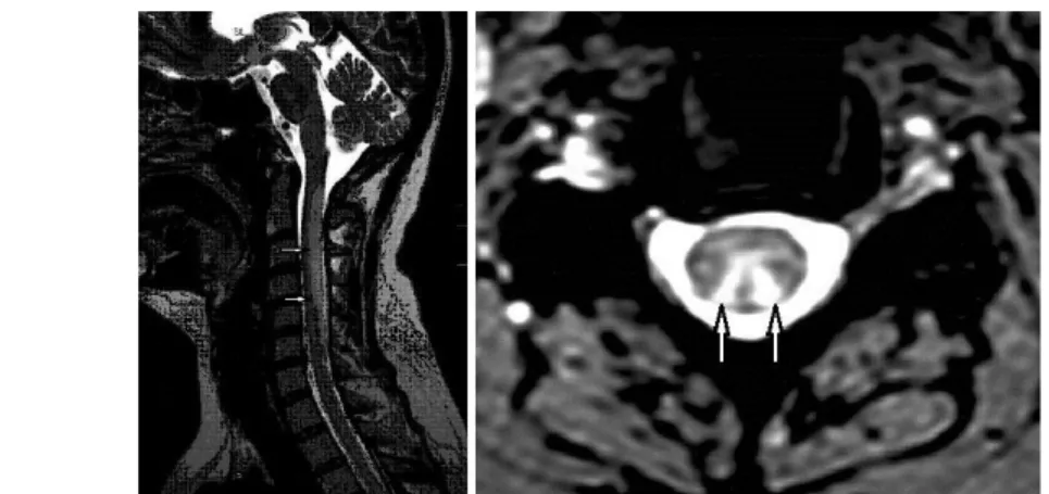
homocystein and methyl-malonic acid levels), Schilling test, gastroscopy with biopsy, cervical and thoracic spinal MRI (showing increased signal intensity in the posterior columns), ENG to prove polyneuropathy, and evoked potentials (VEP, SSEP, TMS).
Diagnostic difficulties
Neurological signs often precede hematological changes. It is possible that normal or slightly decreased vitamin B12 level is detected in the presence of complex and typical clinical signs. One previously administered vitamin B12 injection is enough to disturb the diagnosis.
Treatment
diagnostic difficulties
Subacute combined degeneration is treated by a life-long, parenteral administration of vitamin B12. Progression of the disease is halted with treatment, but clinical improvement is not to be expected after a long-term untreated B12 deficiency.
Fig. 18: MRI image in subacute combined degeneration: a long high-intensity spinal lesion is seen in the dorsal column on T2-weighted image