FROM CULTURED CELLS
AND THEIR CONTRIBUTION TO THE STUDY OF DISEASE*
DAVID SNARY MICHAEL J. CRUMPTON
National Institute for Medical Research London, United Kingdom
PETER GOODFELLOW WALTER F. BODMER
Genetics Laboratory Department of Biochemistry
University of Oxford Oxford, United Kingdom
I. INTRODUCTION
The major human histocompatibility system HLA was originally defined as a series of antigenic specificities identified by sero- logical techniques using peripheral blood lymphocytes. These
*The abbreviation SDS is used for sodium dodecylsulphate.
247
specificities were assigned to two separate series corresponding to two closely linked loci called A (formerly LA) and Β (formerly 4) each with multiple alleles. Recent studies, especially on the H-2 system, the mouse analogue of HLA, as well as in man have shown that the genetic region identified by these two loci also contains many other genes controlling cell surface determinants, immune response differences, some components of the complement system and perhaps other related functions connected in general with cell-cell recognition (Shreffler and David, 1975; Kissmeyer Nielsen, 1975). The original impetus behind the analysis of the HLA system was its use for matching for transplantation; this has turned out to be more complex than was at one time hoped. The demonstration of genetic linkage between the murine major histo- compatibility complex H-2 and resistance to virus induced leuke- mogenesis (Lilly et al., 1964) and specific immune responses
(Benacerraf and McDevitt, 1972) has however stimulated an exten- sive search for association between antigens of the HLA system and specific diseases. Though early data showed only weak associa- tions with Hodgkins and some other diseases, subsequent studies have shown very striking associations between antigens especially of the HLA-B locus and a number of diseases having a presumptive or suspected autoimmune etiology including for example, ankylosing spondylitis, coeliac disease, myaesthemia gravis and multiple sclerosis. In the case of ankylosing spondylitis the association is so strong that it is already of some possible diagnostic value
(McDevitt and Bodmer, 1974; Transplantation Reviews, 1975).
These associations have been interpreted in terms of the ef- fects of linked immune response genes in the HLA region rather than direct effects of the antigens of the HLA-B or A loci them-
selves. It has thus already been established that mixed lympho- cyte culture determinants controlled by the HLA-D locus show more significant association with multiple sclerosis than do antigens of the HLA-A or Β loci. This emphasizes the importance of devel- oping direct methods for identifying genes of the HLA region which may be more directly involved in disease association.
The complete characterisation of gene products and functions depends ultimately on a detailed analysis of their chemical structure. Much progress has recently been made in the elucida- tion of the chemical structure of the HLA and H-2 serological de- terminants and the aim of this chapter is to review some of our own studies in this direction. Among the gene products recently identified in the HLA and H-2 region are a new set of serological specificities called la for immune associated. Unlike the H-2D and H-2K, or HLA-A or Β antigens the la antigens have a tissue distribution that is relatively specific, including especially Β but excluding Τ lymphocytes. (Shreffler and David 1975; Jones et al., 1975). These antigens seem to be closely associated with mixed lymphocyte culture determinants of the HLA-D locus and prom- ise to become a powerful tool in disease association and related studies (Kissmeyer Nielsen, 1975). There is in addition the def- inite possibility that the la specificities will be of more direct importance for the problem of clinical transplantation than the specificities of the original defined HLA-A and Β loci. The chem- ical characterisation of these products which is an obvious con- comitant of the work on the HLA-A and Β locus antigens, may there- fore turn out to be of considerable interest. Apart from funda- mental advances which follow from a further understanding of the chemical nature of these products, it seems most probably that valuable antisera for HLA typing may be produced by immunisation with the purified antigens.
II. SOURCE OF HLA
It is preferable that the tissue used for the isolation of HLA antigens should satisfy the following requirements: CD it should be readily available; (2) it should contain large numbers of cells with a high content of HLA antigens; (3) it should express select- ed HLA specificities; and (4) it should be fresh (i.e., not au- topsy material) in order to reduce possible degradation. The readily available tissues such as red blood cells (Doughty et al., 1973) and placenta (Goodfellow et al., 1977) contain low levels of HLA antigens only. Tissues such as liver, spleen, white cells and leukemic cells have higher levels (Amos and Ward, 1975) but, be- cause of the polymorphism of HLA antigens, are basically unsuit- able except for the isolation of small amounts of antigen. In contrast, lymphoblastoid cells grown in culture express high lev- els of HLA antigens on their surface, and represent a reproducible antigen source. They can be grown in suspension to relatively high densities (2 to 4 x 1 06 cells/ml) and, given mass culture facilities, in unlimited numbers. Lymphoblastoid cell-lines also offer the opportunity of choosing particular HLA specificities and of developing homozygous cell-lines to promote the separation of allogeneic activities as well as doubling the antigen yield. The studies on HLA antigens described here were carried out using the lymphoblastoid cell-line BRI8 (HLA-A1, A2, B8, B13, 4a and 4 b ) .
III. ISOLATION OF HLA
HLA antigens are intimately associated with the cell surface (plasma) membrane (Wilson and Amos 1972; Snary et al,, 1974) and are insoluble in conventional aqueous solvents. Two approaches to solubilisation have been used namely, partial degradation and de- tergent extraction. The former method using proteolysis with pa- pain (Sanderson 1969; Cresswell et al,, 1973) or 3M KCl extraction
(Reisfeld et al., 1971) yields antigenically active but degraded products. Although such materials are suitable for examination of the molecular basis of HLA specificity, they provide no informa- tion on the antigen's mode of integration into the cell membrane.
Fragments of molecules are also generally less immunogenic than the whole molecules and may induce a different spectrum of immune responses (for example, humoral versus cellular immunity). In contrast, detergents solubilise the complete molecule.
Two approaches to detergent solubilization are available.
Firstly, direct solubilisation of the surface membrane of whole cells by non-ionic detergents such as Nonidet P-40 and Triton X- 100 which fail to solubilise the nuclear membrane (Schwartz &
Nathenson 1971)' and secondly, detergent solubilization of the iso- lated cell surface membrane (Snary et al., 1974; Springer et al., 1974). The direct approach gives good solubilisation of HLA anti- gens without significant losses in HLA activity although the con- comitant release of proteolytic enzymes from the cell (Grayzel et al., 1975) may lead to degradation. Detergent solubilization of a crude cell membrane preparation has the same associated prob- lems, whereas a purified plasma membrane preparation greatly re- duces the possibility of enzymic degradation, and has the added advantage that it gives a significant degree of purification.
HLA antigens are known to be glycoproteins (Sanderson et al., 1971; Snary et al., 1974) and advantage can be taken of this fact during fractionation. Thus, lectins which bind carbohydrate resi- dues can be used to separate detergent solubilised membrane glyco- proteins from the non-glycosylated proteins (Allan et al., 1972;
Hayman and Crumpton, 1972).
A. Plasma Membrane Preparation
BR18 cells were broken in a mechanical pump by shear (Stansted Fluid Power Ltd., Stansted, Essex, CM24 8HT, U . K . ) . The cells were pumped past a ball bearing held in a small orifice by a spring the tension on which was adjusted so that the plasma mem-
brane was stripped from the cell (Crumpton and Snary, 1974). The plasma membrane was separated from the cell homogenate by differ- ential centrifugation and purified by discontinuous sucrose densi- ty gradient centrifugation. The sucrose densities were selected by analysis of a continuous sucrose density gradient (Crumpton and Snary, 1974). The plasma membrane preparation was essentially free from other subcellular organelles as judged from assays of various marker enzymes. Evidence for the surface membrane local- ization of HLA antigens was obtained by comparing the HLA antigen distribution amongst the subcellular fractions with the distribu- tion of marker enzymes for the plasma membrane (5'-nucleotidase and N a+/ K+ ATPase) (Crumpton and Snary, 1974). The preferential location of HLA antigens on the cell surface is also supported by the similar distribution of HLA antigens and 5'-nucleotidase on continuous sucrose density gradient centrifugation of the micro- somal fraction (Snary et al., 1974). A representative example of the purification and recovery of the plasma membrane and its as- sociated HLA antigens is shown in Table I. A relatively high re- covery of plasma membrane was achieved (average of 10 experiments about 35°/o), together with a 45-fold purification of the HLA antigens.
B. Fractionation of Solubilized Membrane
The BRI8 plasma membrane (4mg of protein/ml) was solubilised in 2°/o (w/v) sodium deoxycholate-lOmM tris buffer, pH 8.2. After centrifuging for 3 x 1 06g min greater than 86°/o of the HLA acti- vity and 90°/o of the membrane protein were present in the super- natant fraction. Non-ionic detergents such as Triton X-100, Lubrol-PX and Nonidet P-40 were found to be much less efficient solubilising agents (less than 60°/o of the HLA activity being solubilised even at lmg of membrane protein/ml).
The sodium deoxycholate solubilised BRI8 plasma membrane was fractionated by gel-filtration on a column of Ultragel AcA 34 (LKB Instruments Ltd.). A single peak of HLA activity was recovered
TABLE I Distribution of HLA-A2 Antigenic Activity and 51-Nucleotidase on Fractionation of Disrupted BRI8 Cells'
51-Nucleotidase HLA-A2 Protein
(°/o of homo- (specific activity Fraction genate) relative to homogenate)
Homogenate 100 1.0 1.0
Nuclei and 61.8 0.5 0.7
mitochondria
Cytosol 39.7 0.2 0.3
Endoplasmic 0.98 9.1 11.1
reticulum
Plasma membrane 0.6 51 45
experimental details are described in Crumpton and Snary (1974) and Snary et al. (1974). BRI8 cells were grown by Searle Diag- nostics, High Wycombe, Bucks., U.K.
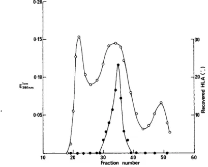
which accounted for 93°/o of the HLA activity loaded onto the column (Fig. 1 ) . The degree of purification of HLA antigens in the fraction recovered from gel-filtration was 135-fold relative to whole cells (Table I I ) .
The recovered material was further fractionated by affinity chromatography in 0.5°/o sodium deoxycholate on a column of Lens culinaris lectin attached to Sepharose 4B. This lectin binds the majority of lymphocyte plasma membrane glycoproteins (Hayman and Crumpton, 1972) including HLA antigens (Snary et al., 1974). The bound glycoproteins were eluted with methyl-a-D-mannopyranoside
(Fig. 2 ) . The glycoprotein fraction accounted for 19°/o of the HLA activity of the original cells. The degree of purification was 1240-fold relative to the whole cells (Table I I ) .
Since HLA antigens contain ^2^m^-cro^°^DU^n (Grey et al., 1973; Solheim and Thorsby, 1974) they can be selectively sepa- rated from other components by using immunoadsorbents against $2"
microglobulin. The above glycoprotein fraction was passed down a column containing the immunoglobulin fraction of a rabbit anti-
0-20r
10 20 30 40 50 60 Fraction number
FIGURE 1 Fractionation of BRI8 plasma membrane solubilised in sodium deoxycholate by gel-filtration on Ultragel AcA 34. The column (91 x 1.6cm) was eluted with 0.5°/o sodium deoxycholate at a flow-rate of 4.9ml/h. Fractions (2.45ml) were assayed for pro- tein (°'E280nm^ ad H L An ~A 1 activity (·).
serum to human 32-microglobulin attached to Sepharose 4B. The bound HLA antigens were subsequently eluted with 3M potassium thiocyanate in 0.5°/o sodium deoxycholate.
C. Characterisation of HLA Antigen Fractions
Figure 3 shows the polypeptide compositions of the above HLA antigen fractions revealed by Polyacrylamide gel electrophoresis in SDS after reduction with dithiothreitol. The material which was adsorbed by and eluted from the a n t i- 3 2-mic r o9l ob u l i n column, comprised primarily two polypeptide chains of molecular weights 43 000 and 12 000. The larger chain most probably corresponds to the glycosylated polypeptide of HLA antigens that carries the polymorphic allogeneic specificities, whereas the smaller chain
TABLE II Purification of HLA Antigens from BRI8 C e l l sa
Degree of purification Protein (°/o) HLA (°/o) of HLA
BRI8 cells 100 100 1
Plasma membrane 0.6 27 45
Na deoxycholate 0.56 22.8 41
solubilised membrane
Gel-filtrationb 0.156 21.2 136
fraction
Glycoprotein 0.0153 19.1 1240
fraction0
aResults are expressed relative to the initial BRI8 cells.
bS e e Fig. 1.
cS e e Fig. 2.
represents ^2"m^cro(3^0^u^n (Tanigaki and Pressman, 1974) . The minor bands are probably due to elution of some immunoglobulin from the column. The glycoprotein fraction possessed both of these polypeptide chains together with polypeptides of molecular weights 39 000, 33 000 and 28 000. The latter polypeptides are of unknown origin and function, although the la antigens expressed on the surfaces of B-lymphocytes and lymphoblastoid cell-lines
(Jones et al., 1975) are known to co-fractionate with the HLA an- tigens on gel-filtration and by affinity chromatography on Lens culinaris lectin-Sepharose (Snary and Goodfellow, unpublished ob- servations) . Furthermore, mouse la antigens have been shown to comprise two or three polypeptide chains with slightly different molecular weights of about 30 000 (Sachs et al., 1975). These results suggest that the polypeptide chains of molecular weight 28 000 and 33 000, and possibly 39 000, (Fig. 3) could be the human counterparts of the mouse la antigens.
The above preparations of HLA antigens isolated from BRI8 cells expressed four private specificities (Al, A2, B8 and B13).
15 p
FIGURE 2 Separation of glycoprotein by affinity chromatog- raphy on Lens culinaris lectin-Sepharose. The HLA antigen frac- tion from gel-filtration (Fig. 1) was added to a column (6 x 10cm) of Lens culinaris lectin attached to Sepharose 4B (lmg of lectin/
ml of Sepharose) in 0.5°/o sodium deoxycholate. The adsorbed glycoproteins were eluted with 2°/ο (w/v) methyl-a-D-mannopy-
ranoside in 0.5°/o sodium deoxycholate (commencing at fraction no.
12). The fractions were assayed for protein (o; E^QQnm) and HLA- A2 activity (·) .
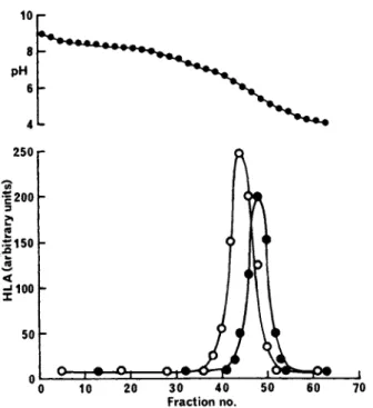
Some separation of these specificities has been achieved by iso- electric focusing in l°/o Triton X-100. Figure 4 shows that the HLA-A2 antigen (isoelectric point, pH 6.2) separates partly from the other three HLA types (isoelectric point, pH 5.5). However, no separation of the HLA-A1, -B8 and -B13 antigens or of the la antigens was detected. The problem of the separation of different HLA specificities may be in part circumvented by careful selection of homozygous cell-lines with HLA antigens of different isoelec- tric points.
FIGURE 3 Polyacrylamide gel electrophoresis patterns of HLA antigen fractions in SDS. Samples were reduced with dithiothrei- tol and run on 10°/ο (w/v) gels using the tris-glycine buffered system of Laemmli (1970). The gels were stained with Coomassie blue. Sample A, the glycoprotein fraction recovered in Fig. 2.
Sample B, the glycoprotein fraction was added to a column (10 χ 1.2cm) of rabbit anti-human 32"iH^c'roSrloJbuIin - Sepharose 4B (15mg of immunoglobulin/ml) and the adsorbed protein was eluted with 3M potassium thiocyanate in 0.5°/o sodium deoxycholate. Molecular weights were calculated from the mobilities of transferrin, bovine serum albumin, ovalbumin, immunoglobulin L chain, soybean trypsin inhibitor and cytochrome c.
Abbreviations: LcH, Lens culinaris lectin; L- and H-chain, immunoglobulin G L- and H-chains.
IV. THE MOLECULAR STRUCTURE OF HLA ANTIGENS
Recently, it has been proposed that HLA antigens have an im- munoglobulin- like structure in which two basic units of one each of the polypeptide chains of molecular weight 43 000 and 12 000 are linked either noncovalently (Rask et al., 1974) or via a di- sulfide bridge (Strominger et al., 1974; Cresswell & Dawson, 1975). A considerable amount of information on the molecular structure of HLA antigens can be obtained directly even though they are not available in a pure form. Thus, biological activity can be used to measure their location on gel-filtration and su-
1 0 r
FIGURE 4 Distribution of HLA antigenic activities on iso- electric focusing. BRI8 plasma membrane solubilised in l°/o Tri- ton X-100 was fractionated on a pH 3 to 10 gradient in a column
(55 x 1cm) containing a sucrose gradient from 5 to 60°/o (w/v).
The electrode buffers were l°/o ascorbic acid in 60°/o sucrose, and l°/o ethanolamine. Fractions were assayed for HLA-A1 (·) and HLA-A2 (Ö) activities.
crose density gradients of deoxycholate-solubilised plasma mem- brane, and their size and shape can be calculated from these measurements (Siegel and Monty, 1966).
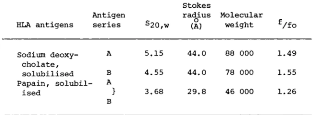
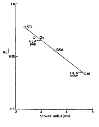
Gel-filtration of sodium deoxycholate-solubilised BRI8 plasma membrane showed (Fig. 1) a single peak of HLA-A1 activity with no evidence of aggregates or gross polydispersity. HLA activities of the A and the Β series and the HLA antigens of the plasma mem- brane fraction from a human spleen occupied identical positions with that shown for HLA-A1. The Stokes radius of the detergen- solubilised HLA molecule (Table III) was calculated (Siegel and Monty, 1966) using the calibration curve shown in Fig. 5. The
TABLE III Molecular Nature of HLA Antigens in Sodium Deoxycholate
HLA antigens
Antigen
series s2 0 , w
Stokes
radius Molecular (A) weight
Sodium deoxy- A 5.15 44.0 88 000 1.49 cholate,
solubilised Β 4.55 44.0 78 000 1.55 Papain, solubil- A
ised } 3.68 29.8 46 000 1.26 Β
sedimentation coefficients of the HLA antigens were obtained by analysis of deoxycholate-solubilised BRI8 membrane on sucrose density gradients (Fig. 6 ) . The A and Β series antigens possessed slightly different S2of W v a l us (Table III). Molecular weights e calculated for the A and Β series antigens from the Stokes radii and S2o , w v alu e s were 88 000 and 78 000 respectively. Provided dissociation had not occurred, these molecular weights are too low to accommodate a four chain, immunoglobulin-like dimer of molecu- lar weight 110 000 (based on the ß2~mic r 09l 0 Du l i n a nd H LA poly- peptide chains having sizes of 12 000 and 43 000 respectively as determined by Polyacrylamide gel electrophoresis in SDS). Dis-
sociation was, however, considered unlikely for two reasons.
Firstly, so far as can be determined the HLA antigens had not been subject to reducing conditions during the isolation, solubilisa- tion and fractionation of the BRI8 plasma membrane. Secondly, sodium deoxycholate failed to promote the dissociation of non- polar interactions between subunits in multimeric proteins such as hemoglobin (Snary et al., 1975). The most likely explanation for the difference between the calculated values for the molecu- lar weights of the deoxycholate-solubilised antigens and those es- timated by Polyacrylamide gel electrophoresis in SDS (minimum molecular weight 55 000) is that HLA antigens resemble other inte-
10 p
S t o k e s ' radius ( n m )
FIGURE 5 Relationship between élut ion positions (K^3) of standard proteins from Ultragel AcA 34 in 0.5°/o sodium deoxycho- late and Stokes radii. s calculated as described by Siegel w a and Monty (1966). The Stokes radii were those reported by Andrews
(1970) except for soybean trypsin inhibitor which was calculated from the data of Birk et al. (1963). Al, aldolase; BSA, bovine serum albumin; Ov, ovalbumin; STI, soybean trypsin inhibitor. The arrows indicate the measured K^3 for the whole HLA antigens (mem) and for the papain solubilised HLA antigens (pap).
gral membrane proteins, such as cytochrome b^ (Spatz and Stritt- matter. 1 9 7 3 )t in possessing a hydrophobic domain which binds de- tergent (Helenius and Simons, 1975). As a result the deoxycho- late-solubilised HLA antigens will possess anomalously high mo- lecular weights when assessed relative to water-soluble globular
FIGURE 6 Sucrose density gradient centrifugation of BRI8 plasma membrane solubilised in l°/o sodium deoxycholate. Solubi- lised membrane (lml) was layered onto a linear sucrose gradient
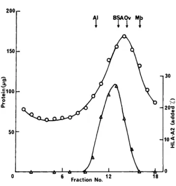
(9 to 28°/o sucrose) containing l°/o sodium deoxycholate. Marker proteins: aldolase (Al) 7.5S; bovine serum albumin (BSA) 4.41S;
ovalbumin (Ov) 3.55S; sperm whale myoglobin (Mb) 2.08S were added to identical gradients. The gradients were centrifuged at 4°C in a Beckman SW 41 rotor for 48h at 38 000 r.p.m. Fractions (0.67 ml) were pumped from the bottom of the gradients and were assayed for protein (0) and HLA-A2 activity (L).
proteins. In contrast, examination of papain-solubilised HLA an- tigens by gel-filtration and sucrose density gradient centrifuga- tion in sodium deoxycholate gave a molecular weight of 46 000
(Table III) which is in close agreement with the value found by Polyacrylamide gel electrophoresis in SDS (Turner et al., 1975).
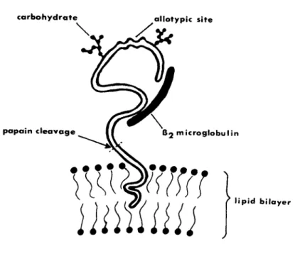
This result implies that the hydrophobic domain which is inserted in the lipid bilayer and which binds detergent is cleaved by pa- pain (Fig. 7 ) , whereas the antigenically-active region binds lit- tle or no detergent. An analogous situation has been previously reported for cytochrome b5 (Spatz and Strittmatter, 1973). De-
FIGURE 7 Model of membrane-bound HLA antigen.
tergent binding is also consistent with the higher molecular weights reported for HLA and H2 antigens in detergents of higher micelle size than sodium deoxycholate (460 000 and 380 000 for Brij 99 and Nonidet P40 respectively; Springer et al., 1974 and Schwartz et al., 1973). since these probably express the different micelle sizes of the bound detergent. Estimates of the amount of deoxycholate bound by the HLA antigens gave 0.57 and 0.39g of de- oxycholate/g of protein for the A and Β series antigens respec- tively. These values are in accord with those determined for mem- brane proteins (within the range 0.3-0.7g of deoxycholate/g of protein; Helenius and Simons. 1975 and Snary et al., 1975). The difference between the amounts of deoxycholate bound by the A and Β series antigens probably arises from a difference in hydropho- bicity, in which case it may indicate that the A series antigens are inserted deeper into the lipid bilayer than Β series antigens.
In contrast to previous work (Strominger et al,, 1974;
Cresswell and Dawson, 1975), no evidence was obtained for the
presence of dimers. The reason for this difference is not known.
However, one possible explanation is that the disulfide-bonded dimers found by the other workers arose by disulfide-interchange.
Thus, the gel-filtration pattern of dimers and larger aggregates reported by Cresswell and Dawson (1975) for deoxycholate-solubi- lised HLA antigens resembles that given by an aged sample of bo- vine serum albumin which had undergone aggregation through di- sulfide- interchange (Hellsing, 1969). The crude membrane prepa- rations used by Strominger et al. (1974) and Cresswell and Dawson
(1975) most probably contain relatively large amounts of endoplas- mic reticulum which is rich in an enzyme catalysing disulfide- interchange (de Lorenzo et al., 1966). If the above interpreta- tions are valid, then the 4-chain, dimeric, immunoglobulin-like model that has been proposed for membrane-bound HLA antigens is unlikely to be correct.
V. SUMMARY
Cultured lymphoblastoid BRI8 cells provide a very convenient source of HLA antigens. The antigens were isolated from a puri- fied preparation of the cell surface membrane by solubilisation in sodium deoxycholate, gel-filtration and affinity chromatography on Lens culinaris lectin attached to Sepharose 4B. The product contained primarily HLA and IA antigens which were separated by using an immunoadsorbent against 32"" microglobulin.
HLA antigens are composed of a glycosylated polypeptide chain of molecular weight 43 000 that is non-covalently bonded to $2~
microglobulin (mol. wt. 12 000). Estimates of the molecular weights of deoxycholate-solubilised HLA antigens by sucrose densi- ty gradient centrifugation and gel filtration gave values of 88 000 and 78 000 for the A and Β series antigens respectively.
These values most probably reflect the binding of deoxycholate, presumably to a hydrophobic domain which is normally inserted into
the lipid bilayer of the membrane and which is removed by papain (Fig. 7 ) . No evidence for the presence of dimers was obtained.
It was concluded that the present results cast doubt upon the va- lidity of the dimeric, immunoglobulin-like, model for HLA anti- gens. On the other hand, the absence of a dimeric, immunoglobu- lin-like structure does not rule out the possibility of a common evolutionary origin for HLA antigens and immunoglobulin.
ACKNOWLEDGMENTS
We are grateful to Dr. A. R. Sanderson for a gift of papain solubilised HLA. The work was suported in part by a grant from the Medical Research Council to The Genetics Laboratory, Oxford.
P. Goodfellow is the recipient of a Research Scholarship from the Medical Research Council.
REFERENCES
Allan, D., Auger, J., Crumpton, M. J., (1972), Nature New Biol.
236, 23-25.
Amos, D. B., Ward, F. Ε., (1975), Physiological Rev. 55, 206-246.
Andrews, P., (1970), Meth. Biochem. Anal. 18, 1-53.
Benacerraf, Β., McDevitt, Η. Ο., (1972), Science 175, 273-279.
Birk, Y., Gertler, Α., Khalef, S., (1963), Biochem. J. 87, 281- 284.
Cresswell, P., Turner, M. J., Strominger, J. L., (1973), Proc.
Nat. Acad. Sei. U.S.A. 70, 1603-1607.
Cresswell, P., Dawson, J. R. , (1975), J. Immunol. 114, 523-525.
Crumpton, M. J., Snary, D., (1974), in "Contemporary Topics in Molecular Immunology," (Ada, G. L. e d . ) . Vol. Ill, pp. 27-56, Plenum Press, New York.
de Lorenzo, F., Goldberg, R. F., Steers, E., Givol, D., Anfinsen, C. Β., (1966), J. Biol. Chem. 241, 1562-1567.
Doughty, R. W., Goodier, S. R., Geisthorpe, Κ., (1973), Tissue Antigens 3, 189-194.
Goodfellow, P. Ν., Barnstable, C. J., Bodmer, W. F., Snary, D., Crumpton, M. J., (1977), Transplantation, in press.
Grayzel, A. I., Hatcher, V. Β., Lazarus, G. S., (1975), Cell.
Immunol. 18, 210-219.
Grey, Η. M., Kubo, R. T., Colon, S. M., Poulik, M. D., Cresswell, P., Springer, T., Turner, Μ., Strominger, J. L., (1973), J.
Exptl. Med. 138, 1608-1612.
Hayman, M. J., Crumpton, M. J., (1972), Biochem. Biophys. Res.
Comm. 47, 923-930.
Helenius, Α., Simons, Κ. , (1975), Biochem. Biophys. Acta 415, 29- 80.
Hellsing, Κ., (1969), Biochem. J. 114, 145-149.
Jersild, C., Dupont, Β., Fog, T., Hansen, G. S., Nielsen, L. S., Thomsen, M., Svejgaard, Α., (1973), Transplant. Proc. 5, 1791- 1798.
Jones, Ε. Α., Goodfellow, P., Bodmer, J. G., Bodmer, W. F., (1975), Nature 256, 650-652.
Kissmeyer Nielsen, F., (ed), (1975), "Histocompatibility Testing 1975," Munksgaard, Copenhagen.
Laemmli, U. Κ., (1970), Nature 227, 680-685.
Lilly, F., Boyse, Ε. Α., Old, L. J., (1964), Lancet ii, 1207- 1209.
McDevitt, H. O., Bodmer, W. F., (1974), Lancet i, 1269-1275.
Rask, L., Ostberg, L., Lindblom, Β., Fernstedt, Υ., Peterson, P.
Α., (1974), Transplant. Rev. 21, 85-105.
Reisfeld, R. Α., Pellegrino, Μ. Α., Kahan, B. D., (1971), Science 172, 1134-1136.
Sachs, D. H., Cullen, S. Ε., David, C. S., (1975), Transplant, 19, 388-393.
Sanderson, A. R., (1969), Nature 220, 192-194.
Sanderson, A. R., Cresswell, P., Welsh, Κ. I., (1971), Nature New 'Biol. 230, 8-12.
Schwartz, B. D., Nathenson, S. G., (1971), J. Immunol. 107, 1363- 1367.
Schwartz, B. D., Kato, K., Cullen, S. Ε., Nathenson, S. G., (1973), Biochemistry 12, 2157-2164.
Shreffler, D. C , David, C. S., (1975), in "Adv. in Immunol,"
(Dixon, F. J., Kunkel, H. G., eds.). Vol. XX, pp. 125-195, Academic Press, New York.
Siegel, L. M., Monty, K. J., (1966), Biochem. Biophys. Acta 112, 346-362.
Snary, D., Goodfellow, P., Hayman, M. J., Bodmer, W. F., Crumpton, M. J., (1974), Nature 247, 457-461.
Snary, D., Goodfellow, P., Bodmer, W. F., Crumpton, M. J., (1975), Nature 258, 240-242.
Sol'heim, B. G., Thorsby, Ε., (1974), Tissue Antigens 4, 83-94.
Spatz, L, Strittmatter, P., (1973), J. Biol. Chem. 248, 793-799.
Springer, T. Α., Strominger, J. L., Mann, D. L., (1974), Proc.
Nat. Acad. Sei. U.S.A. 71, 1539-1543.
Strominger, J. L., Cresswell, P., Grey, H., Humphreys, R. Ε., Mann, D., McCune, J., Parham, P., Robb, R., Sanderson, Α. R., Springer, Τ. Α., Terhorst, C , Turner, M. J., (1974), Trans- plant. Rev. 21, 126-143.
Tanigaki, Ν., Pressman, D., (1974), Transplant. Rev. 21, 15-34.
Transplantation Reviews (1975), (Moller, G., e d . ) . Vol. XXII, Münk sgaard, Copenhagen.
Turner, M. J., Cresswell, P., Parham, P., Strominger, J. L., Mann, D. L., Sanderson, A. R., (1975), J. Biol. Chem. 250, 4512-4519.
Wilson, L. Α., Amos, D. B., (1972), Tissue Antigens 2, 105-111.