CHAPTER 5
Phospholipids
J. B. Davenport
I . Phospholipid Catabolism 174 A . Phospholipase A 184 B . Phospholipase Β 190 C. Phospholipase C 191 D . Phospholipase D 194 I I . Phospholipid Synthesis 197 I I I . Ion Transport and Phospholipid Metabolism 204
I V . Alternative Pathway for Lecithin Synthesis; Transmethylation In
volving Glycerophosphatides 206
V . Conclusions 209 References 211
Progress in our knowledge of the intermediary metabolism of phospho
lipids has been slower than in the fields of protein and carbohydrate metabolism. Even understanding of the synthesis and breakdown of homolipids has advanced further than that of the phospholipids. The degradation of phosphatidylcholine and phosphatidylethanolamine has been completely studied in only one tissue ( 1 ) . On the biosynthetic side the outstanding work, principally of Kennedy and his co-workers during the last five years (#), has thrown much light on how some phospholipids are synthesized in the cell, but only one of the enzymes concerned has been purified (3, 4 ) , the various reactions having been studied with cell homogenates or particulate subcellular fractions. Thus, not only is there a strictly limited amount of information on the inhibition of these processes, but in very few cases is it possible to pinpoint the enzymic site of the inhibition and to distinguish specific inhibition from more general physico- chemical effects on the whole and usually complex system.
Phospholipids are in general not soluble in water. Hence, in the in vitro study of the enzymes involved in their metabolism, we are concerned with a heterogeneous system of a water-soluble enzyme and an emulsified
173
174 J. Β. DAVENPORT
substrate, essentially a two-phase system. In the case of a particulate enzyme preparation one may be dealing with a three-phase system: the enzyme contained in its natural lipoprotein environment, the substrate insoluble in water, and the aqueous environment in which they are both suspended. It has already been shown in two cases (5, 6) that the principles of colloid science can help our understanding of such systems, and un
doubtedly further work along these lines will prove very revealing.
It remains to point out in this introduction that such heterogeneous systems are not necessarily a disadvantage in our goal of attempting to understand the processes occurring in the living cell. Probably the most exciting advance in biochemistry of the last ten years has been the demon
stration that the cell cytoplasm is a heterogeneous polyphase system and that enzymes are localized specifically in morphologically distinct and recognizable phases. These may be any of the cytoplasmic membranous structures or the aqueous phase which bathes them. The heterogeneous systems with which we shall be concerned in this chapter may represent better models for enzyme reactions in the living cell than in vitro systems involving a soluble enzyme and a soluble substrate.
I. PHOSPHOLIPID CATABOLISM
Although there have been reports in the literature that the fatty acid moiety of phospholipids can be oxidized in situ in the molecule, i.e., that phospholipids are active intermediates of fatty acid catabolism, the bulk of evidence supports the view that phospholipids are first hydrolyzed prior to fatty acid oxidation [see (7) for pertinent references]. There are four points of hydrolytic attack in the phosphoglycerides; the a- and /3-acyl ester linkages and the two phosphate ester linkages to glycerol, on one hand, and the nitrogen base or inositol, on the other. These will be designated phospholipases A, B, C, and D, as shown in Fig. 1. Some American workers have followed the original nomenclature suggested by Contardi and Ercoli (8) and used phospholipase C for the enzyme splitting the phosphate-nitrogen base (or inositol) linkage, and in this system the enzyme splitting the glycerol-phosphate linkage is called phospholipase D.
The nomenclature used here is that recommended by the commission on enzyme nomenclature of the International Union of Biochemistry (9).
That A and Β activity probably reside in the same enzyme, provided it is offered the appropriate physical conditions, has recently been demon
strated by Dawson (10).
Further hydrolytic degradation of the molecule is shown in Fig. 1 and results ultimately in the production of fatty acids, diglycerides, phospho-
CH2OCOR'
CHOCOR"
I ο
CHI n 2OPO-Nbase
OH
Phospholipase A
CH2OCOR'
CHOH + R"COOH
I n
CH2OPO-Nbase
OH
Lysophospholipid
I Phospholipase Β
Phospholipase C
CHoOCOR"
I
2
CHOCOR"
I C H2O H
Diglyceride +
Phosphoryl Ν base
CHJJOH C H O H
I
ο
I
IIC H2O P O - N b a s e
Glycerylphosphoryl— Ν base O H
FI G . 1. The hydrolytic breakdown of glycerophosphatides.
Phospholipase D
Phosphatidic acid Phosphatase
C H2O C O R '
C H O C O R "
I Q
Phosphatidic acid +
Ν base
5. PHOSPHOLIPIDS 175
176 J. Β. DAVENPORT
diesters, phosphomonoesters, and inorganic phosphate. The phosphatidic acid phosphatase will be considered later in the section on synthesis, while the enzymic degradation and metabolism of the remaining products are more appropriate to other chapters of this book. Thus, in this section we will be concerned principally with phospholipase action. In discussing the question of the inhibition of enzymes it is impossible to divorce it from discussion of their activation. Frequently these are two extremes in a spectrum of activity; a reagent or ion may, at one concentration, activate while at a different concentration (usually higher) it may inhibit.
Hanahan first demonstrated the activation by diethyl ether of the at
tack by pancreatic phospholipase A on egg lecithin (11). He showed that the enzyme-substrate complex can be extracted into the solvent phase where the enzymic reaction proceeds. Presumably the enzyme protein is made lipid-soluble by combination with the substrate to form a complex, probably analogous to a proteolipid (12). Kates has also demonstrated diethyl ether activation of the attack on lecithin by a phospholipase D present in plastid particles (IS, H). He was able to observe, by phase contrast microscopy, that solvents which activate the system (e.g., diethyl ether and other linear aliphatic ethers, ketones and esters; mixtures of methanol with diethyl ether, petroleum ether, or benzene) bring about coalescence of the plastid particles and the substrate emulsion particles when the solvent was added to the aqueous suspension. Solvents which did not activate the system (chloroform, petroleum ether) did not produce coalescence. Kates put forward an interesting mechanism to account for the results (6). He suggested that the active solvent molecules can be ad-
T A B L EI
M I N I M U M C O N C E N T R A T I O N OF V A R I O U S AMPHIPATHIC SUBSTANCES N E C E S S A R Y TO A C T I V A T E E N Z Y M I C HYDROLYSIS OF L E C I T H I N B Y THE PHOSPHOLIPASE Β OF Pénicillium notatum A N D THE ELECTROPHORETIC M O B I L I T I E S OF S U C H M I X T U R E S
Critical Critical activation activation
Anion mobility molarity
Activator species (μ s e c
-1
v o l t s
-1
cm) (mole % )
Dicetylphosphoric acid P O 4 - - 1 . 6 8.5
Cardiolipin P O 4 - - 1 . 9 5.5
Monophosphoinositide P O 4 - - 1 . 4 6.0
Sodium l-palmitoyl-4-anisidine-2- S O 3 - - 1 . 6 36.0 sulfonate
Sodium dodecyl sulfate S O 4 - - 1 . 6 0.0011% in bulk
aqueous phase Sodium hexadecyl sulfate S O 4 - - 1 . 7 0.00065% in bulk
aqueous phase
5. P H O S P H O L I P I D S 177 sorbed at the surfaces of both the emulsion substrate particles and the plastids, rendering both lipophilic and thus allowing the particles to coalesce. In the same paper he described activation of the system by anionic, but not by cationic, detergents. Coalescence in this case is prob
ably due to a charge effect, and it suggests that the plastid particles are positively charged at pH 4.7. The influence of surface charge on phospho
lipase action will be discussed in the following sections.
The observations of Bangham and Dawson, however, have cleared the way for a rational approach to the question of the activation and inhibition of phospholipase activity. Dawson (10) showed that the phospholipase Β of Pénicillium notatum, though without action on pure lecithin emulsions, would attack the intact lecithin, molecule when present in the mixed phospholipid fractions isolated from liver. He showed further that this property was due to the presence of small quantities of phosphatidyl- inositol and a polyglycerol phospholipid in the crude mixture. Bangham and Dawson followed up these observations by demonstrating that the
FIG. 2. The effect of the addition of T h
4
+ ions on the electrophoretic mobility and enzymic hydrolysis of lecithin plus dicetylphosphoric acid particles by the phospho
lipase Β of Pénicillium notatum. From A . D . Bangham and R . M . C. Dawson (15).
178 J. Β. DAVENPORT
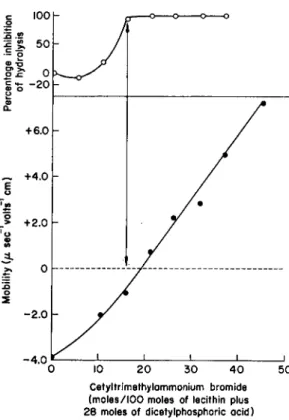
ition 100
lysis
50 -- ο
S? >»
σ χ: 0 C Η- φ ο -20 α.
Cetyltrimethylammonium bromide (moles/100 moles of lecithin plus 28 moles of dicetylphosphoric acid)
FIG. 3. Electrophoretic mobility and inhibition of enzymic hydrolysis (by the phospho
lipase Β of Pénicillium notatum) upon the addition of cetyltrimethylammonium bromide to lecithin plus dicetylphosphoric acid particles. From A . D . Bangham and R . M . C.
Dawson (15).
addition of any amphipathic molecule (molecule having both hydrophobic and hydrophilic groups) which carried a negative charge at the optimum pH (3.3) of the enzyme would induce phospholipase A activity (15). Thus, both naturally occurring substances, such as cardiolipin (polyglycero- phospholipids) and phosphatidylinositol, and synthetic substances, such as dicetylphosphoric acid and long-chain sulfonates and sulfates, induced the enzyme activity when mixed with lecithin in appropriate proportions.
Fatty acids, which are not dissociated at this pH, were without effect.
Although the molar proportion of each activator to produce the optimal effect varied widely, they all produced a constant zeta potential (Table I ) , as measured by direct electrophoretic observations of the active sub
strate emulsions, and it was concluded that the critical parameter was the surface charge density of the substrate particles. This induced activity could be inhibited by reducing the zeta potential to zero, achieved either
5. PHOSPHOLIPIDS 179 by the addition of ions (υθ2+
+
, Ca+ +, Th 4
+) or by the incorporation of a positively charged amphiphatic molecule (cetyltrimethylammonium bro
mide) into the lecithin-activator emulsion. In the former case (Fig. 2), complete inhibition occurred when the zeta potential was zero or slightly positive, while in the latter case (Fig. 3) complete inhibition occurred when the zeta potential was still slightly negative. This, the authors sug
gested, was due to interaction of the aqueous phase ions with the enzyme protein, thus changing its charge density at the same time as the effect on the emulsion zeta potential. The long-chain cation, however, being in
corporated into the emulsion particles and not present in the aqueous phase, did not affect the charge on the enzyme protein. Hence, it is im
portant to emphasize that understanding of the effect of ions upon the phospholipases must take into account the interaction of the ions with both the enzyme protein and the phospholipid substrate. To summarize, the phospholipase A activity of the P. notatum enzyme occurred when the substrate emulsion carried a minimal negative charge and the enzyme was either isoelectric or slightly positively charged. In other words, enzyme activity was dependent upon conditions under which heterocoagulation can occur.
In order to decide whether the activation produced by the negative charge on the surface of the emulsions was directly concerned with the reaction between enzyme and substrate at the interface or whether the induced enzyme activity was due to more efficient dispersion of the sub
strate by virtue of the charge, Bangham and Dawson (16, 17) used an elegant experimental approach, a further development of a technique used by Hughes in 1935 (18). They studied the attack of the phospholipase Β on unimolecular films of lecithin containing radioactive phosphorus. They followed the course of the enzyme reaction by the loss of radioactivity from the surface as the water-soluble product (glycerylphosphorylcholine) diffused into the subnatant. The apparatus was a Langmuir trough with provision for the measurement of the surface radioactivity. With this ap
paratus the authors obtained continuous simultaneous recording of surface radioactivity, surface pressure, and surface potential. The surface pressure is adjustable in the apparatus, and the surface potential can be varied at will by either altering the composition of the monomolecular film (addition of amphipathic molecules), or the ionic composition of the subnatant, or both. With this apparatus they showed that the P. notatum phospholipase Β would hydrolyze films of lecithin below a certain critical pressure (33 dynes/cm) and that the rate of hydrolysis increased with increasing sur
face pressure from 16 dynes/cm to 33 dynes/cm. The hydrolysis ceased abruptly at pressures above 33 dynes/cm, and the phospholipase would only hydrolyze films at pressures greater than this when an anionic amphi-
180 J . Β . D A V E N P O R T
pathie molecule, e.g., dicetylphosphoric acid, was added to the film. This effect is illustrated in Fig. 4. The high pressure films were not hydrolyzed when mixed with a cationic amphipathic molecule such as heptadecylamine.
There was a linear correlation between the threshold pressure above which the hydrolysis did not occur and the amount of dicetylphosphoric acid added to the lecithin; 20-25 mole% produced hydrolysis at the collapse pressure of the film. This same concentration of activator was required to bring about hydrolysis of a lecithin emulsion, and it suggests that the lecithin at the surface of the emulsion particle is at a pressure close to the collapse pressure of a film. The addition of Ca
+
+ or U02+ +
to the sub
natant inhibited the hydrolysis of the high pressure lecithin-dicetylphos- phate films, presumably due to counterion binding of the anionic phos
phate groups. These films were hydrolyzed, however, when the pressure was reduced.
F I G . 4. Recordings showing the surface radioactivity, pressure, and potential of a P
S 2
-lecithin monolayer (A) and a P
3 2
-lecithin-25% dicetylphosphoric acid mixed mono
layer (B) at pressures above 30 dynes/cm before and after the addition of the phospho
lipase Β of Pénicillium notatum; solid line, surface pressure; dashed line, surface po
tential; dotted line, surface radioactivity. From A . D . Bangham and R . M . C. Dawson (17).
The surface potential and the ionic environment near a charged interface or surface and their interdependence is theoretically analyzed in the diffuse double layer theory of Gouy and Chapman with the later modi
fications of Stern and of Grahame. The reader is referred to modern text
books of colloid science (e.g. 19) for the complete mathematical treatment.
The following general points however are worthy of emphasis.
(1) In biological systems the charge at the interface is dependent on pH, i.e., we are dealing with ionogenic surfaces, and the charge at a par-
5. P H O S P H O L I P I D S 181 ticular pH will depend principally on the degree of dissociation of groups such as carboxyl, amine, and phosphate. These dissociable groups are either in protein or lipid molecules.
(2) Close to the surface the concentration of ions of opposite charge to the net charge on the surface, the counterions, is higher than their concentration in the bulk phase. Conversely, the concentration of ions of the same charge is lower close to the surface. This region of differing ionic composition to the bulk solution is known as the diffuse double layer, and its ionic composition, together with the charge on the inter
face, determines the potential at any point on or near the surface. This situation is mathematically analyzed in the theory of Gouy and Chap
man (see 19).
(3) Thus, the zeta potential (i.e., the potential at the plane of shear if the interface is moving relative to the bulk solution, e.g., as in electro
phoresis) and ψο (the surface potential) both depend upon the surface charge density and the ionic composition of the bulk solution. In general, they will be different, and the zeta potential will be less than the surface potential. The surface potential of a flat surface with a Gouy-Chapman- type diffuse double layer is given by the expression :
= W
s m hΖω*
( 1 )where V is the valence of the electrolyte in the bulk aqueous phase; k is Boltzmann's constant; Τ is the absolute temperature; e is the unit elec
tronic charge; A is the area per fixed charge on the surface; and c is the molar concentration of electrolyte. The dielectric constant of the medium, water, is included in the 135 in the numerator. Thus, both the concentra
tion and the valence of the inorganic ions dissolved in the aqueous phase are important. A t low surface charge density and at low electrolyte con
centration the zeta potential will be close in value to the surface potential but always less than the surface potential.
(4) It is thus theoretically possible to calculate and, in general, the calculations should be reliable for a uni-univalent electrolyte. With ions of higher valence than unity, binding of the counterions to the charged groups on the surface is more likely, and a more complicated situation results. It cannot be dealt with by the simple diffuse double layer theory of Gouy and Chapman, and the Stern modification to the theory must be introduced (19). When a colloid particle bears both cationic and anionic groups, the addition of polyvalent ions may bring about a reversal of charge, and the ions can be arranged in order of effectiveness as a charge reversal spectrum (20).
Anderson and Pethica (21) investigated the effects of pH and various metallic ions on the surface pressure and surface potential of unimolecular
182 J . Β . D A V E N P O R T
films of a synthetic lecithin. The surface potential of lecithin spread on 0.1 M sodium chloride does not change between pH 2.0 (corresponding to the pKa of the phosphate group) and pH 9.0. Over this range of pH the lecithin is zwitterionic and has a net charge of zero. This is confirmed by the fact that there is no change in surface potential at pH ~ 5 between 10~
3
and 10 _1
M sodium chloride. Potassium and lithium ions behaved similarly and gave the same surface potentials. However, cations of higher valence produced a positive potential on the film, indicating that they strongly interact with the phosphate group, thus exposing the cationic choline groups. The order of binding efficiency was
U 02 +
+ » A l
3+
» Cs+ > C u
+
+ > M g
+
+ C a
+
+ > N a
+
, K + L i +
The uranyl ion binds very strongly indeed to phosphates, and the inter
action of U 02 ++
and a wide range of other cations with lecithin is dis
cussed by Kruyt (20). Little information is available on the behavior of other pure phosphatides, but their behavior can be predicted qualitatively from a consideration of the pK of the N H2 group in the case of phospha- tidylethanolamine and the — N H2 and —COOH groups in the case of phosphatidylserine (22). Sphingomyelin would behave very similarly to lecithin.
The other side of the picture, the effect of metallic ions on proteins, has been reviewed recently by Gurd and Wilcox (23). These authors state:
"Aside from the alkali metals, it is safe to assume that any metallic cation which finds its way into a living organism will spend an important part of its time bound to proteins before it is excreted or laid down in skeletal tissue." They discuss several categories of metal-binding to proteins, especially readily reversible associations with individual chemical groups in the proteins. Physical properties such as net charge, aggregation, and solubility are affected by the influence of the salt concentration on the electrokinetic potential of the proteins. In general, and apart from specific chelate formation, polyvalent cations will react with ionized carboxyl groups and tend to reduce the net negative charge of the protein molecule.
Although the cations can be listed in the order of their affinity for carboxyl groups, their degree of binding to proteins may not follow this pattern due to chelate formation and other more subtle effects. Thus, although Ca
+ +
and M g + +
have very similar association constants for combination with a carboxyl group, M g
+ +
has a much greater affinity for the glycinate ion than C a
++
due presumably to the smaller size of the M g + +
, which sets it apart from the other alkaline earth metals. Thus, though the inter
action of Ca+
+
and M g + +
with the phosphate group of lecithin is very similar, they may show very different reactivities towards a protein.
An attempt will be made to discuss the activation and inhibition of the phospholipases in the light of these general principles. Conditions which
5. P H O S P H O L I P I D S 183 produce coagulation in the system may stimulate the enzyme activity by bringing the enzyme and substrate into close proximity. On the other hand, under conditions in which both enzyme and substrate bear the same charge, the colloidal repulsive forces will keep the enzyme and substrate apart, and no enzymic reaction can take place. It must be emphasized that conditions of flocculation do not necessarily result in enzyme action, as there may be specific requirements for stages of the enzyme activity following the combination of enzyme and substrate. The simplest representation of the over-all reaction would be as follows
where Ε and S are the usual symbols for enzyme and substrate, and ES*
represents an activated complex prior to the breakdown of the substrate into the products of the reaction. In the preceding discussion we have been concerned only with the first stage of the over-all reaction, and, certainly, inhibition of enzyme activity must result if this reaction cannot occur. The over-all reaction rate will be that of the rate-limiting step in the chain. There seems little doubt that the nature of the ionogenic sur
face charge density at any particular pH, and the valence and concen
tration of ions which are in contact with it are the important parameters controlling the first step of the over-all enzymic reaction. However, the subsequent stages may well be affected in a more specific manner, either inhibitory or stimulatory, by certain ions or reagents. It is in these stages that questions of enzyme specificity and the nature of the active center on the enzyme and its mode of reaction with the appropriate chemical group
ing in the substrate molecule become important.
Little is known about the active centers in phospholipid-hydrolyzing enzymes, and future studies should combine physicochemical examination of the substrate surface and the kinetics of the over-all reaction. Although presenting many technical difficulties, the surface trough technique of Bangham and Dawson appears to be the most suitable tool. Kinetics of the emulsion system could also be studied with a pHstat and the data combined with electrophoretic studies of the substrate emulsion and of the pure enzyme. The kinetics of the ether-soluble system of Hanahan
(11) should be very revealing, as presumably here the enzyme-substrate complex has already been formed. However, no reports on inhibition of such a system have so far appeared. Garvin and Karnovsky (2JÇ) have shown that the apparent dissociation constant of ionizable groups of phospholipids in an organic solvent is very different from the "true" pKa in water. For the system 99% 2-ethoxyethanoI, 1% water, 0.001 M KC), the following relation held:
Ε + S -> ES ç± ES* -> E + products (2)
ρΚα
25
= 0.5 "pKa" + 0.5 (3)
184 J. Β. DAVENPORT
where pKa is the dissociation constant in water, and "pKa" is the apparent dissociation constant in the organic solvent. Thus, in the diethyl ether system conditions of activation and inhibition may be very different from those in an aqueous system. The different dielectric constant of the medium will also influence the system by its effect on the surface potential [see Eq. (1)].
The first stage of the reaction sequence may well be an example of a heterocoagulation process, i.e., the coagulation of dissimilar particles bearing charges of opposite sign or of the same sign but of very different charge densities. Though fairly intensive studies, both theoretical and ex
perimental, have been made of the coagulation of particles bearing the same charge densities, very little work has been done on heterocoagulation.
Some of the possible cases have been treated theoretically by Derjaguin {25).
A. Phospholipase A
Phospholipase A occurs in the venom of the cobra and other snakes, in bee stings, and in animal, plant, and fungal tissue. The reader is referred to a recent review (26) for references to the original literature. However, only a few of the studies of phospholipase A from the various sources have dealt with inhibition of the enzyme; viz., mocassin snake venom, a pan
creatic enzyme, an enzyme from the intestinal mucosa, and the phospho
lipase Β of P. notatum which can, under certain circumstances, show phospholipase A activity.
1. VENOM PHOSPHOLIPASE A
A number of phospholipases A from snake venoms have been purified (27j 28). Crotoxin, from the venom of the rattlesnake (Crotalus terrificus), was crystallized by Slotta and Fraenkel-Conrat (29) and shown to be electrophoretically homogeneous, with an isoelectric point of 4.71 (30).
A subsequent report (31) has shown that the electrophoretically ho
mogeneous material has more than one protein component, but neverthe
less it would provide an excellent enzyme for a physicochemical study of conditions for activation and inhibition of phospholipase A activity, es
pecially in view of its remarkable heat stability. The crude venom of the cottonmouth moccasin (Agkistrodon piscivorus) has been used in two de
tailed studies of phospholipase A activity by Long and Penny (82) and by Rosenthal and Geyer (33). In both studies the enzyme was activated by C a +
+
and the assay carried out in ethereal solution. Calcium was essential for enzyme activity, and Long and Penny showed that E D T A inhibited the system, presumably by binding the calcium.
5. PHOSPHOLIPIDS 185 The optimum concentration of C a
++
required for activation was de
pendent on the amount of lecithin in the assay system and was about one mole of calcium for 40 moles of lecithin. Also, the optimum concentration of C a +
+
was independent of the amount of venom used. This suggests that it exerts its effect on the lecithin micelles and is not concerned in a stoichiometric reaction with enzyme and substrate. It is likely, therefore, that the activation is a physicochemical effect probably analogous to the systems studied by Bangham and Dawson. Other divalent cations, viz., Ba+ +, Sr
+ +, Mg+
+
, and Cd+ +, which may be expected to react similarly to C a
++
with lecithin micelles were, however, inactive, and C u ++
and Zn+
+
were inhibitory. The order of interaction of several ions with lecithin in an aqueous system was given in the introduction. How cations in general react with lecithin and protein in an ethereal system is, however, unknown.
The above pattern of activation and inhibition does, however, suggest more specific effects than a purely physicochemical relation between two charged particles, and the inhibition by Cu
+
+ and Z n +
+ may well involve a specific interaction with the active center of the enzyme. Also, the effect of the many other proteins in the crude venom, their charge relationships, and their ability to chelate some of the above metals is an unknown factor.
Despite the inhibition by Cu + +
, substances which also react with sulfhydryl groups, such as iodoacetate and p-chloromercuribenzoate, were without effect, indicating that sulfhydryl groups are not concerned in the active center. The reaction was inhibited by 5% ethanol in the ether but not by 1% ethanol. This is probably due to denaturation of the enzyme at the higher alcohol concentration, although more subtle effects, such as shifts in the pii's of involved ionogenic groups and changes in the dielectric constant of the medium are not ruled out.
Although crude ox-brain cephalin fractions were hydrolyzed by the calcium-activated enzyme, albeit more slowly than lecithin, there was no hydrolysis of a pure sample of phosphatidylethanolamine prepared from egg by silicic acid chromatography. Furthermore, the addition of 1% of pure phosphatidylethanolamine to the lecithin-resulted in over 50% in
hibition of the hydrolysis of the lecithin. If the function of C a ++
is to make the lecithin particles slightly positively charged, then this inhibition could be explained by the introduction of negatively charged phosphatidyl
ethanolamine molecules. The amount of phosphatidylethanolamine re
quired to produce inhibition is approximately of the same order as the number of positive charges produced by the optimum amount of Ca+ + and should balance the calcium-induced positive charge. It is possible that the addition of further amounts of Ca+ + to the mixture of phospha
tidylethanolamine and lecithin would have allowed the hydrolysis to proceed. Long and Penny then showed that the addition of ammonia
186 J. Β. DAVENPORT
(which is soluble in ether) increased the rate of hydrolysis of lecithins, allowed the enzyme to attack pure phosphatidylethanolamine, and over
came the inhibition of lecithin hydrolysis by the phosphatidylethanolamine.
Although the pH of such an ethereal system is difficult to determine and may indeed be experimentally inaccessible, the authors interpreted the results in terms of the pH optimum of the enzyme. They suggested that the pH optimum of the enzymic attack on phosphatidylethanolamine is higher than the "pH" attained in the ethereal system to which ammonium hydroxide has not been added. This, however, would not explain the in
hibition of lecithin hydrolysis by phosphatidylethanolamine, and if this inhibition is due to the negative charge introduced with the phosphatidyl
ethanolamine and if the ethereal system behaved in an analogous manner to an aqueous suspension, then the higher " p H " achieved by the addition of ammonium hydroxide would result in both enzyme and substrate becoming more negatively charged, and enzymic reaction would be even less likely. An alternative explanation is that the ammonium ions in a medium of low dielectric constant are capable of forming ion pairs with the negatively charged groups on either substrate or enzyme protein or both rather than behaving merely as counterions of the diffuse double layer as they would in an aqueous system. Enhanced ion-pair formation in organic solvents is a well-known phenomenon (3JÇ). An investigation of the stoichiometry of the ammonium hydroxide activation would prob
ably reveal such a mechanism, as the optimum activation would depend on a specific ratio of ammonium ions to the amount of enzyme or substrate present. A direct comparison of the action of mocassin venom on sub
strates in aqueous emulsions with these data in ethereal systems would be very interesting. However, this interpretation, that the enzyme requires a positively charged substrate, does not explain why the calcium-activated enzyme attacks phosphatidylethanolamine in crude cephalin mixtures. It appears that probably the orientation of the molecules in the micelle is important and that this is profoundly affected by the presence of other lipids. It may be an analogous situation to the attack by the P. notatum phospholipase Β on low pressure films of lecithin but not on high pressure films unless an activator (a negatively charged substance in this case) is present. Also, Long and Penny showed that although lecithin prepared by Pangborn's procedure (35) and containing some lysolecithin was at
tacked by the calcium-activated venom, pure lecithin was not attacked unless activated by ammonium hydroxide. Here, as with the impure cephalin, dilution with an impurity, lysolecithin, even though bearing the same ionogenic group as the lecithin itself, stimulates the enzymic ac
tivity. A synthetic DL-a-lecithin was 50% hydrolyzed by the venom at a slower rate than egg lecithin, suggesting that the D-isomer may be in-
5. PHOSPHOLIPIDS 187 hibitory. Although the venom was without action on the diacylated β-glycerophosphorylcholine analogue of lecithin, the latter compound was not tested to see if it inhibited the hydrolysis of the a-lecithin.
Rosenthal and Geyer (88) tested the effect of cetylcholine chloride, cetylpyridinium chloride, and a synthetic nonphosphorylated, quaternary ammonium analogue of lecithin, viz., DL-2 ,3-distearoyloxy propyl (di
methyl) -β-hydroxyethylammonium acetate ( I ) on the hydrolysis of lecithin by the calcium activated mocassin venom. They carried out their
CHI 2O.COCi7H35
C H O . C O C i7H35
C HI 20 C H\ l 3
Ν — C H2C H2O H I
C H3 +
" O O C . C H3
( I )
assays in diethyl ether containing 5% of chloroform. Cetylcholine chloride had no effect on the hydrolysis of saturated lecithin and very slightly inhibited the hydrolysis of unsaturated lecithins. Cetylpyridinium chloride slightly activated the hydrolysis of saturated lecithin and inhibited the hydrolysis of unsaturated lecithins. The lecithin analogue was a powerful inhibitor of both saturated and unsaturated lecithins. It is clear that these results cannot be completely explained in terms of the addition of positively charged molecules to the substrate. Obviously, more subtle effects probably related to the geometry of the molecules and the way in which they pack with the lecithin into micelles are involved. If we accept the thesis that the calcium activation is due to the induction of a positive charge on the substrate micelle, then the addition of further positively charged molecules to the system might be expected to bring about further activation and not inhibition. It has been shown, however, that both with the P. notatum enzyme (15) and with the Clostridium perfringens α-toxin (86) maximum activation of the enzyme occurs with an optimum surface charge density on the lipid micelle and that at surface charge densities higher than this optimum enzymic activity decreases again. This will be discussed more fully in the section on phospholipase C. Thus, inhibition of the mocassin venom phospholipase A by these synthetic compounds may be due to the very high surface charge density resulting from the addition of, in this case equimolar, quantities of long-chain quaternary ammonium compounds. It does not explain however why in some cases the synthetic compounds had little effect and could even be slightly
188 J. Β. DAVENPORT
activating. Obviously, the fact that compound (I) has a closer geometrical affinity to the substrate than the cetylcholine or cetylpyridinium com
pounds is significant either to the structure of the mixed micelles or to the specific interaction of the enzyme active center with the inhibitor.
Once again, a comparison of the ethereal system with an aqueous system in which more physicochemical data are accessible would throw more light on the problem. Also, the synthetic inhibitor ( I ) was used as its acetate, whereas the cetylcholine and cetylpyridinium compounds were used as their chlorides. In the ethereal system there may be differences in the behavior of these anions resulting in differing charge relationships on the micelles.
2. PANCREATIC PHOSPHOLIPASE A
Rimon and Shapiro (37) studied a purified extract of ox pancreas which showed phospholipase activity towards lecithin, phosphatidylethanol
amine, phosphatidylserine, and phosphatidic acid. The extract was soluble at pH 9.8 but not at 7.0, the pH at which the assays were conducted. It appeared to be a particulate preparation and hydrolyzed lecithin, phospha
tidylethanolamine, and phosphatidylserine only in the presence of calcium ions and was much more active toward the latter two substrates than toward the lecithin. It hydrolyzed the phosphatidic acid, however, in the absence of calcium ions, and the addition of calcium inhibited the hy
drolysis, probably by precipitating the substrate. It would seem that the extract probably contained at least three different enzymes; one specific for lecithin and requiring a positively charged substrate; one specific for phosphatidylethanolamine and phosphatidylserine, requiring an un
charged or slightly negative substrate; and one specific for the phospha
tidic acid, which at the pH at which the assay was conducted (6.5) would be highly negatively charged. A comparison of the activity of the extract towards the different substrates at different pHs would provide additional evidence for the heterogeneity of the system and would throw further light on the charge relationship of enzymes and substrates if these are, indeed, the critical factors controlling the activation and inhibition.
3. MUCOSAL PHOSPHOLIPASES
The hydrolysis of phosphatidylethanolamine by a mitochondrial fraction of rat intestinal mucosa was studied by Schmidt et al. (88). After ho- mogenization of the tissue they centrifuged at 24,000 g and found enzymic activity in the sediment, which hydrolyzed phosphatidylethanolamine in brain cephalin mixtures (about 6 0 % phosphatidylethanolamine) to glyceryl- phosphorylethanolamine and free fatty acid. This suggests that both phospholipase A and Β activities were present. The enzymic activity had
5. PHOSPHOLIPIDS 189 a narrow pH optimum at 6.7. Acetal phospholipids were not broken down, and lecithin was attacked at a much slower rate than cephalin, although in a mixture of the two substances the release of hydrolysis products was greater than that from the equivalent amounts of substrates incubated separately. It would seem that the mitochondrial enzyme requires a nega
tively charged substrate, thus explaining the low activity towards lecithin and the enhancement of this activity in the presence of cephalin. A study of the effect of added soaps of increasing chain length on the hydrolysis of cephalin gives some support to this concept but indicates a more complex situation. Saturated soaps up to hexanoate had little effect; those ranging between heptanoate and decanoate were inhibitory; and laurate and oleate were stimulatory. The stimulating effect of the long-chain fatty acid anions is probably due to their ability to pack into the phospholipid micelle and confer on it a negative charge. However, bile salts and cetyl sulfate, which would all be negatively charged at this pH, were inhibitory.
They would probably have a profound effect on the structure of the mitochondria, probably disrupting them, and it would have been inter
esting had the authors attempted to sediment the mitochondria which had been so treated.
Epstein and Shapiro (89) studied the activity of the same system on lecithin. They examined a "mitochondrial" fraction (sedimenting at 12,000 g) and a "microsomal" fraction (sedimenting at 40,000 g) from rat intestinal mucosa and found the highest activity in the latter. This, coupled with the fact that Schmidt et al. located the activity in a 24,000-0 fraction, suggests that the hydrolytic activity may well be a property of a particulate fraction analogous to the lysosomes of liver and intermediate in size be
tween mitochondria and microsomes. In the absence of added fatty acids there was an extremely long lag period for the hydrolysis of lecithin, but this was greatly reduced by fatty acids, of which oleic and linoleic acids were most effective; in general, shorter-chain fatty acids were least ef
fective. Thus, it would seem that conferring a negative charge on the lecithin emulsion is a requirement for activation. A range of divalent cations of which H g
+ + , C d
+ +
, and Ca++ were the most effective was inhibitory to the oleic acid-activated system, probably by neutralizing the negative charge. The Ca+
+
inhibition could be reversed by EDTA.
It is not known whether the phospholipase A and Β activities of these particles reside in the same enzyme or in different proteins. It may be a situation analogous to the P. notatum enzyme where the phospholipase A activity is induced by negatively charged amphipathic molecules in the substrate, and good evidence was provided to suggest that the two ac
tivities were associated with one protein. A final answer to this question must await further purification of the mucosal enzyme or enzymes.
190 J. Β. DAVENPORT Β. Phospholipase Β
Phospholipase Β activity is present in a wide range of living tissues (26).
The phospholipase Β of pancreas has been purified by Shapiro (40), and inhibition of the enzyme has been studied in a glycerinated extract of acetone-dried liver (41) and in liver homogenates (42). Both the pan
creatic enzyme and the enzyme in the glycerinated liver extract were in
hibited by high substrate concentration, but this effect was not observed with the liver homogenate. In the latter case the enzyme is probably present in a particulate fraction, whereas the substrate inhibition occurred with a soluble enzyme. The pH optimum of these various enzymes lies be
tween 6.0 and 6.5, and Dawson demonstrated that the liver enzyme at
tacked lysolecithin at a much faster rate than lysophosphatidylethanol- amine and that the latter partially inhibited the hydrolysis of the former.
This suggests that optimum conditions of enzyme activity may depend on uncharged substrate micelles (48) and that the lysophosphatidylethanol- amine is attacked more slowly and can inhibit by virtue of its negative charge. Calcium had no effect on the soluble enzyme or on the enzymic activity of the homogenate, and E D T A was slightly stimulatory, pre
sumably due to the binding of traces of heavy metal ions. This suggests that induction of a positive charge of the substrate does not influence the enzyme reaction. Magnesium was slightly inhibitory to the homogenate but not to the soluble enzyme. Both, however, were inhibited by Hg+
+ , Cu++, and Z n +
+
. This, however, does not appear to be due to the in
volvement of —SH groups in the active center of the enzyme but does indicate a specific inhibition. In contrast to phospholipase A, saturation of the incubation medium with diethyl ether or the addition of 6% (v/v) ethanol gave large inhibitions. As the enzyme is far less stable than venom phospholipase A, this may be due to denaturation. Bromoacetophenone, diphenylchloroarsine, and fluoride were all slightly inhibitory, and cyanide, although without effect on the soluble enzyme, slightly inhibited the homogenate activity.
This mammalian enzyme is without action on lecithin, but it would be interesting to know if phospholipase A activity could be induced in a manner analogous to the P. notatum phospholipase B. An answer to this question and indeed the combined phospholipase A and Β activities of the pancreatic and mucosal subcellular particles discussed in the previous section must await the purification and demonstration of homogeneity of the enzymes concerned. Fairbairn has reported (44) the complete inhibition of the P. notatum phospholipase Β by cyanide, although Dawson (45) was not able to repeat this observation. Copper and silica ions were slightly inhibitory, and Ca+ +, M g
+ +
, and C o +
+ were without effect. The physico-
5. PHOSPHOLIPIDS 191 chemical conditions for the induction of phospholipase A activity in this enzyme were discussed in the introduction.
C. Phospholipase C
Bangham and Dawson have purified the α-toxin, a phospholipase C, of C. perfringens and investigated the electrokinetic requirements for its interaction with lecithin and phosphatidylethanolamine (36). The enzyme was isoelectric at a pH below 6.8, the pH at which the enzymic assays were conducted, and thus at this pH the enzyme protein bore a net negative charge. It hydrolyzed lecithin emulsions which had a positive zeta poten
tial, and this explains the early observation of Macfarlane and Knight (4-6) that C a
++
was required for enzyme activity; the C a + +
, by counter- ion binding to the phosphate groups of the lecithin, renders it positively charged. Macfarlane and Knight also tested the effect of a variety of sub
stances on the calcium-activated system; inhibition by fluoride, citrate, and phosphate was dependent on the Ca
+ +
concentration, suggesting that their inhibitory effect was due to the reduction of effective Ca
+ + concentration below a certain threshold value. Of the other substances tested, only sodium dodecyl sulfate was an effective inhibitor, apparently by balancing the Ca
+ +
-induced positive charge. They also investigated the inhibition of enzyme activity by antitoxin. The inhibitory effect of antibodies on the enzymic activities of their antigens is the subject of an excellent review by Cinader (47), in which he describes a considerable amount of work on the C. perfringens lecithinase, and the reader is re
ferred to this for references to the original literature and an account of Cinader's own work in this field. This is an important aspect of enzyme inhibition and is undoubtedly significant in antibody protection of the host against an invading microorganism. The degree of inhibition appears to depend on the relative sizes of the enzyme (antigen) and the substrate and on subtle features of the interaction of the enzyme (antigen) and inhibitor (antibody), a complete understanding of which must depend on more detailed knowledge of the structure of the antigen and antibody proteins, and the relation of the enzymic active center of the antigen to the antibody combining site or sites.
The inhibition and activation of the phospholipase C activity of the α-toxin by a series of divalent cations was studied by Zamecnik et al. (48).
The most active stimulator was C a ++
while Mg++, Co++, Zn++, and Mn+ + were less effective; Cu++, Sr++, Fe++, Cd++, Ba++, Al
3 + and F e
3+
were all inhibitory the trivalent ions being the most effective in
hibitors. A wide range of divalent ions have been shown to induce a posi
tive charge on a lecithin surface by counterion binding (20). The trivalent
192 J. Β. DAVENPORT
ions should be more effective in this respect, and the divergent pattern of activation and inhibition may be due to the enzyme's requirement for an optimum surface charge density on the substrate. The order of strength of counterion binding as deduced from the charge reversal spectra (20) is as follows:
Al*+ > C d
+ +
> Cu+
+
> Zn+ + > C a
+ +
> Co+ + > M n + + > B a
+ +
> Sr+ +
It is significant that the activating ions lie in the center of this spectrum, whereas the inhibitory ions lie at either extreme, suggesting that they produce either too high or too low a surface charge density for optimum enzyme activity. The addition of inhibitory ions resulted in slower hy
drolysis than that of the lecithin with no addition of polyvalent cations, and specific inhibition cannot be ruled out. Further studies of the physico- chemical effects of these ions on lecithin and on the purified α-toxin are required before the question can be satisfactorily answered.
Bangham and Dawson (36) have demonstrated that whether the positive charge in the lecithin emulsion particles is attained by the addition of amphipathic cations (cetyltrimethylammonium, stearylamine, or docos- anylpyridinium) or by the addition of divalent cations, enzymic hydrolyses increased with electrophoretic mobilities up to a value of 0.45 μ sec
-1 volts""
1
cm, and with higher mobilities the activity declined. Uranyl and magnesium ions, as well as Ca
+
+ produced positive mobilities and acti
vated the enzyme, whereas B a ++
inhibited and was comparatively in
effective at producing a positive charge on the lecithin micelle. The rate of hydrolysis with added Ca
+
+ was many times greater than when the same net positive charge was achieved by the addition of positively charged amphipathic molecules, and this the authors ascribe to the C a
++
being released from the surface and available to activate further molecules once the hydrolysis of a lecithin molecule has occurred. The positively charged amphipathic molecules, however, are relatively immobile, being fixed in the micelle and only able to activate the hydrolyses of surrounding lecithin molecules. This is further supported by experiments with unimolecular films of lecithin, which were hydrolyzed at surface pressures below 30 dynes/cm without additions, but at higher pressures were hydrolyzed only in the presence of Ca
+ +
or UO2"
1
"
+
but not in mixed monolayers of lecithin and docosanylpyridinium ions.
The effect of pH on the enzymic activity was tested by preparing a series of emulsions rendered positively charged by the addition of docosanyl
pyridinium and a series rendered negatively charged by the addition of dicetylphosphoric acid. In each series the bulk pH was varied from 4.5 to 7.4, and determinations were made of the susceptibility of the particles to enzymic attack. No enzymic hydrolysis of negatively charged particles
5. P H O S P H O L I P I D S 193 occurred at any pH. With the positively charged particles hydrolysis oc
curred between pH 5 and 6 (about the isoelectric point of the enzyme) and increased steadily up to pH 7.4, as is shown in Fig. 5. The absence of hydrolysis when the enzyme was positively charged (pH below 5.0) and the substrate was negatively charged and when, presumably, hetero
coagulation of substrate particles and enzyme protein would occur, sug
gests that the orientation of the enzyme on the substrate surface is ex
tremely important. It could also mean that at this pH ionogenic groups in the active center of the enzyme necessary for the hydrolysis of the phosphate ester bond are not ionized.
50 r 2 « > μ
<L 20 Ι
α
* 10h
'ϋ •0.5h-
"ο
« oh
• -05 h
LECITHIN +DOCOSANYL-
PYRIDINE
LECITHIN +DICETYL- PHOSPHORIC ACID
LECITHIN
— O - + DOCOSANYL—
PYRIDINE
LECITHIN +DICETYL- PHOSPHORIC ACID
F I G . 5. The hydrolysis of positively charged (open circles) and negatively charged ( · ) emulsions of lecithin by the phospholipase C of Clostridium perfringens at various pH's. From A . D . Bangham and R . M . C. Dawson (86).
The inhibition of lecithin hydrolysis produced by the addition of nega
tively charged dicetylphosphate ions could be overcome by the addition of Ca
+ +
, although hydrolysis commenced when the particles were slightly negatively charged, and the curve relating hydrolysis with sign and mag
nitude of charge was a complex one, having two points of inflection. This probably reflects interaction of Ca
+ +
with the enzyme protein as well as with the micelles. Also, the hydrolysis of lecithin activated by docosanyl- pyridium ions or Ca
+
+ could be inhibited by ferricyanide ions, which at the same time caused a charge sign reversal on the micelles. The amount of ferricyanide required for complete inhibition was directly related to the initial surface charge density on the micelles.
194 J. Β. DAVENPORT
Bangham and Dawson also demonstrated a very interesting example of product inhibition with this system. Hydrolysis of lecithin activated with docosanylpyridinium ions ceased abruptly when about 60-70% of the phosphorylcholine had been released. Mixtures of lecithin and increasing amounts of a diglyceride, the other product of the reaction, and containing a constant molar proportion of docosanylpyridinium bromide have a constant surface charge density up to 60% of the diglyceride. With higher diglyceride concentrations there is a sharp reversal of charge, and this explains the abrupt cessation of hydrolysis. The evidence suggests that during the earlier stages of hydrolysis the diglyceride accumulates within the micelle, so that initially there is no change in the zeta potential. Then, above a critical concentration of diglyceride, it appears quite dramatically on the surface of the micelle, causing a sudden reversal of charge.
The earlier reports that the α-toxin was without action on phosphatidyl
ethanolamine (49, 50) are most likely due to the fact that at pH's above 4.0 phosphatidylethanolamine is negatively charged. Bangham and Daw
son were able to demonstrate some hydrolysis of phosphatidylethanolamine by the enzyme in the presence of Ca+ + or U 02
+ +
or by diluting the sub
strate with lecithin. It was difficult to reverse the charge of the phospha
tidylethanolamine, although there was slow hydrolysis of high pressure films of phosphatidylethanolamine in the presence of U 02
+ +
. There have been reports of the hydrolysis of phosphatidylethanolamine by the α-toxin when the substrate is present in crude lipid and lipoprotein mixtures, such as brain thromboplastin (51), the red cell membrane (52), and egg low- density lipoproteins (36).
D. Phospholipase D
The characterization of an enzyme in cabbage leaves which splits choline from intact lecithin was first described by Hanahan and Chaikoff (58). The enzyme has a wide distribution in plant tissues but has not been found in animals or microorganisms. Kates showed that in spinach and cabbage the enzyme occurs in the chloroplasts (54) while Tookey and Balls studied a soluble enzyme isolated from cottonseed (55). The soluble enzyme had a pH optimum between 5 and 6 and released ethanolamine from cephalin as well as choline from lecithin. The influence of varying sodium chloride concentrations was complex, sometimes inhibitory and sometimes stimulating, but is difficult to interpret owing to the fact that the substrate used was a complex phospholipid mixture. It does suggest however, the probable importance of the electrokinetic potential in con
trolling the reaction. Diethyl ether, in contrast to its effect on the chloro- plast enzyme, slightly inhibited the soluble enzyme. There was present in
5. PHOSPHOLIPIDS 195 the cabbage leaf extract a substance which inhibited the cabbage par
ticulate enzyme but not the cottonseed soluble one. The high concentration of this inhibitor needed to exert its effect suggests that it is not of physio
logical importance. It was unstable to heating, and its ash had a stimulat
ing effect on the cabbage leaf enzyme, probably due to Ca + +
and other divalent cations.
Kates has made a very thorough study of the phospholipase D activity of chloroplasts. He showed (56) that the chloroplasts were able to split choline, ethanolamine, and serine from their respective phosphoglycerides when the medium was saturated with diethyl ether. With the exception of dioleyl lecithin, very little activity occurred in the absence of ether. When a soybean phosphatide mixture was used as substrate, there was consider
able hydrolysis in the absence of ether, but the activity was further stimu
lated by ether. The soybean mixture contained choline, ethanolamine, and serine phosphatides and phosphoinositide and hence would have a net negative charge. This result is probably explained by Kates subsequent observation (6) that negatively charged amphipathic molecules such as sodium dodecyl sulfate and sodium deoxychol'ate gave a maximum stimu
lation of lecithinase D activity at concentrates of 0.02-0.03 moles/liter.
Higher concentrations were inhibitory. In the presence of ether, however, the negatively charged molecules were inhibitory. Thus, it would seem that in an aqueous system lecithin is not attacked because it has no charge and either phosphatidylethanolamine or phosphatidylserine is not attacked because they have too high a negative charge. However, with the mixture of the three in the soybean phospholipids the surface charge density may well be near the optimum charge density for coalescence with plastid particles. Although Kates demonstrated coalescence microscopically in solvent-activated systems (14), he did not make observations on the dodecyl sulfate- or deoxycholate-activated aqueous systems. Consistent with the interpretation of these results in terms of charge is the fact that a positively charged amphipathic molecule, cetylpyridinium chloride, in
hibited the ether-stimulated system.
Kates also studied the effect of a wide range of solvents on the reaction system (6*), and solvents producing stimulation could be divided into two groups: (a) solvents possessing a disubstituted oxygen functional group to which hydrocarbon chains of an appropriate length are attached, e.g., aliphatic ethers, ketones, or esters, and (b) mixtures of methanol with ethyl ether, petroleum ether, or benzene. The activating solvents were shown by microscopic examination to produce coalescence of the plastid particles and the lecithin emulsion particles. With solvents such as benzene, petroleum ether, and chloroform, there was no coalescence and very little, if any, stimulation of activity, so that they could be considered as in-
196 J . Β . D A V E N P O R T
hibitory to the system. Kates explained these results by assuming absorp
tion of the activating solvent molecules at the surface of the particles, the oxygen function acting as an attractant to polar groups on the surface with one hydrocarbon chain penetrating the lipid surface and the other projecting outwards so that it rendered the surface lipophilic and thus allowed coagulation to occur. This mechanism of coagulation is inde
pendent of that induced by charge relationships, but both phenomena probably produce coagulation and resultant enzymic activity. With the plastid particles we are concerned with the interaction of two lipid surfaces and it is not surprising that diethyl ether, which activated the plastid system, inhibited the soluble enzyme from cottonseed (δδ) where the interaction is between a lipid surface and a protein molecule. Kates also studied the inactivation of the enzyme itself by various solvents. Iso- propanol and n-propanol were powerful inactivators of the enzyme and were also poor stimulators of enzymic activity. The early isolation of phosphatidic acid from plant sources (57) has been shown to be due to the action of a phospholipase D on the various N-containing phosphatides (δ8), probably stimulated by the solvents used for extraction. One useful way of avoiding this degradation would be the use of the propanols to ex
tract the lipid.
Davidson and Long have shown that the ether-activated system can be further stimulated by calcium ions (59), and Einset and Clark (60) have published an assay procedure which dispenses with diethyl ether and relies on the addition of Ca
+
+ to achieve adequate reaction rates. The latter authors showed that in the presence of ether, C a +
+ , N i
+ + , Co++, Mg++, Mn++ all stimulated the system while Zn++, NH4+, Li+, K+, N a
+
, Cu+ +, and F e 3+
were without effect. In the absence of ether, Ca+
+ and to a much lesser extent Ni+ + stimulated activity, whereas the other ions were inactive. The data of Davidson and Long, working with the Savoy cabbage, suggest that there are two phospholipases D : one in the chloroplasts, active in the absence of C a
+ +
, and a soluble enzyme in nonphotosynthetic tissue, which is greatly stimulated by C a
+ + . They purified this latter enzyme and showed, in contrast to the soluble cotton
seed enzyme of Tookey and Balls, that it was stimulated by diethyl ether as well as by Ca
+
+. It had, however, the same pH optimum (5.6) as the chloroplast enzyme and the cottonseed-soluble enzyme. The activation by Ca+ + was dependent on the ratio of substrate to addedCa
+ +
and was independent of the enzyme concentration, suggesting that the Ca
+ + func
tions by inducing a positive electrokinetic potential in the substrate. In contradiction to this, however, they found that diphenylphosphate, which presumably would behave like an amphipathic molecule, activated. Also the activation of a soluble phospholipase D from cabbage by phospha-
5. PHOSPHOLIPIDS 197 tidylinositol at pH 5.8 has been recently described (61). The requirement for Ca
+
+ was, however, not affected by the presence of the activator. Thus, it would seem impossible at the present time to offer a rational explana
tion, in terms of electrokinetic potentials, of the activation and inhibition of plant phospholipase D activity. Obviously, the particulate enzyme activity is controlled by different physicochemical conditions from those operating in the case of the soluble enzyme, and much more needs to be known about the behavior of ion-colloid interactions in a diethyl ether medium.
II. PHOSPHOLIPID SYNTHESIS
Our current knowledge of the synthesis of phospholipids in living tissue has been reviewed recently by Rossiter (26). In Fig. 6 is a summary of the reactions concerned in the synthesis of phosphatidylcholine, phospha
tidylethanolamine, and phosphatidylinositol (monophosphoinositide). Less certain is the step leading to the plasmalogens (phosphatidalcholine and phosphatidalethanolamine). Figure 7 gives the series of reactions leading to sphingomyelin. Two outstanding features of these systems are the central position occupied by phosphatidic acid (and possibly its plas- malogen analogue) in the synthesis of the phosphoglycerides and the importance of the cytidine coenzymes in the introduction of the bases into the intact phospholipids. Practically all these reactions have been studied with tissue homogenates or cell particle preparations, and it would seem that phospholipid synthesis is carried out in both the mitochondria and the endoplasmic reticulum (microsomes). Thus, these important cellular structures both rich in phospholipids may both synthesize their own phospholipid. Each step will be discussed separately, as numbered in Figs. 6 and 7.
1. GLYCEROKINASE
Bublitz and Kennedy (4) studied a purified soluble glycerokinase from rat liver. The activity was stabilized by glycerol, cysteine, and E D T A added to the stored solutions. Various sulfhydryl compounds stimulated the activity, and preincubation of the enzyme with —SH-reacting sub
stances such as iodoacetamide and p-chloromercuribenzenesulfonate pro
duced considerable inhibition. Magnesium at a concentration of 0.003 M was necessary for optimum activity, but higher concentrations were dis
tinctly inhibitory. The magnesium could be replaced by manganese, but calcium was inhibitory. The enzyme can transfer phosphate from both A T P and U T P to glycerol, and in both cases the reaction is inhibited by added ADP, suggesting product inhibition of the reaction.
Phosphoryl
choline CDP. choline
Cytidine diphosphate diglyceride
" Inositol
•CMP
(10)
G
( 9 )Phosphatidyl- inositol
(Monophosphoinositide)
GLYCEROL (1) \ A D P L-α- Gly cerophosphate
^oA L-α-Phosphatidic acid
(3) M P i ^-1 D-a, β-Diglyceride
(4)
j^CMP.
L-α-Lecithin
(or phosphatidyethanolamine) FI G . 6. Reactions involved in the biosynthesis of the phosphoglycerides.
FATTY ACID (5) /"ATP +
VAMP + Fatty acyl CoA
ATP + CoA PPi
Choline (or ethanolamine) (6) ATP
AD Ρ Phosphorylcholine
(7) j V P P i
• Cytidine diphosphate choline (or ethanolamine)
(8) Plasmalogen diglyceride Plasmalogen + CMP
198 J. Β. DAVENPORT