List of Abbreviations
Introduction
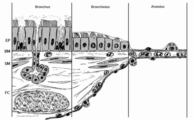
- Phases of lung development
- Histology of the airway epithelium
- Classification of lung tumors
- Adenocarcinoma of the lung
- Neuroendocrine (NE) lung tumors
- Surfactant proteins
- Clara cell specific protein (CCSP)
- Thyroid transcription factor 1 (TTF-1)
- Forkhead box A2 (Foxa2)
Submucous glands of the minor salivary type are found in the submucosa of the trachea and bronchi (but not bronchioles). SP-A is required for the formation of tubular myelin and has various roles in lung defense functions [23-26].
Objectives
Materials and Methods
- Fetal and neonatal lung tissue
- Tumor tissues
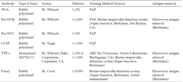
- Antibodies
- Immunohistochemical methods
- In situ hybridization probes
- In situ hybridization procedures
- Statistical analysis
The orientation of the subcloned cDNA relative to the T3 and T7 promoters was determined by restriction mapping analysis. The orientation of the subcloned fragment relative to the T3 and T7 promoters was assessed by restriction mapping analysis.
Results
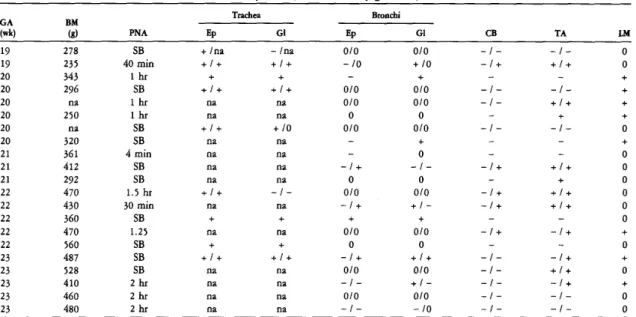
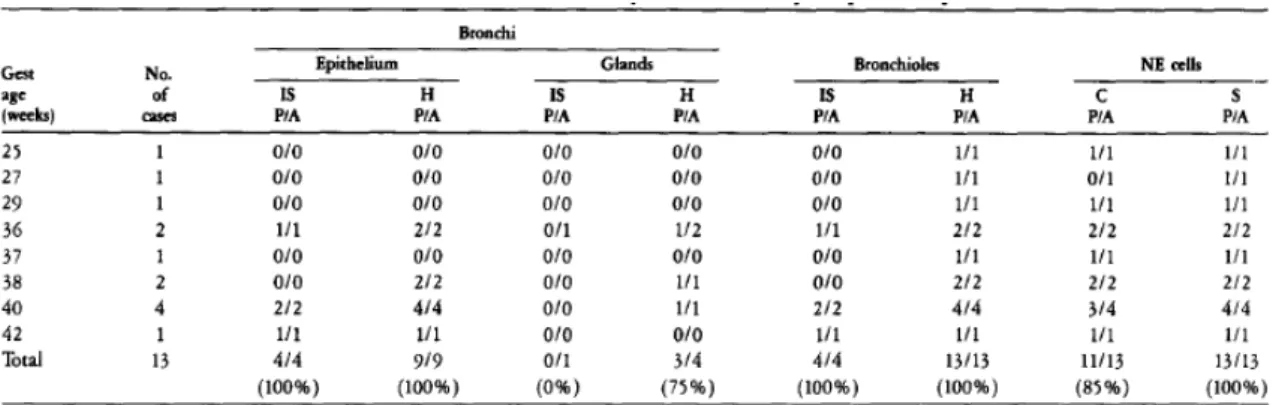
Expression of SP-A and SP-A mRNA in the developing lung
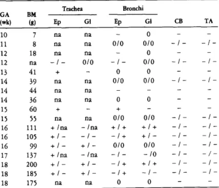
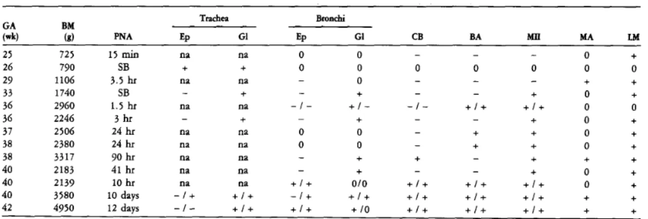
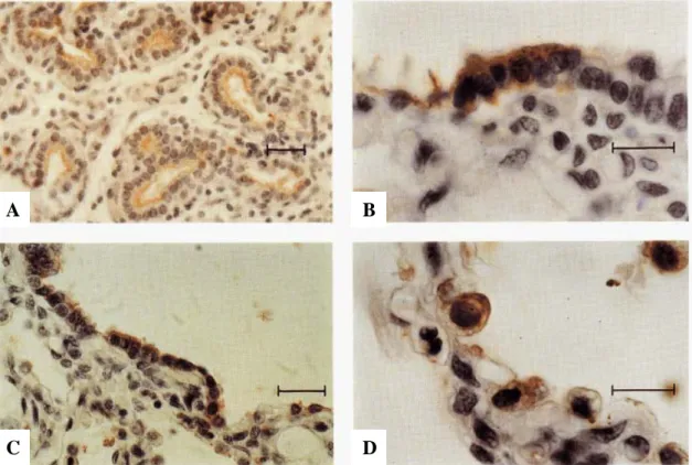
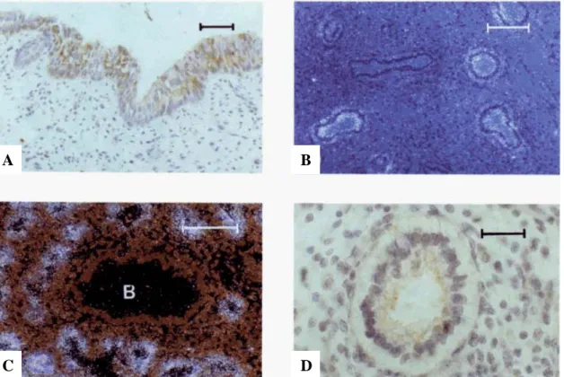
GA, gestational age; BM, birth weight; PNA, postnatal age; Ep, epithelium; Gl, glands; CB, ciliated bronchioles; TA, terminal airway cells; LM, luminal material. Many cells in submucosal glands are stained for SP-A. B) Serial section of the same field shown in A. A few cells from the epithelial lining in the depths of folds are immunostained for SP-A. Many cells in submucosal glands are also stained.
Cuboid cells of the terminal airways are weakly immunostained for SP-A. B) Lung from a term infant who survived 10 days and died of non-pulmonary causes. Cells from a bronchioloalveolar portal are stained for SP-A. D) Lung from a full-term infant who survived 10 days and died of non-pulmonary causes. Lung tissue was hybridized in situ with an anti-sense SP-A probe and photographed with (A) bright field and (B) dark field illumination.
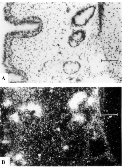
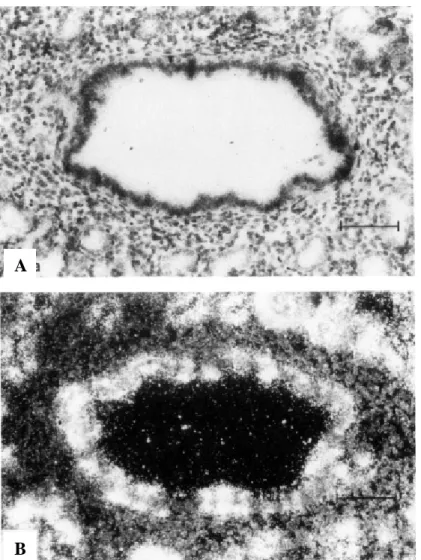
SP-A mRNA in scattered columnar epithelial cells and submucosal glands in the trachea of a 23-week fetus. A section of the trachea was hybridized in situ with an anti-sense SP-A probe and photographed with (A) brightfield and (B) darkfield illumination.
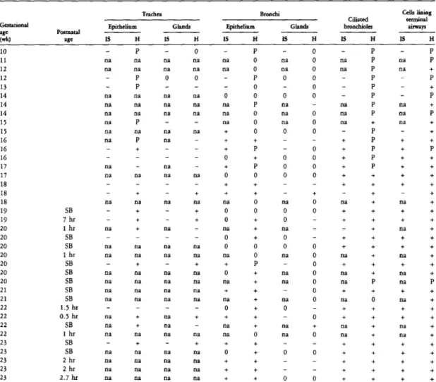
Expression of pro-SP-B and SP-B mRNA in the developing lung
IS, in situ hybridization; H, immunohistochemistry; P, peripheral staining; SB, stillborn; 0, the structure is not present in the section; on, not available. Fetal lung tissue at 23 weeks of gestation was in situ hybridized with an antisense SP-B probe and photographed using dark-field illumination. Fetal lung at 16 weeks of gestation hybridized in situ with the SP-B antisense probe and photographed with dark-field illumination.
Lung of a 40-week-old infant. pregnancy hybridized in situ with the antisense SP-B probe and photographed using dark-field illumination. There is immunolabeling of non-ciliated bronchiolar cells and numerous type II cells in the terminal airways.
Expression of pro-SP-C and SP-C mRNA in the developing lung
IS, in situ hybridization; 0, the structure is not present in the section; H, immunohistochemistry; no stained cells found; on, not available. The terminal airway of a 15-week-old fetus shows weak immunoreactivity for pro-SP-C. A) Fetal lung at 20 weeks of gestation, immunolabeled for pro-SP-C in the epithelial cells lining the terminal airways. SP-C mRNA is localized to type II alveolar cells and bronchiolo-alveolar portal cells (arrow).
Expression of CCSP and CCSP mRNA in the developing lung
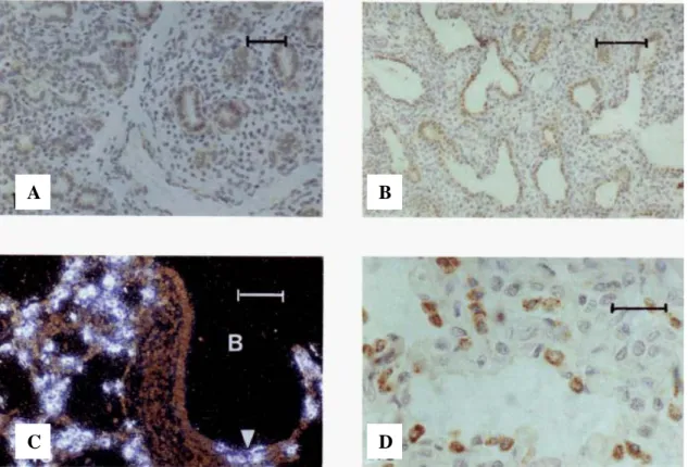
The egg-shaped luminal surface of the NEB at the point of branching is covered by a "crown" of immunolabeled cuboidal cells. Four NEBs, some of which are at branch points, are surrounded by immunolabeled cells of the epithelial lining. Bronchioles from fetal lungs at 22 weeks of gestation hybridized in situ with the CCSP antisense probe and photographed under dark-field illumination.
CCSP mRNA is expressed in scattered cells of the epithelial lining and in clusters associated with the NEB. At the sites of branching (arrows), there are two NEBs associated with an immunolabeled epithelial lining. D) Adjacent section of the same lung as shown in (C), immunostained for bombesin. The second NEB shows only a trace of bombesin (arrowhead). A) Lungs of an infant at 31 weeks' gestation who had a lobectomy 28 days postpartum are immunostained for CCSP.
Immune Labeled columnar epithelial lining cells are scattered over the surface of the folds, but are not seen in the flanking areas in the depth of the folds.
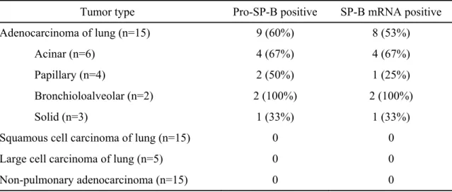
Differential expression of pro-SP-B and SP-B mRNA in NSCLCs and non-
Squamous and large cell lung carcinomas did not stain with the pro-SP-B antibody. The sensitivity and specificity of pro-SP-B for lung adenocarcinomas were 60% and 100%, respectively. The distribution of SP-B mRNA in these tumors was similar to that of pro-SP-B.
Similar to pro-SP-B, SP-B mRNA was detected in adenocarcinomas with acinar, papillary, bronchioloalveolar, and solid growth patterns. Squamous and large cell carcinomas of the lung and nonpulmonary adenocarcinomas were without any in situ hybridization signal. The sensitivity and specificity of SP-B mRNA signal for adenocarcinoma of the lung were 53% and 100%, respectively.
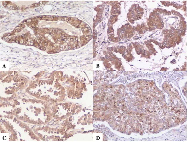
Pro-SP-B immunoreactivity in lung adenocarcinoma with an acinar growth pattern (biotin-streptavidin technique with DAB chromogen; original magnification, 175X). Usefulness of pro-SP-B and TTF-1 in differentiating lung adenocarcinoma from malignant mesothelioma.
The utility of pro-SP-B and TTF-1 in differentiating adenocarcinoma of the lung
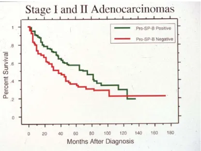
Prognostic value of pro-SP-B and TTF-1 in early-stage lung adenocarcinoma.
The prognostic value of pro-SP-B and TTF-1 in early stage adenocarcinoma of
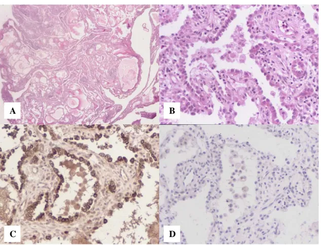
There is a significantly longer survival for patients with pro-SP-B positive tumors versus negative tumors (p=0.0310). Use of TTF-1, Cdx2, CK7, and CK20 in defining the primary site for metastatic brain adenocarcinomas. Expression of TTF-1, Cdx2, CK7 and CK20 in 38 brain metastatic adenocarcinomas and performance of these immunocarcinomas. identification of a primary site are summarized in Tables 17 and 18 [55]. Fifty-five percent of pulmonary adenocarcinomas expressed TTF-1, whereas none of the breast or gastrointestinal primaries did.
As expected, 100% of lung and breast adenocarcinomas expressed CK7, with only 1 gastrointestinal tumor showing positivity. Performance of immunohistochemical markers for identification of a primary site of metastatic adenocarcinoma to the brain. Brain metastasis from a breast carcinoma showing immunoreactivity for CK7 only; hematoxylin-eosin (A) and CK7 (B); original magnifications x400.
Brain metastasis from a pulmonary adenocarcinoma, showing immunoreactivity for TTF-1 and CK7; hematoxylin-eosin (A), TTF-1 (B) and CK7 (C); original magnifications x400. Brain metastasis from a colon adenocarcinoma, showing immunoreactivity for Cdx2 and CK20; hematoxylin-eosin (A), Cdx2 (B) and CK20 (C); original magnifications x400.
Expression of TTF-1 in malignant pleural effusions
Differential expression of TTF-1 and CK20 in SCLC and Merkel cell tumor . 61
The tumor cells show nucleation, fine granular chromatin and scanty cytoplasm; mitoses are frequent (H&E, x130). The tumor cells show strong nuclear immunoreactivity for TTF-1 (biotin-streptavidin detection system with DAB and hematoxylin, x190). The tumor cells are negative for TTF-1 (biotin-streptavidin detection system with DAB and hematoxylin, x 190).
In the non-neoplastic lung, nuclear immunoreactivity to Foxa2 was present in Type II cells; other cell types were unreactive. Panels C and D, original magnification, x400; panels A and C, H&E stain; panels B and D, biotin-streptavidin detection system with DAB chromogen, cobalt chloride enhancement and nuclear fast red counterstain.).

Expression of pro-SP-B, pro-SP-C and TTF-1 in alveolar adenoma
Positive cytoplasmic staining for pro-SP-B (Figure 39C) and pro-SP-C was observed in most of the epithelial cells in all six cases analyzed, confirming the presence of Type II pneumocytes.
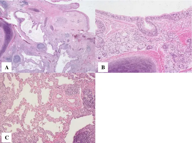
Expression of SP-A, pro-SP-B, pro-SP-C and CCSP in mature teratoma of the
Mature teratoma of the uterine cervix with pulmonary differentiation (H&E stain). with cartilage and submucosal glands.
Discussion
Our first study found the presence of pro-SP-B and SP-B mRNA in lung adenocarcinoma [53]. Pro-SP-B and SP-B mRNA were detected in all major subtypes of lung adenocarcinoma, including acinar, papillary, bronchioloalveolar, and solid. The sensitivity and specificity of pro-SP-B immunoreactivity for lung adenocarcinoma compared with malignant mesothelioma were 57% and 100%, respectively.
The sensitivity and specificity of TTF-1 for lung adenocarcinoma versus malignant mesothelioma were 76% and 100%, respectively. Taken together, our data indicate that immunostaining for pro-SP-B and/or TTF-1 may be useful in differentiating lung adenocarcinoma from malignancy. While both pro-SP-B and TTF-1 were highly specific for lung adenocarcinomas in our study, the sensitivity of TTF-1 exceeds that of pro-SP-B.
Moreover, the combined sensitivity of pro-SP-B and TTF-1 is only slightly higher than that of TTF-1 alone. These data suggest that both pro-SP-B and TTF-1 are positive markers of survival in patients with early-stage lung adenocarcinoma.
Conclusions
Expression of pro-SP-B, pro-SP-C and TTF-1 in lining cells of alveolar adenoma confirms its differentiation towards Type II cells. Expression of SP-A, pro-SP-B, pro-SP-C and CCSP in mature cervical teratoma with pulmonary differentiation confirms this diagnosis.
Civitareale D, Castelli MP, Falasca P, Saiardi A (1993) Thyroid transcription factor 1 activates the thyrotropin receptor gene promoter. Liu C, Glasser SW, Wan H, Whitsett JA (2002) GATA-6 and thyroid transcription factor-1 directly interact and regulate surfactant protein C gene expression. Zhang L, Whitsett JA, Stripp BR (1997) Regulation of Clara cell secretory protein gene transcription by thyroid transcription factor-1.
Besnard V, Xu Y, Whitsett JA (2007) Sterol response element binding protein and thyroid transcription factor-1 (Nkx2.1) regulate Abca3 gene expression. Khoor A, Whitsett JA, Stahlman MT, Olson SJ, Cagle PT. Khoor A, Byrd-Gloster AL, Nicosia SV (2010) Expression of Thyroid Transcription Factor-1 in malignant pleural effusions.
Byrd-Gloster AL, Khoor A, Glass LF, Messina JL, Whitsett JA, Livingston SK, Cagle PT (2000) Differential expression of thyroid transcription factor 1 in small cell lung carcinoma and Merkel cell tumor. Zhou L, Lim L, Costa RH, Whitsett JA (1996) Thyroid transcription factor-1, hepatocyte nuclear factor-3beta, surfactant protein B, C, and Clara cell secretory protein in developing mouse lung.
Candidates publications related to the PhD thesis
Khoor A, Stahlman MT, Johnson JM, Olson SJ, Whitsett JA (2004) Forkhead box A2 transcription factor is expressed in all types of neuroendocrine lung tumors. Strickland-Marmol LB, Khoor A, Livingston SK, Rojiani A (2007) Usefulness of tissue-specific transcription factors thyroid transcription factor 1 and Cdx2 in determining the primary site of metastatic adenocarcinomas to the brain.
Candidates publications unrelated to the PhD thesis
Khoor A, Myers JL, Tazelaar HD, Swensen SJ (2001) Pulmonary Langerhans cell histiocytosis presenting as a solitary nodule. Cheung OY, Muhm JR, Helmers RA, Aubry MC, Tazelaar HD, Khoor A, Leslie KO, Colby TV (2003) Surgical pathology of granulomatous interstitial pneumonia. Ott MC, Khoor A, Leventhal JP, Paterick TE, Burger CD (2003) Pulmonary toxicity in patients receiving low-dose amiodarone.
Khoor A, Myers JL, Tazelaar HD, Kurtin PJ (2004) Amyloid-like pulmonary nodules including localized light chain deposition: clinicopathologic analysis of three cases. Regala RP, Weems C, Jamieson L, Khoor A, Edell ES, Lohse CM, Fields AP (2005) Atypical protein kinase C iota is an oncogene in human non-small cell lung cancer. Tun HW, Wallace KH, Grinton SF, Khoor A, Burger CD (2005) Etanercept therapy for late-onset idiopathic pneumonia syndrome after nonmyeloablative allogeneic hematopoietic stem cell transplantation.
Waller EA, Roy A, Brumble L, Khoor A, Johnson MM, Garland JL (2006) The expanding spectrum of Mycobacterium avium complex-associated lung disease. Regala RP, Davis RK, Kunz A, Khoor A, Leitges M, Fields AP (2009) Atypical protein kinase C{iota} is required for bronchoalveolar stem cell expansion and lung tumorigenesis.
Acknowledgements