ALZHEIMER’S DISEASE: THE ROLE OF EXERCISE AND MICROBIOME IN A
TRANSGENIC MICE MODEL
PhD Thesis
Dóra Ábrahám
Doctoral School of Sport Sciences University of Physical Education
Supervisor: Dr. Zsolt Radák, professor, DSc
Official reviewers: Dr. László Tretter, professor DSc Dr. László Virág, professor DSc
Head of the Final Examination Committee:
Dr. József Tihanyi rector emeritus, DSc
Members of the Final Examination Committee:
Dr. Levente Rácz senior research fellow, PhD Dr. Andor Molnár college associate professor, PhD
Budapest 2020
DOI: 10.17624/TF.2021.5
1
Table of contents
Abbreviations ... 3
List of figures and tables ... 5
1.1. Alzheimer’s disease ... 7
1.1.1. Clinical features ... 8
1.1.2. Epidemiology ... 10
1.1.3. Pathology- What are the molecules that are underlying AD? ... 11
1.2. Mouse models in AD ... 17
1.3. Oxidative stress in AD ... 18
1.4. Microglial activation in AD ... 19
1.5. Methods of prevention ... 19
1.5.1. Role of exercise ... 20
1.5.2. Effects of environmental enrichment ... 22
1.6. Microbiome ... 22
1.6.1 Microbial changes during life ... 23
1.6.2. Healthy host means a healthy gut? ... 25
1.6.3. Leaky gut ... 26
1.6.4. Gut-brain axis ... 27
1.6.5. Microbiome and exercise... 29
1.6.6. Microbiome and diet ... 30
1.6.7. Microbiome and AD ... 31
2. Objectives of the study ... 33
3. Materials and methods ... 35
3.1 Origin of the animals ... 35
3.2 Protocols in the animal house ... 35
3.2.1. Cognitive tests ... 37
3.2.2. Immunohistochemistry ... 42
3.2.3. Western blotting ... 43
3.2.4. Library preparation and identification of prokaryotic species ... 44
3.3 Statistics ... 45
3.3.1. Microbial analytical methods ... 45
2
3.3.2. Bioinformatics analysis of the microbiome ... 46
4. Results ... 47
4.1. Results from the animal experiments ... 47
4.1.1. Animal weight change during the experiment ... 47
4.1.2 Cognitive test results ... 48
4.2. Results from brain tissue investigation ... 57
4.3. Results from microbiome analysis ... 60
5. Discussion ... 70
6. Conclusions ... 75
7. Summary ... 76
7.1. Summary in English ... 76
7.2. Summary in Hungarian- Összefoglalás ... 77
8. Bibliography ... 79
9. Bibilography of own Publications ... 93
10. Acknowledgments ... 94
3 Abbreviations
AD Alzheimer’s disease
ANOVA Analysis of variance APP Amyloid Precursor Protein
Aβ β-amyloid
BBB Blood brain barrier CNS Central nervous system
CV Cardiovascular
CVD Cardiovascular Disease
DSM Diagnostic and Statistical Manual of Mental Disorders ENS Enteric Nervous System
GI Gastrointestinal
GIT Gastrointestinal tract
HIIT High Intensity Interval Training
HM Human Microbiome
HO- 1 Heme Oxigenase-1
HPA Hypothalamic-pituitary-adrenal HRP Horseradish peroxidase
ISP Ion Sphere Particles LAB Lactic acid bacteria
MCI Mild Cognitive Impairment
MICT Mild Intensity Continuous Training
MWM Morris Water Maze
NEP Neprilysin
NEP2 Neprilysin-2
NFT Neurofibrillary tangles NOR Novel Object Recognition
NRF2 Nuclear factor erythroid 2-related factor 2
OFT Open Field Test
OGG1 8 oxoguanine DNA glycosylase 1
oxoG 8-oxoguanine
4
PB Phosphate buffer
PFA Paraformaldehyde
PS1 Presenilin 1
PS2 Presenilin 2
ROS Reactive oxygen species SCFA Short chain fatty acid
WT Wild type
5 List of figures and tables
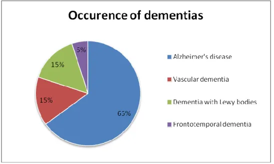
Figure 1. Percentage distribution of dementias ... 10
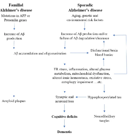
Figure 2. Amyloid cascade hypothesis ... 12
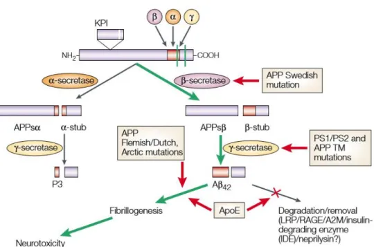
Figure 3. Route of amyloid processing ... 14
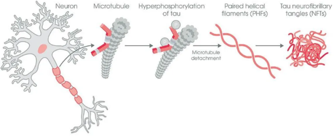
Figure 4. Neurofibrillary tangle formation ... 16
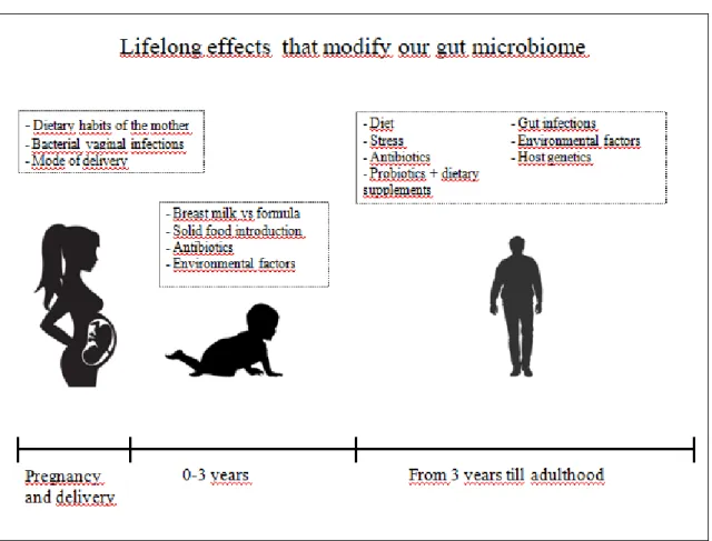
Figure 5. Lifelong effects that modify our microbiome ... 25
Table 1. Dietary and lifestyle guidelines to prevent AD. ... 32
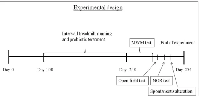
Figure 6. Experimental timeline ... 37
Figure 7. MWM test ... 38
Figure 8. Open field arena ... 39
Figure 9. NOR test arena ... 40
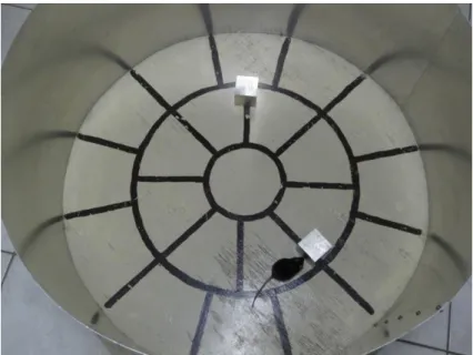
Figure 10. Y maze for spontaneous alternation test ... 41
Figure 11. Body mass change during experimental period ... 47
Figure 12. MWM test results including WT ... 49
Figure 13.A. Open field test exploratory activity ... 50
Figure 13.B. Latency time in OFT ... 51
Figure 13.C. Total exploration in OFT ... 52
Figure 14. NOR test ... 53
Figure 15.A. Spontaneous alternation test, APP/PS1TG-C vs WT ... 54
Figure 15.B. Spontaneous alternation test, transgenic groups ... 55
Figure 16. Spontaneous alternation, number of entries ... 56
6
Figure 17. Amyloid plaque depositions in hippocampus ... 57
Figure 18. Microglia number and area in the hippocampus ... 58
Figure 19. OGG1 levels in hippocampus ... 59
Figure 20. Microbiome distribution in phylum level ... 60
Figure 21.A. Strains with effects on mucn production WT vs APP/PS1TG-C ... 62
Figure 21.B. Butirate producer strains WT vs APP/PS1TG-C ... 63
Figure 21.C. Butirate producer strains, transgenic groups ... 64
Figure 21.D. Effect of probiotic lysate on Lactobacillus spp. ... 65
Figure 22.A. Species differences among WT and APP/PS1TG-C... 67
Figure 22. B. Species differences among transgenic groups I... 68
Figure 22. C. Species differences among transgenic groups II. ... 69
7 1. Introduction
1.1. Alzheimer’s disease
There is this old joke:
Grandpa: Grandson, do you know, what is the name of that German officer, who always hides my belongings?
Grandson: Yes, grandpa I know, that is Alzheimer.
Unfortunately, this is not a joke, Alzheimer’s disease (AD) which was named after the German researcher –Alois Alzheimer-, who reported it in 1907, is the most common form of dementia. He described this condition as the patient was disoriented in time and space, she had serious breakdowns, memory impairments and she had difficulties with writing and speaking as well. Postmortem investigation has shown atrophic brain, with especially changed neuron tangles and a serious neuron loss in the upper layer of the cortex (1). Even though it is the cause of 60-80% of dementias, the underlying mechanism is one of the less understood ones, and regrettably, it is still without effective medical treatment (2).
It is not only the most frequent type of dementia, but it rises to one of the leading causes of death in western countries approaching the number of deaths caused by cardiovascular diseases (CVD) and cancer. Not the disease itself is so deadly, but it makes the body weak against illnesses. In most of the cases people with AD die from pulmonary diseases such as pneumonia and in this case, AD is not listed as the main cause of death. Because of this reason exact percent of death caused by AD itself is unclear. It is undoubtedly hard to precisely get AD diagnosed. In 2011 the National Institute on Aging has made new diagnosing criteria in which not only memory loss needs to be proved, but deeper data including biomarker tests are needed (3). Computed tomography scans can be performed to visualize beta-amyloid plaques but a simple and effective blood test to detect the disease with 100% certainty is still needed to be developed.
8 1.1.1. Clinical features
AD is a degenerative central nervous system disease which affects mostly the elderly population, namely people aged 65 or over. It is more and more commonly diagnosed as life expectancy increases, however, there is a tendency that in western countries the number of patients will be lower due to better cardiovascular (CV) prevention. There is an emerging evidence that poor heart function and CV state which is paired with lower cerebral blood circulation velocities and decreased ejection fraction can be a serious risk factor for developing AD (4). As the population ages, AD will put a higher burden to the society, not only medical wise but economically as well. It is estimated that by 2050 the number of patients with this disease will triple (www.alz.org). The fact that there isn’t any medical treatment available which cures the disease, nor any that slows it down, elderly populations will be affected with AD in higher numbers. As a consequence of the disease, individuals will lose the capacity to live their life alone, they will need help continuously, twenty-four hours a day, seven days a week.
AD research has become popular in the last 30 years because governments realized that due to ameliorating medical treatments, the life expectancy increased, so did the number of patients with AD. Hence elevated financial inputs will be required to fulfill AD related care giving costs. Nowadays health care institutes are not prepared to supply this emerging need of patient care. Dementia is described as an acquired condition which has a serious impact on everyday activities and it consists of progressive cognitive impairment (5). Affected persons are not allowed to be left alone, because they are unable to complete everyday tasks such as dressing up, meal preparation or going on a short walk. AD shows different signs, firstly short term-, later long-term memory loss, struggle in day by day activities, speaking, writing and spelling difficulties, decline in problem-solving and navigation skills(6). As neuron loss and cerebral cortex shrinkage develops the ventricles become enlarged. Mostly right and left lateral ventricles are affected from this modification (7).
As time elapses elderly people need more personal assistance. It is usually realized by family members, but in some cases, it is not enough. A more reliable social
9
network is needed to fulfill the care giving needs. According to the latest Diagnostic and Statistical Manual of Mental Disorders (DSM) criteria dementia is a major neurocognitive disease (8).
AD has six stages, each stage usually lasts 5 years. It ranges from early symptoms until serious cognitive impairment and this process normally involves 20-25 years. When the disease begins, it is usually not diagnosed as AD, because symptoms can be the consequences for several other diseases and typical dementia-related signs are missing. Although this period could be the best for medical intervention, any product or method which could prevent Abeta formation, or limit the aggregation of β- amyloid (Aβ) could be disease modifying. As the symptoms are worsening, the disease will be more and more recognizable, but still it can be confused with other types of dementia. The fourth stage is the first one where mild cognitive impairment (MCI) can be investigated, and hippocampal atrophy is evident on a magnetic resonance imaging scan. Only with physical and pathological signs can be approved that it is AD, clinical diagnosis regularly happens in stage V (9). Usually when a patient goes to the doctor with the symptoms it is already too late, the disease is already in a progressive state. In the last phase cognitive decline arises from mild to severe, full-time surveillance is necessary. Typical symptoms are difficulties in eating and swallowing, inability to walk and to move, and the state of immune system becomes fragile.
10 1.1.2. Epidemiology
AD is the main cause of dementia, it accounts for 50-75% of all cases, affecting aging populations. (Figure 1.) In 2018, around 50 million people lived with dementia worldwide. The incidence is rising in low or middle-income countries while it shows a decreasing tendency in high-income countries. Still every 3 seconds a new case will be registered globally (10).
Figure 1. Percentage distribution of dementias
Occurrence of different types of dementia, showing that AD is responsible for the majority of the cases.
AD has two incidences. Sporadic or late-onset form is underlying 99% of the cases, it affects elderly people and it is due to a spontaneous gene mutation. While familial form is less common, it is hereditary, pathogenic mutations are mainly in genes APP, PS1, PS2 and it causes early onset dementia. AD is observed to be more prevalent in women than in man (11).
The reason for this gender dependent distribution is still unclear. Longer life expectancy for woman plays a role in it, or maybe because men have a higher mortality rate from other diseases. In the aging brain sex hormones have a crucial, defending role.
Hormonal changes in females can also play a role in AD development. Estrogen
11
adjustment at the time of menopause may reduce the risk for AD. As for females the estrogen loss, the testosterone loss in males is also a normal, age-related phenomenon which automatically brings a higher risk for the disease (12).
1.1.3. Pathology- What are the molecules that are underlying AD?
It is still unclear why the disease develops, whether it is the intracellular amyloid deposition or the extracellular tau aggregates that cause AD or they are only consequences? The individuals suffering from AD have thinner cerebral cortex in the temporal, orbitofrontal and parietal brain regions compared to healthy individuals (13).
Patients with this disease have senile plaques extracellularly mainly in the cortex and in the hippocampus, and neurofibrillary tangles (NFT) can be observed intracellularly. The amyloid cascade hypothesis (Figure 2. (14)) was originally declared back in 1992, suggesting that amyloid plaque deposition is the very first step to develop AD. Amyloid plaque depositions were declared as the main cause for the development of NFTs, cell loss and dementia (15). This hypothesis is partly accepted as an explanation for the developing disease. Familial AD evolves because abnormal formation and number of Aβ is present in the brain of the patients and combined with this, insufficient clearance takes place in disease specific brain regions. This imbalance can also be one of the factors underlying this disease. In contrast to this, it was proved, that in sporadic AD no correlation can be found between the plaque density and memory loss, still Aβ can be the initial step towards AD (16). So far scientific studies were unable to completely explore the mechanism underlying this disease.
12
Figure 2. Amyloid cascade hypothesis
According to the amyloid cascade hypothesis Aβ accumulation and aggregation generates amyloid plaques and initiates a cascade mechanism (14). Later on this causes the dysfunction of BBB leading to inflammation, NFT formation and neuron loss. These changes are ending in cognitive impairment and dementia.
1.1.3.1. APP
The above mentioned hypothesis suggests that Aβ plaque deposition is crucial to develop the disease. Amyloid precursor protein (APP) is encoded on chromosome 21, and it is also present in healthy humans. It’s neuronal function is still unknown, but it might have a role in synaptic plasticity (17). Aβ is the product of 2 cleavages made on APP, the first cleavage happens on the extracellular domain by β- secretase, the second which is made by γ-secretase happens on the transmembrane region (18). These 2
13
cleavages are necessary to form the 37-43 amino acid length Aβ (19). (Figure 3.) When APP has no mutation, it is usually cleaved by α- secretase resulting in a secreted APPα which is the amino terminal fragment of APP. Carboxy- terminal fragment is called CTF83 which is cleaved by γ-secretase but at the end, the remaining molecules P3 extracellularly and the amino-terminal APP intracellular domain is nontoxic (20). For example, Swedish mutation in APP initiates the cleavage by the β- secretase instead of α, resulting in Aβ that can aggregate in the extracellular space. It is suggested that extracellular deposits of Aβ are the triggering factor for neuroinflammation, microglial activation and also for tau tangle formation. Plaques are insoluble, and after their formation is finished, they are not so toxic. Latest inspections propose that soluble Aβ40 and Aβ42 which can be found in the plasma and central nervous system (CNS) of the patients trigger toxicity. Most mutations that affect APP are resulting in an elevated Aβ42 to Aβ40 ratio in the plasma, and the patient usually has an elevated presence of Aβ42 in CNS (21).
14
Figure 3. Route of amyloid processing
The route of amyloid processing (22) APP could be cleaved by α,β and γ secretase. In a healthy state cleavage happens initially by α- secretase followed by a cleavage made by
γ secretase generating a final nontoxic 2 products, P3 extracellularly and a nontoxic APP fragment intracellularly. If Swedish mutation alters APP cleavage happens firstly by β-secretase followed by a γ secretase cleavage thus generating the toxic Aβ42 which
is able to aggregate and this mechanism will result in neurotoxicity.
1.1.3.2. Presenilin 1 and Presenilin 2
Presenilin 1(PS1) protein is the proteolytic subunit of the γ-secretase. Mice that overexpress a mutant PS1 are developing increased levels of Aβ42. More than 50 PS1 gene mutations were discovered in patients with AD. Mutations in this gene are the biggest cause of early onset dementia. Presenilin 2 (PS2) also plays a role in APP cleaving, and also can be blamed for early-onset AD, still, it is only responsible for around 5 % of the cases while PS1 is responsible almost for 70% of the cases (23).
15
Mutations in PS1 and PS2 are resulting in an increased ratio of Aβ42 to Aβ40 as well as mutant APP.
1.1.3.3. Neprilysin and neprilysin-2
Insufficient Aβ clearance can be one of main causes of AD. Several proteolitic enzymes are found to take part in Aβ degradation, from which neprilysin is considered to be the most important one. Neprilysin (NEP) and neprilysin-2 (NEP2) are both zinc metalloendopeptidases and are part of the metalloprotease13 family (24). NEP is present in the periphery and in the central nervous system with a function of small peptide degrading (25). Even though NEP is responsible for Aβ degradation, elevated levels of this enzyme are still insufficient for Aβ elimination. Other NEP like enzymes such as NEP2 is also an important participant in Aβ degradation. It was shown that NEP2 knockout mice had elevated accumulation of Aβ species in the hippocampus (26).
1.1.3.4. ApoE
ApoE gene is the most important risk factor for familial AD. It has 3 alleles ApoE2 which is considered to be protective, ApoE3 is considered to be neutral and last but not least ApoE4 is considered to be a genetic risk factor for AD. In vivo and in vitro studies suggest that ApoE4 has an impact on Aβ clearance. (27). ApoE4 binds to Aβ peptides at residues 12-28, which can modulate Aβ accumulation, if this binding site is blocked amyloid plaque deposition can be prevented in the affected brain areas (28).
1.1.3.5. Tau protein
Tau protein is present in the healthy brain mainly in neurons, with the role of microtubule stabilization and polymerization. Abnormal phosphorylation of microtubule-associated protein tau results in a paired helical filament structured tau and NFTs. (Figure 4.) It was observed that hyperphosphorylated tau is present in the brain of patients suffering from AD, while healthy individuals do not have this mutation (29).
16
NFTs are one of the main pathophysiological characteristics of AD, they are responsible for the neuron loss. Phosphorylated tau appears in neurons well before tangle formation is present. This suggest that previous to disease recognition signaling pathways are already damaged (30). Mutations which affect tau function and/or isoform expression are more likely to get phosphorylated. Phosphorylated tau destructs microtubules and forms tangles, this modified structure can intrude axonal transport thus causing cell death (31). It was revealed that not only phosphorylation can convert tau into NFTs but truncation of the N or C terminal can also end in tau aggregation. It is still a question weather phosphorylated tau or truncated tau is more toxic to the cell (32).
Figure 4. Neurofibrillary tangle formation
Tau protein takes part in microtubule stabilization. When it gets
hyperphosphorylated, tau proteins destruct microtubules and aggregates forming NFTs (33).
17 1.2. Mouse models in AD
Mouse models can be used to study the pathology and progression of AD because they are able to mimic the effects of the disease such as Aβ plaques, tau tangles simultaneously with cognitive decline. Adversely none of these mouse models can represent other significant changes caused by the disease (34).
The first successful transgenic mouse model, using human APP was able to represent amyloid plaques within the mouse brain. Moreover, they have displayed several pathological features of AD. Model animals displayed a 5-14 fold higher Aβ presence than non transgenic littermate controls (35). Shortly after the first model, another human APP mutant mice was developed (36). After that presenilin mutant mice came to sight, to show that mutations in APP cleaving enzymes can also lead to Aβ accumulation (37). As knowledge expanded about AD the number of models representing different features of the disease increased. From the first transgenic model, scientific community arrived to a point where several different model types are available to choose from in close accordance with the investigation needs. Mice which can develop not only Abeta aggregates but tau tangles as well are available, allowing a more profound discovery on the mechanisms regulating the evolvement of AD.
Different model types are available according to the method of how the transgenic animal was made. Two different options can be used to gain a transgenic animal. The first option is to introduce a genetic mutation on top of existing genetic background. Second is to modify an existing gene of interest in its original position (38). Most common transgenic models contain the following modified genes: human APP, presenilin, tau and human ApoE (39). Even though numerous models and possibilities are available to study this disease in mice, unfortunately, these models cannot mimic exactly the disease pathology, as it happens in the human body. The main cause for this is that the immune system reacts differently in mice, it cannot recognize perfectly the human peptides applied in the models.
18 1.3. Oxidative stress in AD
Damage made by oxidative stress happens before the first hallmark of AD is present in the human brain (40). Neurons are more affected from oxidative stress than other cells in the brain because their metabolic rate is higher. Neurons on one hand contain polyunsaturated fatty acids, which enables them to be more responsive to reactive oxygen species (ROS), generating lipid peroxidation and molecular destruction.
On the other hand glutathione content, which is a known antioxidant is really low in neurons (41). Oxidative stress contributes to lipid, protein and/or nucleic acid oxidation, mitochondrial dysfunction, metal accumulation, tau pathology and inflammation (42).
Mitochondrial dysfunction is undoubtedly one of the main ROS producer in AD, with signs of abnormal mitochondrial axonal trafficking, and with disrupted glucose metabolism. These problems lead to an elevated ROS production which contributes to early stages of AD. However there are several factors which contributes to ROS production, there is no available antioxidant cure to prevent oxidative damage in AD (43).
Nuclear factor erythroid 2-related factor 2 (Nrf2) is an important gene taking role in the fight against oxidative stress. Nrf2 levels are usually low in AD. It was observed that Nrf2 regulates other enzymes that are taking part in redox regulation such as heme oxigenase-1 (HO-1) and glutathione cysteine ligase modulatory subunit.
Elevated expression of Nrf2 in AD models reduces oxidative stress moreover if it was overexpressed in the territory of hippocampus it results in better memory and learning.
On top of that the activation of HO-1 improves learning and memory in mouse models of AD (44).
DNA damage caused by oxidative stress is also a main symptom in AD.
Oxidative DNA damage removal is accomplished by 8-oxoguanine DNA glycosylase 1 (OGG1) (45). The level of this glycosylase is significantly lower in AD patients than is controls (46). OGG1 is involved in the first step of base excision repair, which is the most common pathway in DNA damage removal. It excises 8- oxo- 2’-deoxyguanisine from DNA. OGG1 enzyme has numerous variants and it seems that there is a great significance of which genotype is present in the plasma of AD patients (47).
19 1.4. Microglial activation in AD
Microglia is a phagocyte within the CNS and plays a critical role in Aβ degradation, it can interact with the soluble forms and also with the insoluble forms of Aβ. This interaction is different in both cases (48). Microglia is found to be in close relation with amyloid plaques showing an activated phenotype. Their size and number is in correlation with plaque size. Even though they are phagocytes, CNS resident microglia is unable to remove Aβ plaques with phagocytosis (49). Uncontrolled microglia activation can lead to chronic inflammation and to the release of several inflammatory cytokines thus causing neurodegenerative diseases such as AD. Still these cells are the only ones to mediate immune responses in the CNS. In addition to inflammatory cytokine production, microglia is able to produce anti inflammatory cytokines and reactive oxygen intermediates (50). In late onset AD there is emerging evidence that activated microglia is responsible not only for continuous inflammation but also for neurodegeneration and synapse loss In AD microglial activity has two faces. Firstly they are cells responsible for brain health and tissue homeostasis with a sufficient Aβ clearance serving as a protective factor against AD. Secondly if Aβ accumulation occurs and microglial activation is continuous, neuron loss and inflammatory cytokine production happens helping the disease to develop. These results suggest that microglial activation as a therapeutic strategy is only suitable in the early stages of AD (51).
1.5. Methods of prevention
So, what can be done to prevent this disease? Not much in fact. It is believed that regular physical exercise, a healthy diet, and continuous learning can help. Several types of research have proved that physical exercise as for the body and as for the brain can be beneficial. Lifelong learning can keep the mind refreshed, and in good condition.
Also, it is believed that a healthy diet can help to postpone the disease onset. It is observed that people who received better education and live in better circumstances are less affected by AD.
20 1.5.1. Role of exercise
The effects of regular exercise have been in focus of research for a long time, and it is proved to be beneficial in several diseases and in numerous health conditions.
Practiced at any age, for any duration and in any type, it is hard to say any disadvantage about exercising. It can help fight against negative stress, create an esthetic body, establish a more positive thinking, keep a healthy lifestyle and on top of all that it can prevent numerous diseases. There are some obvious examples such as high blood pressure, diabetes and excess weight caused health problems like back and knee pain, insomnia and hampered respiration where exercise can be a reassuring solution.
Exercise can also cause some not so obvious changes in the body, it is liable for neurogenesis in the hippocampus which is the most important area in the brain for learning and memory forming (52). Also, it can ameliorate the survival of newborn neurons in the dentate gyrus (53).
Activity of OGG1 is also upregulated by the effects of exercise thus reducing DNA damage (45). This enzyme is responsible for the repair of 8-oxoguanine (8-oxoG), the most abundant DNA lesion caused by oxidative stress. These lesions become more abundant with age, and they can be responsible for inflammation, and can be related to CNS diseases such as AD, and Parkinson’s disease (54). Interestingly, exercise is not altering the ROS levels in the brain (55).
It is believed that regular physical activity can also ameliorate CNS related diseases, not just memory improvement but it could delay the progression of AD as well as lower the incidence of it (56). Furthermore, exercise can reduce the risk of developing AD (57). However the exact mechanism underlying this phenomenon is still unclear, it is believed that exercise can increase the metabolic function of the brain and can reduce the effect of oxidative stress in AD (58).
There are several studies that prove that voluntary or forced exercise can slow down disease progression and can amend physical symptoms caused by the disease.
Voluntary exercise is known to have more beneficial effects than forced exercise however it is mainly because of stress-related factors. Voluntary and forced exercise differs in training time and in running speed. If the goal is, to achieve the same distance during the training period, animals which were allowed to run voluntarily, accomplished
21
the distance with several short running sessions for a faster speed, than animals that were forced to run. At the end, voluntarily exercising animals reached the distance goal first. At the end of training months, only anxiety related experiments showed an advantage for animals in the voluntary exercise group. In open field test these animals were more active and crossed more inner areas, while there weren’t any differences neither in Morris water maze test (MWM) nor in body weight measurements (59).
Interval training is a time efficient way to do daily exercise. It is believed that high-intensity interval training (HIIT) is a more efficient way for weight loss than moderate intensity continuous training (MICT). HIIT is composed of two different intensity sections, one of them is a high-intensity period, and the other is low-intensity period. These two sections are changing during the training period creating more challenging cardiovascular training. It turned out that this method is more efficient only if we take time into consideration (60). It has no more beneficial effects than MICT in weight loss or in body composition.
Treadmill running reduced Aβ42 levels in the brain of Neuron specific enolase/APPswe tg mice, also it reduced the escape latency time in the MWM compared to the sedentary group (61). As a prevention method in APP/PS1 mice, regular voluntary physical activity is proved to slow down the disease development (62). It has also been observed, that forced treadmill running can significantly improve the memory deficit and the learning abilities of APP/PS1 transgenic mice (63). When exercise was used in a progressed, irreversible state of AD, unfortunately no improvement was shown in the cognitive function of the animals due to the training. There was no improvement in the escape latency time in MWM nor decreased number in the Aβ plaque burden in motor-related brain regions of APP/PS1 mice at the age of 10 months (64). It seems that in the progression of AD exercise alone cannot play a protective role, maybe human microbiome can have a similar retentive effect on disease pathology. It is well known that exercise can reversibly alter the microbiome, not only the composition but the function as well (65). The only question is whether changes in microbiome can help delay the onset of the disease?
22 1.5.2. Effects of environmental enrichment
The role of environmental enrichment is controversial, because this type of housing differs from conventional housing. Cage with enrichment could contain a running wheel, toys for the animals and sometimes the cage has a bigger area collectively. Thus, it is hard to observe the effects of environmental enrichment solely.
Positive effects regarding enrichment consistently come from the beneficial effects of exercise. In mouse models of AD it was proved that enriched housing results in reduced Aβ level in the brain together with decreased number of senile plaques. Moreover, the enzyme responsible for Aβ degradation, neprilysin, was observed in a higher quantity (66). Both factors together, physical activity and environmental enrichment can trigger cognitive improvement in models of CNS diseases (67). Even so environmental enrichment alone may induce synaptogenesis, and can have beneficial effects on synaptic plasticity (68).
1.6. Microbiome
The human microbiome (HM) was not in the center of research until very lately.
In the last decade there is emerging evidence that the microbiome is far more essential than it was imagined earlier. Its composition and its function can be the underlying cause for numerous illnesses. HM consists of various participants such as archaea, protozoa, viruses, eukaryotes, and mainly bacteria. These organisms inhabit different parts of our body for instance skin, mouth, vagina, oesophagus and gut where they are present in a different composition. The biggest part, around 95% of the HM is in our gastro intestinal (GI) tract which plays a major role in nutrition, inflammation, immunity and its composition can affect neuronal function(69). Although the content of the microbiome not only differs from person to person, from age and from the ethnical origin, the main characteristics can be generalized and observed. It was believed for a long time that microbes in our body are outnumbering the number of human cells in a tenfold magnitude.(70) A recent study from 2016 revealed that this ratio is almost 1:1, to be exact is 1.3:1 (71). If we consider gene number this ratio is even much higher
23
150:1 (72) which is such an enormous quantity, that it makes complicated to investigate every participant and every function of the HM. Lately gut microbiome research became a hot topic, numerous researchers suggest that there must be a lot on our gut microbiome if we talk about health or disease. Functional relevance of the microbiome is really wide, it is fundamental for normal GIT (Gastrointestinal tract) motility, digestion and host metabolism, it takes part in the maintenance of barrier function, it is responsible for epithelial cell repair after injury, it takes part in immune function and pathogen recognition (73). A bidirectional pathway is suggested to exist between the gut and the brain in contrast to the previously believed only one direction pathway, that CNS regulates the enteric nervous system.
1.6.1 Microbial changes during life
The human gut microbiome is an interesting and continuously changing environment. During pregnancy, in the intrauterine life, a part of the maternal microbiota is already inherited through the placenta. Lactobacillus and Bifidobacterium DNA was isolated from human placenta suggesting an evidence against the uterus being sterile (74). When a baby is born, normally it inherits the microbiome from the mother’s vaginal microbiome, which composition allows easier breast milk digestion. In contrast to this if someone was born through C section, the primary bacterial colonization happens from the mother’s skin flora. In newborns the majority of bacteria which colonizes the GI tract is Lactobacilli, whose function is to prepare the environment in the gut for further bacteria until total GI maturation is reached (75). Later on, bacteria are obtained from the environment. It is observed that the microbiome of babies fed by breast milk is mainly different from the microbiome of babies fed by formula. If the formula contains prebiotics, the microbiome composition of formula fed babies can be more similar to the microbiome of babies fed by breast milk (76). Gut microbiome changes a lot within the first 3 years of age, the number and the variety of species become larger. The GI tract composition can mirror the food intake as well as antibiotics can transform the original composition of it. (Figure 5.) These differences can be observed even several years later since early life microbiome is the foundation of
24
the adult microbiome. It is suggested that aberrant microbiome between ages 0-3 years can lead to diseases, most probably due to the altered development of immune system (77). After early years the gut microbiome reaches a balanced state, it still has shifts and changes, but it is more stable, renews and adapts time to time during adulthood.
Microbial shifts can occur due to an illness, antibiotic usage, or even due to new type of diet, but normally the HM can remain stable for months in some cases even for years (78). The composition of the GIT on its functional level doesn’t change a lot during a lifespan. On the contrary on species level the actual composition transforms doubtlessly.
Changes in the GI tract are slowing down by age 65, elderly people have less diverse microbiome and differences between individuals are blanching (79).
The gut microbiome is a self regulating system, the human body attempts to keep it in a balanced state, maintaining all necessary functions, without diverging from the original setup. Each person has a 150-400 species in their gut, which is mainly composed of 4 phyla: Firmicutes, Bacteroidetes, Actinobacteria and Proteobacteria.
Individual bacterial diversity depends on age, diet, weight and geographical factors (80).
If the microbiome of two individuals is compared, a huge difference can be observed between them. Interestingly, during a one year observation period, the composition of the microbiome of the individuals do not varies significantly so the differences between the 2 individuals will not disappear or become even more different. In contrast to the microbiota stability, it can be observed how external factors have a huge role in the microbiome formation. Traveling to a different country, consumption of food from different gastro culture and gut infections can easily destabilize the gut balance. These changes can perturb the structure up to the phylum level. Reversibility of perturbation can have 2 types, environmental and community disturbance model. When environmental perturbation happens, changes being done in the microbiome will go back to the original structure when the environment goes back to the original state, for example when we return home after a long journey. Community perturbation results in altered microbiome composition which can be stabilized, and later on, when perturbation vanishes this newly formed community remains stable and functional stability can become permanent (81).
25
Figure 5. Lifelong effects that modify our microbiome
It is easily noticeable, that a set of factors are responsible for the gut microbiome development. Most aggressive effects are affecting the microbiome in the first 3 years
of our life.
1.6.2. Healthy host means a healthy gut?
As it was mentioned previously, every individual has a continuously changing microbiome, but somehow this system is still able to keep a balanced state. Sometimes, this stability is broken. How can it be known, that for the proper microbiome function help is needed to restore the balanced state? Is it possible to know, what should be done to improve the state of the gut microbiome? What can be considered as a healthy microbiome? If the host is healthy does that mean that the host microbiome is healthy too, or the host can be healthy only if the gut is healthy? It was proven before that the
26
state of microbiome can affect the central nervous system of the host. It was observed on patients suffering from autism or Parkinson’s disease (82) (83) (84).
If the microbiome is considered to be healthy, a higher and ticker mucus layer is observed together with higher short chain fatty acid (SCFA) and antimicrobial signal production. This elevated SCFA level helps to reduce food intake and improve glucose absorption (85). Main SCFAs produced by gut bacteria are butyrate, propionate and acetate from which butyrate is the best known for its beneficial effects. After a diet rich in fiber elevated levels of butyrate and acetate were observed together with reduced apetite and with improved glucose tolerance. While increased butyrate level is beneficial to the host, increased fecal propionate level predicts a higher risk for type 2 diabetes (86). On the contrary, low diversity of the gut microbiota can be in association with an unwell health state including allergy, obesity, bowel diseases and even neurological disorders. For a healthy body the gut microbiome must be in contact with the host’s immune system (87). When the microbiome is investigated it is essential to measure not only the presence of several bacterial species but their activity as well. The activity measurement is not that easy as it seems. In one hand there is no easy and cost efficient way to quantify the metabolite production of all bacterial species. On the other hand the microbiome of every individual should be examined separately. There is no current understanding on how the activity of microbes vary within the same disease (88). Recent findings ensure that every person has a unique microbiome, it presents a new possibility to develop personalized medications. Individual medication usage will bring less side effects, reduced disease risk together with a more effective, non invasive and affordable disease treatment (89). Considering these facts, it is impossible to declare what kind of microbiome composition can be stated as a healthy microbiome.
1.6.3. Leaky gut
Leaky gut is a common and summarizing nomenclature for increased epithelial permeability in the GI tract, most commonly because of mucosal damage caused by stress or inflammatory factors. Stress factors can be endurance training, non-steroidal anti-inflammatory drug administration and pregnancy. These factors can cause mucosal
27
damage and modify intestinal permeability. Stress caused damaged can be reversed by an appropriate diet without any pharmacological intervention. On the contrary damage caused by some inflammatory event such as allergy, ageing, CNS disorders cannot be restored with diet because the intestinal barrier function normalization will not repair the modification. Still it is not proven that increased gut permeability can cause any serious problems (90). Mucosal permeability may activate a mucosal immune response.
This activation can depend on the severeness of mucosal deregulation and also on the circumstances. It seems that the state of mucosa is in active connection and cooperation with the immune system so inflammatory response is not obligatory (91). One of the most common bacteria in the gut is Bacteroides thetaiotaomicron which is a well known acetate producer, it hydrolyzes polysaccharides delivered from diet or from the host and it increases goblet cell differentiation. Under normal circumstances it is part of the normal gut flora and it can promote mucus production. If an acetate consumer and butyrate producer bacteria is present in association with Bacteroides thetaiotaomicron this effect can diminish and can lead to thinner mucus layer (92). Acetate overproduction in the GIT usually happens if a diet high in fat and calorie is followed.
Acetate can trigger an elevated insulin secretion in the gut causing insulin resistance and later on obesity (93). Also it can promote the secretion of a hunger hormone called ghrelin which can cause increased food intake creating a continuous loop of eating, acetate overproduction, ghrelin production and then eating again (94). It is suggested that leaky gut can underlie some CNS disorders by influencing blood brain barrier function (BBB). If this is so, small bacterial components can pass the BBB (95).
1.6.4. Gut-brain axis
It was observed in the last century that emotional changes can have different effects on the GI tract. But it is a recent finding that alterations in microbiota composition can affect brain function. It is suggested that variations in the microbiome can also have several effects on distinct diseases such as autism, anxiety, depression, irritable bowel disease and even memory related dysfunctions can be due to the microbiome. Gut-brain axis is a bidirectional connection between the CNS and enteric
28
nervous system (ENS). Microbes not only have effects on ENS but on CNS as well.
Communication is mediated through neural, neuroendocrine, and neuroimmune pathways. Neuroendocrine pathways are mediated through the hypothalamic-pituitary- adrenal (HPA) axis. For the normal function of this axis the development of an appropriate stress response is essential. This stress response is largely dependent on the normal gut colonization in early life (96). Since it is known that early microbial composition can have an effect on CNS development and function, there is urging need to find the mechanisms and patterns responsible for this phenomenon (97). Most of the findings are based on animal models, mainly with germ free mice where anxiety related behavior is reduced compared to conventional mice (98). Not just antianxiety- but antidepression like behavior was observed in germ free mice (99). First results arrived when stress related factors were investigated, but there is huge hope to find relations between dysfunction of gut microbiome and gut disorders such as irritable bowel disease (100). Nervus vagus was the first hypothetical connection between these two interfaces, and it is partly true that it has an important role in the communication. It is still unclear if the bacterial composition of gut is important when we talk about the effects on CNS, or the secreted metabolites are the responsible for all the changes. The nervous system is able to detect an infection without any immune response just because a pathogen is present in the gut. Also some neuropsychiatric disorders can be improved with antibiotic registration. In these cases there is no proven role of the secreted metabolites, only the presence or absence of bacteria is important. When we talk about metabolites it is even a more complex topic. There is no comprehensive knowledge available about what type of metabolites are secreted from specific bacteria. One type of metabolite can be a product of several different bacterial species, and metabolite secretion can be altered by environmental factors such as diet or infections(101). If there will be a reliable method to investigate bacterial metabolite secretion, most probably that will be the route to follow in the scientific exploration of gut-brain axis.
29 1.6.5. Microbiome and exercise
If the role of exercise on the microbiome is investigated, several evidences can be found suggesting that physical activity can modify the composition of the microbiota. Various questions are arising such as what kind of exercise is able to initiate modifications, how long does it need to be made, what age is optimal to start exercising, how is possible to perceive the changes caused by activity? Will anything be observed outside like bigger muscles, or quicker performance development, or only inside differences will be made such as improved nutrition utilization?
If only training effects are taken into consideration, it is a challenging task to investigate its impact on the microbiome, still there is an arising need to specify the results achieved by workout. It has been reported that physical activity during childhood is able to make the microbiome more diverse and can promote the growth of butyrate producing bacteria. Furthermore, exercise in childhood has a higher influence on microbiome modification than in adulthood. Changes made in childhood can last until adulthood, while changes occurred in adulthood are considered to be more unstable.
These changes might affect body fat content and skeletal muscle mass furthermore in the long run metabolic health can be improved (102). Even though exercise in childhood has enormous beneficial effects on health state, the existing positive effects of regular physical activity in the elderly population cannot be forgotten. It can raise bacterial diversity which may result in a better immunological state, together with an exercise driven anti-inflammatory effect (103). If the fecal samples of sedentary or control animals are compared, it can be observed that their microbiome composition is undoubtedly different, samples from the exercise group showed a higher n-butyrate concentration (104). N-butyrate is a well known SCFA which has an inhibitory effect on tumor development, also its low levels are observed in inflammatory bowel disease.
One of its main roles is to modulate NF-κB activation (105). Elevated butyrate levels were also observed in individuals with a better cardiovascular fitness state, if level of VO2 max is higher the amount of produced butyrate is also higher (106). Butyrate is secreted by bacteria when dietary fiber is processed. Butyrate is essential in the colon, it produces about 70% of the energy used by colonocytes. It has anti-inflammatory effects
30
and it induces regulatory T-cell proliferation. Indeed, SCFAs are important mediators against inflammation. SCFAs are fatty acids that contain less than six carbon atoms, most common forms are butyrate, propionate and acetate. They take part in fiber digestion, and they are important molecules because they can cross the BBB. In a mouse model of AD it was presented that the number of SCFA in the fecal samples was significantly lower than in control animals, which can result in the perturbation of several metabolic pathways, therefore opening new possibilities to therapy (107). Most probably SCFAs can mediate the effects of probiotics and prebiotics, and they can be involved in the communication between the GIT and CNS (108).
1.6.6. Microbiome and diet
Diet is one of the main modulators of the microbiome. Dietary intake alters the microbiome on a daily basis. It can modify its components on both the short and long run. A one-off change can have a serious impact on its state, while a continuous life change can modify the roots of the microbiome composition. It is believed that human nutrition intake is mainly dependent on gut bacterial composition, namely the food preferences are in accordance with bacterial needs. Specific carbohydrates are proved to be the main nutrition to them. Some of the food carvings should be due to our microbiome needs, microbes may manipulate our eating behavior in favor to their advantage. It is suggested that due to the fight for nutrition and habitat in a higher population diversity may lead to a more colorful composition without the opportunity that any particular participant can overgrow the others, resulting in an easy host dietary manipulation (109). There is also an abundant number of evidence that microbiota of obese and lean individuals is different, if their microbiome is exchanged the obese becomes lean and vice-versa. Can the use of pro- and prebiotics modify the pattern of our microbiota? Probiotics are live microorganisms mostly from phyla Lactobacillus and Bifidobacterium often referred to as healthy bacteria because they provide health benefits. Prebiotics are substances that support the growth of probiotics (110). Some lactic acid bacteria (83) is able to produce water soluble vitamins mainly from the group of vitamin B. These are mainly folic acid, riboflavin and B12 vitamin. Some members of
31
Lactobacillus genus can have a positive effect on the host health state (111). Riboflavin is essential for cellular metabolism, it serves as a coenzyme for hydrogen carrier molecules in redox reactions. Riboflavin is produced by Bacillus subtilis (112). The de novo synthesis of B12 vitamin is not very common in the human intestine, only few bacterial strains are able to do that. One of them is Lactobacillus reuteri (113). B12 vitamin is essential for healthy neurological development, deficiency in this vitamin may lead to cognitive decline including AD, depression and stroke (114). These observations are suggesting a new methodology in medical sciences. What if every disease can be cured through the GIT? What if the GIT can be colonized with beneficial bacteria? Will the average health state become better? Shifts in microbial composition can affect behavior, can have an effect on mood and maybe it can make changes in the CNS.
1.6.7. Microbiome and AD
Microbiome most probably has a role in AD as well. One of the main products of bacteria is different amyloids which can underlay amyloid accumulation in the brain (115). It was investigated in numerous studies what effects can be observed if probiotics were administered to patients with AD. Since AD usually occurs at a senior age when diet is changed and it turns more one-sided, instability in microbiome composition occurs, thus leaky gut syndrome can develop which can easily lead to unexpected inflammation. Probiotic supplementation can activate the immune response to inflammation in patients with AD (116). Furthermore, it was observed that not only probiotic supplement composition matters but the duration of regular use of probiotics is also important. There is no evidence that probiotics can help in severe stages of AD.
Meanwhile it was shown that in early stages this kind of supplementation can lead to an increase in some antioxidant factors, and a slight improvement in cognition. Further investigation is needed about the longitude of administration, and about the most effective composition of the supplement (117). Healthy microbiome is fundamental for the integrity of BBB (118). Stress can have harmful effects on the microbiota and thus on the brain, these modifications can cause a more severe impact on ageing brain (119).
32
Aging and stress together can weaken the gastrointestinal barrier as well. Unfortunately a detailed analysis of microbiome of patients with AD is still lacking, but several evidence suggest that the composition of microbiome can play a bigger role on the disease progression than it was believed previously (120).
Seven dietary and lifestyle guidelines were proposed to help prevent AD at the International Conference on Nutrition and the Brain, from which 6 relate to dietary habits and 1 relates to regular exercise (121). (Table 1.)
Table 1. Dietary and lifestyle guidelines to prevent AD.
Possible dietary and lifestyle guidelines to prevent Alzheimer's disease
Reduce Add
Minimize saturated fat intake Instead of meat and dairy products vegetables, fruits and whole grains should
be eaten
Reduce iron consumption Dietary intake of B12 vitamin is necessary Avoid the use of materials which contain
aluminium
Do aerobic exercise at least 3 times a week
Vitamin E should come from food instead of dietary supplements
33 2. Objectives of the study
The main objective of the study was to investigate if interval treadmill running and specific probiotic lysate supplementation can delay the onset of dementia in APP/PS1 transgenic mice.
We have found several publications with the positive effects of marathon type exercise on the development of AD and we were interested if high intensity interval exercise can have similar positive effects on the cognitive functions. Moreover it was proved that higher running speed results in greater metabolic demand for the brain.
Because we wanted to test interval training effects, which relies on continuous speed control we could not use voluntary exercise model.
We also wanted to reveal that a specific probiotic lysate supplementation can significantly modify the composition of the gut microbiome. And if yes, can these changes make such a big difference in cognition? Is it possible that changes in microbiome can enlarge the positive effects of exercise?
It was at the center of our interest that these two types of treatments can be effective alone, or they can have more beneficial effects if they are applied together. Is it possible, that these preventive methods can postpone the development of AD?
34 Hypotheses:
1. Regular physical activity and probiotic lysate treatment will enhance the cognitive function in AD transgenic mice.
2. We hypothesized that the suggested beneficial effects of exercise and probiotic treatment have different mechanism, therefore these effects can be summarized and thus reduce the accumulation of Abeta plaques
3. Accordingly to our hypothesis training and/or probiotic treatment will positively modify gut microbiome composition.
4. HIIT will have positive effect on the cognition and will cause a delayed progression of AD.
35 3. Materials and methods
3.1 Origin of the animals
We have worked with male transgenic mice which were originally obtained from the University of Valencia. Breeding was maintained by the department of Biophysics and Radiation Biology in Semmelweis University under the control of Krisztián Szigeti.
With their help we could obtain thirty-two male APP/PS1 transgenic mice (B6C3-Tg (APPswe, PSEN1dE9)85Dbo/Mmjax) which were randomly assigned to four groups (n=8 per group) control, exercise, nutrition and combined (exercise and nutrition) group. An additional wild type control group was added to our experiment (n=10). Investigations were performed according to the requirements of
“The Guiding Principles for Care and Use of Animals, EU”, and were approved by the Semmelweis University Ethics Committee under the number of PEI/001/2105-6/2014.
3.2 Protocols in the animal house
We have transferred the mice to our animal house when they have reached age 100days. After the transportation has been made, animals had a week of adaptation period. All animals were caged individually and as mice usually lives in colonies we needed to use environmental enrichment. The animals were provided water ad libitum, and they have got 5 grams of classic rodent chow daily. We kept a 12:12 hour light-dark cycle where the light cycle coincided with daytime. All experiments were carried out during the light phase. We have used interval treadmill running and specific probiotic lysate supplementation to test their effects on AD. In our study we have used APP/PS1 transgenic mice in which Abeta plaques are developing from as soon as 6 months, showing cognitive impairment as well. Tau aggregates are not present in our model.
Animals were housed individually, because we needed to measure the probiotic intake per animal day by day. Hence for the well being of the animals we needed to use environmental enrichment which consisted of a big cage equipped with a plastic tunnel.
36
Further than this water was given from a bottle where a rolling ball was in the way of water providing other stimuli to the mice. Running wheels were not implemented because that would interrupt the study design.
All treatments were carried out for 20 weeks. (Figure 6.) Interval treadmill running was applied for the exercise and combined group. Previously all exercising animals were habituated with the motor-driven treadmill (Columbus Inst. Columbus Ohio) and the running speed for 2 weeks. Training was performed four times a week, for 60 minutes. Each training session lasted 10 cycles, each cycle consisted of 4 minutes of high intensity and 2 minutes of low intensity running. Low intensity running speed was permanent during the experiment meaning a speed of 10m/min. While high intensity running speed started at 16m/min and was elevated every third week with 1 m/min until 20m/min was reached. Control and Nutrition group were also habituated with the treadmill and stayed there for 5 minutes/day on a standing treadmill.
Nutrition supplement called Framelim® were given 5 times a week 120mg/day for 20 weeks along with the rodent chow. Framelim® contains Bifidobacterium longum and Lactobacillus acidophilus lysate along with B1, B3, B6, B9, B12 vitamins resolved in cod-liver oil. The positive effects of this exact supplement in irritable bowel syndrome and improvements in bowel- and neuropsychiatric symptoms have been reported (122). We have monitored daily food and probiotic lysate uptake, probiotic lysate supplementation did not influence the eating and drinking habits of the animals.
After the 20 week long treatment, animals were exposed to 2 weeks of cognitive testing. We have performed Morris water maze test, Y maze test and open field test.
After all the cognitive tests were finished, animals were anaesthetized with an intraperitoneal injection of ketamine (Richter, concentration: 100mg/ml) /xylazine (Produlab Pharma, concentration: 20mg/ml) cocktail in a dose of 0.1 ml/ 10g bodyweight and transcardially perfused with heparinized ice-cold saline.
Brain was removed and measured rapidly, afterward dissected in half along corpus callosum. One hemibrain was postfixed in 4% paraformaldehyde (PFA) for immunohistological staining, the other hemibrain was dissected into 3 parts (frontal, parietal and occipital), and furthermore hippocampus was taken out. All parts were collected, frozen in liquid nitrogen and stored in -80°C until further biochemical analysis.
37
Fecal samples were also collected for microbiome analysis.
Figure 6. Experimental timeline
Treatments lasted for 20 week from day 100 until day 240. After that cognitive tests were performed.
3.2.1. Cognitive tests
3.2.1.1. Morris water maze test
Morris water maze test was developed by Richard G. Morris in 1984 and since then it is one of the gold standards in behavioral testing. It is used to test spatial memory and learning in rodents. Hippocampus takes part in spatial memory forming, so it is a good way to test hippocampal function in AD transgenic mice (123). In this test, animals can use spatial clues to find the platform, for example posters on the wall, or other objects placed in the testing room.
The test was performed on four consecutive days, each day consisted of four trials. A circular pool with 60 cm in height and 100 cm in diameter were filled with water, and then a six cm wide circular platform was placed in the center of the northwest quadrant of the pool, just one cm below the water surface. Water temperature was maintained between 22° and 23° C throughout all sessions. The test was conducted in darkness so there was no need of water opaquisition. (Figure 7.)
38
Four starting points were used (north, south, west, or east) and mice were allowed to find the platform for a 60 second period. Each day the order of starting points were mixed. After the 60 second trial animals were allowed to rest on the platform for 30 seconds. This protocol was used for every animal in the experiment.
Animal order was randomly assigned. The platform was in the same place on every trial. The latency time to find the hidden platforms was recorded.
Figure 7. MWM test
Morris water maze test in our animal house. Mouse is resting in the hidden platform. 4 starting points are marked with A, B, C and D.
Mouse resting on the hidden platform.
39 3.2.1.2. Open field test
With this general test it is possible to measure locomotor and exploration activity as well as anxiety. A circular field is used, where outer and inner areas were set in order to measure these spontaneous activities. (Figure 8.) The mouse was placed in the middle of the circle as a starting point, afterwards for a 5-minute time period inner and outer area crossing, rearing, grooming, and latency time was measured and then analyzed.
Figure 8. Open field arena
Open field arena, starting point is in the middle, outer and inner areas are marked.
40 3.2.1.3. Novel Object Recognition test
This test is performed in the same arena as the Open field test. It can be used the day after the open field test was performed, thus open field test can serve as a habituation test as well. Here the recognition capacity of the mice is evaluated as well as their tendency to restart exploring when they are presented to a novel environment. This trail consists of 2 consecutive test days. On the first day 2 items which are the same size, color and material are placed in the arena. Mice can observe these 2 objects for a certain period of time, and time spent with object observation is recorded. (Figure 9.) On the second test day 1 item is changed to another one which differs in size and material a so called new item. Observation time within old and new object is measured, than analyzed (124).
Figure 9. NOR test arena
Novel object recognition task, part 1. Two equally designed objects are placed in the arena.
41 3.2.1.4. Spontaneous alternation test
With this test it is possible to investigate the natural behavior of mice, the willingness to explore new areas. In healthy animals it is observed that they are exploring new arms of the maze instead of previously visited ones. Maze arms are set 120 angles apart, starting point is in the middle. Animals are placed individually in the starting point of the maze (Figure 10.) then they could freely explore the arms. We have used a 5-minute trial with a check point at 3 minutes, where latency time and correct alteration was measured. It is counted as a correct alteration when the current entry of the mice differs from the two previous ones, and the 2 previous ones are also different.
High rate in alteration refers to sustained cognition. Entry is counted when all 4 limbs are inside the arm as shown in the picture.
Figure 10. Y maze for spontaneous alternation test
Spontaneous alternation test in an Y maze. Mouse is entering one of the arms, all 4 limbs are inside the arm.
42 3.2.2. Immunohistochemistry
After conservation in 4% PFA for a day brain hemispheres were washed in 0,1%
phosphate buffer (PB) in room temperature. Then hemispheres were put into 15%
saccharose solution for 4 hours and then placed in 30% saccharose solution overnight in 4° Celsius. Tissue was than embedded in cryoprotectant (Tissue Tek, Sakura Finetek Europe Ref. 4583) over liquid nitrogen.
Brains were than sectioned in a Leica Sliding Microtome (Model SM2000R) to 40 μm coronal sections and stored in PB with sodium-azide.
3.2.2.1. β- amyloid and OGG1 staining
Every sixth free- floating brain section of groups was immunostained for amyloid plaques (6E10, Anti-β- amyloid, 1-16 antibody, BioLegend #803015). Beta- amyloid was detected with an affinity-purified antiserum from a mouse immunized with human antigen. This antibody (1:15000 for 3,3’-diaminobenzidine) was applied overnight at room temperature followed by incubation of the sections in biotinylated antimouse secondary antibody 1 h (1:1000 Vector Laboratories #BA 2000) and then in avidin-biotin-peroxidase complex (1:500, VECTASTAIN Elite ABC-Peroxidase Kit, PK-6100) for 1h. Subsequently, sections were treated with 0.06% diaminobenzidine (DAB, sigma) 0.08% nickel (II) sulfate and 0.003% H2O2 in Tris-hydrocloride buffer (0.1M, pH8.0) for 2.5 min, mounted and coverslipped.
DNA repair enzyme (Anti-OGG1 Abcam #ab22766) was visualized using the same method described above. The antibody was used in a dilution of 1:2000. A biotinylated antirabbit secondary antibody was used (Jackson ImmunoResearch #711-065-152)
3.2.2.2. Double labeling 6E10 and Iba1
Brain sections of animals were processed for double labeling with 6E10 and Iba1. Every sixth free-floating section was first stained for Iba1 (AIF/Iba1 Novus biologicals # NB100-1028) by using FITC-tyramide amplification fluorescent