ORIGINAL ARTICLE
Nectar‑ and stigma exudate‑specific expression of an acidic chitinase could partially protect certain apple cultivars against fire blight
disease
Anita Kurilla1 · Timea Toth2 · Laszlo Dorgai3 · Zsuzsanna Darula4 · Tamas Lakatos2 · Daniel Silhavy1,4 · Zoltan Kerenyi1,5 · Geza Dallmann1
Received: 11 July 2019 / Accepted: 25 October 2019 / Published online: 28 November 2019
© The Author(s) 2019
Abstract
Main conclusion Certain apple cultivars accumulate to high levels in their nectar and stigma exudate an acidic chi- tinase III protein that can protect against pathogens including fire blight disease causing Erwinia amylovora.
Abstract To prevent microbial infections, flower nectars and stigma exudates contain various antimicrobial compounds.
Erwinia amylovora, the causing bacterium of the devastating fire blight apple disease, is the model pathogen that multiplies in flower secretions and infects through the nectaries. Although Erwinia-resistant apples are not available, certain cultivars are tolerant. It was reported that in flower infection assay, the ‘Freedom’ cultivar was Erwinia tolerant, while the ‘Jonagold’
cultivar was susceptible. We hypothesized that differences in the nectar protein compositions lead to different susceptibility.
Indeed, we found that an acidic chitinase III protein (Machi3-1) selectively accumulates to very high levels in the nectar and the stigma exudate of the ‘Freedom’ cultivar. We show that three different Machi3-1 alleles exist in apple cultivars and that only the 5B-Machi3-1 allele expresses the Machi3-1 protein in the nectar and the stigma exudate. We demonstrate that the 5B-Machi3-1 allele was introgressed from the Malus floribunda 821 clone into different apple cultivars including the
‘Freedom’. Our data suggest that MYB-binding site containing repeats of the 5B-Machi3-1 promoter is responsible for the strong nectar- and stigma exudate-specific expression. As we found that in vitro, the Machi3-1 protein impairs growth and biofilm formation of Erwinia at physiological concentration, we propose that the Machi3-1 protein could partially protect 5B-Machi3-1 allele containing cultivars against Erwinia by inhibiting the multiplication and biofilm formation of the patho- gen in the stigma exudate and in the nectar.
Keywords Acidic chitinase · Antibacterial effect · Erwinia amylovora · MYB305 · Nectar- and stigma-specific transcription · Promoter repeat
Abbreviations
EPS Exopolysaccharide JA Jasmonic acid Machi3-1 Malus chitinaseIII-1
Introduction
Plants secrete rewarding, sugar-rich fluids such as stigma exudates and nectar to attract pollinators (Tanveer et al.
2014). Based on their stigma, Angiosperms can be divided into two groups, plants with dry (e.g.: Arabidopsis thaliana) and wet stigma (e.g.: Malus domestica, apple and Nicotiana tabacum, tobacco). While the surface of the dry stigma is covered with a proteinaceous extracuticular layer (pellicle), the wet stigma is covered with stigma exudates (Edlund et al. 2004). This fluid secretion plays a critical role in pol- len capture, hydration, growth of pollen tube and serves as a reward for the pollinators. The exudate can also be found at the intercellular spaces of the stigmatic zone and trans- mitting tissue in mature pistils (Rejón et al. 2014). Floral
To the memory of Dr. Tamás Bubán.
Electronic supplementary material The online version of this article (https ://doi.org/10.1007/s0042 5-019-03303 -2) contains supplementary material, which is available to authorized users.
* Daniel Silhavy silhavy@brc.hu
Extended author information available on the last page of the article
nectars are secreted by specific glands called nectaries (Heil 2011; Roy et al. 2017). Nectaries have evolved indepen- dently multiple times, and they can differ in their position, morphology, and secretion mechanism (De la Barrera and Nobel 2004). Despite these differences, nectary development is conserved in most angiosperms (Min et al. 2019). The C-lineage genes regulate the expression of the Crab Claws transcription factor, which is essential for nectary develop- ment (Bowman and Smyth 1999; Lee et al. 2005; Morel et al. 2018). Jasmonic acid (JA) is required for nectar secre- tion, while auxin mainly controls the volume of the nectar (Radhika et al. 2010; Bender et al. 2013; Roy et al. 2017).
JA induces nectary-specific gene expression by stimulating the degradation of JAZ proteins (Kelley and Estelle 2012), thereby releasing the JAZ repressed transcriptional factor MYB305 or the homologs of MYB305 (Liu et al. 2009; Liu and Thornburg 2012; Stitz et al. 2014; Schmitt et al. 2018a).
MYB305 directly and indirectly promotes the transcrip- tion of nectary-specific genes, including nectarins, genes whose proteins products are secreted into the nectar (Liu and Thornburg 2012).
The chemical composition of the stigma exudate and the nectar is relatively different, but both secretions are highly nutritious containing high levels of sugars. The stigma exu- date is rich in complex sugars and proteins and contains at lower concentration free sugars, amino acids and lipids (Pusey et al. 2008). In contrast, the nectar is rich in sucrose and hexoses and free amino acids. It also contains additional components such as phenolics, secondary metabolites and nectarins (Heil 2011). The proteome of the floral nectars is relatively simple and frequently contains only a few (some- times only one or two) dominant proteins (Roy et al. 2017).
The protein profile of the stigma exudates is more complex suggesting that it is a physiologically more active extracel- lular fluid (Rejón et al. 2013). Both secretions are excel- lent medium for microbes. Microbial infection of the flower secretions is harmful as microbes can alter the chemical composition of the fluids, and mainly because plant patho- gens can infect efficiently by multiplying in these nutritious fluids and then by entering into the plant through the sto- mata of the nectaries. Thus, it is not surprising that plants accumulate antimicrobial components including antimicro- bial proteins (such as chitinases and glucanases) in these secretions (González-Teuber et al. 2009, 2010). Indeed, in the proteome of stigma exudates, the defense and stress- response proteins are the dominant GO categories (Sang et al. 2012). Flower nectars frequently contain antimicrobial proteins in very high concentrations (Zha et al. 2016; Ma et al. 2017; Nogueira et al. 2018; Schmitt et al. 2018b) or accumulate nectarins that generate antimicrobial hydrogen peroxide in the nectar (Carter et al. 2007). It was shown that nectar of wild squash is antibiotic and efficiently reduces the symptoms of the bacterial wilt (Sasu et al. 2010).
The Gram-negative bacterium Erwinia amylovora, that is one of the most devastating bacterial pathogens of apple, is the classical example of pathogens that multiply in flower secretions and infects through the nectaries (Farkas et al. 2012;
Malnoy et al. 2012). Erwinia first colonizes the stigma and multiplies in the stigma exudates, then the pathogen is washed down by rain or dew into the nectar. The pathogen further multiplies in the nectar and finally enters into the plant through the stomata of the nectaries (Bubán et al. 2003). E. amylo- vora produces exopolysaccharides (EPS) that are involved in biofilm formation (Koczan et al. 2009). Pear fruit and apple shoot inoculation assays show that mature biofilm formation is needed for full virulence of Erwinia (Koczan et al. 2011;
Piqué et al. 2015). Although Erwinia-resistant apple cultivars are not available, certain cultivars are tolerant (Gusberti et al.
2015). These cultivars are infected less frequently and develop reduced symptoms. For instance, after inoculation of the stig- mas of the tolerant ‘Freedom’ and the susceptible ‘Jonagold’
cultivars, ‘Freedom’ was less infected, much less bacteria were detected on the surface of the nectaries and tissue color- ing symptoms were much weaker on the ‘Freedom’ (Mihalik et al. 2004). It was assumed that the chemical composition of the ‘Freedom’ and ‘Jonagold’ nectars was identical, and proposed that the rough surface of the ‘Jonagold’ nectary was responsible for the more efficient colonization and the stronger symptoms (Mihalik et al. 2004). However, accumulating data indicate that nectarins can play important antimicrobial role (Heil 2011; Roy et al. 2017). Therefore, we wanted to test an alternative (but not mutually exclusive) hypothesis that the nectar protein profiles of the tolerant and susceptible cultivars are different. Indeed, we found that an acidic chitinase III pro- tein (Machi3-1) accumulates to high level in the nectar and the stigma of the tolerant ‘Freedom’ cultivar, but not in the susceptible cultivars.
We show that different Machi3-1 alleles are present in
‘Freedom’ and ‘Jonagold’ cultivars and that the presence of five direct repeats in the promoter of ‘Freedom’ Machi3-1 allele is responsible for the strong nectar- and stigma-spe- cific expression. We demonstrate that the strongly express- ing Machi3-1 allele was introgressed from Malus floribunda 821 into different cultivars including ‘Freedom’. Relevantly, we found that Machi3-1 protein can inhibit the growth and biofilm formation of E. amylovora in vitro at physiological concentration. How the stigma- and nectar-specific expression of Machi3-1 could contribute to the Erwinia tolerance and in general to plant defense will be discussed.
Materials and methods
Bacterial strain and plant materials
The bacterial strain Erwinia amylovora ref T was grown in TSB (Tryptic Soy Broth) medium at 28 °C overnight. The plant materials were collected from the cultivar collection of the Research Institute for Fruitgrowing and Ornamentals (Újfehértó, Hungary) from 2005 to 2015. We used various scab-resistant and susceptible cultivars. Malus domestica Borkh. cultivars ‘Jonagold’, ‘Sampion’, ‘Golden Delicious’,
‘Gala’ ‘Idared’, ‘Redwinter’, ‘Red Rome’ and the Hungarian landrace ‘Simonffy’ are scab susceptible, while ‘Releika’,
‘Resi’, ‘Remo’, ‘Rewena’ (Germany) ‘Rajka’, ‘Selena’,
‘Topaz’, ‘Rubin’, ‘Rubinola’ (Czech Republic), ‘Hesztia’
(Hungary) are scab resistant cultivars. F1 hybrids of ‘Free- dom’ × ‘Redwinter’ and ‘Freedom’ × ‘Red Rome’ derived from earlier crossbreeding program.
Collection of nectar and stigma exudate
Apple nectars were collected from field grown plants. Nec- tars from transgenic tobaccos, which were grown in the green- house, were collected in the morning (9–10 am). Nectars from the same cultivar were usually pooled and stored at − 70 °C.
Stigma samples were pooled from five stigmas of the same plant. Apple stigma exudates were collected in the morning, for each sample, 30 flowers were pooled. Apple exudates were prepared as described (Pusey et al. 2008). Briefly, stigmas were submerged in water, vortexed for 2 min and then centrifuged at 13,000×g for 5 min. The supernatant was filtrated through membrane (Millex-GS; Sigma, SLGS033). Stigma exudates from tobaccos were prepared as described with some modi- fication (Verhoeven et al. 2005). Five mature stigmas from one plant were soaked for 30 min in 200 μL of 50 mm NaAc, pH 4.5, centrifuged for 20 min, and then the supernatant was collected. Stigma exudates were frozen and kept in − 70 °C.
Nucleic acid techniques and protein extraction Genomic DNA was purified with Quick-DNA Plant/Seed Miniprep kit (Zymo Research D6020). RNA extraction was carried out as described (Szittya et al. 2002). To purify plant protein extract, 100 mg plant tissue was homogenized with 400 µL extraction buffer (100 mM NaCl, 100 mM glycine, 10 mM EDTA, 2% SDS), incubate at 95 °C for 5 min and centrifuged. Protein concentration was measured at 280 nm.
Stain‑free protein profiles and Western‑blot assays Nectars and the protein extracts were separated by stain- free 1D SDS-PAGE (Bio-Rad’s Mini PROTEAN® TGX
Stain-Free™ Gels). For Western-blot assays, samples were separated by SDS-PAGE, blotted onto Amersham Protran membrane (GE Healthcare, 10600008) and hybridized with rabbit polyclonal antibody serum raised against Machi3-1.
ECL Anti-Rabbit IgG Horseradish Peroxidase linked (GE Healthcare, NA934-1ML) secondary antibody was used for detection. Actin antibody (Anti-Actin Plant MerckA0480) was used for control. Chemiluminescent protein detections were conducted with ECL Western Blotting Substrate (Pro- mega, W1001), according to the manufacturer’s instructions.
Western blots were scanned with ChemiDoc MP System and analyzed with ImageLab 5.0 software (Bio-Rad).
Protein sequencing
The dominant protein band of ‘Freedom’ nectar was par- tially sequenced (described in details in Supplementary Materials and methods S1). Briefly, the excised protein band was in-gel digested as described (Migh et al. 2018).
Peptides were analyzed by data-dependent LC–MS using a Waters Q-TOF Premier mass spectrometer online coupled to a nanoAcquity uHPLC system. Raw data were converted into a peaklist using the ProteinLynx PLGS software and the data were searched using the Batchtag Web software of the Protein Prospector search engine. As automated protein identification did not yield high confidence identifications, MS/MS data were inspected manually and high-quality MS/
MS spectra were evaluated manually. Protein segments were used for degenerate PCR primer designing.
PCR cloning of the Machi3‑1 gene from ‘Freedom’
and ‘Jonagold’ cultivars
Degenerated oligonucleotides (aldegf and aldegr, respec- tively) were designed for the predicted N-proximal ADYI- WNNF and the C-proximal WNRFYDN peptide segments.
cDNA was prepared from total RNA isolated from the nec- tary rich tissues of ‘Freedom’. PCR product was amplified, subsequently cloned and sequenced. Based on this informa- tion, specific oligonucleotides were synthesized to clone the genomic region by inverse PCR (invj1 for, invj2 for, invba1 rev, invba2 rev). The ‘Jonagold’ Machi3-1 gene was PCR amplified from the genomic DNA with the SaFreeFor and Machi3-1-stopRev primers. 5B-Machi3-1 and 5B-Machi3-1 GenBank accession numbers are BankIt2254060 5B-Machi3-1 MN496127 and BankIt2265752 2B-Machi3-1 MN496128, respectively.
RT‑PCR assays
For quantitative RT-PCR, total RNAs were treated with DNase I (Thermo Fisher Scientific, EN0525), and cDNAs were transcribed using a RevertAid First Strand cDNA
Synthesis kit (Thermo Fisher Scientific, K1621). qRT-PCR was carried out with Fast Start Essential DNA Green Master Mix (Roche, 06402712001) in a Light Cycler 96 Real-Time PCR instrument (Roche). For semi-quantitative RT-PCR assays, the same cDNAs were used in conventional PCR reactions using DreamTaq Green PCR Mastermix (Thermo Fisher Scientific, K1081) in a T-Personal thermal cycler (Biometra).
Machi3‑1 genotyping
The promoter regions of Machi3-1 alleles were amplified with the SaFreeFor and SaFreeRev primers and separated on 1.5% agarose gel.
Cloning of the transgenic constructs
To generate 5B-Machi3-1 and 2B-Machi3-1 transgenic con- structs, the promoter and coding region of 5B-Machi3-1 and 2B-Machi3-1 genes were amplified with the 5B2BproFor and Machi3-1-stopRev primers, and then the PCR products were cloned into HindIII and BamHI cleaved Bin61S vec- tor (Silhavy et al. 2002). To create promoter deletion con- structs, promoter segments were amplified using one of the 1.2kbFor, 1kbFor, 0.9kbFor, 0.6kbFor, 0.4kbFor, 0.2kbFor forward primers and the Machi3-1stopRev reverse primer.
The fragments were cloned into HindIII and HpaI cleaved 5B-Machi3-1 transformation vector to replace the original promoter regions. The constructs were sequenced. The list of primers is shown as Supplementary Table S1.
Plant transformation
Leaf disc transformation was carried out to generate trans- genic N. tabacum plants (Bevan et al. 1985). Transgenic tobaccos were selected on kanamycin containing media and then the regenerants (T0 plants) were grown in the greenhouse.
Expression of recombinant proteins in Pichia pastoris, analysis of protein expression
Machi3-1 protein was expressed with Pichia Expression Kit (Invitrogen K1710-01) using pPICz vector. The pro- tein was expressed according to the manufacturer’s pro- tocol. The signal peptide of Machi3-1 transports the pro- tein to the extracellular space, allowing the purification of the protein from the supernatant. The secreted protein was purified from the supernatant by precipitating with ammonium-sulfate (60% saturation). The precipitate was pelleted (25,000×g, 10 min, 4 °C), resuspended in 10 mM sodium-acetate (pH 5.0) and concentrated with Amicon Ultra-4 Centrifugal Filter Units (Merck Millipore, 10 K) ~
tenfold, reaching the final protein concentration ~ 3 mg/mL Empty pPICz vector transformant P. pastoris was grown and induced as the test strain, and then, its supernatant was similarly treated (ammonium-sulfate precipitated, centri- fuged, resuspended in 10 mM Sodium-acetate and con- centrated to ~ 3 mg/mL). The purified supernatant of the empty vector transformant P. pastoris was used as negative control in activity assays. SDS-PAGE assay was used to test that the background of the negative control and the purified Machi3-1 was similar.
Chitinase and lysozyme activity assay
Chitinase activity was measured by Schales’ reagent method (Ferrari et al. 2014). Colloid chitin was prepared according to Shen et al. (2010) with minor modifications. 6 g chitin was suspended in 200 mL 37% HCl and agitated overnight at 4 °C. One L of distilled water was added followed by centrifugation at 8000×g for 20 min. The pellet was washed with water till the pH reached 5.0.
Colloid chitin (at 3 mg/ml final concentration) was incu- bated in 200 µL of 50 mM KPO4 (pH 6.0.) with increasing amounts (50–400 ng) of Machi3-1 protein. The reactions were rotated at 30 °C for 1 h. Samples were briefly centri- fuged (10 s) and 100 µL supernatant was transferred to a new tube. 100 µL Schales’ reagent (0.5 M sodium carbonate and 0.5 g/L potassium ferricyanide in water) was added and then boiled at 97 °C for 15 min. After cooling down to RT, absorbance was measured at 420 nm. Chitinase from Strep- tomyces griseus (Sigma, 9001-06-3) was used for positive control. Purified supernatant of the empty vector transfor- mant P. pastoris was used as a negative control.
Lysozyme activity was measured by agar diffusion plate method. Micrococcus lysodeikticus (Merck, 4698) was used as the substrate (0.05 mg/mL). 1% agarose gel containing 1 mg M. lysodeikticus in 10 mM sodium-acetate buffer (pH 5.0.) was made (20 mL per plate). After solidification, lysozyme (Merck L6876) or Machi3-1 were loaded into the wells (4 mm diameter). Plates were incubated at 30 °C for 24 h.
In vitro Erwinia growth inhibition assay
Bacterial in vitro growth inhibition assay was carried out with minor modifications as described (Nash et al. 2006).
Approximately, 102 E. amylovora cells were suspended in 200 µL of 10 mM sodium-acetate buffer (pH 5.0.). The sus- pension was incubated without shaking for 24 h at 28 °C with different amount of purified Machi3-1 protein, or with purified supernatant of empty vector transformant P. pastoris as a negative control. Viable cells were counted by plating.
Enzymatic detachment of Erwinia biofilm
In vitro biofilm detachment assay was monitored with crys- tal violet staining (Koczan et al. 2009; O’Toole 2011). E.
amylovora overnight culture was diluted in LB to 1:100 and 130 µL of the diluted culture was incubated at 30 °C in a 96-well TC-treated Tissue culture polystyrene plate (1 × 10⁶ cells per well) to allow biofilm formation. After 24 h the sus- pension was removed, and then 130 µL of purified Machi3-1 diluted in 50 mM KPO4 buffer (pH 6.0) was added to the biofilm-covered plates. The reactions were kept at 28 °C for 3 h. Wells were washed three times with dH2O, then 150 µL 0.1% crystal violet (CV) was added. After 15 min CV was removed, then the plate was washed three times with dH2O, and dried for overnight. For quantification, 30% acetic acid was added to each well, incubated for 15 min and the OD was measured at 550 nm.
Statistics
Bacterial growth inhibition assays were repeated four times in independent experiments. Biofilm detachment experi- ments were performed in octuplicate and repeated three times. Comparisons between groups were done by ANOVA and Tukey test to determine P values. Spearman correlation coefficient with a P value was calculated in R Statistical Environment. Statistical significance was set at *P < 0.05,
**P < 0.01 and ***P < 0.001.
Bioinformatical analysis
Sequence analysis was made by BLASTN and BLASTP softwares. Transcription factor-binding sites were predicted using PlantTFDB (Plant Transcription Factor Database) (Jin et al. 2017). Protein and DNA sequences were aligned by ClustalW method using the MegAlign program. Structural alignment and homology modeling of Hevamine as the template and the Machi3-1 protein was carried out by the SPDBV (Swiss-PdbViewer) program. Phylogeny tests were made using Bootstrap method (No. of Bootstrap Replica- tions = 1000) and analyzed by UPGMA statistical method using the MEGA6 software.
Results
A class III chitinase‑like protein accumulates
in the nectar of the Erwinia‑tolerant ‘Freedom’ apple cultivar
To test our hypothesis that the nectar composition of the fire blight tolerant and susceptible apple cultivars is differ- ent, nectar protein profiles of the tolerant ‘Freedom’, the
susceptible ‘Jonagold’ and ‘Sampion’ cultivars were studied by 1D SDS-PAGE. None of the nectar proteins accumu- lated to high levels in the susceptible cultivars, while the nectar of ‘Freedom’ contained a 29 kDa-dominant protein (Fig. 1a). Although this protein was present at very high concentration (~ 50–80 ng/µL) in the ‘Freedom’ nectar, it was not detectable in the nectars of the susceptible culti- vars (Fig. 1a). The analysis was repeated in 4 consecutive years with the same results, therefore, the presence of this dominant protein in the ‘Freedom’ nectar was not due to any environmental condition. The 29 kDa protein was iso- lated, partially sequenced, then primers were designed and inverse PCRs were conducted to clone the genomic copy of the gene from the ‘Freedom’ cultivar. The amplified region contained an 894 nucleotide (nt) long intronless coding sequence, a long (1417 nt) upstream and a short (77 nt) downstream regions. Sequence analysis revealed that the predicted ‘Freedom’ nectar protein is a class III chitinase (will be referred to as Machi3-1 for Malus chitinaseIII-1) (Fig. 1b). Class III chitinases belong to the GH18 endochi- tinase family (Adrangi and Faramarzi 2013). Machi3-1 is an acidic class III chitinase (calculated isoelectric point is 4.4), which shows strong sequence similarity (66.4%) to the well- characterized class III chitinases as PSC (pomegranate seed chitinase) and Hevamine (64.18%) (Terwisscha Van Schel- tinga et al. 1996; Lv et al. 2011; Masuda et al. 2015). The critical catalytic amino acids and the cis-peptides (involved in chitin binding) are all conserved (Fig. 1b). Moreover, homology modeling predicts that the structure of Machi3-1 protein is highly similar to the structure of Hevamine (Fig.
S1). Machi3-1 contains an N-terminal signal peptide that destines proteins towards the secretory pathways (Chung and Zeng 2017). These data suggest that the Machi3-1 protein is a functional, secretable acidic chitinase.
Machi3‑1 is an active chitinase
Basic class III chitinases frequently have dual chitinase and lysozyme activities, while the acidic class III chitinases have strong chitinase but only weak or no lysozyme activity (Ma et al. 2017). To characterize the Machi3-1 protein, it was expressed in P. pastoris and then it was purified from the supernatant. The chitinase and lysozyme activities of the purified Machi3-1 were tested in vitro.
To measure the chitinase activity of purified Machi3-1 protein, Schales’ procedure using colloidal chitin for a sub- strate was carried out (Ferrari et al. 2014). S. griseus chi- tinase and the supernatant of empty vector transformed P.
pastoris were used as positive and negative controls, respec- tively. Machi3-1 proved to be a relatively efficient chitinase;
its activity was ~ 25% of the S. griseus chitinase (Fig. 1c).
The Machi3-1 had a barely detectable lysozyme activity in Micrococcus lysis assays (Fig. S2). Thus, we concluded that
Machi3-1, like most acidic chitinase III proteins, has strong chitinase and very weak lysozyme activity.
Expression of Machi3‑1 gene in ‘Freedom’
and ‘Jonagold’ cultivars
To analyze the expression pattern of Machi3-1 gene, polyclonal antibody was produced and accumulation of the Machi3-1 protein was studied in different tissues of the ‘Freedom’ and ‘Jonagold’ cultivars (Figs. 2a, S3). Confirming our earlier data, the Machi3-1 protein
accumulated to very high levels in the ‘Freedom’ nec- tar, but it was barely detectable in the ‘Jonagold’ nectar (Fig. 2a). In the ‘Freedom’ cultivar, the Machi3-1 pro- tein accumulated to low levels in the nectary, leaf, petal, stamen and ovary samples and to moderate levels in the stigma. In the ‘Jonagold’ cultivar, the Machi3-1 protein accumulated to low levels in all samples (Fig. 2a). The findings, that Machi3-1 has a signal peptide and that, it is abundant in the nectar indicate that Machi3-1 is secreted into the nectar. We hypothesized that the protein is also secreted from the stigma into the stigma exudates. Indeed,
Sam.
M. Jon. M. Free.
Machi3-1
a
b
Machi3-1 PSC Hev.
Machi3-1 PSC Hev.
Machi3-1 PSC Hev.
Machi3-1 PSC Hev.
0 0.05 0.1 0.15 0.2 0.25 0.3 0.35 0.4
50 100 200 400
A420
Protein (ng) c Chitinase assay
▲ + control
● Machi3-1
■ - control
50 40
30 25 20 kDa
Fig. 1 Machi3-1 acidic chitinase III protein accumulates to high lev- els in the nectar of ‘Freedom’ apple cultivar. a The nectar protein profile of ‘Jonagold’ (Jon.), ‘Sampion’ (Sam.) and ‘Freedom’ (Free.) cultivars were studied by SDS-PAGE. Note that Machi3-1 protein accumulates only in the ‘Freedom’ nectar. M. shows size marker. b Multiple sequence alignment of Machi3-1 with PSC (Pomegranate seed chitinase) and Hevamine. The N-terminal signal peptide regions were omitted from the alignment. Substrate binding cleft are shown
as filled circle, while the catalytic residues are marked with open circle. The three magnesium-binding sites of PSC (3–5 amino acid/
binding site) are shown by empty, light green and dark gray columns.
c Machi3-1 is an active chitinase. In vitro chitinase assay was con- ducted with purified Machi3-1 protein and with S. griseus chitinase as positive and with supernatant from empty vector transformed strain as negative control. Note that the positive control is ~ 4 times more effective than the Machi3-1
Machi3-1 was very abundant in the stigma exudate of the
‘Freedom’ cultivar, but it accumulated to low levels in the
‘Jonagold’ exudate (Fig. 2b). Next, we studied the expres- sion of Machi3-1 mRNAs in the nectary, stigma and leaf samples of the two cultivars (Fig. 2c). In the ‘Freedom’
cultivar, Machi3-1 transcript expressed to very high levels in the nectary and the stigma but accumulated to low lev- els in the leaves. In contrast, Machi3-1 mRNA expressed to low levels in all ‘Jonagold’ samples (Fig. 2c).
Taken together, these data indicate that the Machi3-1 mRNA is efficiently expressed in the nectary and stigma tissues of the ‘Freedom’ cultivar and then, its protein product is secreted into the nectar and the stigma exudate.
The promoter regions of the ‘Freedom’
and ‘Jonagold’ Machi3‑1 alleles are different
We postulated that variations in the Machi3-1 promoters are responsible for the strikingly different mRNA expression between ‘Freedom’ and ‘Jonagold’ cultivars. Therefore, the coding and the promoter regions of the Machi3-1 gene were also cloned from the ‘Jonagold’ cultivar, and then the
‘Freedom’ and ‘Jonagold’ Machi3-1 genes were compared.
The nucleotide sequences of the coding regions and the pre- dicted protein sequences are almost identical (only 4/894 nt and 2/298 amino acids are different) indicating that the cod- ing region of Machi3-1 is well conserved in different apple
+con.
Leaf Sti.
Nec.
Ova.
Sta.
Sep.
‘Freedom’
Actin +con.
Leaf Sti.
Nec.
Ova.
Sta.
Sep.
‘Jonagold’
Machi3-1
a
Actin Machi3-1 Wes.
**
b Stigma extracts Stigma samples Free.
Jon.
Free.
Jon. Free. Jon. Jon.
Free.
0 1000 2000 3000 4000 5000
1 2 3 4 5 6
‘Freedom’
‘Jonagold’
Stigma Nectar Leaf
c
1973.75
12.66 2598.5
1 8.01 27.14 Machi3-1mRNA expression
P=0.073
0
50 15.05
1 12.66
27.14
Stigma Nectary Leaf
** *
Machi3-1mRNAlevel
Fig. 2 Expression of Machi3-1 in ‘Freedom’ and ‘Jonagold’ apple cultivars. a, b Machi3-1 protein accumulates to moderate levels in the stigma and to high levels in the nectar and the stigma exudate of ‘Freedom’ cultivar. Western-blot assay (Wes.) was conducted to monitor the expression of Machi3-1 protein a in leaf, stigma (Sti.), nectary (Nec.), ovary (Ova.), stamen (Sta.) and sepal (Sep.) and b in the stigma and stigma exudate samples in ‘Freedom’ and ‘Jonagold’
cultivars. ‘Freedom’ nectar was used as positive control (+con.) for the Machi3-1 blot. Actin probe was used as loading control. Note that actin, which lacks signal peptide, does not accumulate in neither the
nectar nor the stigma exudate. **Shows a non-specific band in the stigma exudates of both cultivars. c Expression of Machi3-1 mRNA.
Quantitative RT-PCR assay was conducted to study the expression of Machi3-1 mRNA in different organs of ‘Freedom’ and ‘Jonagold’
cultivars. Machi3-1 mRNA level in the ‘Jonagold’ nectary sample was taken as one and the other expressions were calculated relative to it. Significance levels: *P value < 0.05, **P value < 0.01, ***P value < 0.001. At non-significant differences, the calculated P values are shown
cultivars (Fig. S4). However, while the promoters show strong overall similarity, the ‘Freedom’ Machi3-1 promoter is longer than the ‘Jonagold’ promoter (1417 nt and 1202 nt, respectively). The main differences were found in the mid- dle regions of the promoter (Figs. 3a, S4–7). The ‘Freedom’
contains a 38 nt long insertion and a 15 nt long deletion rela- tive to the ‘Jonagold’ promoter (Fig. 3a). More interestingly, both promoters contain 59–64 nt long direct repeat segments (referred to as boxes). However, the ‘Jonagold’ promoter contains only two boxes, while five boxes are present in the promoter of the ‘Freedom’ Machi3-1 gene (Figs. 3a, S4).
The box1 and box5 of the ‘Freedom’ promoter resemble to the box1 and box2 of the ‘Jonagold’ promoter, respectively, while the ‘Freedom’ box2, 3 and 4 are more similar to each other (Fig. S5). Moreover, in silico studies show that four potential MYB-binding sites are present in the five-box pro- moter region, while the two-box region contains only one predicted MYB-binding site (Fig. S6). As MYB305 regu- lates nectary-specific gene expression (Liu and Thornburg 2012), this finding might explain why Machi3-1 mRNA is
so abundant in the nectary of the ‘Freedom’ cultivar (also see later).
Further studies revealed that Machi3-1 was present in heterozygous form in both ‘Freedom’ and ‘Jonagold’ cul- tivars (Figs. 3a, S7), the second allele in both cultivars was a putative pseudogene. The promoter of the pseudo- gene (ps promoter for pseudogene promoter) contained two boxes. The three alleles will be referred to as 5B-Machi3-1, 2B-Machi3-1 and ps-Machi3-1, respectively (Figs. 3a, S7).
5B‑Machi3‑1 allele is responsible for the high Machi3‑1 protein level in the nectar
We cannot exclude the possibility that Machi3-1 gene is pre- sent in multiple different copies in the ‘Freedom’ and that the Machi3-1 protein that accumulates in the nectar and the stigma exudate is not produced from 5B-Machi3-1 allele. To confirm that the 5B-Machi3-1 allele is responsible for the specific expression of Machi3-1 protein, the genotype and nectar composition of F1 hybrids from ‘Freedom’ × ‘Red
5B-Machi3-1
2B-Machi3-1
Machi3-1 38 nt 5 BOX
Machi3-1 15 nt
2 BOX
a
2 BOX ps-Machi3-1
38 nt 15 nt
e
Free. Flo.821 Jon.
ps-Machi3-1 2B-Machi3-1
5B-Machi3-1 3B-Machi3-1
f
Fre. Pri. Raj. Rel. Sel. Top. Sam. R. W.
ps-Machi3-1 2B-Machi3-1
Machi3-1 5B-Machi1-3
d
Sta.
Free. R.R.
5B-Machi3-1 ps-Machi3-1 M.
F1 ‘Freedom’ X ‘Red Rome’
2 3 4 5 6
1
0.5 0.7 1.0 1.5 kb
Machi3-1 Machi3-1 Sta.
Wes.
Free. R.R.
F1 ‘Freedom ‘ X ‘Red Rome’
2 3 4 5 6
1
b
c
Fig. 3 The 5B-Machi3-1 allele is required for the expression of the Machi3-1 protein in the nectar. a Non-proportional schematic rep- resentation of the three Machi3-1 alleles. White box shows the cod- ing region. Black triangles indicate the allele-specific insertions. The differently colored boxes represent the direct repeats. The arrows show the primers that were used for PCR genotyping. Note that ps- Machi3-1 is a pseudogene. b, c Nectar expression of Machi3-1 co- segregates with the 5B-Machi3-1 ‘Freedom’ allele. b F1 progenies from ‘Freedom’ × ‘Red Rome’ crossing were PCR genotyped for the Machi3-1 alleles. ‘Freedom’ and ‘Red Rome’ genotypes (Free. and R.R., respectively) are also shown. M, DNA size marker (1.5, 1.0, 0.7
and 0.5 kb bands are shown). c Accumulation of the Machi3-1 pro- tein in the nectars of the flowering F1 plants (others did not flower).
Machi3-1 expressions were studied by stain-free gel visualization (Sta.) and by Western-blot assay (Wes.). d, e The Machi3-1 protein is abundant in the nectar of 5B-Machi3-1 allele containing apple cul- tivars. d Apple cultivars were PCR genotyped for Machi3-1 alleles.
(Fre.-‘Freedom’, Pri.-‘Prima’, Raj.-‘Rajka’, Rel.-’Releika’, Sel.-
‘Selena’, Top.-‘Topaz’, Sam.-‘Sampion’, R.W.-‘Red Winter’). e Their nectar samples of the genotyped apple cultivars were stain-free visu- alized (Sta.) f M. floribunda 821 contains a 5B-Machi3-1 allele
Rome’ and ‘Freedom’ × ‘Red Winter’ (Free. × R.R. and Free. × R.W.) crosses and of various apple cultivars were studied.
The Machi3-1 protein was not detectable in the nectars of the ‘Red Rome’ and ‘Red Winter’ cultivars and both were homozygous for the ps-Machi3-1 pseudogene alleles (Figs. 3b, S8). The ‘Freedom’ harbors one 5B-Machi3-1 and one ps-Machi3-1 allele and contains Machi3-1 protein in the nectar. The F1 progenies segregated close to the 1:1 for 5B-Machi3-1/ps-Machi3-1 heterozygous and for ps- Machi3-1/ps-Machi3-1 homozygous plants (Free. × R.R.
F1 segregated for 7:7, while Free. × R.W. F1 hybrids segre- gated for 11:13). Only six Free. × R. R. and eight Free. × R.
W. F1 plants developed flower in the year of the study. We found that Machi3-1 protein accumulated to easily detect- able levels in the nectar of all F1 progenies that inherited the
‘Freedom’ 5B-Machi3-1 allele (Figs. 3c, S8), while the prog- enies that inherited the ‘Freedom’ ps-Machi3-1 allele did not accumulate the protein in their nectar (stigma samples were not collected). These results indicate that the Machi3-1 gene is present in a single copy in ‘Freedom’, and that the 5B-Machi3-1 allele is required and sufficient for the intense nectar-specific (and likely stigma exudate-specific) protein expression.
Several apple cultivars were analyzed to clarify how wide- spread is the 5B-Machi3-1 allele and to study the association between the presence of 5B-Machi3-1 allele and the accu- mulation of Machi3-1 protein in the nectar (Figs. 3, S9). We found that the 5B-Machi3-1 allele is present in heterozygous or homozygous form in the genome of many (but not all) Vf scab resistance gene containing apple cultivars (Gessler and Pertot 2012), while it was not found in the cultivars that did not harbor the Vf gene (Figs. 3d, S9). The Machi3-1 protein was present in the nectar of all 5B-Machi3-1 allele containing cultivars, but it was not detectable in the nectar of cultivars lacking the 5B-Machi3-1 allele (Figs. 3e, S9).
These data further confirm that the 5B-Machi3-1 allele is responsible for the accumulation of Machi3-1 protein in the nectar.
The 5B‑Machi3‑1 might be introgressed
from the Malus floribunda 821 to different cultivars The Vf scab resistance gene was introgressed from the M.
floribunda 821 clone into various apple cultivars (Gessler and Pertot 2012). As the 5B-Machi3-1 is associated with Vf resistance gene, we postulated that the 5B-Machi3-1 allele also derived from the M. floribunda 821 ancestor (Fig. S9). We found that the M. floribunda 821 contains a 5B-Machi3-1 allele in heterozygous form (Fig. 3d), the second Machi3-1 allele has a specific promoter with three boxes (3B-Machi3-1). M. floribunda 821 5B-Machi3-1 allele is almost identical to the ‘Freedom’ 5B-Machi3-1
allele, 295/298 amino acids of the predicted proteins are identical. The promoter of M. floribunda 821 5B-Machi3-1 is also highly similar to the promoter of the ‘Freedom’
5B-Machi3-1 gene, it contains the five boxes and all four MYB-binding sites are present (Figs. S10, S11). These data indicate that the 5B-Machi3-1 allele was introgressed from the M. floribunda 821 clone into different apple cultivars.
Unfortunately, flowers were not available to test the logical assumption that Machi3-1 protein is also abundant in the nectar and the stigma exudates of M. floribunda 821.
The 5B‑Machi3‑1 promoter can confer nectary‑
and stigma‑specific mRNA expression in tobacco If the trans factors that are responsible for the nectary- and stigma-specific expression of the 5B-Machi3-1 allele in apple are also present in other dicot plants, transgenic approach can be used to identify the critical promoter regions. Transgenic tobacco lines were generated with con- structs containing the promoter and the coding region of the 5B-Machi3-1 or the 2B-Machi3-1 alleles (Fig. 4a). We found that the Machi3-1 protein was easily detectable in the nectar of 4 out of 16 5B-Machi3-1 transgenic tobacco lines (although the non-expressing 5B-Machi3-1 transgen- ics were not further studied, we assume that many of them were silenced). In contrast, the transgenic protein could not be detected in any of the 2B-Machi3-1 (0/17 plants) trans- genic tobacco nectars (Fig. 4a, b). We have also studied the accumulation of the Machi3-1 protein in the stigma and the stigma extract of two 5B-Machi3-1 plants that expressed the Machi3-1 protein in their nectars and in two 2B-Machi3-1 plants. In 5B-Machi3-1 transgenic plants, the Machi3-1 pro- tein accumulated to high levels both in the nectar and the stigma exudate (Fig. 4b) and to low levels in the stigma (Fig.
S12). In contrast, in the 2B-Machi3-1 transgenic plants, the Machi3-1 protein expressed to low levels in all three samples (Figs. 4b, S12). We have also analyzed the accumulation of the transgenic mRNAs. In 5B-Machi3-1 transgenics, the Machi3-1 transcripts expressed to low levels in leaves but accumulated to moderate levels in the nectary and to very high levels in the stigma tissues. In the 2B-Machi3-1 trans- genic plants, the Machi3-1 mRNA expressed weakly in the nectary, moderately in the stigma and to very low levels in leaves (Fig. 4c). These results suggest that all trans fac- tors that (1) are required for the nectary and stigma-specific expression of 5B-Machi3-1 promoter and those factors that (2) are important for the efficient export of the Machi3-1 protein into the nectar and the stigma exudate are all present in tobacco. Thus, the 5B-Machi3-1 promoter can be used as a valuable tool for efficient nectary- and stigma-specific expression in various dicot plants. Moreover, by utilizing the signal peptide of the Machi3-1, the transgenic proteins can be effectively secreted into the nectar and stigma exudate.
The five‑box region of the Machi3‑1 promoter is required for efficient expression
Multiplication of a repeat region in a promoter can dramat- ically increase transcriptional activity (Espley et al. 2009).
We assumed that the five-box-containing 5B-Machi3-1 promoter is responsible for the strong nectary-, and stigma- specific expression of Machi3-1 mRNA and indirectly for the nectar- and stigma exudate-specific accumulation of Machi3-1 protein. To test this assumption, plants were transformed with several deletion constructs generated from the 5B-Machi3-1 plasmid (Fig. 4a, bottom panel),
and then we studied the accumulation of the Machi3-1 protein in the nectars of the transformants (stigma sam- ples were not collected in this experiment). 15/48 plants expressed Machi3-1 protein in the nectar (Fig. 4a) when the five-box region was present in the promoter (combin- ing the results of 1.2, 1.0 and 0.9 constructs). In contrast, 1 out of 52 plants accumulated Machi3-1 protein in the nectar at detectable levels when constructs lacking the five boxes were used (we combined the results of 0.6, 0.4 and 0.2 constructs, see Fig. 4a). These results indicate that the five-box region is essential for the efficient nectary- specific expression of 5B-Machi3-1 promoter.
Machi3-1 35S 38 nt 5 BOX
5B-Machi3-1
Machi3-1 35S 15 nt
2B-Machi3-1
2 BOX
a
0/17 4/16 Nectar Machi3-1 +/- Transgenic constructs
Machi3-1 35S 1,2 kb
Machi3-1 35S 1,0 kb
Machi3-1 35S 0,9 kb
Machi3-1 35S Machi3-1 35S Machi3-1 35S
8/17
5/16
2/15
0/13
0/19
1/20 0,6 kb
0,4 kb
0,2 kb
Constructs lacking 5 Box Constructs containing 5 Box
15/48
1/52
b
5B-Machi3-1 2B-Machi3-1
non-trans.
Wes.
Sta.
Machi3-1 Machi3-1
* *
Nec.
Nec.
Nec.
Nec. S. Ex. S. Ex. S. Ex.
S. Ex.
Nectar Machi3-1 +/-
c Machi3-1mRNA expression in transgenic tobaccos 5B-Machi3-1
2B-Machi3-1
-50 0 50 100 150 200
1 2 3 4 5 6 0
20
108.66
6.88 6.0
1,00 0.901.97 Nectary Leaf
p=0.102
Stigma
*
*
Machi3-1mRNAlevel
kDa
29
29
Fig. 4 5B-Machi3-1 transgenic tobacco plants express the Machi3-1 protein in the nectar and in the stigma exudate. a The five-box pro- moter region of the 5B-Machi3-1 allele is required for efficient expression of the protein in the nectar of the transgenic tobaccos.
Non-proportional schematic representation of the 5B-Machi3-1, the 2B-Machi3-1 (upper panel) and the various 5B-Machi3-1 pro- moter deletion (bottom panel) constructs that were used to generate transgenic tobacco lines. The ratio of transgenic plants expressing/
non-expressing Machi3-1 protein to detectable levels in the nectar is shown at the right side (nectar Machi3-1 ±). b The Machi3-1 protein is abundant in the nectar (Nec.) and the stigma exudate (S.ex.) of the 5B-Machi3-1 transgenic tobaccos. The accumulation of the Machi3-1 protein was studied by Stain-free gel visualization (Sta.) and by
Western-blot assay (Wes.). *Marks a non-specific band in the stigma exudates of non-transgenic (non-trans.) negative control as well as in the 5B-Machi3-1 and the 2B-Machi3-1 transgenic plants. c Machi3- 1 mRNAs accumulates to high levels in the stigma and nectary cells of 5B-Machi3-1 transgenic plants. qRT-PCR was carried out to moni- tor the expression of the Machi3-1 mRNAs in different tissues of the 5B-Machi3-1 and the 2B-Machi3-1 transgenic tobaccos. For both transgenic tobaccos, 3–3 plants from two independent transformants were analyzed. mRNA level of the nectary sample of 2B-Machi3-1 was taken as 1 and the other expressions were calculated relative to it. Significance levels: *P value < 0.05, **P value < 0.01, ***P value < 0.001. Non-significant P values are shown
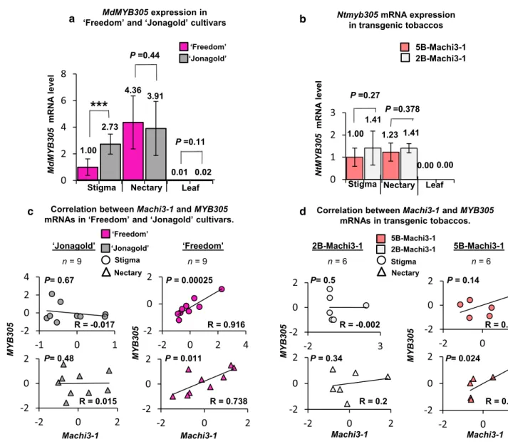
The MYB305 and 5B‑Machi3‑1 mRNA expressions are positively correlated
We found that four potential MYB-binding sites are present in the five-box region of the 5B-Machi3-1 allele but only one site is present in the two-box promoter region and that the five-box promoter region is required for the efficient nectary- and stigma-specific expression of the 5B-Machi3-1 mRNA in both apple and tobacco (Figs. 3, 4). As the MYB305 directs the nectary-specific expression of many genes in tobacco (Liu et al. 2009), we hypothesized that in tobacco the MYB305 and in apple, the MdMYB305 (the putative apple orthologue of MYB305, gene: MDP0000344978, Fig. S13a) transcription factors stimulate the expression of 5B-Machi3-1 in the nectary cells. We hypothesized that MYB305 and MdMYB305 also promote the expression of the 5B-Machi3-1 in the stigma cells. If these assumptions are correct, MdMYB305 and MYB305 mRNAs are co-expressed with 5B-Machi3-1 (but not 2B-Machi3-1) transcripts in apple and in transgenic tobacco, respectively. To test this, qRT-PCR and semi-qRT-PCR assays were conducted to monitor the expression of MdMYB305 in ‘Freedom’ and
‘Jonagold’ cultivars, and the expression of MYB305 in transgenic tobacco lines (Fig. 5a, b). Then, the expression of MdMYB305 and MYB305 and the 5B-Machi3-1 and 2B-Machi3-1 mRNAs were compared (Figs. 2c, 4c, 5). The MdMYB305 mRNA expressed similarly in both ‘Freedom’
and ‘Jonagold’ cultivars, it was abundant in the nectary and stigma samples but was barely detectable in leaves (Fig. 5a). Thus, MdMYB305 and 5B-Machi3-1 mRNAs (but not 2B-Machi3-1 transcripts) are co-expressed, both transcripts are abundant in the nectary and the stigma cells (Figs. 4, 5). To analyze the connections between Machi3-1 and MdMYB305 mRNA levels, Spearman correlation coef- ficients and P values were calculated for nectary and stigma samples of ‘Freedom’ and ‘Jonagold’ cultivars (Fig. 5c).
Relevantly, a strong positive correlation was observed in the stigma and nectary tissues of the ‘Freedom’ cultivar between the Machi3-1 and MdMYB305 mRNA expressions (R = 0.916 and 0.738, respectively), while correlation was not found in these tissues in the ‘Jonagold’ cultivar (Fig. 5c).
To examine if 5B-Machi3-1 and MYB305 transcripts are also co-expressed in transgenic tobacco, MYB305 mRNA levels were studied in the stigma, nectary and leaf tissues of 5B-Machi3-1 and 2B-Machi3-1 transgenic tobaccos (Fig. 5b). As expected, MYB305 mRNA expressed simi- larly in both types of transgenic tobacco lines. In accord- ance with previous report (Liu et al. 2009), we found that the MYB305 mRNAs were abundant in the nectary sam- ples (Fig. 5b). Moreover, we show that MYB305 mRNAs also expressed to high levels in stigma and to low levels in the leaf cells. Relevantly, we found that the 5B-Machi3-1 transgenic mRNA also accumulated to very high levels in
the stigma and to moderate levels in the nectary and that the 2B-Machi3-1 mRNA expressed to much lower levels in these tissues (Fig. 4c). Correlation analysis suggests that MYB305 and 5B-Machi3-1 transgenic mRNA expressions are also positively correlated in the stigma and the nectary of transgenic tobaccos (Fig. 5d). The correlation was strong for the nectary (R = 0.812, P = 0.024) and less convincing for the stigma (R = 0.526, P = 0.14). The 2B-Machi3-1 mRNA expression did not correlate with either the MdMYB305 or the MYB305 transcript. These results that 5B-Machi3-1 expressions positively correlate with the expressions of MdMYB305 and MYB305 are consistent with the model that these transcription factors promote the expression of 5B-Machi3-1 in the nectary and stigma cells in apple and transgenic tobaccos (also see Discussion). Unfortunately, we failed transiently overexpress MYB305 protein in N.
benthamiana, therefore, we could not directly test whether MYB305 activates 5B-Machi3-1 expression.
Machi3‑1 inhibits growth and biofilm formation of E. amylovora in vitro
The Machi3-1 chitinase accumulated to high levels in the nectar and the stigma exudates of the ‘Freedom’ apple cul- tivar. As chitinases are antimicrobial proteins and because during infection, E. amylovora multiplies in the nectar and the stigma exudate, we hypothesize that Machi3-1 can partially protect ‘Freedom’ from E. amylovora. As a first step towards understanding the role of Machi3-1 in defense against E. amylovora, we tested the effect of Machi3-1 on the growth and biofilm formation of the pathogen in vitro.
Erwinia cultures were incubated with Machi3-1 protein purified from the supernatant of Machi3-1 expressing P.
pastoris. We found that at high-concentration (40–80 ng/
µL) Machi3-1 significantly reduced the growth of E. amylo- vora (Fig. 6a). Relevantly, the Machi3-1 protein is present in the nectar of ‘Freedom’ cultivar in similar ~ 50–80 ng/
µL concentration (Fig. S2b). As a negative control, Erwinia cultures were incubated with supernatant of empty vector transformant P. pastoris.
Biofilm formation is required for successful Erwinia infection (Koczan et al. 2011). As chitinases can impair biofilm formation (Chung et al. 2014), we wanted to study if Machi3-1 modifies biofilm formation of E. amylovora.
Pre-formed E. amylovora biofilm was treated with purified Machi3-1 protein, and then biofilm detachment was quan- tified by crystal violet staining assay (O’Toole 2011). As Fig. 6b shows, the Machi3-1 efficiently detached the pre- formed E. amylovora biofilm at physiological concentration.
Taken together, the Machi3-1 acidic chitinase III protein efficiently impairs the growth and biofilm formation of E.
amylovora in vitro at physiological concentration.
Discussion
Here, we show that the 5B-Machi3-1 allele encodes an acidic chitinase III protein that accumulates to very high levels in the stigma exudate and the nectar, the primary niches of the Erwinia infection. As the Machi3-1 protein inhibits the growth and biofilm formation of Erwinia at physiological concentration in vitro, we hypothesize that the Machi3-1 protein could partially protect apple against Erwinia infec- tion and defend nutritious flower secretions from microbial
infections. We propose that the MYB305 protein homologs activate the transcription of the 5B-Machi3-1 mRNAs in the nectary and stigma cells and then the exported protein prod- ucts accumulate in the nectar and stigma exudate.
Machi3‑1 protein might interfere with Erwinia infection at two different steps
We found that the Machi3-1 acidic chitinase III protein is present at very high concentration in the nectar and the
-2 0 2
-2 3
-2 0 2
-2 0 2 -2
0 2
-2 0 2
0 2 4 6 8
a
Stigma Nectary Leaf 1.00
2.73
4.36 3.91
0.01 0.02
‘Freedom’
‘Jonagold’
MdMYB305expression in
‘Freedom’ and ‘Jonagold’ cultivars P=0.44
P=0.11
***
b
-2 0 2
-2 0 2
-2 0 2
-2 0 2
-2 0 2
-2 0 2
c Correlation between Machi3-1and MYB305
mRNAs in ‘Freedom’ and ‘Jonagold’ cultivars. Correlation between Machi3-1and MYB305 mRNAs in transgenic tobaccos.
Machi3-1
MYB305
MYB305
Machi3-1
MYB305
Machi3-1
‘Freedom’
‘Jonagold’ 2B-Machi3-1 5B-Machi3-1
n= 6 n= 6
-2 0 2 4
-1 0 1
Machi3-1
-2 0 2
-2 0 2 4
P= 0.00025
P= 0.011 P= 0.67
P= 0.48
P= 0.14
P= 0.34
MYB305
n= 9 n= 9
P= 0.024 Nectary
Stigma P= 0.5
Nectary Stigma
‘Freedom’
‘Jonagold’ 5B-Machi3-1
2B-Machi3-1
R = -0.017
R = 0.015
R = 0.916
R = 0.738 R = 0.2
R = 0.526
R = 0.812 R = -0.002
0 1 2 3
Ntmyb305 mRNA expression in transgenic tobaccos
5B-Machi3-1 2B-Machi3-1
P=0.27
1.00 1.41
1.23 1.41
0.00 0.00 Nectary Leaf Stigma
P=0.378
d
MdMYB305 mRNAlevel NtMYB305 mRNAlevel
Fig. 5 5B-Machi3-1 and MYB305 expressions show strong cor- relation. a, b qRT-PCR was carried out to monitor the expression of MdMYB305 and MYB305 transcripts. a MdMYB305 is highly expressed in the nectary and the stigma but not in the leaf cells of the
‘Freedom’ and the ‘Jonagold’ cultivars. mRNA level of the stigma sample of ‘Freedom’ was taken as 1 and the other expressions were calculated relative to it. b MYB305 mRNAs levels in different tis- sues of the 5B-Machi3-1 and the 2B-Machi3-1 transgenic tobaccos.
mRNA of the stigma sample of 5B-Machi3-1 was taken as 1 and the
other expressions were calculated relative to it. c, d To assess the con- nections between MYB305 and Machi3-1 mRNA expressions, Spear- man correlation coefficients (R) and the corresponding P values were calculated from the qRT-PCR results shown at A and B. panels. c 5B-Machi3-1 and MdMYB305 mRNA expressions positively cor- relate in both nectary and stigma samples of ‘Freedom’ cultivar. d 5B-Machi3-1 and MYB305 mRNA expressions positively correlated in the nectary and stigma samples in the 5B-Machi3-1 transgenic tobacco
stigma exudate of the 5B-Machi3-1 allele containing apple cultivars (Fig. 2). As chitinases have wide spectrum anti- microbial effect (Cletus et al. 2013), the high concentration of Machi3-1 protein could effectively protect the nectar and the stigma exudate from various microbial infection.
Thus, it can keep the optimal composition of these impor- tant fluid secretions, thereby enhancing the efficiency of pollination and fertilization. We propose that the high level of Machi3-1 in the stigma exudate and the nectar could partially protect 5B-Machi3-1 allele containing apple cul- tivars against Erwinia. During apple infection, Erwinia propagates first in the stigma exudate, then in the nectar and finally enters into the plant through the stomata of the nectary (Bubán et al. 2003; Farkas et al. 2012). As bio- film formation is critical for the fruit and shoot infection of Erwinia (Koczan et al. 2011), it is likely that biofilm formation is also required for efficient flower infection.
In vitro, the Machi3-1 protein inhibits growth and bio- film formation of Erwinia at a concentration in which it is present in the nectar of 5B-Machi3-1 allele containing apple cultivars (Figs. 6, S2). These findings suggest that the Machi3-1 protein can interfere with the propagation and infection of Erwinia in the nectar of the 5B-Machi3-1 allele containing apples. Unfortunately we cannot directly measure the concentration of Machi3-1 protein in the stigma exudate. However, as Machi3-1 is one of the most
abundant proteins in the ‘Freedom’ stigma exudate (Fig.
S14.), it is conceivable that the Machi3-1 protein is also present in inhibitory concentration in the exudate. Thus, we propose that accumulation of Machi3-1 protein could protect apples from Erwinia infection by forming two bar- riers, it interferes with the replication and biofilm forma- tion of the Erwinia in the stigma exudate as well as in the nectar. This model would explain why the 5B-Machi3-1 expressing ‘Freedom’ was more tolerant in flower inocula- tion assay against Erwinia than the non-expressing ‘Jona- gold’ cultivar. Importantly, if 5B-Machi3-1 contributes to the Erwinia resistance, this effect is not detectable in the frequently used shoot inoculation assays.
The molecular mechanism how Machi3-1 inhibits the growth and biofilm formation of Erwinia is not known.
Chitinase treatment modulated the bacterial adhesion and inhibited the biofilm formation of the Gram-negative Francisella novicida. It was hypothesized that chitinases alter the EPS composition of F. novicida thereby impairing the biofilm formation and altering adhesion (Chung et al.
2014). As amylovoran and levan EPSs play a critical role in biofilm formation and pathogenicity of Erwinia (Koc- zan et al. 2009), we speculate that Machi3-1 modulates the EPS of Erwinia, which results in slower growth and inefficient biofilm formation.
Fig. 6 Machi3-1 has an antibacterial effect against E.
amylovora. a Machi3-1 inhibits the growth of E. amylovora.
Erwinia culture was treated with Machi3-1 protein that was purified from the supernatant of Machi3-1 expressing Pichia and with supernatant from empty vector transformed Pichia as a negative control. b Biofilm detachment assay shows that Machi3-1 impairs biofilm formation. Note that, although at 80 ng/µL concentration even the negative control has a mild activity, this effect can be separated from the effect of Machi3-1, because Machi3-1 inhibited significantly more effectively the biofilm formation than the negative control and at lower concentrations only the Machi3-1 inhibited the biofilm formation. Significance levels: *P value < 0.05, ***P value < 0.001. c Model of the regulation of 5B-Machi3-1 expression (for details, see the main text)
0.00 20.00 40.00 60.00 80.00 100.00 120.00 140.00
0.00 20.00 40.00 60.00 80.00 100.00 120.00 140.00
a
*** ***
*** *
***
0 20 40 80 20 40 80
Machi3-1 ng/µL
- control Machi3-1 Growth inhibition assay b
Biofilm detachment assay
ng/µL Machi3-1 ng/µL
cFlowering
Nectary & Stigma
5B-Machi3-1
Nectar & Stigma exudate
JA Myb
305
Machi3-1
CFU % of control CFU % of control