doi: 10.3389/fsufs.2021.745865
Edited by:
Ravinder K. Goyal, Agriculture and Agri-Food Canada (AAFC), Canada
Reviewed by:
John Laurie, Agriculture and Agri-Food Canada, Canada Patricia Corral-Martínez, Polytechnic University of Valencia, Spain
*Correspondence:
Ewa Dubas e.dubas@ifr-pan.edu.pl Iwona ˙Zur i.zur@ifr-pan.edu.pl
Specialty section:
This article was submitted to Crop Biology and Sustainability, a section of the journal Frontiers in Sustainable Food Systems
Received:22 July 2021 Accepted:17 September 2021 Published:21 October 2021 Citation:
Dubas E, ˙Zur I, Morav ˇciková J, Fodor J, Krzewska M, Surówka E, Nowicka A and Gerši Z (2021) Proteins, Small Peptides and Other Signaling Molecules Identified as Inconspicuous but Possibly Important Players in Microspores Reprogramming Toward Embryogenesis.
Front. Sustain. Food Syst. 5:745865.
doi: 10.3389/fsufs.2021.745865
Proteins, Small Peptides and Other Signaling Molecules Identified as Inconspicuous but Possibly
Important Players in Microspores Reprogramming Toward
Embryogenesis
Ewa Dubas1*, Iwona ˙Zur1*, Jana Morav ˇciková2, József Fodor3, Monika Krzewska1, Ewa Surówka1, Anna Nowicka1and Zuzana Gerši4
1Department of Cell Biology, The Franciszek Górski Institute of Plant Physiology, Polish Academy of Sciences, Kraków, Poland,2Department of Biotechnology, Faculty of Natural Sciences, University of St. Cyril and Methodius in Trnava, Trnava, Slovakia,3Plant Protection Institute, Centre for Agricultural Research, Budapest, Hungary,4Department of Biology, Faculty of Natural Sciences, University of St. Cyril and Methodius in Trnava, Trnava, Slovakia
In this review, we describe and integrate the latest knowledge on the signaling role of proteins and peptides in the stress-induced microspore embryogenesis (ME) in some crop plants with agricultural importance (i.e., oilseed rape, tobacco, barley, wheat, rice, triticale, rye). Based on the results received from the most advanced omix analyses, we have selected some inconspicuous but possibly important players in microspores reprogramming toward embryogenic development. We provide an overview of the roles and downstream effect of stress-related proteins (e.g., β-1,3-glucanases, chitinases) and small signaling peptides, especially cysteine—(e.g., glutathione, γ -thionins, rapid alkalinization factor, lipid transfer, phytosulfokine) and glycine-rich peptides and other proteins (e.g., fasciclin-like arabinogalactan protein) on acclimation ability of microspores and the cell wall reconstruction in a context of ME induction and haploids/doubled haploids (DHs) production. Application of these molecules, stimulating the induction and proper development of embryo-like structures and green plant regeneration, brings significant improvement of the effectiveness of DHs procedures and could result in its wider incorporation on a commercial scale. Recent advances in the design and construction of synthetic peptides–mainly cysteine-rich peptides and their derivatives–
have accelerated the development of new DNA-free genome-editing techniques. These new systems are evolving incredibly fast and soon will find application in many areas of plant science and breeding.
Keywords: double haploids, microspore embryogenesis (ME), pathogenesis-related protein (PR), small signaling peptides, stress
INTRODUCTION
By 2035, the human population is expected to reach around 8.9 billion, and by the middle of the 21st century nearly 10 billion (https://www.prb.org/international/geography/world/).
This demographic tendency, coinciding with environmental change, increases the demand for food supply (Calicioglu et al., 2019). In 2050, food production will need to be increased by about 50% compared to that in 2012 (Global monitoring report 2015/2016). This is a big challenge for agricultural producers and breeders, especially that the progress is hampered by the exacerbated climate change and substantial loss of biodiversity.
Meeting this demand would require that agricultural sector including plant breeders increase sustainable agricultural production by introducing more efficient methods of obtaining improved varieties of high quality and stable yield in a short time. Conventional breeding is labor intensive and time- consuming process, which needs to be complemented with modern biotechnological approaches to improve its efficiency.
The abovementioned problems point to the necessity to deliver new plant varieties with enhanced productivity and improved adaptation toward abiotic and biotic stresses. For this purpose, microspore embryogenesis (ME), known also as androgenises, seems to be an unrivaled biotechnological tool to speed up the progress of plant breeding. During ME, the differentiation of immature male gametophyte cells (microspores) into pollen grains is blocked and redirected toward embryo development.
This process is triggered by stress and requires in vitro culture techniques. From one side, the difficulties associated with optimization of in vitro culture conditions are often limiting factors in ME incorporation as a research model or tool in biotechnology or breeding, on a larger scale. On the other hand, the use of in vitro culture provides direct insight into the process
Abbreviations: BSO, buthionine sulfoximine the inhibitor of GSH synthesis;
CAT, catalase; CHIT1, CHIT2, endochitinases; CRISPR/Cas9, CRISPR-related endonuclease Cas9; CRP, cysteine-rich peptides; Cys, cysteine; DH, doubled haploid; DNMT, DNA methyltransferases; ECA, arabinogalactan-like proteins;
ECM, extracellular matrix; ELS, embryo-like structures; EXP, expansins;
EXT, extensins; FLA, fasciclin-like arabinogalactan proteins; GlcNAc, N-acetyl glucosamine; Glu, glutamine; Gly, glycine; GPX, glutathione peroxidase; GRP, glycine-reach proteins; GSH, reduced form of glutathione; GSSG, oxidized form of glutathione; GST, glutathione S-transferase; H2O2, hydrogen peroxide; HECATE, transcription factor; HMT, histone methyltransferases; LT, low temperature;
LTP, lipid transfer proteins; MAN, mannitol; MAP, mitogen-activated protein kinases; MAS, marker assisted selection; ME, microspore embryogenesis; MYB5, transcription factor; MYB75, transcription factor; NGGS, genome sequencing;
1O2, singlet oxygen; O2·–, superoxide anion; OH·, hydroxyl radical; OTC, L- 2-oxothiazolidine-4-carboxylic acid precursor of GSH; PCD, plant cell death;
PR, Pathogenesis-related proteins; PR12,γ-thionins, the family of PR proteins;
PR13, defensins, the family of PR proteins; PR2,β-1,3-glucanases, the family of PR proteins; PR3, chitinases, the family of PR proteins; PSK, phytosulfokine;
PTMP, small post-translationally modified peptides; PTMs, post-translational modifications; RALF, rapid alkalinization factor; ROS, reactive oxygen species;
SAM, S-adenosyl-methionine; S-GlcNAc, acylated S-linked N-acetyl glucosamine;
SOD, superoxide dismutase; SPATULA, transcription factor; SSNs, sequence- specific nucleases; TALEN, transcriptional activator-effector nucleases; TFs, transcription factors; TrX, thioredoxin; TSA, trichostatin A the histone deacetylase inhibitor; WAK, wall-associated kinases; WGS, shotgun sequencing techniques;
WRKY, the zinc-finger transcription factors; ZFN, zinc finger nucleases.
of ME, which follows a pattern very similar to the development of the zygotic embryo in planta.
The final products of ME are doubled haploids (DHs), homozygous at all loci, what brings significant benefits to many basic research areas and plant breeding. Elimination of heterozygosity simplifies genome sequencing, reverse breeding, quantitative genetic research and discovering of recessive, dominant and deleterious mutations. That’s why DHs are interesting objects of studies in many research areas including physiology, molecular biology, genetics and epigenetics (Maluszynski et al., 1996; Castillo et al., 2001; Touraev et al., 2001; Forster et al., 2007; Szarejko and Forster, 2007 and references therein; Dirks et al., 2009; Dunwell, 2010; Chauhan and Khurana, 2011; Ferrie and Möllers, 2011; Krzewska et al., 2012, 2017; Marathi et al., 2012; ˙Zur et al., 2014a,b, 2015a,b;
Barakat et al., 2017; Ren et al., 2017; Song et al., 2017; Shchukina et al., 2018; Tyrka et al., 2018; Nowicka et al., 2019; Shi et al., 2019; Testillano, 2019; Wajdzik et al., 2019; Bilichak et al., 2020;
Malaga et al., 2020). Furthermore, DH technology combined with marker assisted selection (MAS) enables precise identification of plants with enhanced/silenced expression of even one gene of interest within the genome. Due to the fact that total homozygosity is received in one generation, incorporation of DH technology into breeding programmes saves time necessary to develop and release new, improved cultivars (reviewed in Kasha and Maluszynski, 2003; Germanà, 2006, 2011; Forster et al., 2007; Wedzony et al., 2009; Dwivedi et al., 2015). DH lines can be considered as a new variety when self-pollinated or can be used as a parental inbred line for the production of hybrid varieties after cross-pollination and in germplasm conservation.
DH technology is also used to fix traits obtained through transformation and mutagenesis and to develop genetically-fixed molecular mapping populations.
ME is one of the simplest and most effective methods available for haploids/DHs production for a wide range of crops (Wedzony et al., 2009; Ferrie and Möllers, 2011; Germanà, 2011). Established protocols for the ME induction are used in various species, varieties, breeding lines and are based on different experimental approaches, what limits the ability to identify universal solutions leading to redirection of microspores toward sporophytic development, formation of embryo-like structures (ELS) and differentiation into haploid plantlets.
Finally, green and fertile DH plants are obtained by spontaneous or chemically induced genome doubling. Our understanding of the processes that occur during ME, come generally from two dicotyledonous species: rapeseed (Brassica napus) and tobacco (Nicotiana tabacum), as well as three monocots: wheat (Triticum aestivum), barley (Hordeum vulgare) and rice (Oryza sativa) (Hosp et al., 2007; Ferrie and Möllers, 2011; Germanà, 2011;
Soriano et al., 2013; Wedzony et al., 2014; Seifert et al., 2016;
Bélanger et al., 2018, 2020; Shahmir and Pauls, 2021). However,
the majority of information regarding the control and regulation
of ME induction is based on studies on single, highly responsive
genotypes within a given species, like cv. Igri (Jacquard et al.,
2009) and cv. Gobernadora (Bélanger et al., 2018) in barley and
wheat cv. Svilena (Seifert et al., 2016), and rapeseed DH line 4,079
(Joosen et al., 2007) or producing the embryogenic callus DH line
12,075 (Soriano et al., 2013, 2014; Corral-Martínez et al., 2020).
Therefore, a large-scale examination, comparing genotypes of high embryogenic potential with genotypes recalcitrant to ME under the same inducible conditions is of great value and could provide direction for more effective approaches to DH production and its wider incorporation into breeding programmes. Recently, the group of crop species that has been considered as a subject of ME has been extended to hexaploid triticale ( × Triticosecale Wittm.) and its parental species, rye (Secale cereale L.). Both species belong to cereals economically valuable in Northern Europe and North America, due to its yield potential, specific nutritional values of grain and high tolerance to environmental conditions. Studies conducted on several DH lines (triticale) and F1 breeding lines (rye) highly differentiated in respect of ME effectiveness, gave us the possibility for more accurate identification of factors related to ME effectiveness (˙Zur et al., 2008, 2009, 2012, 2014a, 2015a,b, 2019; Krzewska et al., 2012, 2017; Nowicka et al., 2019; Zieli´nski et al., 2020, 2021).
The phenomenon of ME was for the first time observed in 1964 (Guha and Maheshwari, 1964), but its complex and multifaceted nature makes it difficult to investigate, so still the mechanisms of molecular control and regulation have not yet been precisely described (Maraschin et al., 2005; Hosp et al., 2007; Elhiti et al., 2013; Seifert et al., 2016). It is known that the effectiveness of ME is determined by many internal and external factors and their complex interactions. Induction of microspore reprogramming and then initiation of its embryogenic development is accompanied with many changes in molecular, biochemical, physiological and cytological processes (e.g., gene expression, DNA methylation, chromatin organization, redox and hormonal homeostasis) (Pauls et al., 2006; Seguí-Simarro and Nuez, 2008; El-Tantawy A.-A. et al., 2014; ˙Zur et al., 2014a,b, 2015a,b; Solís et al., 2015; Berenguer et al., 2017).
Microspore reprogramming can be induced by various stress factors (e.g., low temperature, heat, starvation, chemical agents and their combinations), which can be applied to the whole plants, harvested spikes, isolated anthers or microspores.
However, the effect of the treatment depends on the donor plant genotype, its physiological condition and growing season—all of which influence the level of cell stress tolerance ( ˙Zur et al., 2019).
The exposure to these stress factors leads to oxidative stress that is induced by increased levels of reactive oxygen species (ROS) including free radicals such as superoxide anion (O
·−2) and hydroxyl radical (OH
·), and non-radicals like hydrogen peroxide (H
2O
2), singlet oxygen (
1O
2) and lipid peroxides (LOOH). It was hypothesized, that recalcitrance to ME could be caused by low ability to counter the oxidative stress induced during initiation of ME and/or transfer of microspores to in vitro culture conditions (˙Zur et al., 2008, 2009, 2014a; Jacquard et al., 2009). ROS together with other reactive compounds derived from ROS-mediated oxidative damage to cellular macromolecules (oxylipins, peptides, mRNAs, DNA) modulate cellular signal transduction and the post-transcriptional gene expression associated with secondary metabolism, stress responses and cellular detoxification (e.g., genes encoding phosphatases, kinases and transcription factors) (Chmielowska- Bak et al., 2015; Schnaubelt et al., 2015; He et al., 2018).
These oxidative modifications result in changes in expression, structure and/or function of the proteins. Aggregation or fragmentation of the polypeptide chains activate proteolysis and cell damage, which can lead finally to the cell death (Shan et al., 2007).
When ROS generation overwhelms the antioxidative capacity in plant cells, various signaling pathways are activated that trigger physiological, biochemical, and molecular responses of cellular metabolism, probably necessary for microspore reprogramming and embryogenesis initiation (˙Zur et al., 2008, 2009, 2012, 2014a, 2015a, 2019). What is more, even high level of ROS does not endanger microspore viability as long as the cells exhibit high activity of ROS-scavenging enzymes (˙Zur et al., 2014a, 2019, 2021).
There are two main cellular systems controlling ROS:
non-enzymatic, low molecular weight antioxidants (e.g., ascorbate, tocopherols, reduced glutathione, flavonoids, etc.) and antioxidative enzymes such as superoxide dismutase (SOD), catalase (CAT) and various peroxidases (Foyer and Noctor, 2003; AbdElgawad et al., 2016). All these antioxidants support sustaining the redox balance in the cell and play a crucial role in the defense against oxidative stress.
The capability of antioxidants to counteract the effect of ROS is related to their structure and chemical properties. It may arise from the presence of: (i) metal ions (Mn, Cu/Zn and Fe in SOD;
Fe–in CAT; S–in other metalloproteins, glutathione peroxidase (GPX) and glutathione S-transferase (GST); (ii) conjugated double bonds like in ascorbic acid, tocopherol and β-carotene;
(iii) aromatic rings like in flavonoids; and (iv) the thiol (or
“sulfhydryl”) group like in glutathione and thioredoxin (TrX) (Flora, 2009; Rahantaniaina et al., 2013). The thiol/thiolate group of the redox-reactive cysteine (Cys) gives the ability to form disulfide bonds with nearby cysteines (–S–S–) or undergo further oxidation to sulfinic (–SO
2H) or sulfonic (–SO
3H) acid. Any oxidation-reduction (redox) modifications of cysteinyl residues lead to post-translational modifications (PTMs) that impact on molecular functions important to cellular processes, including signal transduction (Poole, 2015). Both PTMs and the amino acids sequence determine protein features and functions (Zhang et al., 2021).
To learn more about molecules which belong to the most
important factors enhancing ME induction efficiency, scientists
developed a suite of highly advanced research methods
and biotechnological tools e.g., next generation sequencing
(NGS), improved biochemical isolation procedures, gene
prediction/annotation bioinformatics tools and genome-
editing approaches (Sahu et al., 2020). The manipulation
of metabolic pathways by non-GMO genetic engineering is
a strategy that may increase the tolerance of plants against
abiotic stress. Recent technological advances in crop genomics
allow to discover genetic variation in breeding material and
permit genome-based breeding to deliver cultivars for the
projected food requirements for 2050 (Rasheed and Xia,
2019). Due to the development of modern techniques, several
potential candidates for stimulation of induction and proper
development of ELS–mainly among cysteine-rich or small
signaling peptides and proteins–have been reported in recently
published papers. These results allow us to address the roles of these inconspicuous but possibly important molecular players in the regulation of ME. Inspired by previous and recent findings, we review how stress tolerance-related proteins (e.g., β-1,3- glucanases, chitinases) and small signaling peptides, especially cysteine-(Cys, e.g., glutathione, γ -thionins, rapid alkalinization factor, lipid transfer, phytosulfokine) and/or glycine-rich peptides and other proteins (e.g., fasciclin-like arabinogalactan protein), particularly those involved in the regulation of cellular redox potential or the cell wall reconstruction could be promising tools for improving DH production in crop plants.
STRESS-RELATED PROTEINS
The balance between ROS generation and scavenging determines the level of stress tolerance in plants by modifying the profile of defense-related genes coding for molecules like pathogenesis- related (PR) proteins as β-1,3-glucanases (PR2), chitinases (PR3) or γ -thionins (PR13) (Chinnusamy and Zhu, 2009; Sahu et al., 2013; Wojtasik et al., 2019).
β-1,3-glucanases (EC 3.2.1.39) are hydrolytic enzymes (GH17) that catalyze the cleavage of 1,3-ß-D-glucosidic linkages in ß-1,3- glucan, commonly referred to as callose (Levy et al., 2007; Chen et al., 2009; Wu et al., 2018). Callose is a component of cell walls or cell walls associated structures; and forms the barriers that regulate intercellular trafficking (Chen and Kim, 2009; Wu et al., 2018). In higher plants, callose plays important roles in many biological processes as well as in plant defense response (Chen et al., 2009; Wu et al., 2018; Wang et al., 2021). β-1,3-glucanases together with callose synthases regulate callose homeostasis (Chen et al., 2009; Wu et al., 2018). Based on their structure, plant β-1,3-glucanases have been classified into four classes (I–
IV) (Doxey et al., 2007). The class I comprises basic vacuolar β- 1,3-glucanases whereas classes II–IV include acidic extracellular enzymes (Grover, 2012). In plants, these hydrolases are called PR2 proteins, as they are expressed when plants are exposed to biotic (Moravˇcíková et al., 2004; ˙Zur et al., 2013) and/or abiotic stresses (Mészáros et al., 2011; ˙Zur et al., 2013; Gregorová et al., 2015). Moreover, these enzymes are undoubtedly essential for plant growth and development including microsporogenesis, pollen development or seed germination (Leubner-Metzger and Meins, 1999; Michalko et al., 2017). Their activities, leading to the production of (1 → 3)-linked β-glucan oligosaccharides with signaling properties were reported in both zygotic (Petrovská et al., 2010), somatic (Helleboid et al., 2000; Fráterová et al., 2013) as well as in microspore embryogenesis (Borderies et al., 2004; Muñoz-Amatriaín et al., 2009; Leljak-Levani´c et al., 2015;
Zieli´nski et al., 2021). Several β-1,3-glucanases were detected in stress pre-treated anthers of several plant species (Table 1).
Recently, Zieli´nski et al. (2021) identified an anther-specific and stress-responsive β-1,3-glucanase fraction of 26 kDa and several acidic isoforms in rye. Since direct correlation between the activity of β-1,3-glucanases and the final efficiency of the ME was not observed, it was supposed that β-1,3-glucanases mediate defense responses in the early stages of ME induction.
We also assume that their activity is important only for
the inhibition of gametophytic pollen development, because no activity of β-1,3-glucanases was found in competent and embryogenic microspores in the rye line responsive to ME (Zieli´nski et al., 2021). It might be related to the microspore wall remodeling via a rapid production of abnormal, callose- rich sub intimal layer of cell walls (Dubas et al., 2013;
Parra-Vega et al., 2015; Rivas-Sendra et al., 2019). Thickening of the cell wall with an extra osmoprotective sub intimal callose deposition protects the microspores from cell death and physically isolates them from the outer environment (Parra- Vega et al., 2015; Rivas-Sendra et al., 2019). It stimulates the rearrangements of microspore structure, such as location of the nucleus (central), vacuolization (numerous small vacuoles) and the distribution of cytoplasmic bands (numerous cytoplasmic bridges). Microspores, with poor callose layer are more exposed to the risk of blocked reprogramming (Rivas-Sendra et al., 2019). β-1,3-glucanases activity might be important later for the regulation of embryo differentiation (Helleboid et al., 2000;
Borderies et al., 2004).
Besides β-1,3-glucanases, chitinases are also expressed in a response to stress-initiated microspore reprogramming (Zieli´nski et al., 2021). Plant chitinases (PR3, EC 3.2.1. 14) are glycoside hydrolases that cleave the β-1,4 glycosidic linkages of chitin. As chitin is a structural component of most fungal cell walls (Fesel and Zuccaro, 2016), chitinases were mainly studied in relation to defense response against fungal pathogens (Moravˇcíková et al., 2004; ˙Zur et al., 2013). Structurally, plant chitinases are categorized into six classes (I–VI). The classes I, II, IV and VI belong to the subfamily GH19 that is exclusively present in plants. The classes III and V comprise chitinases of the subfamily GH18 that are found in bacteria, fungi, viruses, animals, and few plant species (Minic, 2008; Grover, 2012). Like β-1,3-glucanases, chitinases are upregulated under both biotic (˙Zur et al., 2013) and abiotic stress (Mészáros et al., 2011; ˙Zur et al., 2013; Gregorová et al., 2015) and their synergistic effect in plant defense has been observed in many plant species (Moravˇcíková et al., 2004; ˙Zur et al., 2013). These enzymes are also active in healthy plants in an organ-specific and developmentally regulated pattern (Kasprzewska, 2003).
Activities of chitinases have been reported to be associated with embryogenesis: zygotic (Sánchez-Díaz et al., 2013), somatic and ME (De Vries et al., 1988; Coutos-Thevenot et al., 1992;
Hilbert et al., 1992; Nielsen and Hansen, 1992) (Table 1). At least three chitinase fractions ( ∼ 30, ∼ 34, and ∼ 95 kDa) were identified in stress pre-treated anthers of two rye breeding lines regardless of the type of the stress treatment (Zieli´nski et al., 2021). Although, these chitinases were not anther-specific, their presence (especially 28–35 kDa fractions) was associated with the plant defense responses according to literature data (Ferreira et al., 2007; Kuwabara and Imai, 2009; ˙Zur et al., 2013).
Chitinases of a ∼ 25 and ∼ 28 were detected in the induction
medium, obviously secreted by maize microspores during
embryo generic development (Borderies et al., 2004). In the
earlier stages of ME in rye anther culture, chitinases were also
detected although their activity was very low (Zieli´nski et al.,
2021). The presence of chitinases may result in the generation of
molecules that have a stimulating effect on changing microspore
TABLE 1 |Proteins and peptides in microspore embryogenesis of crop species.
Plant species Source of material Expression/predicted function Type of analyses Literature Arabinogalactan proteins AGP
Brassica napus L. Embryogenic microspores of four cultivars, heat stress
AGP associated with the transition to embryo formation.
Transcriptome analysis
Malik et al., 2007
Brassica napus L. Embryogenic
microspores, heat stress
Inactivation of AGP-inhibition ofBrassica embryogenesis.
JIM8, JIM13–crucial role in initiation of microspore embryogenesis, maintenance in cell differentiation.
Yariv Reagent Immunofluorescent labeling (JIM4, JIM8, JIM13)
Tang et al., 2006
Brassica napus L. Embryogenic
microspores, heat stress
JIM13, JM14–detected in 2–4 cell stage of embryo cell walls, considered as early marker of microspore embryogenesis.
Immunofluorescent labeling Immuno dot blot assay (JIM13, JIM14, MAC207, LM2)
El-Tantawy A. A. et al., 2013
Brassica napus L. Embryogenic
microspores, heat stress
JIM13–detected in cell apoplast, associated with cell wall totipotency.
Immunogold labeling (JIM8, JIM14, JIM16, JIM13)
Corral-Martínez et al., 2019
Capsicum annuum L. Microspores of seven cultivars
Exogenous AGP in induction medium improved microspore embryogenesis in all cultivars.
- Pourmohammad et al.,
2021 Secale cereale L. Anthers of two breeding
lines after 21 days of (LT, Mn and/or GSH) pre-treatments
Increased accumulation of AGP in the androgenises-responsive line.
Yariv Reagent Zieli ´nski et al., 2021
Secale cereale L. Anthers of two breeding lines after 21 days of (LT, Mn_GSH) pre-treatment
LM2, JIM14 and JIM4–higher fluorescence in the androgenises-responsive line.
JIM13, JIM4–associated with androgenic induction of rye.
JIM13–detected only in the
androgenises-responsive line (in the vesicles and inner cell walls of the microspores and in the cell walls of the anther cell layers).
Dot blot assay Immunofluorescent labeling (LM2, MAC207, JIM16, JIM14, JIM13, JIM8, JIM4)
Zieli ´nski et al., 2021
Triticum aestivum L. Microspores of two cultivars
Exogenous AGP in induction medium strongly affected microspore embryogenesis of both cultivars.
– Letarte et al., 2006
Zea mays L. Conditioned microspore culture
JIM14, JIM13, LM2, JIM16 secreted in the conditioned medium, concentration increased during the time of culture.
Immunodetection (JIM14, JIM13, LM2, JIM4, MAC207, JIM16 and JIM8)
Borderies et al., 2004
Glutathione
Brassica napus L. Embryogenic
microspores, one cultivar;
heat stress
Exogenous application of GSH in the culture medium increased the effectiveness of ME.
– Hoseini et al., 2014
Brassica napus L. Globular–heart transition stage embryos of cv Topaz
Exogenous application of GSH, GSSG and BSO (an inhibitor of glutathionede novosynthesis) in the culture medium showed that a lowering of the glutathione redox status improved the structure of canola ELS and their ability to convert into viable plants.
Spectrophotometrically GSH and GSSG measurements
Belmonte et al., 2006
Brassica oleracea L. Embryogenic microspores of three hybrid cultivars, heat stress
Exogenous application of GSH in the culture medium significantly decreased microspore mortality, and had a strong effect on the number of embryos produced and influenced positively the rate of plant regeneration.
– Zeng et al., 2017
Secale cereale L. Tillers pre-treatment of two breeding lines (LT, Mn and/or GSH)
Exogenous GSH enhanced microspore vitality associated with an increased number of embryogenic microspores.
– Zieli ´nski et al., 2021
Secale cereale L. Tillers pre-treatment of two breeding (LT, Mn and/or GSH)
Exogenous GSH resulted in higher accumulation of AGP in anthers and higher effectiveness of embryo-like structures formation.
– Zieli ´nski et al., 2021
(Continued)
TABLE 1 |Continued
Plant species Source of material Expression/predicted function Type of analyses Literature
×Triticosecale Wittm. Tillers pre-treatment of five DH lines (LT, LT+GSH)
Exogenous GSH protects cells from oxidative damages, which influences microspore yield, viability and the effectiveness of ME. Increased endogenous level of GSSG promotes further ELS development in isolated microspore cultures.
Spectrophotometric measurement of GSH and GSSG
˙Zur et al., 2019
Triticum aestivum L. Microspores of four spring genotypes after LT tillers pre-treatment
Exogenous GSH in induction medium increased embryo and green plant production.
– Asif et al., 2013
Triticum turgidum L. Embryogenic microspores of two cultivars
Exogenous application of GSH in the culture medium increased the number of ELS.
– Cistué et al., 2009
Glycine-reach proteins GRP
Brassica napus L. Embryogenic microspores of DH12075 line
GRP ensure further embryo growth and differentiation
GFP reporter line proGRP:GFP-GUS
Li H. et al., 2014
Brassica napus L. Embryogenic microspores of DH12075 line
GRP initially expressed throughout the embryo clusters while they were still enclosed by the exine;
GRP marks the basal pole of the embryo and the future columella at the site, where the pollen wall remnants are present
GFP reporter line proGRP:GFP-GUS
Soriano et al., 2014
Lipid transfer protein LTP
Hordeum vulgare L. The early stages of microspore
embryogenesis of cv. Igri
ECLTP(early culture lipid transfer protein) had homology to LTP, and had an expression pattern similar to that of an LTP known to be a marker of the early stages of embryogenesis (3 days of culture).
Characterization of cDNAs and Northern hybridization analysis
Vrinten et al., 1999
Phytosulfokine PSK Triticum aestivum L.;
×Triticosecale Wittm.
Embryogenic microspores of two Canadian spring wheat (AC Carberry and AC Peace) and two triticale (AC Ultima and Sunray) cultivars
Exogenous PSK-αin induction medium (in absence of co-cultured ovaries) increased embryo and green plant production.
– Asif et al., 2014
Oryza sativa Embryogenic anther cultures
Exogenous PSK-αpre-treatment at low
temperature in induction medium improved seedling growth from callus of anther.
– Chen et al., 2010
Pathogenesis-related proteins(PR2 and PR3) Hordeum vulgare L. Anthers after 4 days of Mn
treatment
Twoβ-1,3-glucanase genes. Transcriptome analyses
Muñoz-Amatriaín et al., 2009
Hordeum vulgare L. Anthers after 4 days of Mn treatment
Basic endochitinase gene. Transcriptome analyses Muñoz-Amatriaín et al., 2006
Secale cereale L. Microspores after 21 days of (LT, Mn and/or GSH) pre-treatments
Chitinase gene Chit1 (AF280438.1) Chitinase gene Chit 2 (AF280437.1)
β-1,3-glucanase gene Glu2 (GAM181307.1)-not expressed
β-1,3-glucanase gene Glu3 (AM181306.1)-not expressed
RT-qPCR analyses Zieli ´nski et al., 2021
Secale cereale L. Microspores after 21 days of (LT, Mn and/or GSH) pre-treatments
β-1,3 glucanases-no activities or under detection limit
chitinases–no activities or under detection limit
In gel enzyme activity assays
Zieli ´nski et al., 2021
Secale cereale L. Anthers after 21 days of (LT, Mn and/or GSH) pre-treatments
26 kDa (anther-specific)β-1,3-glucanases 30 kDa chitnases
34 kDa chitinases
95 kDa chitinases–allosamidin sensitive
In gel enzyme activity assays
Zieli ´nski et al., 2021
Secale cereale L. Anthers after 21 days of (LT, Mn and/or GSH) pre-treatments
Chitinase geneChit 1(AF280438.1) Chitinase geneChit 2(AF280437.1)
β-1,3-glucanase geneGlu2(GAM181307.1)–not expressed
β-1,3-glucanase geneGlu3(AM181306.1) –not expressed
RT-qPCR analyses Zieli ´nski et al., 2021
(Continued)
TABLE 1 |Continued
Plant species Source of material Expression/predicted function Type of analyses Literature
×Triticosecale Wittm. Microspores 21 days after LT treatment
Chitinase geneCHI3 RT-PCR analyses Dubas et al., 2014a,b
Triticum aestivum L. Anthers after 10–20 days of Mn pre-treatments
Chitinase geneCHI3 RT-PCR analyses
Sánchez-Díaz et al., 2013 Zea mays L. Conditioned medium after
21 days microspore cultivation
30 kDaβ-1,3-glucanases 28 kDa chitinases 25 kDa chitinases
2D isofocusing electrophoresis
Borderies et al., 2004
AC, anther cultures; GSH, reduced glutathione; LT, Low temperature; LT+GSH, Low temperature and reduced glutathione; Mn, mannitol; Mn_GSH, mannitol and reduced glutathione.
structure which precede an ELS development (Matthys-Rochon, 2005).
The morphological changes are a consequence of molecular events and are connected with modified expression of genes encoding endochitinases (CHIT1, CHIT2) (Zieli´nski et al., 2021), arabinogalactan-like proteins (ECA) and lipid transfer proteins (LTP) (Vrinten et al., 1999; Hosp et al., 2007; Malik et al., 2007), which may be involved in membrane and cell wall remodeling at the initial phases of ME and during an ELS development (Malik et al., 2007). Lower plasma membrane fluidity in microspores of B. napus line of high embryogenic potential seems to maintain proper cell protection and may lead to efficient embryogenesis induction (Dubas et al., 2013). The cell wall arabinogalactan proteins (AGP) contain N-acetyl glucosamine residues can be a target for endochitinase cleavage (van Hengel et al., 2001; van Hengel and Roberts, 2002; Kasprzewska, 2003; Minic, 2008) and thereby involved in both wall architecture and cellular regulatory processes (Pfeifer et al., 2020).
The monosaccharide derivative of glucose, N-acetyl glucosamine (GlcNAc) is cross-linked by short peptides that may have functions in cell signaling (Konopka, 2012;
Naseem et al., 2012). Intracellular GlcNAcylation of serine and threonine residues is a well-known and widely investigated post-translational modification in plant cells. Modifications of cysteine (Cys) S-linked N-acetyl glucosamine (S-GlcNAcylation) were recently found as a new post-translational modification in mammals (Maynard et al., 2016). S-GlcNAc is a sulfur-linked analog of O-GlcNAc, which modification is enzymatically stable at both peptide and protein levels (Olszewski et al., 2010; De Leon et al., 2017). The presence of such process is a subject of intense research in plants.
Thionins, peptides composed of 45 to 48 amino acid residues with the molecular weight of ∼ 5 kDa, containing six or eight cysteines, and three or four disulfide bonds belong to the other molecules undergoing PTMs (Melo et al., 2002; Lyapina et al., 2019; Li et al., 2021). Due to their three-dimensional structure, thionins (sulfur-containing cysteine residues) are divided into α- thionins, β-thionins, and γ -thionins (belonging to PR13 and to the part of the defensins PR12) (Lacerda et al., 2014; Nawrot et al., 2014; Plattner et al., 2015; Salas et al., 2015; Tam et al., 2015). As thionins are involved inter alia in signaling, their possible involvement in ME will be detailed described in the chapter below.
CYSTEINE-RICH TRIPEPTIDE (GLUTATHIONE)
The most abundant small thiol molecule—the tripeptide glutathione (γ -glutamyl cysteinyl glycine; GSH) is synthesized in all living cells and belongs to the major redox regulators in plant cells (Poole, 2015). The availability of Cys, produced in mitochondria, is the rate limiting step of GSH synthesis (Foyer and Rennenberg, 2000; Noctor et al., 2012), which takes place in the cytosol by the sequential action of γ -glutamylcysteine synthase (γ -GCS) and GSH synthase (GS) (Queval et al., 2011;
Zechmann, 2020). Strongly influenced by day/night illumination, GSH content varies among different developmental stages, organs, tissues or cell compartments (Diaz Vivancos et al., 2010a,b; Chiu and Dawes, 2012) but its relatively high and stable level is important for plant development. GSH occurs in millimolar concentrations (0.3–15 mM) in vegetative tissues, with the highest concentration in mitochondria, followed by nuclei, peroxisomes, cytosol and plastids (Zechmann et al., 2008;
Zechmann and Müller, 2010). Almost 7.2% of cellular GSH can be detected in mitochondria according to Queval et al. (2011).
Although GSH can be found at much lower concentrations in immature pollen grains (vacuolated microspores), it plays a pivotal role in pollen germination (Zechmann et al., 2011).
The compartmentation of GSH, maintaining intracellular redox homeostasis, is of great importance for many physiological processes and metabolic regulation (Bowsher and Tobin, 2001;
Hartmann et al., 2003). Under stress conditions, excess ROS (e.g., H
2O
2) may stimulate the oxidation of the thiol group of Cys residue to glutathione disulfide (GSSG), which in turn is reduced back to GSH by glutathione reductase (GR), which utilizes NADPH as a reductant (Edwards et al., 1990; Schwarzländer et al., 2008; Marty et al., 2009; García-Quirós et al., 2017).
GSH is also used as a substrate in reactions catalyzed by GPXs in the ascorbate–glutathione cycle (Vanderauwera et al., 2011;
Tuzet et al., 2019). Moreover, GSSG reacts non-enzymatically
with protein thiol groups creating protein–SSG mixed disulfides
(Kalinina and Novichkova, 2021). An appropriate GSH/GSSG
ratio is crucial for cellular redox homeostasis and regulates cell
metabolism including the accumulation and transport of Cys,
and participates in the regulation of gene expression, DNA and
protein synthesis, regulation of cell cycle, cell differentiation, and
PCD (Foyer et al., 2001; Foyer and Noctor, 2005; Maughan and
Foyer, 2006; Szalai et al., 2009; Queval and Foyer, 2012; Deponte, 2013; Schnaubelt et al., 2015).
Regarding the regulatory role of GSH in cellular processes, recent work has identified several GSH-responsive genes in Arabidopsis thaliana including transcription factors (SPATULA, MYB5, MYB75) and proteins involved in the regulation of redox potential (e.g., thioredoxins, glutaredoxin), cell divisions, auxin biosynthesis, its transport and transcriptional response (HECATE) (Schnaubelt et al., 2015). In vitro-cultured microspores challenged by stress factors during ME seem to be a perfect model for investigation of GSH homeostasis in single cells and microspore-derived embryos. Increasing number of data revealed the expression of genes encoding GSTs during the initiation of ME by exogenous stress (Maraschin et al., 2006;
Jacquard et al., 2009; Sánchez-Díaz et al., 2013; ˙Zur et al., 2014b;
Bélanger et al., 2018). The up regulation of GST genes has been identified during ME in both the initial phase (Vrinten et al., 1999; Maraschin et al., 2006; Muñoz-Amatriaín et al., 2006, 2009; Jacquard et al., 2009; Sánchez-Díaz et al., 2013; ˙Zur et al., 2014b) and throughout the multicellular embryo formation (Joosen et al., 2007; Malik et al., 2007; Tsuwamoto et al., 2007).
The maintenance of a reduced cellular environment (high GSH availability) during the early phases induces microspore reprogramming, promotes cell proliferation, and increases the number of produced ELSs by enhancing nucleotide synthesis and mitotic activity (Stasolla, 2010; ˙Zur et al., 2021). Presumably, high GSH level in nuclei of microspores is required for the G1 to S phase (phases preceding mitosis) transition. However, a more oxidative environment where glutathione pool is switched toward an oxidized state favors the continuation of embryo development.
Increased expression of GSTs functioning as GPXs is important for protection against oxidative injuries especially to detoxify harmful organic hydroperoxides of fatty acids (Dixon et al., 2002; Dixon and Edwards, 2009; Kim et al., 2011; Kayum et al., 2018). Such multifunctionality of GSTs, some of which have been differentially expressed during ME induction, has prompted scientists to manipulate GSTs expression, but also to look for new, more specific gene candidates.
In our studies, in order to manipulate GSH biosynthesis or GPXs activities for more effective ME induction, exogenous tillers pre-treatment with GSH, specific GSH precursors (L-2- oxothiazolidine-4-carboxylic acid; OTC) and inhibitor of the rate-limiting enzyme in GSH synthesis (buthionine sulfoximine;
BSO) were used. Exogenous application of GSH or OTC may support antioxidant defense and alter the redox status regulating cell proliferation what was shown in several studies on stress- induced non-zygotic embryo development ( ˙Zur et al., 2019 and references therein; ˙Zur et al., 2021). However, the effect of the treatment was strongly dependent on the genotype-specific activity of endogenous antioxidative system. As some threshold level of ROS is necessary for microspore reprogramming (˙Zur et al., 2008, 2009, 2012) excessive elimination of these signaling molecules could increase microspore vitality but simultaneously diminish the frequency of ME initiation (˙Zur et al., 2012, 2019;
Zieli´nski et al., 2020). Recent studies conducted on triticale and rye revealed that ME-recalcitrance could be to some extent
overcome by a treatment with a combination of low temperature (LT), mannitol (MAN) and GSH (˙Zur et al., 2019, 2021; Zieli´nski et al., 2020). The applied 0.3–0.7 M MAN can be considered as either a mild osmotic stress inducing agent, an osmoprotectant or a quencher of ROS (Meena et al., 2015). However, the effect of the treatment was influenced by various endogenous and environmental factors and fluctuated significantly (see the detailed description below).
Embryogenic competence of plants may be linked to the endogenous glutathione content and its estimation in flag leaves and anthers of the pre-treated tiller can be used as a marker of embryogenic potential. Although the reduced form of glutathione (GSH) predominates over the oxidized form (GSSG) in typical eukaryotic cells, representing more than 99% of the total glutathione pool, the predominant form of glutathione in rye leaves was the oxidized form (GSSG), at average concentrations ranging from 1.463 to 2.149 µM g
−1FW
−1in ME-recalcitrant and ME-responsive lines, respectively (Zieli´nski et al., 2018). The GSSG content was about 68% of the total glutathione content [GSH + GSSG] in the recalcitrant lines, 55.5% in the moderately responsive lines and 50% in the responsive lines. It was shown that high GSH concentration in rye anthers associated with an increase in ROS production and an increase in GSH:GSSG ratio enables relatively efficient induction of ME. It was observed that the concentration of GSH in anthers of ME-recalcitrant lines was 1.13-fold lower than in ME-responsive lines and 1.22-fold lower than in lines characterized as moderately responsive. Increase in GSH level could be achieved by MAN treatment of tillers, which decreases the rate of GSH oxidation, reduces the environment of the cytoplasm and stimulates ME induction (Zieli´nski et al., 2019).
As redox state regulates epigenetic mechanisms represented by DNA methylation, and histone acetylation and methylation (Wang et al., 2016; Locato et al., 2018), this raises the possibility that the main cell redox regulator—GSH, might affect the expression of genes coding for cell cycle regulators (Locato et al., 2015, 2018). In mammalian cells, GSH levels influence the chromatin structure by means of glutathionylation of histone H3 (García-Giménez et al., 2013; García-Giménez and Pallardo, 2014; Huang et al., 2019). In addition, GSH can work in the opposite way, inhibiting the activity of enzymes involved in the synthesis of S-adenosyl-methionine (SAM, sulfur containing methionine), important for cell functioning and survival. This molecule is used by DNA methyltransferases (DNMTs) and histone methyltransferases (HMTs) as a substrate for DNA and histone methylation, respectively (García-Giménez and Pallardo, 2014; García-Giménez et al., 2017). Moreover, SAM could be also utilized in trans-sulfuration reactions (conversion of methionine into cysteine) and is an intermediate in the biosynthesis of polyamines, nicotianamine, biotin and ethylene (Roeder et al., 2009). Finally, based on the transcriptional profiling of barley microspores (embryogenic cv. Igri and non-embryogenic cv.
Golden Promise; GP), it can be speculated that MAN may also
participate in epigenetic processes, inhibiting the activity of the
enzymes involved in the synthesis of SAM (our unpublished
results). Our interpretation is partially supported by the work
of Castillo et al. (2020), who found that the histone deacetylase
inhibitor trichostatin A (TSA) together with MAN treatment led to microspore reprogramming, increased the number of embryogenic microspores and regenerated green plants in bread wheat. However, this hypothesis needs to be further investigated.
OTHER CYSTEINE-RICH AND THE SMALL POST-TRANSLATIONALLY MODIFIED PEPTIDES
The interplay between ROS and acylation might play important roles in the PTMs of peptides and proteins associated with many metabolic processes during plant growth and environmental stress responses (Simon and Dresselhaus, 2015; Zhou et al., 2018). Among small and secreted peptides involved in plant signaling and cell-to-cell communication there are two major classes: (i) the cysteine-rich peptides (CRPs) and (ii) the small post-translationally modified peptides (PTMPs) derived from a proteolytic processing (Murphy et al., 2012; Albert, 2013; Czyzewicz et al., 2013; Matsubayashi, 2014; Simon and Dresselhaus, 2015; Tavormina et al., 2015; De Coninck and De Smet, 2016). Proteolytic breakdown of proteins leads to the formation of smaller than 10 kDa polypeptides (many amino acids), oligopeptides (e.g., 2 to 20 amino acids), or amino acids differently regulating the activities of other molecules within the cell. Many of these peptides usually harbor certain sequence patterns or motifs i.e., rich in a Cys, glycine or tyrosine. For example, CRPs ( ∼ 5 kDa) which contain 2 to 16 Cys residues have been shown to form disulfide bridges and mainly function as antimicrobial peptides (van der Weerden et al., 2013; Tavormina et al., 2015; reviewed by De Coninck and De Smet, 2016). The CRs also regulate stomatal patterning and density, symbiosis and a wide range of reproductive processes such as pollen tube germination, guidance and burst, gamete activation, and seed development (Hara et al., 2007; Sugano et al., 2010; Maróti et al., 2015; Bircheneder and Dresselhaus, 2016; De Coninck and De Smet, 2016).
Among signaling peptides possibly involved in ME, a special focus should be given to the γ -thionins, rapid alkalinization factor (RALF), lipid transfer proteins (LTP) from CRPs and phytosulfokine (PSK) as well as glycine-reach proteins (GRPs) from PTMPs.
It is especially interesting to consider the possible involvement of γ -thionins in ME induction and ELS development since, due to their positive charge, thionins are able to interact with glycolipids, glucoceramides and sphingolipids (Yamuna et al., 2019). Plant γ -thionins can occur in all tissues including the female gametophyte, flowers, pollen, shoots, cotyledons, leaves, roots, bark, the endosperm of immature kernels and fruits. Their functional roles depend on the specific interactions with the plasma membrane (reviewed by Carvalho and Gomes, 2009, 2011; De Coninck and De Smet, 2016; Nikte et al., 2020). Several plant γ -thionins have been shown to induce the accumulation of intracellular ROS and to initiate PCD that are part of regulatory cascades leading to the protection or tolerance against pathogens (Hegedüs and Marx, 2013). The involvement of γ -thionins in other defense reactions, including responses to cold, salt and
drought stresses has also been reported (reviewed by De Coninck et al., 2013). Thionin gene silencing in plants is associated with enhanced susceptibility to pathogens, while its overexpression confers improved resistance (Chan et al., 2005; Lee et al., 2008). It was also suggested, that the regulations between thionins and WRKY transcription factors (TFs) are important for pollen development and functioning. WRKY TFs have been demonstrated to play critical roles in plant development and stress responses (Lei et al., 2017). WRKY33 negatively regulates ABA signaling (Liu S. et al., 2015), WRKY34 is required in the early stages of pollen development (Guan et al., 2014), whereas WRKY2 plays a role in embryo development (Ueda et al., 2011).
Transcriptional profiling of responsive and recalcitrant barley genotypes (cv. Igri and cv. Golden Promise, respectively) revealed a novel HORVU.MOREX.r2.5HG0368810.1 gene coding for a thionin family protein which possibly contribute to the acquisition of embryogenic potential by microspores under osmotic stress. We found a significantly higher (4.7-fold) expression of this microspore-specific thionin gene in highly responsive cv. Igri in comparison with the recalcitrant cv. GP, what suggests its important role in stress defense mechanism leading to ME induction. Its possible function may be associated with modifications in the hydrophobic region of the membrane of the microspore (Gene Ontology ID; GO: 0016021).
Another interesting candidate selected through barley transcriptome analysis is a Cys-rich peptide hormone RALF.
It is processed from a larger precursor by a Golgi-localized subtilisin-like protease activity (Morimoto and van der Hoorn, 2019) and possibly released into the extracellular matrix like in animals and yeast (Covey et al., 2010). It is produced in response to rapidly changing environmental conditions and found to be biologically active in many developmental processes, including ME. RALF precursors are found to be encoded by single genes or members of multigene families and expressed in different tissues (leaves, elements of flowers) and organs from several species like Arabidopsis, soybean, Primula vulgaris, tomato, Solanum chacoense, Solanum lycopersicum, broccoli and Brassica campestris. RALF is a 49 amino acid peptide (5 kDa) with four cysteine residues that form two disulfide bridges. It is ubiquitous and has been associated with stress responses and cell elongation by controlling vacuolar expansion (Dünser et al., 2019; Blackburn et al., 2020). When added to the medium of suspension cultures of tobacco, RALF causes a pH increase followed by changes in proton flux and MAP kinase activation (Pearce et al., 2001), as well as changes in ROS generation and cytoplasmic Ca
2+level (Guerreiro et al., 2013 and citations therein). By that way RALF may regulate diverse receptor kinase complexes during growth and development, or for environmental sensing (Stegmann et al., 2017). Exogenous pollen-specific tomato RALF (SlPRALF) inhibits pollen tube growth (Covey et al., 2010).
Recently, we have found HvRALF gene
(HORVU.MOREX.r2.3HG0195730.1) as one of the 48 most
abundant transcripts in embryogenic microspores of ME-
responsive barley cv. Igri. The same transcripts were found at
lower level in embryogenic structures after the first symmetrical
divisions in in vitro culture, what suggests its involvement in the
acquisition of embryogenic potential by microspores.
Changes in pH affect not only stiffness of the cell wall, but also the entry of auxin and other pH-responsive hormones into cells, and the activity of many enzymes. For example, RALFs may modulate cell wall by influencing the activity of pH-sensitive cell wall modification proteins, including pectin methylesterases, exo-β-glucanases and/or expansins. We found that up regulation of RALF gene may be associated, with lower expression (21.7- fold) of expansin gene (HORVU.MOREX.r2.2HG0156970.1) in barley embryogenic microspores. Expansins (EXP), extensins (EXT), and as mentioned before, AGPs are well-characterized in regulating cell wall expansion. EXP belongs to cell wall-loosening proteins, stimulating wall expansion at acidic pH, similar to AGPs that specifically accumulates at pH 6.0 (Li et al., 2012).
Acidification can be one of the factors necessary for ME initiation through cascade of chemical and structural changes of wall polysaccharides leading to weaker cellulose–pectin interactions and excessive hydration of both cellulose micro fibrils and matrix polysaccharides. These changes lead to the cell wall loosening and expansion, and may occur both independently or as a result of protein-mediated wall loosening (Cosgrove, 2000; Phyo et al., 2018). Because AGPs contains endochitinase cleavage sites (van Hengel et al., 1998; Showalter, 2001), these enzymes can split AGP into small oligosaccharides which may be used as signaling molecules involved in various processes, among others in induction of embryogenic development.
AGPs belong to the subfamily of hydroxyproline-rich glycoproteins that are generally located in cell walls, plasma membranes or secreted into the apoplast (Mareri et al., 2018;
Leszczuk et al., 2019; Testillano, 2019). AGPs play a key role in many developmental processes. They are associated mainly with the proliferation, expansion, elongation and differentiation of cells, pollen tube growth, root formation or preventing PCD (Tang et al., 2006; Leszczuk et al., 2019). AGPs have been shown to stimulate both zygotic (Pennell et al., 1991; Paire et al., 2003;
Qin et al., 2007) and non-zygotic embryogenesis (Seguí-Simarro et al., 2011; El-Tantawy A. A. et al., 2013; Shu et al., 2014;
Duchow et al., 2016; Corral-Martínez et al., 2019). It was assumed that AGPs might be directly involved in ME induction (Seguí- Simarro et al., 2011; Corral-Martínez et al., 2019). Expression of AGPs is strongly affected by stress and their accumulation can be interpreted as plant defense response associated with changes in plasma membrane fluidity and cell–cell communication (Brown et al., 2005; Seifert and Roberts, 2007; Mareri et al., 2018). They also serve as Ca
2+binding molecules (Lamport and Várnai, 2013) and might be involved in the regulation of the specific stages of plant development and adaptation of cells to stress conditions. Since stress is a prerequisite for ME, up-regulated AGP serve as a potential source of signal molecules that are important in the following steps of microspore reprogramming.
A high level of Ca
2+is a prerequisite for the formation of a callose-rich sub intimal layer that is associated with the efficiency of somatic and microspore embryogenesis (Rivas- Sendra et al., 2019). An exogenous application of cell-surface- released oligosaccharides has been shown to be an effective stimulus for initiation of somatic embryogenesis (Leljak-Levani´c et al., 2015). Several authors have reported an involvement of Ca
2+in the induction of chitinases (Schneider-Müller et al., 1994;
Saito et al., 2003; Stressmann et al., 2004) whose possible role in ME was discussed above.
AGP expression and distribution during ME have been studied using monoclonal antibodies (JIM4, JIM8, JIM13, JIM14, JIM16, MAC207, LM2 or LM6) specific for cell wall components (Konieczny et al., 2007; El-Tantawy A. A. et al., 2013; Corral- Martínez et al., 2019; Zieli´nski et al., 2021). In several studies, AGPs recognized by JIM13, JIM14 or JIM4 were associated with early stages of ME (El-Tantawy A. A. et al., 2013; Corral-Martínez et al., 2019; Zieli´nski et al., 2021). AGPs recognized by JIM13 and JIM14 were identified as early markers of Brassica ME (El- Tantawy A. A. et al., 2013). Corral-Martínez et al. (2019) and also showed a massive expression of JIM13 epitopes in Brassica embryogenic microspores. It is assumed that high levels of JIM13 epitopes could be related to cell totipotency and embryogenic competence. Moreover, it is outlined that JIM13 epitopes could act as Ca
2+capacitor that serves as source of cytosolic Ca
2+. As mentioned, Ca
2+is important for formation of the sub intimal layer, an osmoprotective barrier that enhances the viability of induced microspores (Rivas-Sendra et al., 2019). Zieli´nski et al.
(2021) identified epitopes for JIM4 and JIM13 likely involved in rye ME. Epitopes for JIM4 were previously described in the context of somatic embryogenesis and are crucial for embryo development (Šamaj et al., 1990; Stacey et al., 1990; Chapman et al., 2000). As AGPs are heterogeneous in nature, some authors suggest that AGPs should have more than one specific role (El- Tantawy A. A. et al., 2013). One of the AGP subclasses, Fasciclin- like Arabinogalactan Proteins (FLA) are involved in interaction between cell exterior and the cell surface, acting as cell-adhesion molecules and playing important role during plant development and in response to abiotic stress (Pereira et al., 2016). There are at least 36 FLA in the annotated Hordeum vulgare (HORVU.MOREX.r2.) genome and one of them FASCICLIN- Like Arabinogalactan 2 (HORVU.MOREX.r2.2HG0087710.1) detected in barley transcriptome analysis may be important for embryogenic competence acquisition by microspores induced to ME. Similarly, FLA1 and FLA2 transcripts are described as significant factors in the process of competence acquisition during callus formation and in the induction of shoot development in Arabidopsis (Johnson et al., 2003).
Microspore-to-microspore interactions allow the communication by providing signals protecting cells against stress and triggering ME. In embryogenic microspores of Brassica napus, such signals lead to changes in the cell wall and formation of additional callose-rich and cellulose-deficient sub intimal layers (Parra-Vega et al., 2015; Rivas-Sendra et al., 2019).
Pollen wall is primarily composed of the primexine consisting
of the polysaccharide cellulose (Ariizumi and Toriyama, 2011)
and sporopollenin, a highly cross-linked polymer (Paxson-
Sowders et al., 1997), pectins, xylan, and some AGPs that
together form a complex interactive network known as the
extracellular matrix (ECM; Li et al., 2017). It covers an inner
gametophyte-derived intine layer and an outer sporophyte-
derived exine layer upon which a lipid-rich pollen coat is
deposited (Ariizumi and Toriyama, 2011; Quilichini et al.,
2015). Among ECM proteins there are also enzymes (such as
hydrolases, proteases, glycosidases, peroxidases, and esterases),
expansins, wall-associated kinases (WAK), and hydroxyproline (Hyp)-rich glycoproteins (Li et al., 2017). Such ECM composition determines not only the biomechanical properties of cell wall, cell adhesion and tissue integrity, but also enables the transmission of external signals to cells (review in Stavolone and Lionetti, 2017).
A fibrillar ECM-like structure was also observed on the surface of epidermis of the globular microspore-derived embryos of B.
napus at later stages of ME that may regulate the active exchange of information between embryo cells (Dubas et al., 2014a).
The environment via interaction with ECM/cell wall is involved in the modulation of signaling pathways that determine cell fate (Yeats and Rose, 2009). Various secreted peptides are involved in this process, with structures that allow binding of the β-glucosyl Yariv (β-GlcY) and function to facilitate lipid transfer to the cell (Mashiguchi et al., 2004). Among such candidates for the extracellular signaling are Cys rich Lipid-Transfer Proteins (LTP) (Pereira et al., 2016).
The plant LTPs are small (usually below 10 kDa) highly conserved, and soluble extracellular CRPs abundantly expressed in most tissues (Kader, 1996). They possess four or five α-helices, which are stabilized by four conserved disulfide bridges formed by an eight-Cys motif (8 CM). The LTPs are synthesized as pre- proteins with an N-terminal signal peptide that localizes the protein to spaces exterior to the plasma membrane. Several LTPs are involved in a variety of biological processes including growth, reproduction (pollen exine formation) and seed formation (embryo, pericarp, endosperm), adaptations to (a)biotic stress and defense reactions (cutin and suberin deposition) (Kader, 1996; Wang et al., 2005; Chae et al., 2010; review in Salminen et al., 2016). LTPs stabilize membranes and play role in cell wall organization and signal transduction (Liu F. et al., 2015). The importance of LTPs in cell wall loosening, by active phospholipid binding and transferring to membranes, was revealed in tobacco in vitro cultures (Nieuwland et al., 2005). Floral bud and early zygotic embryo-specific genes (VrLTP1.2 and VrLTP1.3) were found to be expressed in mung bean (Liu and Lin, 2003). Some LTPs may be involved in cuticle formation in developing embryos (Sterk et al., 1991). In Arabidopsis, non- specific lipid transfer protein 1 (AtLPT1), rich in eight Cys residues is specifically expressed and binds calmodulin in a Ca
2+- independent manner (Wang et al., 2005). The expression of another AtLTPd9 (END1) gene in dividing nuclei, endosperm nodules and in the developing embryos at the globular stage in Arabidopsis seems to be more specific for embryogenesis (Li M. et al., 2014). The occurrence of LTP1 epitopes in A. thaliana explants differentiated embryogenic from non- embryogenic cells where somatic embryos developed (Potocka et al., 2012). Some indirect data support the possibility that LTPs also play a role in ME initiation in barley (Vrinten et al., 1999 and our study). Transcriptomic experiments have revealed that LTPs associated with embryogenic potential acquisition by barley microspores. The amount of LTPs transcripts appears to be correlated with the growth of microspores achieved by turgor-driven expansion and limited by the extensibility of the sporoderm [a complex extracellular matrix with an intine and an exine layer upon which a lipid-rich coat (tryphine) is deposited; Quilichini et al., 2015].
At later stages of ME initiation, when microspores intensively divide and form embryogenic structures that are able to regenerate green plants, PTMPs, such as PSK and GRP seems to play important roles (Chen et al., 2010; Soriano et al., 2013, 2014;
Asif et al., 2014).
PSK [Tyr(SO
3H)-Ile-Tyr(SO
3H)-Thr-Gln], a disulfated pentapeptide seems to be an interesting molecule. It controls microspore proliferation and differentiation what results in ELS development. PSK increases cytosolic Ca
2+and activates auxin-mediated pathways that enhance resistance and promote cell growth and proliferation (Hanai et al., 2000). A previous study revealed that exogenous PSK-α promoted not only cell division cycle and cell growth but also helped quiescent microspores arrested in G
1stage to re-enter the cell cycle to the S-phase and mitosis (Eun et al., 2003). PSK used as culture medium-supplement in in vitro systems stimulated somatic embryogenesis in Daucus carota and Cryptomeria japonica (Kobayashi et al., 1999; Hanai et al., 2000; Igasaki et al., 2003), cell divisions and regeneration of Brassica oleracea protoplasts (Kiełkowska and Adamus, 2019) and ME in rice, triticale and wheat isolated microspore cultures (Chen et al., 2010; Asif et al., 2014). In the case of ME, supplement of 10
−7M PSK-α promoted production of ELS and green plants regeneration, also in the absence of nursing ovaries (Asif et al., 2014). It also reduced the frequency of albino plant formation but only in wheat regenerants, what is in accordance with earlier reported PSK ability for enhancing chlorophyll synthesis in seedlings of cucumber and Arabidopsis (Yamakawa et al., 1998, 1999).
Because only multicellular and compact ELSs with well- developed protoderm are capable to radial and polar histo- differentiation into all cell types like zygotic embryos produced in planta (Telmer et al., 1995; Yeung et al., 1996; Ili´c-Grubor et al., 1998), GRPs could be considered as a good marker for prediction of microspore derived-embryos to develop successfully (Soriano et al., 2013, 2014).
Glycine-rich proteins (GRP) classes I and II are secreted and localized in the extracellular space. These GRP classes contain N-terminal signal peptides followed by glycine-or cysteine-rich regions. Notably, such signal peptide in class I GRP is followed by a region extremely rich in glycine and containing (GGX)n repeats. In class II, two important regions are present, the first with [GG(X)3GG]n glycine-rich repeats and the second with a specific cysteine-rich motif at the C-terminus (Czolpinska and Rurek, 2018). GRPs are known to participate in post- transcriptional regulation of gene expression (Kim et al., 2005).
Emerging evidence suggests that class I and II GRP members are crucial for the regulation of plant cell and organ growth.
Moreover, they are also active components of the plant cell wall.
Park et al. (2001) reported that class II of GRPs interacts with cell wall-associated kinases, thereby initiating the recognition of environmental stimuli and participating in signal transduction.
The expression of GRP genes is modulated by stress (Czolpinska
and Rurek, 2018) in the key developmental stages (e.g., pollen
and embryo development), however, most of them are expressed
at low levels in pollen in comparison to other tissues. Soriano
et al. (2014) suggested that GRP expression is a suitable marker
for determining the embryo cell fate in ME. Although many
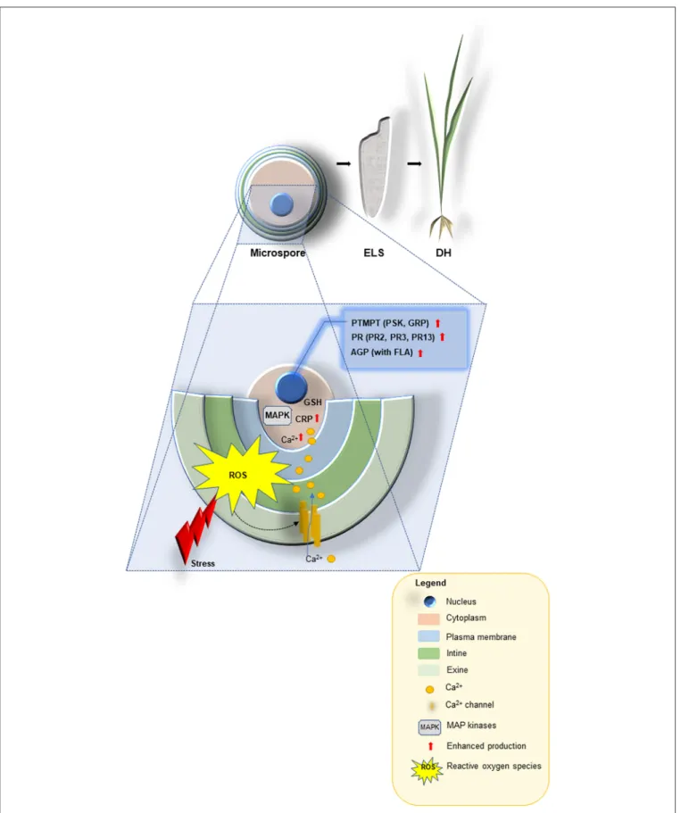
FIGURE 1 |A schematic model describing the proposed functions for stress-related proteins, cysteine-rich tripeptide glutathione, other cysteine-rich and the small post-translationally modified peptides (PTMPs) in microspore embryogenesis signaling and their use as promising tools for improving doubled-haploid (DH) production
(Continued)
FIGURE 1 |in crop plants. Abiotic stresses are perceived at the microspore surface (exine, intine and plasma membrane), and linked to cellular messengers such as ROS, Ca2+, cysteine-rich peptides [CRP e.g., rapid alkalinization factor (RALF), lipid transfer (LTP) and MAP kinases] to render the signals through glutathione (GSH) into a large-scale transcriptional reprogramming that ultimately leads to enhanced production of the most appropriate defense responses: the small PTMPs [e.g., phytosulfokine (PSK), glycine-reach proteins (GRP)], pathogenesis-related proteins (PR2, PR3, PR13) and arabinogalactan proteins AGP [with fasciclin-like arabinogalactan proteins (FLA)] in order to induce microspore-derived embryo-like structures (ELS) able to regenerated green DH lines.